Immunohistochemical diagnosis of human infectious diseases: a review
←
→
Page content transcription
If your browser does not render page correctly, please read the page content below
Oumarou Hama et al. Diagnostic Pathology                 (2022) 17:17
https://doi.org/10.1186/s13000-022-01197-5

 REVIEW                                                                                                                                               Open Access

Immunohistochemical diagnosis of human
infectious diseases: a review
Hamadou Oumarou Hama1,2, Gérard Aboudharam2,3, Rémi Barbieri1,2, Hubert Lepidi2,4 and Michel Drancourt1,2*

  Abstract
  Background: Immunohistochemistry (IHC) using monoclonal and polyclonal antibodies is a useful diagnostic
  method for detecting pathogen antigens in fixed tissues, complementing the direct diagnosis of infectious diseases
  by PCR and culture on fresh tissues. It was first implemented in a seminal publication by Albert Coons in 1941.
  Main body: Of 14,198 publications retrieved from the PubMed, Google, Google Scholar and Science Direct
  databases up to December 2021, 230 were selected for a review of IHC techniques, protocols and results. The
  methodological evolutions of IHC and its application to the diagnosis of infectious diseases, more specifically lice-
  borne diseases, sexually transmitted diseases and skin infections, were critically examined. A total of 59 different
  pathogens have been detected once in 22 different tissues and organs; and yet non-cultured, fastidious and
  intracellular pathogens accounted for the vast majority of pathogens detected by IHC. Auto-IHC, incorporating
  patient serum as the primary antibody, applied to diseased heart valves surgically collected from blood culture-
  negative endocarditis patients, detected unidentified Gram-positive cocci and microorganisms which were
  subsequently identified as Coxiella burnetii, Bartonella quintana, Bartonella henselae and Tropheryma whipplei. The
  application of IHC to ancient tissues dated between the ends of the Ptolemaic period to over 70 years ago, have
  also contributed to paleomicrobiology diagnoses.
  Conclusion: IHC plays an important role in diagnostic of infectious diseases in tissue samples. Paleo-auto-IHC
  derived from auto-IHC, is under development for detecting non-identified pathogens from ancient specimens.
  Keywords: Immunohistochemistry, infectious diseases, past infections, diagnosis

Introduction                                                                             low and serological diagnosis can be difficult; as illus-
Diagnosis of infectious diseases is fundamentally based                                  trated for an example, for the search for the tuberculosis
on the isolation by culture of the causative pathogen and                                agent Mycobacterium tuberculosis in biopsy specimens
isolation by culture of pathogens remains the gold stand-                                such as diseased lymph node biopsies [2, 3]. In recent
ard method for the laboratory diagnosis of infectious dis-                               decades immunohistochemistry has become an indis-
eases [1]. The choice of methods used for isolation by                                   pensable alternative for pathologists due to two major
culture depends in part on the nature of the sample, the                                 technical advances and the use of specific antibodies
microorganism to be identified, and the conditions                                       against various antigens. The application of monoclonal
under which the samples are processed and stored.                                        or polyclonal antibodies to viral, bacterial or fungal anti-
However in some circumstances, rate of isolation of mi-                                  gens in order to characterise infectious agents in immu-
croorganisms from cultures in tissue biopsies may be                                     nohistochemistry is now routinely used in the diagnosis
                                                                                         of many infectious diseases [4–6]. However, like any
* Correspondence: michel.drancourt@univ-amu.fr                                           other diagnostic method, immunohistochemistry re-
1
 IHU Méditerranée Infection, Marseille, France                                           quires quality assurance, reproducibility and sensitivity
2
 Aix-Marseille-Univ., IRD, MEPHI, IHU Méditerranée Infection, Marseille, France
Full list of author information is available at the end of the article                   in order to detect a targeted infectious agent. Thus, to

                                          © The Author(s). 2022 Open Access This article is licensed under a Creative Commons Attribution 4.0 International License,
                                          which permits use, sharing, adaptation, distribution and reproduction in any medium or format, as long as you give
                                          appropriate credit to the original author(s) and the source, provide a link to the Creative Commons licence, and indicate if
                                          changes were made. The images or other third party material in this article are included in the article's Creative Commons
                                          licence, unless indicated otherwise in a credit line to the material. If material is not included in the article's Creative Commons
                                          licence and your intended use is not permitted by statutory regulation or exceeds the permitted use, you will need to obtain
                                          permission directly from the copyright holder. To view a copy of this licence, visit http://creativecommons.org/licenses/by/4.0/.
                                          The Creative Commons Public Domain Dedication waiver (http://creativecommons.org/publicdomain/zero/1.0/) applies to the
                                          data made available in this article, unless otherwise stated in a credit line to the data.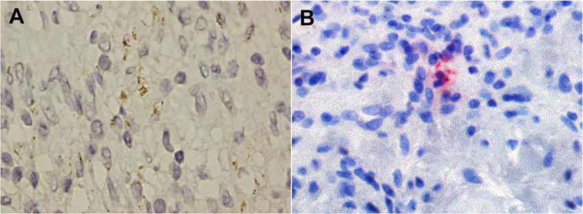
Oumarou Hama et al. Diagnostic Pathology                (2022) 17:17                                                                Page 2 of 15

avoid variations in immunostaining and to maintain the                            December 2021), and the keywords used for the search
immunoreactivity of certain antigens, several factors                             were “immunohistochemistry, immunohistochemical, hu-
need to be taken into account, mainly tissue fixation, tis-                       man infections, diagnosis” (Fig. 1). In addition, for the
sue processing and antigen retrieval [7–9]. Furthermore,                          Google Scholar database, “Publish or Perish” software was
as the antigens had been recovered from ancient paraffin                          used to pre-select the 1,000 best articles associated with
blocks and mummified bodies, the preservation of anti-                            the keywords and, for the Google database, the top 10
genic epitopes dating back at least a century has been                            pages (n=350) were pre-selected. Manual searches were
demonstrated by immunohistochemical staining, despite                             performed for articles outside the keywords and also in
the degradation of certain antigenic determinants in an-                          the reference lists of the pre-selected articles to find other
cient tissues [6, 10]. In this study, we review immunohis-                        relevant sources. Experimental and animal studies were
tochemistry techniques and protocols as applied to the                            excluded from our selection and the final selection of pa-
diagnosis of infectious diseases, including past infections                       pers was based on immunohistochemical methods and
in the context of paleomicrobiology.                                              the crucial contribution that immunohistochemistry can
                                                                                  make to the diagnosis of infectious diseases in humans.
Bibliographical methods
We searched the literature for relevant articles in the                           The historical development of
PubMed, ScienceDirect, Google Scholar and Google da-                              immunohistochemistry and automation
tabases. The pre-selection of articles based on titles and                        Immunohistochemical staining is derived from immuno-
abstracts was complete by July 2020 (and was updated in                           fluorescence, and dates back to 1941, when Coons and
                      Database

                                                                                                            Manual searches
                                                                                                           outside of keywords

                                      Top 10 pages   By Publish or Perish    n=5694                             n=33
                                                                                               n=7121
                     Identification

                                        (n=350 )     software (n= 1000 )

                                                                             n=14198
                     Screening

                                                                       Records screened for                 Records after
                                                                         title and abstract               duplicates removed
                                                                               n= 1615                          n= 1242
                     Inclusion

                                                                                                         230 articles included

 Fig. 1 Summary of the process in a flow diagram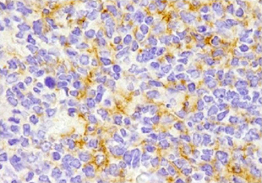
Oumarou Hama et al. Diagnostic Pathology                 (2022) 17:17                                                                       Page 3 of 15

colleagues demonstrated that tissues stained with                                           even greater sensitivity than previous immunohisto-
fluorescein-conjugated antibodies became fluorescent                                        chemical methods [17], due to the fact that the peroxid-
specifically under ultraviolet light [11] (Fig. 2). Although                                ase enzyme is directly covalently bound to the avidin
immunofluorescence has been widely used in immun-                                           molecule (Fig. 2). Epitope retrieval through enzymatic
ology for the diagnosis of diseases, this method has a                                      digestion was another productive step forward in the use
number of limitations related to the use of fluorescein                                     of immunohistochemistry in formalin-fixed and paraffin-
namely the natural autofluorescence of the tissues which                                    embedded tissues [18].
masks the specific fluorescence, the lack of stability of                                      Due to these advances in immunohistochemistry, tis-
the preparations, and the use of ultraviolet microscopes                                    sue antigen analysis became the undisputed gold stand-
which are expensive and difficult to use. In order to                                       ard for pathologists and anatomists for clinical diagnoses
overcome some of these shortcomings, alternative im-                                        or experimental research based on tissue biopsies. The
munostaining methods have been developed. One alter-                                        use of monoclonal antibodies made it possible to detect
native to fluorescent antibodies involves staining the                                      an increasing number use of specific markers against the
tissue by methods of labelling antibodies with enzymes                                      antigens represented in tissues and against infectious
(Fig. 2) that react with non-fluorescent chromogenic                                        agents from formalin-fixed paraffin blocks, and has led
substrates [12]. Preparations stained with antibodies and                                   to increased pressure on industry. The increase in the
conjugated to enzymes such as peroxidase are perman-                                        number of tests each year in research and particularly in
ent and can be observed with ordinary light micro-                                          hospital laboratories resulted in technical problems re-
scopes, enabling the simultaneous observation of antigen                                    lated to the workforce, reagent stability, reproducibility,
localisation and tissue morphology. The use of un-                                          temperature variation of the different steps of an im-
labelled antibodies (peroxidase-antiperoxidase technique:                                   munoassay, and a reliability crisis [19]. A viable auto-
PAP) (Fig. 2) to identify antigens by immunohistochem-                                      mated immunohistochemistry system to detect tissue
istry increased sensitivity [13]. A subsequent develop-                                     antigens was first invented and developed by Brigati and
ment involved using alkaline phosphatase for double                                         colleagues as an alternative to manual methods. This
immunoenzyme labelling (alkaline phosphatase and per-                                       fully automated system (from de-paraffining to nuclear
oxidase), which was capable of detecting two antigens                                       staining) overcame the restrictions on volume and
within the same cell [14]. However, the application of a                                    discrete analysis by using the capillary action with the
secondary labelled antibody to biotin followed by the                                       help of standardised software and a computer. The re-
addition of the avidin-biotin peroxidase complex (ABC)                                      trieval antigen from formalin-fixed, paraffin-embedded
(Fig. 2) proved to be much more sensitive than the un-                                      tissues by heat pre-treatment was a crucial step forward
labelled antibody method [15, 16]. In addition, the                                         in the maintenance and success of immunohistochemis-
peroxidase-labelled avidin-biotin (LAB) method offered                                      try [20]. This important publication resulted in the

                           Secondary                 Peroxidase
                           antibody                  anti-peroxidase complex

                           Primary
                                            Biotin
                           antibody
                       Substrate
                       chromogen       Avidin        Peroxidase

                          A                                                          C                                         E

                                                      Tissue section                                  Tissue section

                                                      Glass slide                                      Glass slide

                              1941                     1966                          1970             1979-1981                      1989

                                        B                                                         D
                                                                    Tissue section                                     Tissue section

                                                                     Glass slide                                       Glass slide

 Fig. 2 Schematic representation of immunohistochemical methods. A: direct method, B: indirect method, C: PAP complex procedure, D: ABC
 procedure, E: LAB procedure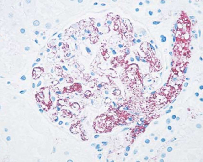
Oumarou Hama et al. Diagnostic Pathology   (2022) 17:17                                                              Page 4 of 15

detection of antigens by immunohistochemical staining            immunohistochemical staining [33, 34]. Prolonged fix-
on formalin-fixed archival tissues dating back at least a        ation can produce a progressive loss of certain tissue an-
century [21–23]. In the years following the automation           tigens used in diagnosis, such as lymphocyte antigens
of immunohistochemistry, considerable improvements               which are lost after three days’ fixation [33]. An alterna-
were observed in the development of the automatons               tive to neutral buffered formalin and a prolonged fix-
i.e., reagent cost management, reagent application, slide        ation time is formalin zinc, which allows better
labelling, traceability of operations and analytical flexibil-   morphological conservation and preserves immunoreac-
ity [24–26].                                                     tivity [30]. Coagulation fixatives are dehydrating agents,
                                                                 which replace water in the tissue environment and thus
Immunohistochemistry procedures                                  destabilise the hydrophobic binding of proteins. This
The examination of hematoxylin and eosin-stained tissue          disruption of tertiary proteins structure mainly causes
sections is the first step in the diagnosis of any infectious    the loss of functions and the insolubility of proteins [35].
disease from tissue samples before any other staining in         This protein denaturation phenomenon does not affect
histopathology [6]. The principal immunohistochemical            all antigens and depends on the alcohol concentration,
methods are direct, indirect, PAP, alkaline phosphatase          the presence of organic and inorganic substances, the
and avidin-biotin techniques [17, 27], which use antigen-        pH and the fixation temperature.
antibody complexes to locate cellular antigens in paraffin          However, regardless of the choice of fixation mode,
sections, frozen tissues, post-mortem tissues and cell           fixation alone does not cause the loss of antigen recognition
preparations.                                                    but rather occurs after a combination of fixation, tissue
  Currently, the labelled streptavidin-biotin method is          treatment and paraffin embedding. Studying the effects of
the most widely used routine laboratory method for               different fixatives under the same conditions of treatment
diagnosing diseases. It has the same principle as the LAB        and embedding, found that dehydrating fixatives such as
method, i.e., the primary antibody against the antigen of        ethanol 95% or methanol 100% and alcoholic formaldehyde
interest is bound to the enzyme-labelled streptavidin            best preserved the immunorecognition of p53 and large
(the enzyme is covalently bound to the streptavidin)             spectrum keratins, while the best fixers for TGFα and p185
                                                                 erbB-2
through the biotin present on the secondary antibody                    were cross-linking fixers (unbuffered 10% formalin
and revealed with a chromogenic substrate [26, 28].              and unbuffered zinc formalin) [36]. After fixation, tissue
                                                                 treatment and embedding the tissue was dehydrated in
Tissue preparation                                               ethanol and then the ethanol was removed by a clarification
Immunohistochemical staining involves different steps            agent such as xylene, which infiltrates the tissue [37]. After
in the technical treatment of tissues that can influence         the hot paraffin dissolves in the xylene, the xylene evapo-
the correct interpretation of the results. These are the         rates and the incorporation of paraffin into the tissue is
procedures of fixation, dehydration and paraffin embed-          complete once the tissue has cooled. This process is carried
ding. Regarding fixation, two types of fixatives are com-        out using an automaton within 12 hours. Thus, using a
monly used, namely reticulation fixatives (e.g. formalin)        heating mould containing molten paraffin, a paraffin block
and coagulation fixatives (alcohol solutions such as etha-       is constructed in which the tissue infiltrated by the paraffin
nol, methanol and acetone) [29]. Formalin is a standard          is trapped. This paraffin infiltration into the tissue allows 4-
fixative that has been used for decades. The increased           μm sections to be cut in order to visualise the structure,
needs of modern histopathological laboratories consid-           but this may also depend on the nature of the sample and
ered standard fixation modes too long for the preserva-          the position of the tissue. The loss of reactivity of some an-
tion of immunoreactivity [30], as somewhere between 12           tigens due to dehydration, paraffin embedding, paraffin wax
and 36 hours is required for complete fixation with 10%          temperature and during fixation of sections on microscopic
neutral buffered formalin, for example [31]. The fixation        slides has previously been discussed [29, 37]. It should be
time in formalin can play an important role in obtaining         noted that the most important problem related to the vari-
a reliable result in immunohistochemistry, such as in the        ation in immunohistochemical staining is inadequate tissue
determination of oestrogen receptors in breast cancer,           dehydration. This can be avoided by renewing solutions
which was at least six to eight hours [32]. A large part of      weekly and applying consistently [9].
variations observed in immunohistochemistry were
caused by the very short fixation time due to a variable         Antigen retrieval
mixture of cross-linking and coagulation fixation during         Antigen retrieval by enzymatic digestion from tissue
the tissue dehydration step by ethanol [9]. In tissue diag-      sections requires only two hours of trypsin treatment
nostics, the fixation time of formaldehyde, which is gen-        to reveal certain immunoreactive sites of the antigens
erally used at 10% in a neutral phosphate buffer, must           [18]. However, some antigens are not revealed by
be minimised for the effective application of the                trypsin digestion, with the exception of cytokeratins
Oumarou Hama et al. Diagnostic Pathology (2022) 17:17 Page 5 of 15
and desmin [38]. This method has been replaced by examination of heart valves mandatory for the diagnosis
microwave heating of formalin-fixed tissue sections. of infective endocarditis [43].
Subsequent methods consist in microwave-heating
paraffin tissue sections at up to 100°C in the presence Yersinia pestis (Y. pestis)
of metallic solutions [20]. This pre-treatment im- Y. pestis is a Gram-negative bacterium responsible for
proves immunohistochemical procedures due to a plague, a zoonosis transmitted by ectoparasites including
considerable improvement in antigen recovery, includ- fleas and possibly lice resulting in lymph node infection
ing in long-term formalin-fixed tissues. Antigens of (so-called bubo), which spreads as septicaemia and pneu-
interest are then revealed using chromogenic sub- monia [44]. Microscopy, culture and polymerase chain re-
strate after several incubation steps with antibodies, action (PCR) are the diagnostic methods used for the
as presented in the Supplementary Material. direct detection of Y. pestis [45, 46]. However, culture and
direct immunofluorescence require specimens (sputum,
blood or aspirations from lymph nodes) which are often
Automation
difficult to obtain and dangerous to handle, hence the use-
The routine use of immunohistochemistry in diagnostic
fulness of immunohistochemical testing to detect Y. pestis
laboratories had resulted in its automation. The staining
in formalin-fixed tissues in order to preserve the morpho-
procedure remains the same as in manual immunohisto-
logical features and to minimise handling of dangerous
chemistry, but it is controlled from a programmed com-
specimens [47]. Indeed Y. pestis was identified intact by an
puter. The advantages and disadvantages of automated
immunohistochemical test inside monocytes and granular
immunohistochemistry vary according to the different
antigen staining in blood vessels (Fig. 3). Immunohisto-
commercially available immunohistochemistry plat-
chemistry was also used to confirm pulmonary plague
forms. The choice of these platforms depends on the
during a plague epidemic in Ecuador [48].
needs of the laboratory, according to the capacity of the
different systems. Unlike the automated immunohisto-
Treponema pallidum (T. pallidum)
chemical method, the manual method provides extensive
Treponematoses such as yaws, bejel and syphilis are infec-
staff knowledge with almost infinite flexibility in the
tions caused by T. pallidum involving the different sub-
choice of reagents and recovery methods as well as the
species T. pallidum pertenue, T. pallidum endemicum,
ability to introduce certain technical variations when
and T. pallidum, respectively [49]. Treponemal infections,
optimising the protocol. However, the automation of im-
more particularly syphilis, have a worldwide impact on
munohistochemistry has made immunohistochemical
human health but remain indistinguishable through rou-
testing reproducible, fast and accurate with processing
tine morphology and serology. Treponemes are fastidious
of multiple slides at the same time, high and consistent
organisms whose culture and maintenance on artificial
labelling quality, analytical flexibility, standardisation of
media remains difficult, despite new techniques in micro-
critical steps, time saving, low cost, and remote oper-
biology [50, 51]. Silver staining of spirochetes in tissue
ation with a user-friendly interface and biosafety [24–26,
39]. Furthermore, despite the development of automated
machines that require periodic monitoring and mainten-
ance, the steps of tissue fixation, paraffin block construc-
tion and tissue cutting remain a human task.
Immunohistochemical detection of bacterial
pathogens
Bartonella quintana (B. quintana)
B. quintana is a Gram-negative bacillus responsible for
trench fever, bacteraemia, endocarditis, bacillary angioma-
tosis and chronic lymphadenopathy [40]. Trench fever
was discovered during the First World War, linked to
poor living conditions and lice infestation (Brouqui et al.,
1999; Maurin and Raoult, 1996). B. quintana endocarditis
is a fatal infection of global importance the current ap-
proach to which involves the Duke diagnostic criteria [41].
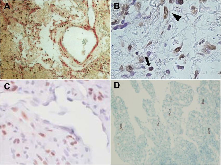
Immunohistochemistry successfully identified B. quintana Fig. 3 Immunohistochemical stain of a kidney sample
demonstrating granular antigen staining delineating the blood
in the heart valves of patients with blood culture-negative
vessels in the glomerulus (anti fraction 1 Y. pestis antibody, ×100)
infective endocarditis [42]; making histopathologicalOumarou Hama et al. Diagnostic Pathology (2022) 17:17 Page 6 of 15
sections was the usual method for identifying T. pallidum, Other bacteria
but the immunohistochemistry technique using a mono- Several microorganisms are responsible for infective
clonal antibody was found to be more sensitive and appro- endocarditis, most commonly Gram-positive cocci (such
priate to avoid marked background staining and to as Staphylococci, Streptococci and Enterococci), B. quin-
facilitate the observation of spirochetes in secondary syph- tana (already described above), Bartonella henselae (the
ilis tissue sections [52]. Immunohistochemistry is more agent of cat scratch disease) Coxiella burnetii (the agent
sensitive and specific than silver staining for detecting T. of Q fever) and Tropheryma whipplei (the agent of
pallidum in tissue sections [53–55], but should be inter- Whipple’s disease) [42, 71–73]. However, when it comes
preted with care, as immunostaining with the T. pallidum to the antimicrobial treatment of patients and certain
antibody (Biocare) also stains some acid-fast bacilli and fastidious or non-cultivable microorganisms, identifica-
Helicobacter pylori [56]. The efficacy of immunohisto- tion of these bacteria by blood culture may be difficult
chemistry for T. pallidum detection has been proven in [74, 75]. Therefore, pathological examination of valve
biopsies of unsuspected oral lesions [57] and in biopsies of tissue sections remains the reference technique for the
skin lesions [58], and its utility has been proven in the diagnosis of infective endocarditis, despite the advances
diagnosis of syphilitic chancre [59, 60] papulonodular sec- in molecular techniques and serological tests [43, 76].
ondary syphilis [61], malignant syphilis [62] and erythema Accordingly, immunohistochemistry is routinely used
multiforme caused by T. pallidum [63]. The combination for the detection of Bartonella spp., T. whipplei and C.
of immunohistochemistry and PCR has been shown to be burnetii, S. aureus, Group A Streptococci, and for the
effective for the diagnosis of secondary syphilis [64] and pathological diagnosis of infectious diseases using spe-
confirmation of syphilitic orchitis in an HIV-infected cific antibodies [42, 72, 73, 77–79]. A related method in-
young man with a false-negative Venereal Disease Re- corporating the patient’s own serum as the primary
search Laboratory test and T. pallidum agglutination test antibody, known as auto-immunohistochemistry, suc-
(Fig. 4A) [65]. cessfully detected bacteria in the heart valves of blood-
culture negative endocarditis patients [71].
Chlamydia trachomatis (C. trachomatis) Despite cross-reactivity between the Mycobacterium tu-
C. trachomatis, an obligate intracellular bacterium, is the berculosis polyclonal antibody and normal eosinophil gran-
most common sexually-transmitted infection in the world ules [80], IHC can be hugely useful in identifying cutaneous
[66, 67]. The detection of C. trachomatis infection is based mycobacteriosis [81–84] and certain bacterial infections as-
on PCR and serology [66]. An immunohistochemical in- sociated with Clostridium species [79], Borrelia burgdorferi
vestigation incorporating a monoclonal antibody reactive [85], Helicobacter pylori [86–90], Rickettsia rickettsii [91–
against C. trachomatis D/K and L2 serovars (Acris Anti- 93], Chlamydia pneumoniae [94, 95], Orientia tsutsuga-
bodies catalogue number AM00660PU-N) detected C. tra- mushi [96–99], Neisseria meningitidis [100], Brachyspira
chomatis in skin lesions in two cases of lymphogranuloma species [101], and Burkholderia pseudomallei [102].
venereum (Fig. 4B) [68]. Despite the greater sensitivity of
DNA detection methods (PCR and ligase chain reaction) Immunohistochemical detection of viral
compared to immunohistochemistry for the detection of pathogens
C. trachomatis from fixed biopsies, the application of im- Human Herpesviruses (HHV)
munohistochemistry remains essential for pathological HHV are intracellular pathogens that have been classi-
diagnosis [69, 70]. fied into three subfamilies and have been divided into
Fig. 4 A Immunohistochemistry for syphilis showing spirochetes in the cytoplasm of histiocytes (1000x, oil immersion). B. Immunohistochemistry
showing C. trachomatis particles in lesional tissueOumarou Hama et al. Diagnostic Pathology (2022) 17:17 Page 7 of 15 eight types based on their biological properties: Alpha- directed against the VZV envelope glycoprotein gpI herpesviridae (herpes simplex virus type 1, herpes sim- [108] and glycoprotein E [109]. plex virus type 2, and varicella-zoster virus) have a Immunohistochemistry has been very useful for the neurocutaneous tropism. They can replicate and spread diagnosis of HSV-1/2 and VZV skin infections from rapidly to many cell systems causing extensive cell lysis fixed biopsies [110–114]. and can establish latent infection primarily in neurosen- Gammaherpesviridae (Epstein-Barr virus and HHV-8) sory ganglia [103]. are the only human herpesviruses with an established Herpes simplex virus (HSV) infections are among oncogenic potential, causing a variety of lymphoprolifer- the most common diseases in humans, largely caus- ative and neoplastic disorders [103]. The viruses of this ing subclinical infections and usually manifesting in subfamily replicate in vitro in lymphoblastoid cells, ulcerative lesions at the site of infection [104, 105]. sometimes in epithelioid and fibroblastic cells, and are Immunohistochemistry using polyclonal or monoclo- specific for T or B lymphocytes in which they can induce nal antibodies has been shown to be a sensitive and latent or lytic infections [103, 104]. specific technique for diagnosing HSV infections Epstein-Barr virus (EBV) was discovered in 1964 by when characteristic intranuclear inclusions or multi- electron microscopy from a Burkitt’s lymphoma cell line, nucleated cells are absent in biopsy specimens [2]. a B-cell derived tumour. EBV is ubiquitous, infecting Discriminating HSV-1 from HSV-2 by immunohisto- more than 90% of adults worldwide by establishing a chemistry is achieved using monoclonal antibodies lifelong persistent infection of B cells characterised by and, in some cases, immunohistochemistry has been excretion of the virus in saliva, with most primary infec- preferable to in situ hybridisation, as in the case of tions occurring sub-clinically in childhood [103, 104]. necrotic lesions, in which immunohistochemical de- Immunohistochemistry identified EBV latent membrane tection has revealed the presence of HSV, while in protein 1 using tissue sections in apical and periapical situ hybridisation was negative (Fig. 5A) [106, 107]. periodontitis lesions (Fig. 5B) [115, 116], in patients with Primary infection with VZV results in varicella and a nasopharyngeal carcinoma [117], in immunocompetent resurgence in zoster [104]. The histological features of adult [118], and in Hodgkin's disease and non-Hodgkin's HSV can be observed in patients with varicella-zoster lymphoma [119–121]. and it has been possible to identify varicella-zoster in Human herpesvirus type 8 (HHV8), also known as immunohistochemistry by using a monoclonal antibody Kaposi's sarcoma-associated herpes virus, is a recent Fig. 5 A HSVI immunoreactivity in the lung (IBD4 monoclonal antibody). B. LMP-1 immunohistochemistry showing positive staining in B cells (arrows) and plasma cells (arrow heads). Scale bar = 50μm. C. Immunohistochemical staining for HHV-8 LNA-1 in cutaneous patch/plaque Kaposi sarcoma. D. Immunohistochemistry of CMV showing nuclear positive cells in ileal tissue. x20
Oumarou Hama et al. Diagnostic Pathology   (2022) 17:17                                                                      Page 8 of 15

member of the herpes family, discovered in 1994 in a            paracortical areas of the lymph nodes, and that atypical
Kaposi’s sarcoma (KS) skin lesion in an AIDS patient, thus      cells were positive for CD3 and CD4 [148, 149]. Further-
establishing a link between HHV-8 infection and the             more, immunohistochemical staining has demonstrated
emergence of KS [122]. Histological diagnosis of HHV8 is        the common presence of HHV6 in renal allografts [150],
problematic because of its broad morphologic spectrum           in bone marrow transplant recipients [151], in the cen-
and the limitation of various benign and malignant vascu-       tral nervous system of neurological pathology patients
lar neoplasms [123]. In such cases, immunohistochemistry        and healthy people [152, 153] and in one infant infected
using an anti-HHV-8 LNA-1 antibody has proven to be a           with HIV and encephalitis [154]. HHV-6 antigens were
reliable marker with high sensitivity and specificity for the   also detected in B- and T-cell lymphoma tissues by im-
diagnosis of KS (Fig. 5C) [124–129].                            munohistochemistry using the polyclonal HHV6 anti-
   Betaherpesviridae (cytomegalovirus, human herpes-            body [155].
virus type 6, and human herpesvirus type 7) replicate in          Another closely related virus, HHV-7, is also known to
a limited number of cellular systems and grow slowly in         be the causative agent of exanthem subitum [103, 156,
cell culture. These viruses cause little cell lysis and can     157]. In 1990, HHV-7 was detected by Frenkel and col-
establish latent infections in secretory glands, the            leagues in a culture of activated CD4+ T cells from a
lymphoreticular system, the kidneys and certain other           healthy individual [158].
tissues [103].                                                    Although rare, the detection of HHV-7 by immunohis-
   The histological diagnosis of cytomegalovirus (CMV)          tochemistry has been successful in brain autopsy samples
in fixed tissues has proven to be the “gold standard” for       from unspecified encephalopathy cases [153] and in fixed
the identification of viral inclusions and other cytopathic     normal tissues, suggesting that HHV-7 causes a persistent
effects, despite the lack of sensitivity in some cases, such    infection rather than a true latent one [153, 159].
as the difficulty in interpreting atypical cytopathic fea-
tures with reactive or degenerative changes [130–132].          Human Immunodeficiency Virus (HIV)
Combined immunohistochemistry and in situ hybridisa-            HIV is a retrovirus belonging to the Retroviridae family,
tion tests compare favourably with culture, allowing for        and is responsible for Acquired Immunodeficiency Syn-
faster diagnosis than immunofluorescence technique or           drome (AIDS) [103]. Biological diagnosis is based pri-
culture for early anti-CMV therapy [133]. In addition,          marily on serology and PCR.
immunohistochemistry is much more sensitive than                  The immunohistochemical technique is rapid and effi-
cytomorphology and in situ hybridisation for the detec-         cient in identifying HIV antigens in fixed surgical and
tion of CMV in smears fixed from bronchoalveolar lav-           autopsy specimens [160]. Immunohistochemistry using
age samples [134]. David and colleagues recommended             an anti-p24 monoclonal antibody has proven useful for
automated in situ hybridisation or immunohistochemis-           detecting HIV in lymph node biopsies of patients with
try for CMV detection in formalin-fixed, paraffin-              unexplained follicular hyperplasia [161], in duodenal and
embedded tissues [135]. Immunohistochemistry using              rectal biopsies of AIDS patients [162], in cervical biopsy
monoclonal antibodies has detected CMV antigens from            samples of HIV-infected women [163], and in a parotid
fixed tissues in septic patients [136], in placental biopsies   gland biopsy from a rare case of HIV-associated benign
[137, 138], in the brain after liver transplantation [139],     lymphoepithelial cysts (Fig. 6) [164]. This approach has
in pulmonary and gastrointestinal tissues (Fig. 5D) [140–
143], in apical periodontitis lesions [116], in tumours
and in peripheral blood [144], in patients with steroid
refractory ulcerative colitis [145] and in one patient with
ischemic colitis [146].
   HHV6 was isolated and characterised for the first time
in 1986 by Salahuddin and collaborators [147], from per-
ipheral blood mononuclear cells in six patients with lym-
phoproliferative disorders, two of whom were co-
infected with human immunodeficiency virus (HIV).
HHV-6 infection usually occurs at the age of two years
and is the aetiological cause of exanthema subitum, also
known as roseola infantum.
   Immunohistochemical analysis with a monoclonal
antibody against HHV-6 envelope glycoprotein (gp60/
                                                                 Fig. 6 Positive immunohistochemical staining for HIV-1 p24 antigen
110 kDa) in two immunocompetent individuals demon-
                                                                 in follicular centres and dendritic cells (original magnification ×400)
strated the presence of numerous viral inclusions in
Oumarou Hama et al. Diagnostic Pathology (2022) 17:17 Page 9 of 15
been recommended for the diagnosis of recent infections Immunohistochemical detection of protozoal
in which anti-p24 antibodies are not yet detectable in pathogens
the serum or if morphological or clinical features suggest The diagnosis of protozoal infections is difficult because
HIV infection [161, 165]. of the distortion of the morphology of the parasite due
to the necrosis or autolysis of tissues or in cases of un-
usual presentation of the disease, hence the usefulness of
Other viruses
immunohistochemistry (Arnold et al., 1997).
Immunohistochemistry has been of great value for the
Despite the higher sensitivity of PCR compared to
laboratory diagnosis of Ebola haemorrhagic fever, espe-
immunohistochemistry for the diagnosis of leishman-
cially during the epidemic in the Democratic Republic
iasis, the use of both tests together has been shown
of Congo in 1995 [166, 167], and also for the elucida-
to be more effective for routine diagnosis [215, 216].
tion of the pathogenesis of influenza A (H1N1) and B
Immunohistochemistry significantly improves the
virus infection from fixed autopsy specimens [168–
diagnosis of cutaneous leishmaniasis in histological
170]. Immunohistochemistry has been one of the main
sections and allows the patient's immune response to
methods used to better understand the pathogenesis of
be evaluated [217–219]. In addition, immunohisto-
Middle East respiratory syndrome coronavirus (MERS-
chemistry has been useful in the diagnosis of
CoV) in humans in fixed tissues obtained from the first
toxoplasmosis [220–222], echinococcosis [223, 224],
autopsy performed on a fatal MERS-CoV case in the
malaria [225] and for the detection of Trypanosoma
world [171]. The use of immunohistochemistry in fixed
cruzi [226, 227].
tissues has been essential as a diagnostic method and
for providing further evidence of enterovirus involve-
Paleoimmunohistochemical detection of pathogens
ment in myocarditis or dilated cardiomyopathy [172–
The application of histological methods for the detection
176]. Immunohistochemistry has been useful in the
of pathogens in ancient tissues has provided information
diagnosis and clinical management of chronic hepatitis
for better understanding the origin and spread of infec-
C [177–179], in the diagnosis of yellow fever [180, 181],
tious diseases [6]. Currently, the representation of past ep-
hepatitis E [182], rabies [183–185], severe acute respira-
idemics relies on molecular biology, which may be an
tory syndrome (SARS) [186], in the identification and
approach limited to pathogens in the event that the al-
pathogenesis of SARS-CoV-2 [187–193], in the identifi-
tered DNA prevents the amplification of pathogen-
cation of infections associated with Zika virus [194],
specific DNA sequences. Immunohistochemistry has been
West Nile virus [195–197], adenoviruses [198–200],
successfully used in a number of paleopathological studies,
hantavirus [201, 202], and in the diagnosis of other cu-
including the detection of bacteria such as T. whipplei and
taneous viral infections by parvoviruses, poxviruses,
R. rickettsii in paraffin blocks at least a century old from
paramyxoviridae [203].
autopsy cases [22, 23], the sporadic typhus agent Rickettsia
typhi in paraffin-embedded tissue blocks from the Second
Immunohistochemical detection of fungal World War (Hamburg, Germany, 1940-1944) [228], and
pathogens the detection of a parasite (Taenia solium) from stomach
The great majority of fungi are easily identified by tissue sections dating from the Ptolemaic period [21].
hematoxylin-eosin staining alone or in combination These studies demonstrated that the antigenicity of pro-
with special stains such as Gomori methenamine teins could be preserved in ancient tissues. We invented
silver stain and Schiff periodic acid stain which are microbial paleoserology, to reveal an epidemic of recur-
used in routine histopathology [204, 205]. However, rent fever in a 16th-17th century French garrison, which
the morphological diagnosis of deep mycoses, ideally had been missed by real-time PCR [229]. This demon-
done by culture, can be time-consuming and often strates the interest of applying other complementary ap-
has to be made from tissue sections for smears, when proaches allowing the detection of antigens or antibodies
cultures are not available [206]. Immunohistochemis- specific to the pathogens. In that pioneering work, total
try can be an accurate and efficient diagnostic tool immunoglobulins previously demonstrated in ancient
for a number of important mycoses in humans and dental pulp [230], were extracted and applied to inacti-
for the localisation of typical or atypical fungal ele- vated pathogens used as antigens in a so-called “mini blot”
ments in lesions from fixed tissue sections. Immuno- format [229]. Extending these observations to the detec-
histochemistry has been used for the diagnosis of tion of unidentified pathogens, we are now applying auto-
pythiosis [207], fungal sinusitis [208], aspergillosis immunohistochemistry used for the detection of patho-
[209–211], sporotrichosis [212], and for the identifica- gens in modern-day tissues, to ancient samples, mainly
tion of Candida albicans [213], and Cryptococcus dental pulp. We call this new, antigen-based detection
neoformans var. gattii [214]. “paleo-autoimmunohistochemistry”.Oumarou Hama et al. Diagnostic Pathology (2022) 17:17 Page 10 of 15
Abbreviations 48–70. http://www.sciencedirect.com/science/article/pii/B9781416039662
IHC: Immunohistochemistry; PAP: Peroxidase-antiperoxidase technique; 000163.
ABC: Avidin-biotin peroxidase complex; LAB: Peroxidase-labelled avidin- 5. Molina-Ruiz AM, Cerroni L, Kutzner H, Requena L. Immunohistochemistry in
biotin; TGF-α: Transforming growth factor alpha; B. quintana: Bartonella the Diagnosis of Cutaneous Bacterial Infections. Am J Dermatopathol. 2015;
quintana; Y. pestis: Yersinia pestis; T. pallidum: Treponema pallidum; C. 37(3):179–96.
trachomatis: Chlamydia trachomatis; HHV: Human Herpesviruses; 6. Raoult D, Drancourt M. éditeurs. Paleomicrobiology: past human infections.
VZV: Varicella-zoster virus; HSV: Herpes simplex virus; EBV: Epstein-Barr virus; Berlin: Springer; 2008. p. 226.
KS: Kaposi’s sarcoma; CMV: Cytomegalovirus; HIV: Human Immunodeficiency 7. Roskams T. The role of immunohistochemistry in diagnosis. Clin Liver Dis.
Virus; AIDS: Acquired Immunodeficiency Syndrome; MERS-CoV: Middle East 2002;6(2):571–89.
respiratory syndrome coronavirus; SARS: Severe acute respiratory syndrome 8. Shi SR, Key ME, Kalra KL. Antigen retrieval in formalin-fixed, paraffin-
embedded tissues: an enhancement method for immunohistochemical
staining based on microwave oven heating of tissue sections. J Histochem
Supplementary Information Cytochem. 1991;39(6):741–8.
The online version contains supplementary material available at https://doi. 9. Werner M, Chott A, Fabiano A, Battifora H. Effect of Formalin Tissue Fixation
org/10.1186/s13000-022-01197-5. and Processing on Immunohistochemistry. Am J Surg Pathol. 2000;24(7):
1016–9.
Additional file 1: Supplementary Material 10. Grillo F, Bruzzone M, Pigozzi S, Prosapio S, Migliora P, Fiocca R, et al.
Immunohistochemistry on old archival paraffin blocks: is there an expiry
date? J Clin Pathol. 2017;70(11):988–93.
Acknowledgements 11. Coons AH, Creech HJ, Jones RN. Immunological Properties of an Antibody
Not applicable. Containing a Fluorescent Group. Exp Biol Med. 1941;47(2):200–2.
12. Nakane PK, Pierce GB. Enzyme-labeled antibodies: Preparation and application
Authors’ contributions for the localization of antigens. J Histochem Cytochem. 1966;14(12):929–31.
H.O.H. and M.D. : performed the conceptualization and methodology of the 13. Sternberger LA, Hardy PH, Cuculis JJ, Meyer HG. The unlabeled antibody
review; H.O.H : software; M.D., H.O.H., G.A. and H.L.: analysis and validation; enzyme method of immunohistochemistry preparation and properties of
H.O.H. and M.D. : writing and original draft preparation; H.L.,G.A. and R.B.: soluble antigen-antibody complex (Horseradish peroxidase-antihorseradish
participated the reviewing and editing; M. D : Supervision. All authors read peroxidase) and its use in identification of spirochetes. J Histochem
and approved the final manuscript. Cytochem. 1970;18(5):315–33.
14. Mason DY, Sammons R. Alkaline phosphatase and peroxidase for double
Funding immunoenzymatic labelling of cellular constituents. J Clin Pathol. 1978;31(5):
This work was supported by the French Government under the 454–60.
“Investissements d’avenir” (Investments for the Future) programme managed 15. Guesdon JL, Ternynck T, Avrameas S. The use of avidin-biotin interaction in
by the Agence Nationale de la Recherche (ANR, fr: National Agency for immunoenzymatic techniques. J Histochem Cytochem. 1979;27(8):1131–9.
Research), (reference: Méditerranée Infection 10-IAHU-03). 16. Hsu SM, Raine L, Fanger H. Utilisation du complexe avidine-biotine-
peroxydase (ABC) dans les techniques d’immunoperoxydase: une
Availability of data and materials comparaison entre les procédures ABC et les anticorps non marqués (PAP).
The data that support of this study are available from the corresponding J Histochem Cytochem. 1981;29(4):577–80.
author upon reasonable request. 17. Elias JM, Margiotta M, Gaborc D. Sensitivity and Detection Efficiency of the
Peroxidase Antiperoxidase (PAP), Avidin–Biotin Peroxidase Complex (ABC),
Declarations and Peroxidase-Labeled Avidin–Biotin (LAB) Methods. Am J Clin Pathol.
1989;92(1):62–7.
Ethics approval and consent to participate 18. Sn H. H M, Jd M. Application of immunofluorescent staining on paraffin
Not applicable. sections improved by trypsin digestion. Lab Invest. 1976;35(4):383–90.
19. Brigati DJ, Budgeon LR, Unger ER, Koebler D, Cuomo C, Kennedy T, et al.
Consent for publication Immunocytochemistry is Automated: Development of A Robotic Workstation
Not applicable. Based Upon the Capillary Action Principle. J Histotechnol. 1988;11(3):165–83.
20. Shi SR, Key ME, Kalra KL. Récupération d’antigène dans les tissus fixés au
Competing interests formol, inclus en paraffine: une méthode d’amélioration de la coloration
The authors have no conflicts of interest to declare. immunohistochimique basée sur le chauffage au four à micro-ondes de
coupes de tissus. J Histochem Cytochem. 1991;39(6):741–8.
Author details 21. Bruschi F, Masetti M, Locci MT, Ciranni R, Fornaciari G. Short report:
1
IHU Méditerranée Infection, Marseille, France. 2Aix-Marseille-Univ., IRD, cysticercosis in an Egyptian mummy of the late Ptolemaic period. Am J
MEPHI, IHU Méditerranée Infection, Marseille, France. 3Aix-Marseille-Univ., Trop Med Hyg. 2006;74(4):598–9.
Ecole de Médecine Dentaire, Marseille, France. 4Laboratoire d’Histologie, 22. Dumler JS. Fatal Rocky Mountain Spotted Fever in Maryland—1901. JAMA.
Faculté de Médecine, Université de la Méditerranée, Marseille, France. 1991;265(6):718.
23. Dumler JS, Baisden BL, Yardley JH, Raoult D. Immunodetection of
Received: 15 July 2021 Accepted: 18 January 2022 Tropheryma whipplei in Intestinal Tissues from Dr. Whipple’s 1907 Patient. N
Engl J Med. 2003;348(14):1411–2.
24. Herman GE, Elfont EA, Floyd AD, Overview of Automated Immunostainers.
References In: Javois LC, editor. Immunocytochemical Methods and Protocols. Totowa,
1. Fournier P-E, Drancourt M, Colson P, Rolain J-M, La Scola B, Raoult D. NJ: Humana Press; 1995. p. 383–403. (Methods in Molecular Bilogy). https://
Modern clinical microbiology: new challenges and solutions. Nat Rev doi.org/10.1385/0-89603285-X:383.
Microbiol. 2013;11(8):574–85. 25. Le Neel T, Moreau A, Laboisse C, Truchaud A. Comparative evaluation of
2. Eyzaguirre E, Haque AK. Application of Immunohistochemistry to Infections. automated systems in immunohistochemistry. Clinica Chimica Acta. 1998;
Arch Pathol Lab Med. 2008;132(3):424–31. 278(2):185–92.
3. Fellag M, Saad J, Armstrong N, Chabrière E, Eldin C, Lagier J-C, et al. Routine 26. Moreau A, Le Neel T, Joubert M, Truchaud A, Laboisse C. Approach to
Culture-Resistant Mycobacterium tuberculosis Rescue and Shell-Vial Assay. Fr automation in immunohistochemistry. Clinica Chimica Acta. 1998;278(2):
Emerg Infect Dis. 2019;25(11):2131–3. 177–84.
4. Hawes D, Shi S-R, Dabbs DJ, Taylor CR, Cote RJ. CHAPTER 5 - 27. Linnoila I, Petrusz P. Immunohistochemical techniques and their
Immunohistochemistry. In: Weidner N, Cote RJ, Suster S, Weiss LM, editors. applications in the histopathology of the respiratory system. Environ Health
Modern Surgical Pathology. 2nd ed. Philadelphia: W.B. Saunders; 2009. p. Perspect. 1984;56:131–48.Oumarou Hama et al. Diagnostic Pathology (2022) 17:17 Page 11 of 15
28. Bratthauer GL, The Avidin–Biotin Complex (ABC) Method and Other Avidin– 53. Martín-Ezquerra G, Fernandez-Casado A, Barco D, Jucglà A, Juanpere-Rodero
Biotin Binding Methods. In: Oliver C, Jamur MC, editors. N, Manresa JM, et al. Treponema pallidum distribution patterns in
Immunocytochemical Methods and Protocols. Totowa, NJ: Humana Press; mucocutaneous lesions of primary and secondary syphilis: an
2010. p. 257–70. (Methods in Molecular Biology; vol. 588). http://link. immunohistochemical and ultrastructural study. Hum Pathol. 2009;40(5):
springer.com/10.1007/978-1-59745-324-0_26. 624–30.
29. Bussolati G, Leonardo E. Technical pitfalls potentially affecting diagnoses in 54. Phelps RG, Knispel J, Tu ES, Cernainu G, Saruk M. Immunoperoxidase
immunohistochemistry. J Clin Pathol. 2008;61(11):1184–92. technique for detecting spirochetes in tissue sections: comparison with
30. Dapson RW. Fixation for the 1990’s: a Review of Needs and other methods. Int J Dermatol. 2000;39(8):609–13.
Accomplishments. Biotechnic Histochem. 1993;68(2):75–82. 55. Tse JY, Chan MP, Ferry JA, Deshpande V, Sohani AR, Nardi V, et al. Syphilis of
31. Ramos-Vara JA, Miller MA. When Tissue Antigens and Antibodies Get Along: the Aerodigestive Tract. Am J Surg Pathol. 2018;42(4):472–8.
Revisiting the Technical Aspects of Immunohistochemistry—The Red, 56. Fernandez-Flores A. Immunostaining for Treponema pallidum: Caution in its
Brown, and Blue Technique. Vet Pathol. 2014;51(1):42–87. Evaluation. Am J Dermatopathol. 2010;32(5):523–5.
32. Goldstein NS, Ferkowicz M, Odish E, Mani A, Hastah F. Minimum Formalin 57. Siqueira CS, Saturno JL, de Sousa SCOM, da Silveira FRX. Diagnostic
Fixation Time for Consistent Estrogen Receptor Immunohistochemical approaches in unsuspected oral lesions of syphilis. Int J Oral Maxillofac Surg.
Staining of Invasive Breast Carcinoma. Am J Clin Pathol. 2003;120(1):86–92. 2014;43(12):1436–40.
33. Leong AS-Y, Gilham PN. The effects of progressive formaldehyde fixation on 58. Quatresooz P, Piérard GE. Skin Homing of Treponema pallidum in Early
the preservation of tissue antigens. Pathology. 1989;21(4):266–8. Syphilis: An Immunohistochemical Study. Appl Immunohistochem Mol
34. Montero C. The Antigen-Antibody Reaction in Immunohistochemistry. J Morphol. 2009;17(1):47–50.
Histochem Cytochem. 2003;51(1):1–4. 59. Calvo DF, Cassarino D, Fernandez-Flores A. Syphilitic Chancre of the Lip. Am
35. Eltoum I, Fredenburgh J, Myers RB, Grizzle WE. Introduction to the Theory J Dermatopathol. 2020;42(10):e143–6.
and Practice of Fixation of Tissues. J Histotechnol. 2001;24(3):173–90. 60. Kenny B, Hamza S, Peermohamed S, Shumilak G, Groot G, Osmond A. A
36. Arnold MM, Srivastava S, Fredenburgh J, Stockard CR, Myers RB, Grizzle WE. case of a pseudoneoplastic primary syphilis chancre on the neck. JAAD
Effects of Fixation and Tissue Processing on Immunohistochemical Case Rep. 2021;16:130–3.
Demonstration of Specific Antigens. Biotechnic Histochem. 1996;71(5):224–30. 61. Veasey JV, Lellis RF, MFF de C B, Porto PL, JCS C. Papulonodular secondary
37. Grizzle WE, Stockard CR, Billings PE. The Effects of Tissue Processing syphilis: a rare clinic presentation confirmed by serologic and histologic
Variables Other Than Fixation on Histochemical Staining and exams. An Bras Dermatol. 2016;91(2):205–7.
Immunohistochemical Detection of Antigens. J Histotechnol. 2001;24(3): 62. Cid PM, Cudós ES, Zamora Vargas FX, Merino MJB, Pinto PH. Pathologically
213–9. Confirmed Malignant Syphilis Using Immunohistochemical Staining: Report
38. Leong AS-Y, Milios J, Duncis CG. Antigen preservation in microwave- of 3 Cases and Review of the Literature. Sex Transm Dis. 2014;41(2):94–7.
irradiated tissues: A comparison with formaldehyde fixation. J Pathol. 1988; 63. Chiang M-C, Chiang F-C, Chang Y-T, Chen T-L, Fung C-P. Erythema
156(4):275–82. Multiforme Caused by Treponema pallidum in a Young Patient with Human
39. Prichard JW. Overview of Automated Immunohistochemistry. Arch Pathol Immunodeficiency Virus Infection. J Clin Microbiol. 2010;48(7):2640–2.
Lab Med. 2014;138(12):1578–82. 64. Buffet M, Grange PA, Gerhardt P, Carlotti A, Calvez V, Bianchi A, et al.
40. Maurin M, Raoult D. Bartonella (Rochalimaea) quintana infections. Clin Diagnosing Treponema pallidum in Secondary Syphilis by PCR and
Microbiol Rev. 1996;9(3):273–92. Immunohistochemistry. J Invest Dermatol. 2007;127(10):2345–50.
41. Li JS, Sexton DJ, Mick N, Nettles R, Fowler VG Jr, Ryan T, et al. Proposed 65. Chu C-Y, Chen W-Y, Yeh S-D, Yeh H-M, Fang C-L. Syphilitic orchitis
Modifications to the Duke Criteria for the Diagnosis of Infective mimicking a testicular tumor in a clinically occult HIV-infected young man:
Endocarditis. Clin Infect Dis. 2000;30(4):633–8. a case report with emphasis on a challenging pathological diagnosis. Diagn
42. Lepidi H, Fournier P-E, Raoult D. Quantitative Analysis of Valvular Lesions Pathol. 2016;11 https://www.ncbi.nlm.nih.gov/pmc/articles/PMC4712524/.
During Bartonella Endocarditis. Am J Clin Pathol. 2000;114(6):880–9. 66. Debonnet C, Robin G, Prasivoravong J, Vuotto F, Catteau-Jonard S, Faure K,
43. Lepidi H, Durack DT, Raoult D. Diagnostic methods current best practices et al. Infection à Chlamydia trachomatis : mise au point. Gynécologie
and guidelines for histologic evaluation in infective endocarditis. Infect Dis Obstétrique Fertilité & Sénologie. 2021;S2468718921000040.
Clin North Am. 2002;16(2):339–61 ix. 67. Unemo M, Bradshaw CS, Hocking JS, de Vries HJC, Francis SC, Mabey D,
44. Barbieri R, Signoli M, Chevé D, Costedoat C, Tzortzis S, Aboudharam G, et al. et al. Sexually transmitted infections: challenges ahead. Lancet Infect Dis.
Yersinia pestis : the Natural History of Plague. Clin Microbiol Rev. 2020;34(1): 2017;17(8):e235–79.
e00044–19 /cmr/34/1/CMR.00044-19.atom. 68. Feltes F, Vallés L, Alcaraz I, Kutzner H, Requena L. Lymphogranuloma
45. Engelthaler DM, Gage KL, Montenieri JA, Chu M, Carter LG. PCR Detection of Venereum: Report of Two Cases with « Bubonulus » As Primary Stage and
Yersinia pestis in Fleas: Comparison with Mouse Inoculation. J Clin Immunohistochemical Demonstration of Chlamydia Trachomatis. 2015;6(1):3.
Microbiol. 1999;37(6):1980–4. 69. Bryan ER, McLachlan RI, Rombauts L, Katz DJ, Yazdani A, Bogoevski K, et al.
46. Ratsitorahina M, Chanteau S, Rahalison L, Ratsifasoamanana L, Boisier P. Detection of chlamydia infection within human testicular biopsies. Hum
Epidemiological and diagnostic aspects of the outbreak of pneumonic Reprod. 2019;34(10):1891–8.
plague in Madagascar. Lancet. 2000;355(9198):111–3. 70. Noguchi Y, Yabushita H, Noguchi M, Fujita M, Asai M, Del Carpio CA.
47. Guarner J, Shieh W-J, Greer PW, Gabastou J-M, Chu M, Hayes E, et al. Detection of Chlamydia trachomatis infection with DNA extracted from
Immunohistochemical Detection of Yersinia pestis in Formalin-Fixed. formalin-fixed paraffin-embedded tissues. Diagn Microbiol Infect Dis. 2002;
Paraffin-Embedded Tissue. Am J Clin Pathol. 2002;117(2):205–9. 43(1):1–6.
48. Gabastou J-M, Proaño J, Vimos A, Jaramillo G, Hayes E, Gage K, et al. An 71. Lepidi H, Coulibaly B, Casalta J-P, Raoult D. Autoimmunohistochemistry: A
outbreak of plague including cases with probable pneumonic infection, New Method for the Histologic Diagnosis of Infective Endocarditis. J Infect
Ecuador, 1998. Trans R Soc Trop Med Hyg. 2000;94(4):387–91. Dis. 2006;193(12):1711–7.
49. Majander K, Pfrengle S, Kocher A, Neukamm J, L du P, Pla-Díaz M, et al. 72. Lepidi H, Fenollar F, Dumler JS, Gauduchon V, Chalabreysse L, Bammert A,
Ancient Bacterial Genomes Reveal a High Diversity of Treponema pallidum et al. Cardiac Valves in Patients with Whipple Endocarditis: Microbiological,
Strains in Early Modern Europe. Curr Biol. 2020;30(19):3788–803 e10. Molecular, Quantitative Histologic, and Immunohistochemical Studies of 5
50. Belkacemi S, Khalil JB, Ominami Y, Hisada A, Fontanini A, Caputo A, et al. Patients. J Infect Dis. 2004;190(5):935–45.
Passive Filtration, Rapid Scanning Electron Microscopy, and Matrix-Assisted 73. Lepidi H, Houpikian P, Liang Z, Raoult D. Cardiac Valves in Patients with Q
Laser Desorption Ionization–Time of Flight Mass Spectrometry for Fever Endocarditis: Microbiological, Molecular, and Histologic Studies. J
Treponema Culture and Identification from the Oral Cavity. J Clin Microbiol. Infect Dis. 2003;187(7):1097–106.
2019;57(10) https://jcm.asm.org/content/57/10/e00517-19. 74. Brouqui P, Raoult D. Endocarditis Due to Rare and Fastidious Bacteria. Clin
51. Edmondson DG, Hu B, Norris SJ. Long-Term In Vitro Culture of the Syphilis Microbiol Rev. 2001;14(1):177–207.
Spirochete Treponema pallidum subsp. pallidum. mBio. 2018;9(3) https:// 75. Lamas CC, Eykyn SJ. Blood culture negative endocarditis: analysis of 63
mbio.asm.org/content/9/3/e01153-18. cases presenting over 25 years. Heart. 2003;89(3):258–62.
52. Hoang MP, High WA, Molberg KH. Secondary syphilis: a histologic and 76. Fournier PE, Raoult D. Nonculture Laboratory Methods for the Diagnosis of
immunohistochemical evaluation. J Cutan Pathol. 2004;31(9):595–9. Infectious Endocarditis. Curr Infect Dis Rep. 1999;1(2):136–41.You can also read


