IGG4-RELATED DISEASE: A DIAGNOSTIC CHALLENGE - AUTOPSY AND ...
←
→
Page content transcription
If your browser does not render page correctly, please read the page content below
Clinical Case Report
IgG4-related Disease: a diagnostic challenge
Rodrigo Díaz Olmos1,2 , Marcelo Arlindo Vasconcelos Miranda Rodrigues1,3 ,
Cristiane Rúbia Ferreira4 , Rita de Cássia Franco Etrusco5 ,
Carla Romagnolli1
How to cite: Olmos RD, Rodrigues MAVM, Ferreira CR, Etrusco RCF, Romagnolli C. IgG4-related Disease: a diagnostic
challenge. Autops Case Rep [Internet]. 2021;11:e2021312. https://doi.org/10.4322/acr.2021.312
ABSTRACT
Immunoglobulin IgG4-related disease (IgG4-RD) is an immune-mediated fibroinflammatory condition with a characteristic
histopathological appearance that can affect almost any organ. The clinical features result from a focal or diffuse
appearance of a tumor-like swelling of the affected organs, identified by physical and/or imaging examination. Herein,
we report the case of a 38-year-old male complaining of a worsening chronic right lumbar pain associated with legs and
scrotum edema. He also had itchy and erythematous cutaneous lesions on the abdominal wall over the last 8 months,
and complained of a diffuse and mild to moderate abdominal discomfort. On examination, the liver was firmly enlarged
and tender. His legs had 2+ symmetrical pitting edema extending from his feet to just above the knees. An abdominal
computed tomography scan showed a large mass (10 x 8 x 4cm) involving the abdominal infrarenal aorta and the iliac
arteries, and compressing the inferior vena cava, with dilated iliac veins, raising the possibility of lymphoproliferative disease.
During the initial investigation, the laboratory workup revealed anemia, without other marked changes. A laparoscopic-
guided biopsy of the peri-aortic mass was undertaken. The histological report associated with IgG4 immunoglobulin
measurement rendered the diagnosis of IgG4-RD. The patient had a favorable outcome after the use of glucocorticoids
with the abdominal mass remission.
Keywords
Immunoglobulin G; Immunoglobulin G4-Related Disease; Lymph Node Excision.
INTRODUCTION
Immunoglobulin IgG4-related disease (IgG4-RD) pancreatitis or pancreatitis related to IgG4), but it
is an immune-mediated fibroinflammatory condition was not recognized as a systemic condition until
with a characteristic histopathological appearance 2003, when extrapancreatic manifestations were
that may involve virtually all organs.1 It was initially identified in patients with type 1-autoimmune
identified in the pancreas in 2001 (type 1-autoimmune pancreatitis. 1 The most frequently affected organs
1
Universidade de São Paulo (USP), Hospital Universitário, Internal Medicine Division, São Paulo, SP, Brasil
2
Universidade de São Paulo (USP), Hospital Universitário, Faculty of Medicine, Internal Medicine Department, São Paulo, SP, Brasil
3
Universidade São Caetano do Sul (USCS), Faculty of Medicine, São Paulo, SP, Brasil
4
Universidade de São Paulo (USP), Hospital Universitário, Anatomic Pathology Service, São Paulo, SP, Brasil
5
Universidade de São Paulo (USP), Hospital das Clínicas, Faculty of Medicine, São Paulo, SP, Brasil
Copyright: © 2021 The Authors. This is an Open Access article distributed under the terms of the Creative
Commons Attribution License, which permits unrestricted use, distribution, and reproduction in any medium,
provided the original work is properly cited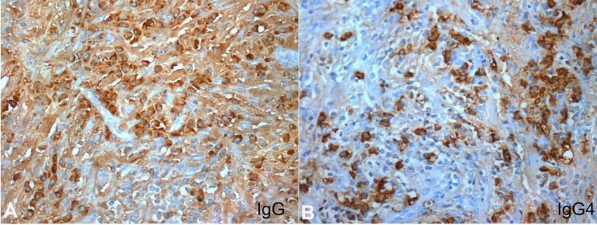
IgG4-related Disease: a diagnostic challenge

include the major salivary and lacrimal glands                transforming growth factor-b) might have a role in
(sclerosing sialadenitis), orbital disease, autoimmune        the progression of fibrosis, which is characteristic of
pancreatitis, retroperitoneal fibrosis (commonly with         this disease.
chronic peri-aortitis and the ureters involvement),                 The diagnosis of IgG4-RD might be easily missed
biliary tract (sclerosing cholangitis), lymph nodes and       by unsuspecting general practitioners, internists,
kidneys (tubulointerstitial nephritis).2,3 The IgG4-related   hematologists, and rheumatologists, as patients may
dacryoadenitis and sialadenitis may be more common            present with a multitude of clinical presentations
in women; atopic dermatitis, asthma, or chronic               that may mimic some hematologic, infectious, and
sinusitis may be present in up to 40% of patients;            rheumatologic diseases such as Castleman disease,
multiple and simultaneous organ involvement occurs            lymphoma, plasma cell neoplasms, hypereosinophilic
in 50% of cases; and it most commonly affects middle-         syndromes, sicca syndrome, systemic lupus
aged to elderly individuals, with a male to female            erythematosus, infectious mononucleosis and so forth.
ratio of 4:1. These multiple organ involvements share         Therefore, it is crucial for the clinicians who first see
several core pathologic features, as well as clinical and     these patients to suspect this entity when facing cases
serologic similarities. In this setting, IgG4-RD could be     with lymphadenopathy, eosinophilia, and polyclonal
compared with sarcoidosis because of the multiorgan           hypergammaglobulinemia together with some of
involvement with a unique histological appearance.            the more common presentations such as lacrimal
      The pathologic features include tumor-like              and salivary glands involvement, orbital disease, and
swelling of the involved organs, a lymphoplasmacytic          tumor-like swelling of the involved organs.4 The clinical
infiltrate with IgG4-positive plasma cells, obliterative      manifestations generally result from a focal or diffuse
phlebitis, and a variable degree of fibrosis that has         appearance of tumor-like swelling of the organs,
a characteristic “storiform” pattern. Furthermore,            identified by physical and/or imaging examination. The
there is often but not always, elevated serum IgG4            clinical course is normally subacute, and constitutional
determination. Table 1 shows the clinical, laboratory,        symptoms are uncommon.
and pathological criteria for the diagnosis of IgG4-RD.2
     In the past few decades, this entity has been            CASE REPORT
recognized as a systemic disease encompassing many
individual organ diseases that were not known to be                A 38-year-old male presented to the emergency
related (and are now part of the spectrum of IgG4-            department with worsening chronic right lumbar
related disease), such as retroperitoneal fibrosis,           pain associated with legs and scrotum edema. He
Mikulicz’s syndrome, Küttner’s tumor, and Riedel              also had itchy and erythematous cutaneous lesions
thyroiditis.                                                  on the abdominal wall over the last 8 months, with
      The pathogenesis of IgG4-RD is unclear; however,        no remission despite numerous therapeutic attempts,
it seems to be involved in an allergic mechanism. The         and complained of diffuse mild to moderate abdominal
T-helper 2 (Th2) cytokine expression and regulatory           pain. He denied weight loss, fever, or night sweats. He
cytokines have been noted to be up-regulated in               was a smoker, and his past medical history included
the affected tissues of patients with IgG4-RD. The            unilateral (right) polycystic kidney disease with
increase in Th2 cytokines (IL-4, -5, -10, and -13, and        functional renal exclusion and a left vicariant kidney (he

Table 1. Clinical, laboratory, and pathological criteria for IgG4-RD
 •
     swelling or mass in one or more organs;
 •
     serum IgG4 greater than 1.35 g/L;
 •
     histopathological features:
 ◦ marked infiltration by lymphocytes and plasma cells,
 ◦ more than ten IgG4-positive plasma cells per high-power field with an IgG4/IgG ratio greater than 40%,
 ◦ storiform fibrosis.
 ◦ obliterating phlebitis

2-7                                                                            Autops Case Rep (São Paulo). 2021;11:e2021312
Olmos RD, Rodrigues MAVM, Ferreira CR, Etrusco RCF, Romagnolli C
was waiting for a right nephrectomy) and pulmonary Abdominal computed tomography (CT) showed a
tuberculosis in 2003. He frequently used diclofenac, large, contrast-enhanced mass (10 x 8 x 4cm) involving
paracetamol, caffeine, and carisoprodol for his chronic the abdominal infrarenal aorta and the iliac arteries
lumbar pain. and compressing the inferior vena cava with dilated
On admission, he had a good general appearance, iliac veins and the left ureter (Figure 1A), raising the
no signs of respiratory distress, no alterations in skin hypothesis of the lymphoproliferative disease. The liver
color. His heart rate was 95 bpm, blood pressure had normal size but had a heterogeneous attenuation.
166 x 104 mmHg, and room air oximetry were 96%. The chest CT showed a diffuse pleural thickening
There were eczematous desquamating skin lesions in the right hemithorax with a small loculated pleural
on his thighs and legs. There were no enlarged effusion, and enlarged mediastinal and peri-esophageal
lymph nodes. Chest auscultation revealed decreased lymph nodes.
breath sounds and dullness to percussion at the
The patient was admitted with the working
right lung base. On abdominal palpation, the liver
diagnosis of a lymphoproliferative disease for a
was enlarged, palpable up to 5 cm below the costal
confirmatory diagnostic workup. A double J ureteral
margin and was slightly tender. His legs had 2+
stent was placed in the left ureter. A CT-guided biopsy
symmetrical pitting edema extending from his feet
of the right pleural thickening was performed, which
to just above his knees. Laboratory workup revealed
lacked a precise diagnosis. The patient was submitted
mild anemia (Hb 10.8 g/dl), normal white blood cell,
to a laparoscopic-guided biopsy of the peri-aortic
and platelet count. The hepatic enzymes, bilirubin,
mass. This biopsy showed a fibro-connective tissue
creatine kinase, lactate dehydrogenase, uric acid, and
exhibiting fibrosis, obliterating phlebitis, and moderate
electrolytes were within the normal range. However,
mixed inflammatory infiltrates with few eosinophils,
the creatinine was 1.47 g/dL (RR: 0.70 - 1.20 g/dL)
and the creatinine clearance estimated by the CKD-EPI histocytes, and small lymphocytes and frequent plasma
equation was 69 mL/min/1.73m2. C-reactive protein cells (Figures 2 and 3).
(32 mg/L; RR: < 3 mg/L) and erythrocyte sedimentation The immunohistochemical examination (Table 2)
rate (46 mm/1h) were increased. HIV, HCV, syphilis, showed an increased number of IgG4 positive plasma
and Hepatitis B virus serologies yielded negative cells. The IgG4/IgG ratio of 30% and approximately
results. The rheumatoid factor and antinuclear 80 IgG4 positive-plasma cells per high-power field
antibodies were negative, and the complement (HPF) in areas of more density. This examination,
was normal. Urinalysis revealed 1+ of protein and together with the morphological and clinical findings,
150 x 103 red blood cells/mm3. is highly suggestive of IgG4-RD.
Figure 1. Abdominal CT axial plane: A – Initial examination on admission. Note large mass involving the abdominal
aorta; B – CT scan performed 7 months after the first examination and under treatment with corticosteroid. Note
the expressive reduction of the periaortic mass.
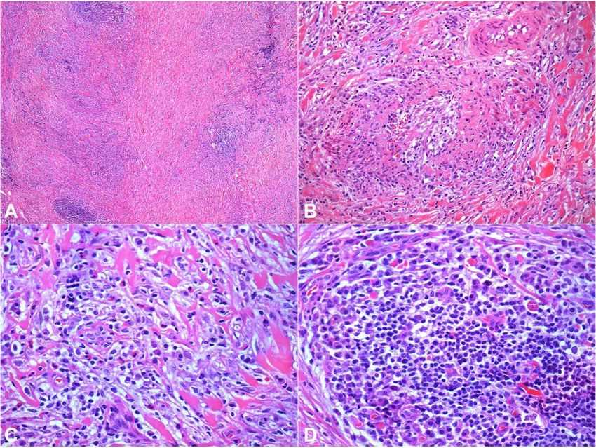
Autops Case Rep (São Paulo). 2021;11:e2021312 3-7IgG4-related Disease: a diagnostic challenge Figure 2. Photomicrographs of the biopsy: A – Fibro conjunctive tissue exhibiting fibrosis, with thick collagen bundles and moderate inflammatory infiltrate with lymphoid aggregates (HE – 40X); B – Obliterating phlebitis (HE – 200X); C – Fibro conjunctive stroma showing thick collagen bundles with few eosinophils, histiocytes and small lymphocytes mixed with plasma cells (HE – 400X); D – Numerous plasma cells surrounding a lymphoid aggregate (HE – 400X). Figure 3. Photomicrographs of the Immunohistochemical staining of the biopsy: A – Numerous IgG positive plasma cells near a lymphoid aggregate (400X); B – Increased number of IgG4 positive plasma cells presenting an elevated IgG4/IgG ratio greater than 30% in areas of more density (400X). 4-7 Autops Case Rep (São Paulo). 2021;11:e2021312
Olmos RD, Rodrigues MAVM, Ferreira CR, Etrusco RCF, Romagnolli C

Table 2. Immunohistochemical examination
  Antibodies                        Result                  Antibodies                          Result
    AE1/AE3                        Negative                   CD68                      Positive (in histocytes)
      S-100                      Inconclusive                Lysozyme                Positive (focal in histocytes)
      SMA                          Positive                   CD1a                              Negative
     Desmin                        Negative                   CD23            Positive (focal in follicular dendritic cells)
     CD117                         Negative                   CD43                 Positive (reactive T-lymphocytes)
      CD20            Positive (reactive B-lymphocytes)       Kappa                           Inconclusive
      CD3             Positive (reactive T-lymphocytes)      Lambda                           Inconclusive
      CD30                         Negative                    IgG                  Positive (frequent plasma cells
      AlK c                        Negative                    IgG4                 Positive (frequent plasma cells

     The patient was started on 1.0 mg/kg/day                  and radiological criteria. The involvement of many
of prednisone for 2 weeks and then tapered for                 organs and the wide range of clinical manifestations,
0.6 mg/kg/day for two months, and to 20 mg/day for             depending on the affected organ, make the diagnosis
3 months, afterward. The outcome was favorable and             even more difficult. Although the serum IgG4
he gradually recovered his previous health status. The         concentration is generally high (and even higher in
abdominal and lumbar pain ceased in about 3 weeks              atopic patients) in patients with IgG4-RD, in up to 40%
after starting treatment, his hemoglobin level returned        of the biopsy-confirmed cases the IgG4 serum level is
to normal and his inflammatory markers decreased.              normal. Thus, the serum IgG4 determination within
Five months after initiation of corticotherapy, a new          the normal range does not exclude the diagnosis9, 15,
abdominal CT scan was done (Figure 1B), and the initial
                                                               16.
                                                                   Similarly, a high serum level of IgG4 is not enough
contrast-enhanced mass had almost disappeared, but             to confirm the diagnosis, because several infectious,
it showed an inferior vena cava thrombosis extending           neoplastic, and inflammatory conditions can also lead
to bilateral external iliac veins. He was started on           to similar results.1,2,4
warfarin. The double J ureteral stent was removed,                   The three main histopathological characteristics
and the patient remains asymptomatic with a good               associated with IgG4-RD are (i) dense lymphoplasmacytic
urine output with no renal function impairment. At             infiltrate; (ii) storiform fibrosis; and (iii) obliterating
the closure of this manuscript, he was asymptomatic            phlebitis. A reliable pathological diagnosis of IgG4-RD
and taking only 5 mg/day of prednisone.                        requires the presence of at least two of these criteria,
                                                               which in most cases are the dense lymphoplasmacytic
                                                               infiltrate associated with the storiform fibrosis. 2 The
DISCUSSION                                                     lymphoplasmacytic infiltrate is rich in IgG4+ plasma
                                                               cells. Other non-specific features associated with
     Retroperitoneal fibrosis/periaortitis, as in this         IgG4-RD are phlebitis without obliteration of the lumen
case, affects approximately 9.6–27.0% of the cases of          and an increased number of eosinophils.
IgG4-RD 5-12. It usually manifests with abdominal, flank,
                                                                       Some studies17–19 recently proposed the diagnostic
or lumbar pain, edema of the lower limbs, decreased            criteria for IgG4-RD: (i) evidence of nodules or masses,
urinary output, low-grade fever, loss of appetite, and         local or diffusely spread in one or multiple organs;
weight loss. Hydronephrosis occurs in 33–67%, being            (ii) elevation of serum IgG4 (> 135mg / dL); and
mostly (75%) unilateral, similar to the involvement of         (iii) tissue infiltration of plasma cells IgG4 +> 10 /high
former idiopathic retroperitoneal fibrosis13,14.               power field and IgG4 + / IgG + cells> 40%. The presence
     The diagnosis of IgG4-RD is challenging. No               of the three criteria indicates a “definitive diagnosis
single marker or clinical feature is specific to make a        of IgG4-RD”. If criteria 1 and 3 are met, even in the
definitive diagnosis of the disease. Thus, the diagnosis       absence of other histopathological findings suggestive
is made based on clinical, laboratory, histological            of the disease, it is defined as “possible diagnosis of

Autops Case Rep (São Paulo). 2021;11:e2021312                                                                                  5-7
IgG4-related Disease: a diagnostic challenge
IgG4-RD” and, in patients with organic involvement response to corticotherapy was favorable, as shown
and with histopathological criteria present, but without in the literature.
increased serum IgG4 concentration, the diagnosis is
considered as “probable diagnosis of IgG4-RD”.
REFERENCES
The treatment of IgG4-RD depends on the
affected organ, ranging from expectant management 1. Stone JH, Zen Y, Deshpande V. IgG4-Related Disease.
(lymphadenopathies and asymptomatic pulmonary N Engl J Med. 2012;366(6):539-51. http://dx.doi.
org/10.1056/NEJMra1104650. PMid:22316447.
nodules) to aggressive treatment with glucocorticoids
in case of vital organs at risk of dysfunction. 1 2. Deshpande V, Zen Y, Chan JK, et al. Consensus
Glucocorticoid is the first-line drugs to induce statement on the pathology of IgG4-related disease. Mod
Pathol. 2012;25(9):1181-92. http://dx.doi.org/10.1038/
remission. The recommended dose and time of modpathol.2012.72. PMid:22596100.
treatment vary in the literature. The treatment result
3. Umehara H, Okazaki K, Masaki Y, et al. A novel
is observed quickly. Symptoms improve in one month,
clinical entity, IgG4- related disease (IgG4RD): general
and the serum IgG4 concentrations decrease rapidly, concept and details. Mod Rheumatol. 2012;22(1):1-
permitting the glucocorticoid withdrawal within a 14. http://dx.doi.org/10.3109/s10165-011-0508-6.
few weeks, in most patients.22–24 The initial dose and PMid:21881964.
duration of glucocorticoid therapy should be carefully 4. Chen LYC, Mattman A, Seidman MA, Carruthers MN.
evaluated based on the comorbidities of each patient IgG4-related disease: what a hematologist needs to know.
Haematologica. 2019;104(3):444-55. http://dx.doi.
and their potential for medication intolerance.22–24 The
org/10.3324/haematol.2018.205526. PMid:30705099.
conventional steroid-sparing agents are ineffective for
the treatment of IgG4-RD, and no benefit in relapse- 5. Yamada K, Yamamoto M, Saeki T, et al. New clues
to the nature of immunoglobulin G4-related disease:
free survival has been observed by adding these a retrospective Japanese multicenter study of baseline
medications together with the glucocorticoids.24 As they clinical features of 334 cases. Arthritis Res Ther.
are ineffective, they cannot be used in other situations, 2017;19(1):262. http://dx.doi.org/10.1186/s13075-017-
1467-x. PMid:29191210.
such as corticosteroid contraindication or refractoriness.
Azathioprine, methotrexate, or mycophenolate 6. Inoue D, Yoshida K, Yoneda N, et al. IgG4-related
mofetil can be used in case of glucocorticoid usage disease: dataset of 235 consecutive patients. Medicine.
2015; 94( 15) : e680. http://dx.doi.org/10.1097/
contraindications21–24. Rituximab is also the option in MD.0000000000000680. PMid:25881845.
recurrent or refractory cases.21,24 Early diagnosis and
7. Chen Y, Zhao JZ, Feng RE, et al. Types of organ
treatment prevent organ dysfunction by fibrosis and
involvement in patients with immunoglobulin G4- related
poor therapeutic response. 20,21 Thus, the degree of disease. Chin Med J. 2016;129(13):1525-32. http://dx.doi.
fibrosis at the beginning of treatment is still the main org/10.4103/0366-6999.184459. PMid:27364787.
predictor of the therapeutic success.20,21,24 During the 8. Sekiguchi H, Horie R, Kanai M, Suzuki R, Yi ES, Ryu
follow-up, after starting treatment, especially with JH. IgG4- related disease: retrospective analysis of
glucocorticoids, it is expected a decrease in the IgG4 one hundred sixty-six patients. Arthritis Rheumatol.
2016;68(9):2290-9. http://dx.doi.org/10.1002/art.39686.
levels; however, there may be clinical remission even
PMid:26990055.
without its fall, as well as recurrence with normal IgG4
9. Wallace ZS, Deshpande V, Mattoo H, et al. IgG4-
levels.1
related disease: clinical and laboratory features in one
hundred twenty-five patients. Arthritis Rheumatol.
2015;67(9):2466-75. http://dx.doi.org/10.1002/
CONCLUSION art.39205. PMid:25988916.
IgG4-RD can be a challenging diagnosis and 10. Lin W, Lu S, Chen H, et al. Clinical characteristics of
immunoglobulin G4-related disease: a prospective study of
be a differential diagnosis of intra-abdominal mass. 118 Chinese patients. Rheumatology. 2015;54(11):1982-
In the case presented herein, it was necessary to 90. http://dx.doi.org/10.1093/rheumatology/kev203.
perform a biopsy of the intra-abdominal mass and PMid:26106212.
IgG4 measurement. It is important to consider this 11. Zen Y, Nakanuma Y. IgG4-related disease: a cross-sectional
differential diagnosis in similar cases. In our case, the study of 114 cases. Am J Surg Pathol. 2010;34(12):1812-
6-7 Autops Case Rep (São Paulo). 2021;11:e2021312Olmos RD, Rodrigues MAVM, Ferreira CR, Etrusco RCF, Romagnolli C
9. http://dx.doi.org/10.1097/PAS.0b013e3181f7266b. 18. Mahajan VS, Mattoo H, Deshpande V, Pillai SS,
PMid:21107087. Stone JH. IgG4- related disease. Annu Rev Pathol.
2014;9(1):315-47. http://dx.doi.org/10.1146/annurev-
12. Fernández-Codina A, Martínez-Valle F, Pinilla B, et al.
pathol-012513-104708. PMid:24111912.
IgG4- related disease: results from a multicenter Spanish
Registry. Medicine. 2015;94(32):e1275. http://dx.doi. 19. Guma M, Firestein GS. IgG4-related diseases. Best Pract
org/10.1097/MD.0000000000001275. PMid:26266361. Res Clin Rheumatol. 2012;26(4):425-38. http://dx.doi.
org/10.1016/j.berh.2012.07.001. PMid:23040358.
13. Mizushima I, Inoue D, Kawano M. Retroperitoneal
fibrosis/periaortitis and hydronephrosis. In: Saito T, 20. Perugino CA, Stone JH. Treatment of IgG4-related
Stone JH, Nakashima H, Saeki T, Kawano M, editors. disease: current and future approaches. Z Rheumatol.
IgG4-related kidney disease. Tokyo: Springer; 2016. p. 2016;75(7):681-6. http://dx.doi.org/10.1007/s00393-
159-71. http://dx.doi.org/10.1007/978-4-431-55687- 016-0142-y. PMid:27431746.
9_14.
21. Perugino CA, Stone JH. IgG4-related disease: an update
14. Kawano M, Saeki T, Nakashima H. IgG4-related kidney
on pathophysiology and implications for clinical care.
disease and retroperitoneal fibrosis: an update. Mod
Nat Rev Rheumatol. 2020;16(12):702-14. http://dx.doi.
Rheumatol. 2019;29(2):231-9. http://dx.doi.org/10.108
org/10.1038/s41584-020-0500-7. PMid:32939060.
0/14397595.2018.1554321. PMid:30499730.
22. Zhang W, Stone JH. Management of IgG4-related
15. Engelhart S, Glynn RJ, Schur PH. Disease associations
with isolated elevation of each of the four IgG subclasses. disease. Lancet Rheum. 2019;1(1):E55-65. http://dx.doi.
Semin Arthritis Rheum. 2017;47(2):276-80. http://dx.doi. org/10.1016/S2665-9913(19)30017-7.
org/10.1016/j.semarthrit.2017.03.021. PMid:28457528. 23. Khosroshahi A, Wallace ZA, Crowe JL, et al. International
16. Della Torre E, Mattoo H, Mahajan VS, Carruthers M, consensus guidance statement on the management and
Pillai S, Stone JH. Prevalence of atopy, eosinophilia, treatment of IgG4-related disease. Arthritis Rheumatol.
and IgE elevation in IgG4- related disease. Allergy. 2015;67(7):1688-99. http://dx.doi.org/10.1002/
2014;69(2):269-72. http://dx.doi.org/10.1111/all.12320. art.39132. PMid:25809420.
PMid:24266692.
24. Hart PA, Topazian MD, Witzig TE, et al. Treatment
17. Umehara H, Okazaki K, Masaki Y, et al. Comprehensive of relapsing autoimmune pancreatitis with
diagnostic criteria for IgG4-related disease (IgG4-RD). immunomodulators and rituximab: the Mayo Clinic
Mod Rheumatol. 2012;22(1):21-30. http://dx.doi. experience. Gut. 2013;62(11):1607-15. http://dx.doi.
org/10.3109/s10165-011-0571-z. PMid:22218969. org/10.1136/gutjnl-2012-302886. PMid:22936672.
This study was carried out at the Hospital Universitário from the University of São Paulo. São Paulo, Brazil.
Authors’ contributions: Rodrigo Díaz Olmos, Marcelo Arlindo Rodrigues, Rita de Cássia Franco Etrusco, and
Carla Romagnolli wrote the manuscript. Cristiane Rúbia Ferreira was in charge of the pathological report and
provided the histological pictures. All authors proofread the manuscript’s final version and approved it for
publication.
Ethics statement: The authors retain informed consent signed by the patient authorizing the data publication.
Conflict of interest: The authors have no conflict of interest to declare.
Financial support: The authors declare that no financial support was received.
Submitted on: May 19th, 2021
Accepted on: June 25th, 2021
Correspondence
Rodrigo Díaz Olmos
Universidade de São Paulo, Hospital Universitário, Internal Medicine Division
Av. Prof Lineu Prestes, 2565, Butantã, 05508-000, São Paulo, SP, Brasil
Phone: +55 (11) 3019-9433
rodrigo.olmos@fm.usp.br
Autops Case Rep (São Paulo). 2021;11:e2021312 7-7You can also read