IC AN Notes The ultramicrotome as a tool for the preparation of ultra-thin samples for TEM investigations
←
→
Page content transcription
If your browser does not render page correctly, please read the page content below
IC AN
Notes

     The ultramicrotome as a tool
 for the preparation of ultra-thin
   samples for TEM investigations

   T. B. Nguyen and M. Heidelmann, ICAN Notes 4, 1 - 4 (2021)

                                         Open - Minded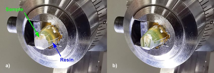
IC AN
Notes
The ultramicrotome as a tool for the preparation of ultra-
thin samples for TEM investigations
by Thai Binh Nguyen* and Markus Heidelmann -
Interdisciplinary Center for Analytics on the Nanoscale (ICAN)
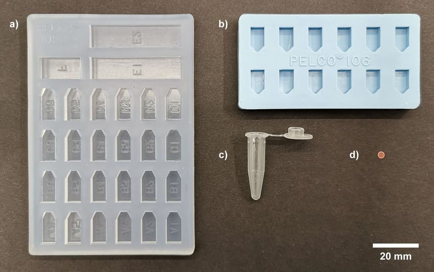
Transmission electron microscopy (TEM) is a powerful tool for the For small, very soft, or thin specimens (i. e. like foils) it is beneficial to
characterization of a wide variety of materials on an atomic scale. Since embed them in epoxy resin. The resin fixates these specimens for the
the electrons which are used for imaging have to pass through the actual cutting. Without embedding, the specimen would move du-
material, however, the limitations regarding the specimen thickness ring the cutting process and an uneven cut would be the result. Fig. 1
are quite strict. Depending on the material and the TEM’s accelerating shows different casting molds for embedding. The molds a) and b) in
voltage, samples with a thickness of 50 to 100 nm are still electron- Fig. 1 are embedding molds for flat samples. These are well suited for
transparent, whereby the attainable spatial resolution decreases with thin specimens such as foils. Alternativelly an Eppendorf container
increasing thickness of the sample. Compared to other microscopy (see Fig. 1c) can also be used for embedding samples.
methods, such as optical microscopy or scanning electron microscopy,
electron-transparency often requires complex sample preparation The embedded samples are cured in the vacuum drying oven
procedures. Furthermore, the necessary preparation depends on the (VT6060, Thermo Scientific) at 80°C for approx. one hour, or
specifics of the material. In the case of hard materials like metals or alternatively at room temperature for at least 24 hours in case
ceramics, the preparation path involves sawing, grinding, polishing the specimens are temperature sensitive. In the ICAN, usually
and finally thinning with an ion beam. If the area of interest itself needs Epoxy 3000 from Cloeren Technology GmbH is used.
to be selected with high precison targeting, Focused Ion Beam (FIB)
methods can be used. For soft materials like polymers or biological Once the epoxy resin has been cured, the specimen has to be
specimens, these methods can cause severe thermal stress or even removed from the mold and than trimmed into the shape of a
destruction of the specimen material. To avoid this, the preparation truncated pyramid shape with the EM TRIM3 trimmer before
technique of choice would be the use of an ultramicrotome. The ultramicrotome cutting. Fig. 2a shows the epoxy pyramid with
ultramicrotome creates ultra-thin sections (usually tenths of embedded sample shows the epoxy pyramid with embedded
nanometers) from the specimens mechanically and with as little stress sample fixed into the specimen holder of the ultramicrotome after
as possible by using glass or diamond knives. Particularly sensitive the initial trimming. The surfaces of the pyramid are still rough.
samples can be nitrogen cooled to avoid damage during cutting. This
note describes the preparation of a TEM sample by ultramicrotome The truncated side of the pyramid should be as flat as possible so that
cutting with the ultramicrotome (EM UC 7 with EM FC 7 module for the pressure from the ultramicrotome blade cuts evenly through
cryo applications, Leica Microsystems) in the ICAN. the specimen. Therefore, the fine trimming of the pyramid is
accomplished with a glass trimming knife, or a diamond trimming
In order to meet the ultramicrotomes size requirements, the specimen knife in the ultramicrotome. Fig. 2b shows the epoxy pyramid fixed
has to roughly fit into a (5 x 5 x 20) mm³ block. Depending on the original into the specimen holder of the ultramicotome after completed fine
size of the specimens, they can be trimmed using razor blades and a trimming.
scalpel. Alternatively, the trimmer (EM TRIM 3, Leica Microsystems)
in the ICAN can be used. Note that the knives used for trimming are different from the
* Corresponding author; eMail: thai.nguyen@uni-due.de
1 T. B. Nguyen and M. Heidelmann, ICAN Notes 4, 1 - 4 (2021); https://doi.org/10.17185/ican.notes/4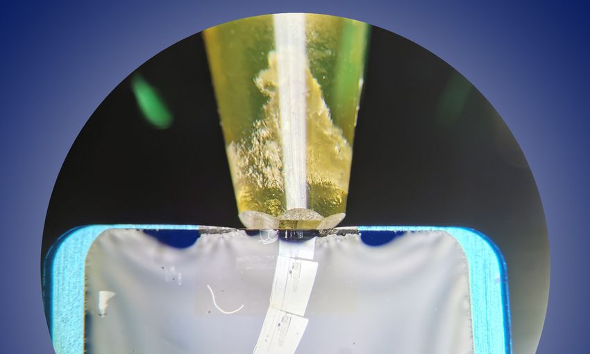
Figure 1: Different embedding molds with a TEM grid (d) for size comparison.

knives used for ultramicrotome cutting. Using the ultramicrotome After fine trimming the sample is ready for the ultramicrotome
knife for trimming is also possible, but it would take more time and cutting. Fig. 3b and 3c show knives for the ultra-thin cutting. Both
would wear out the knife.                                               knives have integrated basins (tubs) that can be filled with distilled
                                                                        water. The knife shown in Fig. 3b is made of glass while the knife in
Re-trimming with the glass trimming knife shown in Fig. 3a leads Fig. 3c is made of diamond.
to very smooth surfaces which later on facilitate approaching the
knife for the ultra-thin cuts to the specimen via the reflection of the Even though diamond knives are very hard, they eventually wear
knife at the tip of the pyramid. Looking through the microscope, out with repeated use. Since re-sharpening is very expensive,
it is difficult to see how close the blade is to the block. This can be it is advisable to first try a freshly broken glass knife with tub for
asseessed with the help of the optical reflection on the trimmed cutting the sample. These are cheaper and they are sufficient for soft
smooth surface. Moving the knife to the block, the distance between materials. If, however, the glass knife shows cutting marks after the
the real blade and the mirrored blade is reduced.                       first cut, it is recommended to switch to a diamond knife.

                                  Figure 2: Truncated pyramid located at the ultramicrotome sample holder a)
                                  after trimming with trimmer and b) after fine trimming with glass trimming

                   T. B. Nguyen and M. Heidelmann, ICAN Notes 4, 1 - 4 (2021); https://doi.org/10.17185/ican.notes/4                        2
Figure 3: a) Glass knife for trimming; b) Glass knife with tub; c) Diatome 35° Ultra with blue metal tub with diamond blade.
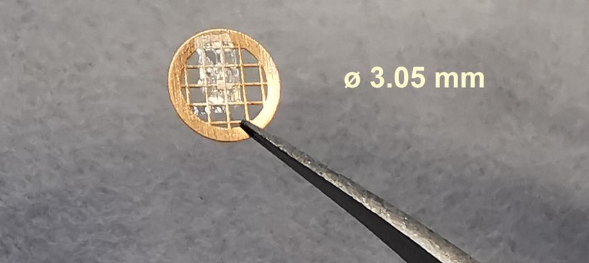
Figure 5: Air dried slice on a TEM grid.
In case of a problematic charging up of the specimen is observed
during irradiation with the electron beam inside the TEM, the
Figure 4: Floating Slices. sample may be additionally coated with a nanometer thin layer of
carbon in a sputter coater as a final preparation step prior to the
TEM investigation.
Once the cutting parameters (requested thickness, cutting speed) are
set and the tub has been filled with distilled water, the cutting process In summary ultramicrotomy is most suitable for the preparation
can be started. During the cutting process, the ultra-thin cuts slide of soft materials and biological samples. Unlike conventional
into the tub onto the water surface. Fig. 4 shows three slices floating on methods including FIB, where only one electron-transparent
water that have been cut from the embedding pyramid at the upper sample can be prepared at a time, the ultramicrotome is capable of
half of the figure. Floating slices are picked up with a TEM grid and creating several electron-transparent samples in a sequence during
dried in air at room temperature. The final result can be seen in Fig. 5. the preparation process. In addition, the remaining block with the
embedded sample can also be used for analysis of cross-sections,
After this procedure the sample is ready for TEM measurements. for example by using SEM/EDX or SAM.
References Acknowledgements
[1] J. Thomas and T. Gemming. Analytische Transmissionselektro- ICAN is a registered open core facility (DFG RIsources reference:
nenmikroskopie - Eine Einführung für den Praktiker. Springer- RI_00313). Funding by the German Research Foundation (DFG,
Verlag Wien, 49 - 55, 2013. grant HA 2769/7-1) is gratefully acknowledged.
[2] K. K. Ohtaki , H. A. Ishii and J. P. Bradley. Combined Focused
Ion Beam-Ultramicrotomy Method for TEM Specimen Prepara-
tion of Porous Fine-Grained Materials. Microscopy and Micro-
analysis 26, 120 - 125, 2020.
3 T. B. Nguyen and M. Heidelmann, ICAN Notes 4, 1 - 4 (2021); https://doi.org/10.17185/ican.notes/4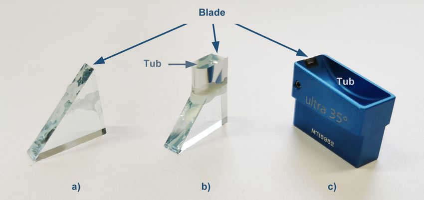
Notes

   Contact
                        ICAN | CENIDE                                      Tel.: +49 (0) 201 379 8080
                        Universität Duisburg-Essen                         Fax: +49 (0) 201 379 8046
                        Carl-Benz-Str. 199                                 eMail: ican@uni-due.de
                        47057 Duisburg                                     www.uni-due.de/ican

        T. B. Nguyen and M. Heidelmann, ICAN Notes 4, 1 - 4 (2021); https://doi.org/10.17185/ican.notes/4   4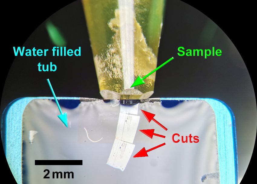
Imprint
                ICAN | CENIDE                                    Published by:
                Universität Duisburg-Essen                       ICAN Scientific Director
                Carl-Benz-Str. 199                               Frank Meyer zu Heringdorf
                47057 Duisburg                                   © 2021, all rights reserved.

          Cover image: View on the cutting process in the ultramicrotome. © ICAN 2021
This text is made available via DuEPublico, the institutional repository of the University of
Duisburg-Essen. This version may eventually differ from another version distributed by a
commercial publisher.

DOI:       10.17185/ican.notes/4
URN:       urn:nbn:de:hbz:464-20210820-191315-2

All rights reserved.
You can also read