Hsa_circ_0007967 promotes gastric cancer proliferation through the miR-411-5p/MAML3 axis
←
→
Page content transcription
If your browser does not render page correctly, please read the page content below
www.nature.com/cddiscovery

                  ARTICLE                  OPEN

                  Hsa_circ_0007967 promotes gastric cancer proliferation
                  through the miR-411-5p/MAML3 axis
                                                                                                                                       ✉
                  Quanbin Zha1,7, Xi Wu2,7, Jingxin Zhang3,7, Tingting Xu2, YongKang Shi1, Yayun Sun4, Yuan Fang2, Yunru Gu2, Pei Ma1,2 ,
                                  1,2,5,6 ✉                     1✉
                  Yongqian Shu              and Shengwang Tian

                  © The Author(s) 2022

                      Circular RNAs are an important kind of noncoding RNAs and involved in cancerogenesis, but the specific mechanism between
                      gastric cancer and circRNAs needs further study. Hsa_circ_0007967 was selected by RNA sequencing. Here, hsa_circ_0007967 was
                      highly expressed in gastric cancer tissues than adjacent normal tissues. Overexpressing hsa_circ_0007967 promoted gastric cancer
                      cell proliferation in vitro and in vivo, while suppression of hsa_circ_0007967 inhibited gastric cancer cell proliferation in vitro and
                      in vivo. Mechanistically, hsa_circ_0007967 sponged miR-411-5p to increase MAML3 expression. Overall, hsa_circ_0007967 is a
                      promising biomarker for gastric cancer diagnosis and a potential molecule for gastric cancer treatment.

                      Cell Death Discovery (2022)8:144 ; https://doi.org/10.1038/s41420-022-00954-1
1234567890();,:

                  INTRODUCTION                                                                                demonstrated that hsa_circ_0007967 promoted GC proliferation
                  Gastric cancer (GC) is a significant public health problem because                           through miR-411-5p/MAML3 axis in vitro and in vivo.
                  of its high morbidity and mortality. Across the world, the
                  prevalence is much higher in Asia, Africa, South America, and
                  Eastern Europe [1]. With the development of advanced technol-                               RESULTS
                  ogy, targeted drugs, and immune checkpoint inhibitors are                                   Identification and characterization of hsa_circ_0007967
                  emerging, but their effect on advanced GC patients is unsatisfac-                           To study how circRNAs involved in GC progression, 5 pairs of GC
                  tory, and the 5-year survival rate of such patients is  5), among which 10 were upregulated and 10 were
                  metastasis. However, the specificity and sensibility of clinical                             downregulated. We evaluated the expression of top 5 upregulated
                  traditional test like serum tumor markers is low [3], so it is                              and downregulated cirRNAs in forementioned 5 pairs of tissues. It
                  meaningful to further investigate mechanisms behind GC                                      was found that only hsa_circ_0007967 exhibited the most
                  progression and provide new sights for diagnosis and treatment.                             significant change (Fig. 1A). Then we evaluated the expression
                     As an important kind of non-coding RNAs, circular RNAs                                   of hsa_circ_0007967 in another 47 pairs of GC tissues and
                  (cirRNAs) exhibit closed circular structures without free 3′ and 5′                         matched normal tissues by RT-qPCR, and the expression of
                  tails, which are resistant to nuclease and make circRNAs stable [4].                        hsa_circ_0007967 was significantly higher in GC tissues (Fig. 1B).
                  Owing to the development of high-throughput sequencing and                                  Consistently, hsa_circ_0007967 was higher expressed in GC cell
                  bio-informatics, more knowledge about circRNAs is being                                     lines (SGC7901, MGC803, BGC823, HGC27, AGS, and MKN87) than
                  excavated. CirRNAs can not only function as miRNA sponges [5],                              in the normal gastric epithelial cell line GES1 (Fig. 1C).
                  but also act as transcription and translation regulaters [6–9], as                             Hsa_circ_0007967 (chr2:175976295-175986268) was back
                  well as become scaffolds to facilitate the interaction between                              spliced from 5 to 10 exons of protein-coding gene ATF2 (Fig.
                  protein [10–13]. What’s more, specific circRNAs could encoding                               1D). Its circular structure was confirmed by sanger sequencing
                  protein [14–16]. Emerging studies suggest that the aberrant                                 (Fig. 1E). As well, RNase R digestion assays and the use of act-D
                  expression of cirRNAs may lead to a variety of diseases, such as                            (1 μg/μl) suggested that hsa_circ_0007967 was more stable and
                  cancers, cardiovascular system diseases, and nervous system                                 had a lower degradation rate compared with the corresponding
                  diseases [17].                                                                              mRNA (Fig. 1F, G). Additionally, hsa_circ_0007967 could be
                     In our study, we performed RNA-seq between GC tissues and                                amplified from cDNA and gDNA by convergent primers, but only
                  matched normal tissues and identified hsa_circ_0007967. We                                   cDNA by divergent primers (Fig. 1H). Finally, fluorescence in situ

                  1
                   Department of Oncology, Jintan Hospital, Jiangsu University, Changzhou 213200, People’s Republic of China. 2Department of Oncology, the First Affifiliated Hospital of Nanjing
                  Medical University, Nanjing 210029, People’s Republic of China. 3Department of General Surgery, the Affifiliated People’s Hospital of Jiangsu University, Zhenjiang Clinic School of
                  Nanjing Medical University, Zhenjiang 212002, People’s Republic of China. 4Department of Neurology, Jintan Hospital, Jiangsu University, Changzhou 213200, People’s Republic
                  of China. 5Department of Oncology, Sir Run Run Hospital, Nanjing Medical University, Nanjing 210029, People’s Republic of China. 6Jiangsu Key Lab of Cancer Biomarkers,
                  Prevention and Treatment, Collaborative Innovation Center for Cancer Personalized Medicine, Nanjing Medical University, Nanjing 210029, People’s Republic of China. 7These
                  authors contributed equally: Quanbin Zha, Xi Wu, Jingxin Zhang. ✉email: mapei@njmu.edu.cn; yongqian_shu@163.com; tswjtyy@163.com

                  Received: 30 December 2021 Revised: 10 March 2022 Accepted: 17 March 2022

                  Official journal of CDDpress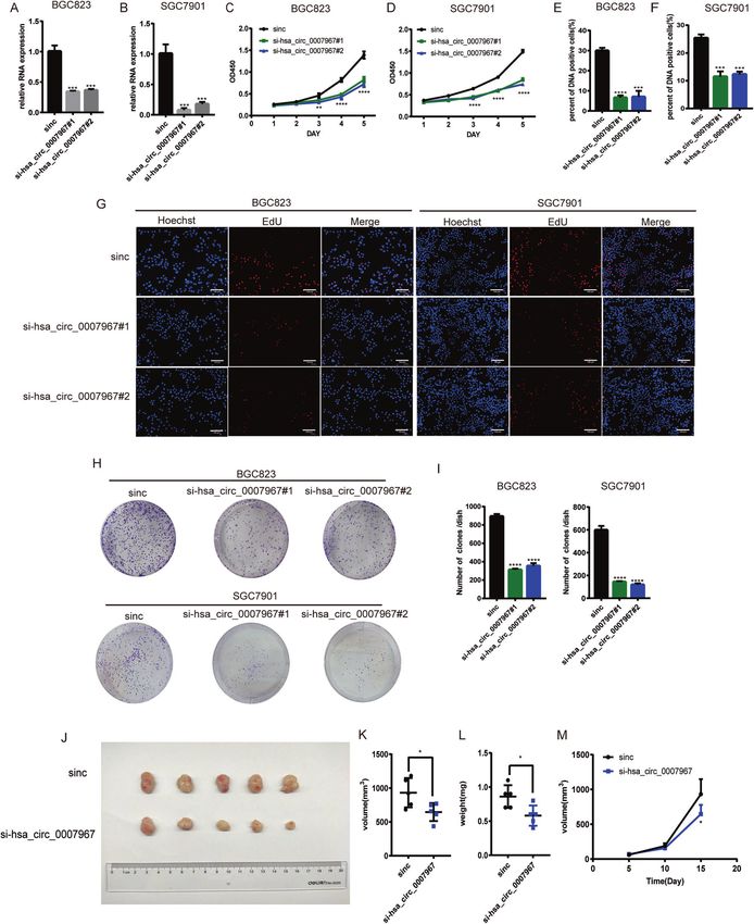
Q. Zha et al.
2

    Fig. 1 Identification and characterization of hsa_circ_0007967. A Expression of five most upregulated and downregulated circRNAs in five
    paired GC tumors and normal tissues. B Expression of hsa_circ_0007967 in 47 pairs of GC tissues and normal tissues. C Expression of
    hsa_circ_0007967 in GC cell lines and normal gastric epithelial cell line. D The location of hsa_circ_0007967 in chromatin and its structure. E
    Sanger sequencing confirming the back splicing junction site. F The RNase R digestion assay suggesting hsa_circ_0007967 is resistant to
    RNase R. G The use of act-D (1 μg/μl) demonstrating that hsa_circ_0007967 is stable than its linear form. H Northern blotting of
    hsa_circ_0007967 and its linear form in cDNA and gDNA amplified by convergent and divergent primers. I Location of hsa_circ_0007967 in
    BGC823 cells detected by FISH. J QPCR analysis of nuclear and cytoplasmic fractions conforming that hsa_circ_0007967 is located in
    cytoplasm.

                                                                                                     Cell Death Discovery (2022)8:144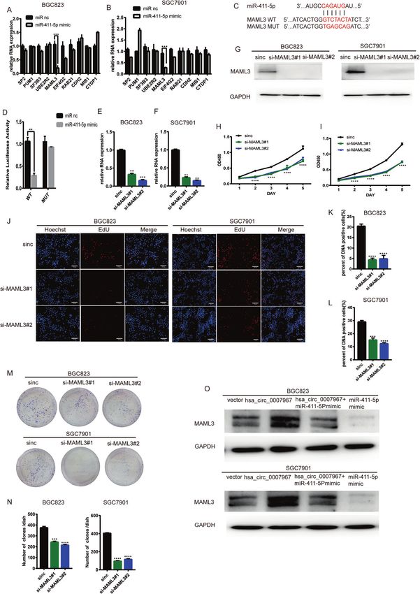
Q. Zha et al.
                                                                                                                                              3

Fig. 2 Overexpressing hsa_circ_0007967 promotes GC proliferation in vitro and in vivo. A, B The expression of hsa_circ_0007967 in
BGC823 and SGC7901 cells transfected with vector and hsa_circ_0007967 plasmids. C, D The growth rate of BGC823 and SGC7901 cells
transfected with vector and hsa_circ-0007967 by cck8 assays. E EdU assays of BGC823 and SGC7901 cells transfected with vector and hsa_circ-
0007967 plasmids. F, G DNA positive cells of BGC823 and SGC7901 cells transfected with vector and hsa_circ-0007967 by EdU assays. H, I The
assessment of proliferation of BGC823 and SGC7901 cells transfected with vector and hsa_circ_0007967 plasmid by colony formation assays.
J–L Tumors from mice in two different groups, and the weight and volume of tumors at the end point. M The weight of tumors measured
every 5 days until the end point.

Cell Death Discovery (2022)8:144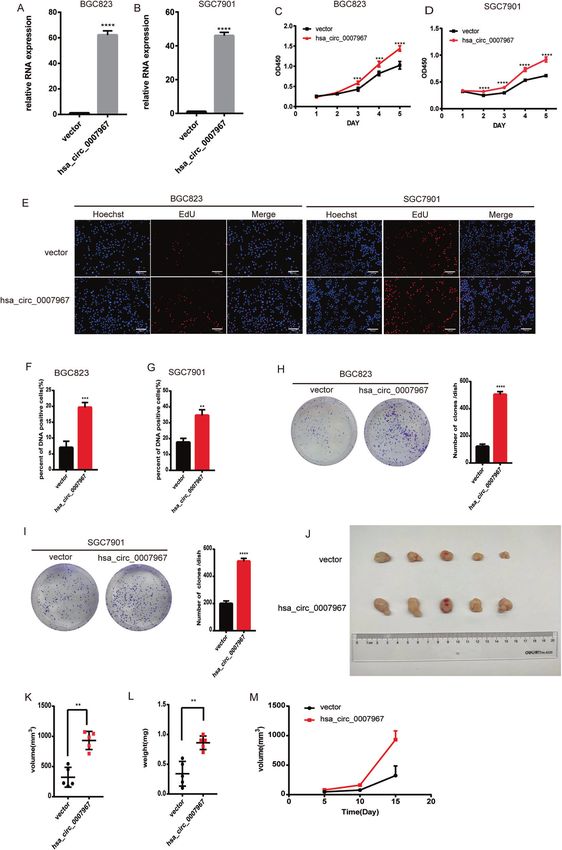
Q. Zha et al.
4

    Fig. 3 Silencing hsa_circ_0007967 inhibits GC proliferation in vitro and in vivo. A, B The expression of hsa_circ_0007967 in BGC823 and
    SGC7901 cells transfected with sinc, si-hsa_circ_0007967#1, and si-hsa_circ_0007967#2. C, D The growth rate of BGC823 and SGC7901 cells
    transfected with sinc, si-hsa_circ-0007967#1, and si-hsa_circ-0007967#2 by cck8 assays. E, F DNA positive cells of BGC823 and SGC7901 cells
    transfected with sinc, si-hsa_circ-0007967#1, and si-hsa_circ-0007967#2 by EdU assays. G EdU assays of BGC823 and SGC7901 cells transfected
    with sinc, si-hsa_circ-0007967#1, and si-hsa_circ-0007967#2. H, I The assessment of proliferation of BGC823 and SGC7901 cells transfected
    with sinc, si-hsa_circ_0007967#1, and si-hsa_circ_0007967#2 by colony formation assays. J–L Tumors from mice in two different groups, and
    the weight and volume of tumors at the end point. M The weight of tumors measured every 5 days until the end point.

                                                                                                  Cell Death Discovery (2022)8:144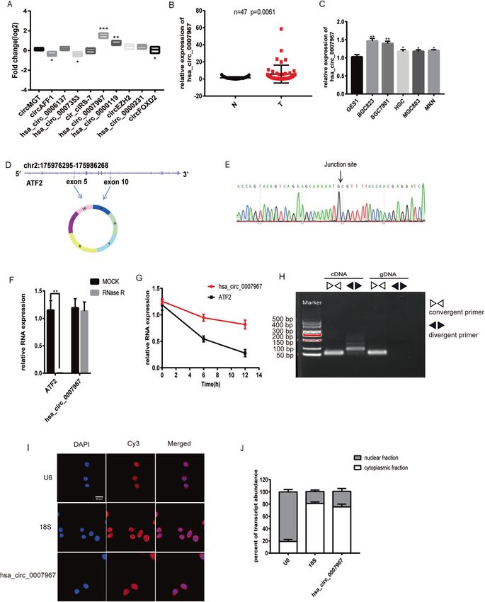
Q. Zha et al.
5
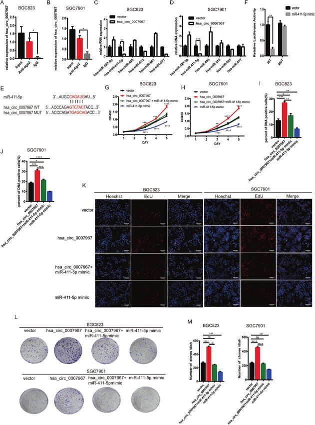
Fig. 4 Hsa_circ_0007967 serves as the sponge of miR-411-5p. A, B Level of hsa_circ_0007967 enriched for Ago2 by RIP assays in BGC823 and
SGC7901 cells. C, D The expression of selected miRNAs in BGC823 and SGC7901 cells after overexpressing hsa_circ_0007967. E The potential
binding site between miR-411-5p and hsa_circ_0007967. F The relative luciferase activity in HEK-293T cells cotransfected with miR-411-5p
mimics and hsa_circ_0007967 plasmids with WT/MUT binding site by Dual luciferase reporter assays. G, H The growth rate of BGC823 and
SGC7901 cells transfected with vector, hsa_circ_0007967 plasmids, miR-411-5p mimics or cotransfected with hsa_circ_0007967 plasmids and
miR-411-5p mimics by cck8 assays. I–K DNA positive cells of BGC823 and SGC7901 cells transfected with vector, hsa_circ_0007967 plasmids,
miR-411-5p mimics or cotransfected with hsa_circ_0007967 plasmids and miR-411-5p mimics by EdU assays. L, M The assessment of
proliferation of BGC823 and SGC7901 cells transfected with vector and hsa_circ_0007967 plasmids by colony formation assays.
Cell Death Discovery (2022)8:144Q. Zha et al.
6
hybridization (FISH) and RT-qPCR assays of nuclear and cytoplas- Hsa_circ_0007967 promotes GC cell proliferation in vitro and
mic fractions confirmed that hsa_circ_0007967 mainly located in in vivo
cytoplasm (Fig. 1I, J). These results suggested that hsa_- To explore whether hsa_circ_0007967 involves in the progression of
circ_0007967 was upregulated in GC tissues, and was a stable GC, we first overexpressed hsa_circ_0007967 in BGC823 and
circRNA in cytoplasm. SGC7901 cells (Fig. 2A, B). Cck8 assays showed that cells with
Cell Death Discovery (2022)8:144Q. Zha et al.
7
Fig. 5 MAML3 is the target gene of miR-411-5p and promotes GC progression. A, B level of selected genes in BGC823 and SGC7901 cells
transfected with miR-411-5p mimics. C The potential binding site between miR-411-5p and MAML3 mRNA. D The relative luciferase activity in
HEK-293T cells cotransfected with miR-411-5p mimics and MAML3 plasmids with WT/MUT binding site by Dual luciferase reporter assays. E, F
The expression of MAML3 in BGC823 and SGC7901 cells transfected with sinc, si-MAML3#1 and si-MAML3#2 by qPCR. G The expression of
MAML3 in BGC823 and SGC7901 cells transfected with sinc, si-MAML3#1 and si-MAML3#2 by WB. H, I The growth rate of BGC823 and
SGC7901 cells transfected with sinc, si-MAML3#1, siMAML3#2 by cck8 assays. J–L DNA positive cells of BGC823 and SGC7901 cells transfected
with sinc, si-MAML3#1, siMAML3#2 by EdU assays. M, N The assessment of proliferation of BGC823 and SGC7901 cells transfected with sinc, si-
MAML3#1 and si-MAML3#2 by colony formation assays. O The protein level of MAML3 of BGC823 and SGC7901 cells transfected with vector,
hsa_circ_0007967 plasmids, miR-411-5p mimics or cotransfected with hsa_circ_0007967 plasmids and miR-411-5p mimics by WB.
hsa_circ_0007967 overexpressed had a faster growth rate (Fig. 2C, of MAML3 may lead to different diseases, like cancers [19]. Then,
D). 5-Ethynyl-2′-deoxyuridine (EdU) assays suggested that the we conducted dual luciferase reporter assays with mutant and
percent of DNA positive cells was higher in hsa_circ_0007967- wild-type MAML3 plasmids (Fig. 5C). Dual Luciferase reporter
overexpressed cells (Fig. 2E–G). Consistently, colony formation assays showed that miR-411-5p mimics reduced the activity of
assays showed that hsa_circ_0007967-overexpressed cells grew into luciferase in the WT group, which suggested that miR-411-5p
bigger clones (Fig. 2H, I). Then, we downregulated the expression of bound to the 3’UTR of MAML3 mRNA (Fig. 5D). To further
hsa_circ_0007967 in BGC823 and SGC7901 with 2 siRNAs (Fig. 3A, B). investigate whether MAML3 was involved in GC proliferation, we
On the contrary, Cck8, EdU, and colony formation assays revealed silenced MAML3 in both BCG823 and SGC7901 cells, which was
that silencing hsa_circ_0007967 could inhibit GC cell proliferation confirmed in mRNA and protein level (Fig. 5E–G). Cck8, EdU, and
(Fig. 3C–I). To further investigate whether hsa_circ_0007967 could colony formation assays suggested that silencing MAML3
accelerate the proliferation of GC cell in vivo, twenty BALB/c-nude inhibited GC proliferation (Fig. 5H–N). What’s more, WB assays
mice were divided randomly into 4 groups and mice in different suggested that overexpressing hsa_circ_0007967 could cause the
groups were subcutaneously injected with differently treated high expression of MAML3 and miR-411-5p mimics downregu-
BGC823 cells (hsa_circ_0007967-overexpressed, hsa_circ_0007967- lated the expression of MAML3. While miR-411-5p mimics
silenced, and the correspond control ones). Mice that were injected suppressed the upregulation of MAML3 caused by hsa_-
with hsa_circ_0007967-overexpressed BGC823 cells bore bigger and circ_0007967 overexpression (Fig. 5O). Overall, hsa_circ_0007967
heavier tumors than those in the control group (Fig. 2J–M). And the promoted GC proliferation through miR-411-5p/MAML3 axis.
weight and volume of tumors in the mice that were injected with
hsa_circ_0007967-downregulated BGC823 cells were significantly
lighter and smaller than those in the control group (Fig. 3J–M). DISCUSSION
These data suggested that hsa_circ_0007967 facilitated GC cells In recent years, emerging studies suggested that circRNAs play an
proliferation in vitro and in vivo. important role in cancerogenesis. Here, we identified the
oncogenic circRNA, hsa_circ_0007967, through RNA-seq, which
Hsa_circ_0007967 serves as the sponge of miR-411-5p was highly expressed in GC tissues than in matched normal
As hsa_circ_0007967 is mainly located in the cytoplasm, we tissues. It was back spliced from ATF2 gene and mainly located in
speculated that hsa_circ_0007967 may act as miRNA sponges. We cytoplasm. Cck8, EdU, and colony formation assays demonstrated
performed RNA-protein immunoprecipitation (RIP) assays and that overexpressing hsa_circ_0007967 promoted GC proliferation
found that more hsa_circ_0007967 was enriched with anti-AGO2 in vitro. Consistently, overexpressing hsa_circ_0007967 promoted
antibody than IgG, which suggested that hsa_circ_0007967 bound subcutaneously injected GC cells proliferation. Hsa_circ_0007967
well to miRNAs (Fig. 4A, B). Then we searched CircInteractome was the sponge of miR-411-5p and finally upregulated MAML3
(circinteractome.nia.nih.gov) and chose six potential miRNAs with expression. Overexpressing miR-411-5p suppressed GC prolifera-
highest score (miR-127-5p, miR-411-5p, miR-495, miR-611, miR- tion and weakened the oncogenic effect of hsa_circ_0007967.
513, and miR-877). Overexpressing hsa_circ_0007967 only MAML3 is an important transcriptional co-activator in the Notch
caused a significant downregulation of miR-411-5p in both signaling pathway and silencing MAML3 suppressed GC prolifera-
BGC823 and SGC7901 cells, suggesting that miR-411-5p may be tion. Overexpressing hsa_circ_0007967 caused the upregulation of
the downstream molecule of hsa_circ_0007967 (Fig. 4C, D). We MAML3, which was resecured by overexpressing miR-411-5p. It
predicted the binding site between hsa_circ_0007967 and miR- was suggested that hsa_circ_0007967/miR-411-5p/MAML3 axis
411-5p (Fig. 4E). Dual luciferase reporter assays showed that involved in GC progression. Consequently, hsa_circ_0007967 is a
miR-411-5p mimics only caused a significant reduction of the promising biomarker for GC diagnosis and prognosis, and is a
relative luciferase activity in hsa_circ_0007967-WT group (Fig. potential therapeutic target for GC treatment.
4F). To further study whether hsa_circ_0007967 promoted GC Though our study demonstrated that hsa_circ_0007967 played an
proliferation by regulating miR-411-5p, we performed rescue oncogenic role in GC, we only investigated that hsa_circ_0007967
experiments. Cck8, EdU, and colony formulation assays showed functioned as an miRNA sponge and just focused on the malignant
that miR-411-5p mimics inhibited GC cell proliferation, and activity of proliferation without further exploration. What’s more, we
suppressed GC cell proliferation caused by hsa_circ_0007967 only evaluated the expression of circRNAs between tumors and
overexpression (Fig. 4G–M). adjacent non-malignant tissues. It would be better to conduct an
appropriate control cohort that reflected the intended use of the
MAML3 is the target gene of miR-411-5p and promotes GC biomarker. We believed that, with the development of technology,
progression circRNA expression will oneday be detected at the single-cell level
MiRNAs generally bind to the 3′ untranslated region of mRNAs and with spatial resolution, which will be essential for better
and lead to mRNA degradation [18]. After searching three understanding circRNA functions in the future.
websites (TargetScan, TargetMiner, and miRDB), we found 12
mutual protein candidates that miR-411-5p targets. While miR-
411-5p mimics only caused MAML3 downregulate in both BGC823 MATERIALS AND METHODS
and SGC7901 cells (Fig. 5A, B). MAML3 is encoded by Mastermind Patient samples
like (MAML) family genes and is an important transcriptional co- A total of 47 pairs of GC tissues and adjacent normal tissues were collected
activator in the Notch signaling pathway. The aberrant expression from the hospital of Zhenjiang according to institutional protocols and this
Cell Death Discovery (2022)8:144Q. Zha et al.
8
study was approved by the Medical Ethics Committee of First Affiliated Cell counting kit-8 assay
Hospital of Nanjing Medical University. Informed consent form was signed After 48 h of transfection, cells (2 × 10³/well) were seeded into 96-well
by every patient. plates (Corning,USA). Then 100 μl of 10% Cell counting kit-8 (CCK8;
Beyotime, China) solution was added to each well at appointed time (8 h,
24 h, 48 h, 72 h, 96 h). After 2 h of incubation at 37 °C, the absorbance at
Cell culture 450 nm was measured with a microplate reader (Pro-11 multiskan FC,
The HEK-293T, MGC803, BGC823, HGC27, SGC7901, AGS, and GES1 cell Thermo Fisher, USA).
lines were purchased from Type Culture Collection of the Chinese
Academy of Sciences (Shanghai, China). The HEK-293T, MGC803, HGC27,
BGC823, SGC7901, and GES1 cells were cultured in RPMI 1640 medium Colony formation assay
(Gibco, USA). The AGS cells were cultured in F12k medium (Wisent, After 48 h of transfection, cells (1 × 10³/well) were seeded into six-well
Canada). All the cell lines were cultured in a 37 °C, 5% CO2 incubator plates (Corning). After incubation for 10 days at 37 °C, cells were fixed with
(Thermo Fisher, USA), and were provided with 100 μg/ml streptomycin methyl alcohol and stained with crystal violet solution.
(Gibco), 100 U/ml penicillin (Gibco), and 10% fetal bovine serum (BI, Iseral).
5-Ethynyl-2′-deoxyuridine incorporation assay
RNA extraction and quantitative real-time polymerase chain EdU assays were performed with the Cell-Light EdU DNA Cell Proliferation
reaction Kit (RiboBio) according to the manufacturer’s instruction. Images were
Total RNA was extracted from the cells or tissues using TRIzol reagent obtained with a Nikon Ti microscope (Nikon, Tokyo, Japan), and the
(Ambion, USA). The nuclear and cytoplasmic RNAs were extracted with number of EdU positive cells was counted.
PARIS™ Kit (Thermo Fisher, USA). Isolated RNAs were reversely transcribed
into cDNAs with HiScript Q RT SuperMix for qPCR (Vazyme, China). RT-qPCR
Western blotting
assays were carried out with SYBR Green PCR Master Mix (Vazyme, China)
Cells were lysed in RIPA buffer (Beyotime, China) with 1% PMSF (Biosharp,
on the Applied Biosystems steponeplus (USA) Real Time PCR system. China). The protein was separated by sodium dodecyl sulfate-
GAPDH and U6 were used as internal controls, and expressions of all polyacrylamide gel electrophoresis (Epizyme, China) and transferred onto
samples were normalized to GAPDH and U6. The primers are shown in PVDF membrane (Millipore, USA). Primary antibodies were applied at 4 °C
Table 1. overnight and HRP-conjugated secondary antibodies were applied for an
hour at room temperature. The immunocomplexes were detected with ECL
SiRNA and plasmid transfection Western Blotting Substrate (NCM Biotech, China), visualized with Tanon
The linear form of hsa_circ_0007967 was inserted into plasmid pcDNA3.1- (5200multi 4600SF, Tanon, USA). GAPDH was used as the internal control.
CMV by Hanbio Biotechnology (Shanghai, China). SiRNAs targeting Primary Antibodies included rabbit antiMAML3 (1:500, Biorbyt, UK), mouse
hsa_circ_0007967 were purchased from RiboBio (Guangzhou, China). antiGAPDH (1:20000, Beyotime, China). Secondary antibodies (A0208 and
SiRNAs targeting MAML3 were purchased from GenePharma (Shanghai, A0216, Beyotime, China) were diluted in 1:1000.
China). The miRNA mimics or inhibitors were purchased from GenePharma.
The plasmids, siRNAs, miRNA mimics, and inhibitors were transfected into Northern blotting
cells with Lipofectamine 3000 (Life Technologies, USA).
DNA was separated using 1% agarose gel electrophoresis for 20 min under
110 v and was detected by BIO-RAD (BIO-RAD Gel Doc XR+, USA)
RNase R treatment
In all, 5 μg of total RNA was incubated for 15 min at 37 °C with or without RNA stability assay
4 U/μg of RNase R (Epicentre Technologies, USA) in 1× reaction buffer, and Cells were seeded into 6-well plates for 12 h incubation and grew to 50%
was then reversely transcribed into cDNA. confluence. Then cells were treated with 1 μg/ml actinomycin D and total
RNAs were collected at 0, 6, 12 h. RNA levels were detected using RT-qPCR,
RNA fluorescence in situ hybridization and the halflife of cirRNAs and mRNAs was evaluated.
FISH assays were conducted with RiboTM Fluorescence In Situ Hybridization
Kit (RiboBio) under the manufacturer’s instruction. Cy3-labeled probes
Dual luciferase reporter assay
targeting hsa_circ_0007967, U6, 18S were purchased from RiboBio. Cells The wild-type sequence of hsa_circ_0007967 and the 3′UTR of MAML3
were seeded into eight-well plate and incubated for 12 h before fixation. which containing predicted binding site of miR-411-5p were subcloned
After 30 min’ fixation, and 10 min’ permeabilization (0.5% Triton X-100), into the luciferase reporter vector GV272 (GenePharma, China). The
cells were prehybridized in prehybridization buffer at 37 °C for half an hour. corresponding ones containing mutant predicted binding site of miR-411-
Then cells were hybridized in hybridization buffer with specific probes at
5p were subcloned into the luciferase reporter vector GV272 (Gene-
37 °C overnight in the dark. 4×SSC (including 0.1% Tween-20), 2×SSC and
Pharma). HEK-293T cells were seeded in 24-well plate (6 × 10⁴ cells/well)
1×SSC were used for washing off hybridization buffer at 42 °C in the dark. for 24 h before transfection. Cells were co-transfected with a mixture of
Confocal images were captured by Zeiss LSM5 confocal microscope (Carl luciferase reporter vectors containing wild-type sequence or mutant
Zeiss Jena, Oberkochen, Germany). sequence along with miRNA mimics. After 24 h incubation, the luciferase
activity was measured with a specific microplate reader (Synergy H1, USA).
Dual-Luciferase®Reporter (DLR™) Assay System was used according to the
manufacturer’s instructions.
Table 1. Primers used in this study.
Names Sequences (5′-3′) RNA-protein immunoprecipitation
The MagnaRIP RNA-Binding Protein Immunoprecipitation Kit (Merk, USA)
Hsa_circ_0007967: forward CCCTGTACCAGGCCCATTTC was employed according to the manufacturer’s instructions. The cell lysate
Hsa_circ_0007967: reverse TGGGACTGCAGCTGGAACA was incubated with beads coated with 5 μg of antibody against
Argonaute-2 (AGO2) (Abcam, USA), and control IgG with rotation at 4 °C
MAML3: forward CCTACCAGCCAACCAGGAATGTA overnight. Total RNA was extracted for the evaluation of circRNA
MAML3: reverse ATGCTCTGACCAAAGCCACTCAC expression by RT-qPCR.
miR-411-5p: forward GGCCGGCTAGTAGACCGTATAG
miR-411-5p: reverse ACTGCAGGGTCCGAGGTATT Animal studies
All animal experiments were approved by the Institutional Animal Care and
GAPDH: forward GGGAGCCAAAAGGGTCAT Use Committee of Nanjing Medical University. Twenty BALB/c-nude mice
GAPDH: reverse GAGTCCTTCCACGATACCAA (female, 4-week-old) were divided randomly into 4 groups with online tool
U6: forward CTCGCTTCGGCAGCACA (Each was given a random number) and mice in different groups were
injected with differently treated BGC823 cells (hsa_circ_0007967-over-
U6: reverse AACGCTTCACGAATTTGCGT expressed, hsa_circ_0007967-silenced and the correspond control ones).
Cell Death Discovery (2022)8:144Q. Zha et al.
9
Cells (5 × 10⁶) were injected into the left back subcutaneously. The body 19. McElhinny AS, Li JL, Wu L. Mastermind-like transcriptional co-activators: emerging
weight and tumor volume (volume = length × width2/2) were measured roles in regulating cross talk among multiple signaling pathways. Oncogene.
every 5 days after injection until mice were killed. At the end of 2008;27:5138–47.
experiments, the mice were killed, and the tumors were dissected and
weighed.
ACKNOWLEDGEMENTS
Statistical analysis We would like to thank the Core Facility of the First Affiliated Hospital of Nanjing
GraphPad Prism software was used for statistical analysis. The data were Medical University for its help in the detection of experimental samples.
presented as the mean ± standard deviation. Student’s t test was used for
the determination of the statistical signifcance. A p value thatYou can also read


