Goose Nephritic Astrovirus Infection of Goslings Induces Lymphocyte Apoptosis, Reticular Fiber Destruction, and CD8 T-Cell Depletion in Spleen ...
←
→
Page content transcription
If your browser does not render page correctly, please read the page content below
viruses
Article
Goose Nephritic Astrovirus Infection of Goslings Induces
Lymphocyte Apoptosis, Reticular Fiber Destruction, and CD8
T-Cell Depletion in Spleen Tissue
Rui Ding † , Han Huang † , Hongyu Wang, Zewen Yi, Siyu Qiu, Yingjun Lv * and Endong Bao

                                             MOE Joint International Research Laboratory of Animal Health and Food Safety, College of Veterinary Medicine,
                                             Nanjing Agricultural University, Nanjing 210095, China; 2019107013@njau.edu.cn (R.D.);
                                             2019807138@stu.njau.edu.cn (H.H.); wanghongyuwhy92@163.com (H.W.); 2020807153@stu.njau.esu.cn (Z.Y.);
                                             2018107009@njau.edu.cn (S.Q.); b_endong@njau.edu.cn (E.B.)
                                             * Correspondence: lyj@njau.edu.cn
                                             † These authors contributed equally to this work.

                                             Abstract: The emergence of a novel goose nephritic astrovirus (GNAstV) has caused economic losses
                                             to the Chinese goose industry. High viral load is found in the spleen of goslings infected with GNAstV,
                                             but pathological injuries to the spleen due to GNAstV are largely unknown. In this study, 50 two-
                                             day-old goslings were infected orally with GNAstV, and 50 goslings were treated with PBS as control.
                                             Spleens were collected at different times following infection to assess damage. GNAstV infection
                                   caused visceral gout and urate deposition in joints, and resulted in 16% mortality. GNAstV was found

                                             in the lymphocytes and macrophages within the spleen. Lymphocyte loss, especially around the
Citation: Ding, R.; Huang, H.; Wang,         white pulp, and destruction and decline in the number of reticular fibers was observed in GNAstV-
H.; Yi, Z.; Qiu, S.; Lv, Y.; Bao, E. Goose   infected goslings. Moreover, in GNAstV-infected goslings, ultrahistopathological examination found
Nephritic Astrovirus Infection of            that splenic lymphocytes exhibited condensed chromatin and apoptotic bodies, and reticular cells
Goslings Induces Lymphocyte                  displayed damage to plasma membrane integrity and swollen mitochondria. Furthermore, TUNEL
Apoptosis, Reticular Fiber
                                             staining confirmed apoptosis of lymphocytes, and the mRNA levels of Fas and FasL were significantly
Destruction, and CD8 T-Cell
                                             increased in the GNAstV-infected goslings. In addition, GNAstV infection reduced the number and
Depletion in Spleen Tissue. Viruses
                                             protein expression of CD8. In conclusion, GNAstV infection causes lymphocyte depletion, reticular
2021, 13, 1108. https://doi.org/
                                             cell necrosis, reticular fiber destruction, lymphocyte apoptosis, and reduction in CD8 levels, which
10.3390/v13061108
                                             contribute to spleen injury.
Academic Editors: Chao-Nan Lin and
Peck Toung Ooi                               Keywords: goose nephritic astrovirus; spleen; pathological changes; apoptosis; CD8

Received: 29 April 2021
Accepted: 5 June 2021
Published: 9 June 2021                       1. Introduction
                                                   GNAstV is a novel pathogen that was first isolated in China in 2018 [1]. GNAstV
Publisher’s Note: MDPI stays neutral         is a small, non-enveloped, single-stranded RNA virus. GNAstV infection in three- to
with regard to jurisdictional claims in      fifteen-day-old goslings causes visceral gout and results in 2–20% mortality. In addition,
published maps and institutional affil-
                                             GNAstV infection has been observed in Cherry Valley ducklings in the clinic, and has also
iations.
                                             been shown to infect chickens in experimental studies [2–4]. At present, most studies have
                                             focused on the isolation and identification of GNAstV, animal regression, and establishing
                                             methods for detecting the virus [5–7]; however, little is known about the pathogenic
                                             mechanisms of GNAstV infection.
Copyright: © 2021 by the authors.                  Goslings that are infected with GNAstV display swelling and urate deposition in
Licensee MDPI, Basel, Switzerland.           kidney tissue, as well as liver damage. Both organs’ lesions are easy to understand because
This article is an open access article       the liver is responsible for the production of uric acid, and the kidney is the main excretory
distributed under the terms and
                                             organ. Additionally, a high viral load and necrosis of splenic lymphocytes was also
conditions of the Creative Commons
                                             observed in infected animals [8,9], indicating that GNAstV may cause damage to the
Attribution (CC BY) license (https://
                                             spleen tissue and induce immunosuppression. Moreover, recent studies show that GNAstV
creativecommons.org/licenses/by/
                                             infection increases pro-inflammatory cytokines, such as IL-1β and IL-8, and decreases
4.0/).

Viruses 2021, 13, 1108. https://doi.org/10.3390/v13061108                                                       https://www.mdpi.com/journal/viruses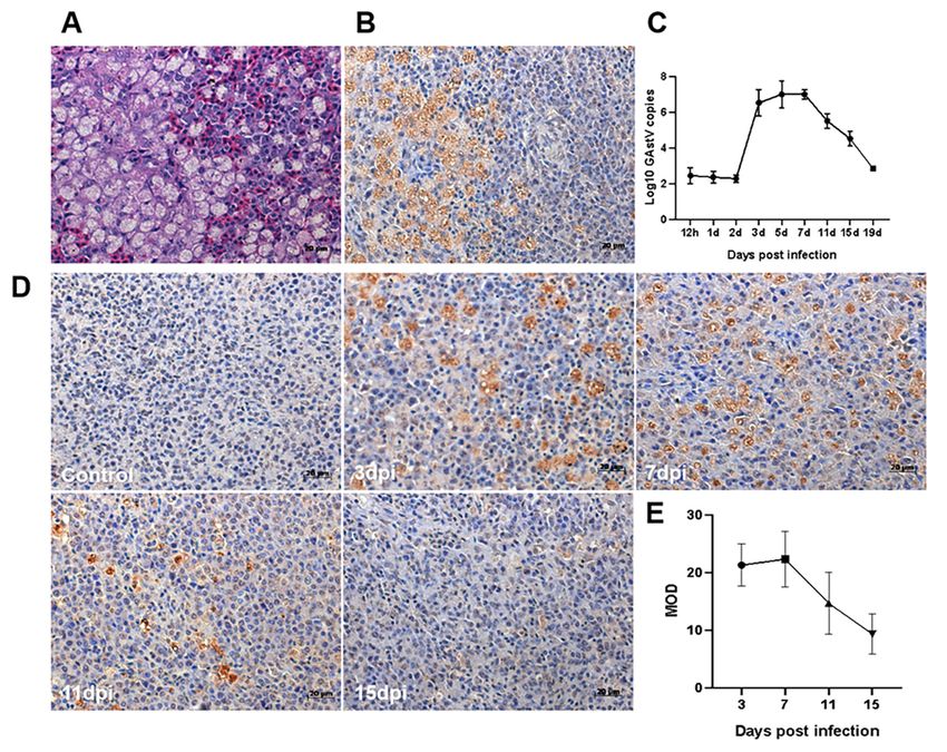
Viruses 2021, 13, 1108                                                                                            2 of 13

                         MHC-II levels in the spleen at the early stages of infection [10,11]. In addition, a case of
                         co-infection with the astrovirus and parvovirus in goslings was also reported [12], although
                         it is not known whether GNAstV contributes to the transmission or spread of the goose
                         parvovirus. Although there are sporadic reports of spleen damage in goslings infected
                         with GNAstV, the specifics of these injuries need to be further investigated. In the present
                         study, two-day-old goslings were experimentally infected with GNAstV, and changes in
                         gross morphology, histopathology, ultrahistopathology, apoptosis, autophagy, and levels
                         of CD4 and CD8 expression in spleen tissue were investigated.

                         2. Materials and Methods
                         2.1. Virues
                              The GNAstV-JSHA isolate (GenBank accession no.MK125058) was isolated and kept
                         in our laboratory, and has been shown to cause visceral gout [13]. The titer of the virus was
                         1 × 104.25 50% tissue culture infective dose (TCID50)/mL, as determined by titration on
                         goose kidney epithelial cells according to the method of Reed and Muench.

                         2.2. Animal Experiment
                               Before the animal experiment, five two-day-old goslings (Yangzhou white goose) were
                         euthanized with intravenous pentobarbital sodium. No gross changes were found in all
                         organs, and no GNAstV RNA was detected from kidneys, spleens and livers using the
                         RT-PCR method. Then, 100 one-day-old goslings were purchased from the same flock and
                         randomly assigned to two groups, namely, the infection group and control group, each with
                         50 goslings per group. These goslings were reared separately in negative-pressure animal
                         isolators without any immunizations and provided with sterilized water and feed without
                         antibiotics ad libitum. The goslings in the infection group were challenged with 0.5 mL
                         (0.5 × 104.25 TCID50/goose) GNAstV by oral infection. The control group was fed with
                         0.5 mL of PBS. All geese were monitored daily for the occurrence of clinical signs for 19 days.
                         During the experiment, five geese from each group were weighed, blood-collected and
                         euthanized with intravenous pentobarbital sodium at 12 h, 1, 2, 3, 5, 7, 11, 15 and 19 days
                         post-infection (dpi). The spleen was collected and weighted, and the relative weights of
                         the spleen were calculated as a proportion of each gosling’s body weight. Then, a portion
                         of the spleen was fixed in 4% paraformaldehyde for histopathological examination. Part of
                         the spleen was fixed in 2.5% glutaraldehyde for ultrahistopathological examination. The
                         rest were stored at −80 ◦ C for further experiments.

                         2.3. Histopathological Examination
                              Hematoxylin and eosin staining was performed according to previous studies with
                         minor revision [14]. Briefly, the spleen samples were fixed in 4% paraformaldehyde and
                         dehydrated by a series of alcohols, clarified in xylene, and embedded in paraffin. Then,
                         samples were sliced serially into 4 µm sections and stained with hematoxylin and eosin
                         by routine methods. Stained sections were examined with a light microscope (Carl Zeiss,
                         Gottingen, Germany).

                         2.4. Silver Staining
                             Silver staining was performed according to the instruction of a commercial Improved
                         Godon and Sweet kit (Yifeixue Bio Tech, Nanjing, China). Briefly, after dewaxing to
                         water, the spleens’ paraffin sections were oxidized by the Gordon and Sweet oxidant for
                         4 min, bleached by oxalic acid for 3 min, mordanted by ferric ammonium alum for 14 min,
                         immersed by silver ammonia by 10 min, reduced by Gordon and Sweet reductant for 4 min,
                         and counterstained by kernechtrot for 20 min.
Viruses 2021, 13, 1108                                                                                          3 of 13

                         2.5. Immunohistochemical (IHC) Examination
                               IHC staining was performed according to the conventional protocol with some mod-
                         ifications [15]. After deparaffinization and rehydration, the spleen tissue slides were
                         inactivated with 3% hydrogen peroxide for 10 min, and then exposed to citric acid buffer at
                         100 ◦ C for 8 min. The sections were blocked with 5% bovine serum albumin for 30 min and
                         incubated with mouse anti-GNAstV Capsid protein monoclonal antibody (gift from Prof.
                         Zongyan Chen, Shanghai Veterinary Research Institute, China), rabbit anti-CD4 polyclonal
                         antibody (Beyotime, Shanghai, China), and mouse anti-CD8 monoclonal antibody (gift
                         from Prof. Bo Ma, Northeast Agricultural University, China) at 4 ◦ C overnight. After
                         washing with PBS and incubation with secondary antibodies for 1 h at 37 ◦ C, the samples
                         were visualized using diaminobenzidine staining and counterstained with hematoxylin.
                         Stained sections were examined with a light microscope. The software Image-Pro Plus
                         (Version 6.0.0.260) was used to conduct semi-quantitative statistics of the positive signals.

                         2.6. Ultrahistopathological Examination
                              The spleens were trimmed into 1 mm3 blocks, and then were dehydrated in ethyl
                         alcohol, infiltrated with a propylene oxide-Araldite mixture, and embedded in Araldite.
                         Then the ultrathin section was stained with uranyl acetate and lead citrate for 15 min.
                         The samples were visualized under a HT7700 transmission electron microscope (Hitachi,
                         Tokyo, Japan).

                         2.7. Virus Determination
                              Viral RNA was extracted from serum and spleen samples using the GeneJET Viral
                         DNA/RNA Purification Kit (Thermo Scientific, Rockford, IL, USA) and transcribed into
                         cDNA using the HiScript Q RT SuperMix Kit (Vazyme Biotech Co., Ltd., Nanjing, China).
                         Then the viral loads were measured using a SYBR Green I-based real-time PCR method
                         established in our lab [16].

                         2.8. Terminal Deoxynucleotidyl Tranferase-Mediated Nick-End Labeling (TUNEL) Staining
                              TUNEL staining was performed according to our previous study [17]. The tissue
                         sections were deparaffinated in xylene and rehydrated in descending concentrations of
                         ethanol, followed by antigen retrieval in sodium citrate buffer for 10 min at room tem-
                         perature and in a microwave oven at 100 ◦ C for 15 min. Endogenous peroxidase was
                         inhibited by incubation with 3% hydrogen peroxide for 15 min at room temperature. Then,
                         tissue sections were stained by TUNEL using the ApopTag kit (Roche, Indianapolis, IN,
                         USA) according to the manufacturer’s protocol. The sections were then stained with hema-
                         toxylin. The apoptotic index was calculated as the percentage of TUNEL-positive cells in
                         spleen cells.

                         2.9. Quantitative Real-Time PCR (qPCR) Analysis
                              Total RNA was extracted using Trizol (Invitrogen, Carlsbad, CA, USA) and reverse
                         transcription of the RNA was conducted using the HiScript Q RT SuperMix Kit (Vazyme
                         Biotech Co., Ltd., Nanjing, China). A Thermocycler (Bio-Rad, Hercules, CA, USA) was
                         used for quantitative PCR. Primer 5.0 software was used for the designation of the related
                         genes, as shown in Table 1. RNA expression was normalized by quantification of GAPDH
                         as a housekeeping gene. Each sample was prepared in three duplicate tubes. Specific gene
                         expression was quantified using the 2−∆∆CT method.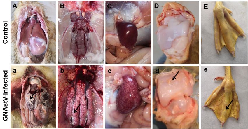
Viruses 2021, 13, 1108                                                                                                      4 of 13

                                            Table 1. Primers used in the study for real-time PCR.

    Primers              Accession Number                Primer Sequences (5 ’-3 ’)                 Host    Product Length (bp)
   Beclin1-F                                      CGCTGTGCCAGATGTGGAAGG
                          XM_013199763.1                                                            Anser         151bp
   Beclin1-R                                   CAGAAGGAATACTGCGAGTTCAAGA[18].
    ATG5-F                                            CCGATTGGTTTGCTCTT
                          XM_013175657.1                                                            Anser         226bp
    ATG5-R                                           ATCCCATCCACAGTTGC
     Bax-F                                        GGACGAGCTGGACAGCAACG
                            KY788660                                                                Anas          159bp
     Bax-R                                         AGGCGGCAGGCGAAGTAGA
    Bcl-2-F                                        TGACCGAGTACCTGAACCG
                          XM_013187395.1                                                            Anser         154bp
    Bcl-2-R                                         GCTCCCACCAGAACCAAA
    cyt C-F                                           AAATGCTCCCAGTGCC
                          XM_013171877.1                                                            Anser         159bp
    cyt C-R                                         CATCAGAGTATCCTCACCC
     Fas-F                                           CACTCCCACAAGTCAAG
                          XM_013171650.1                                                            Anser         163bp
     Fas-R                                           AGTAGGGTTCCATAGGC
    FasL-F                                         ATGGAAGATCACGCAAAGC
                          XM_01319016.1                                                             Anser         152bp
    FasL-R                                          GGTGGGAAGGGAGCAAT

                                    2.10. Western Blotting
                                           The spleen samples were lysed in 500 µL RIPA lysis buffer supplemented with 1%
                                    Phenylmethanesulfonyl fluoride. Following centrifugation at 13,000× g for 15 min at 4 ◦ C,
                                    the concentration of the total protein was quantified using a BCA assay kit (Thermo Scien-
                                    tific, Rockford, IL, USA). The lysates were mixed with 5 × SDS loading buffer and heated
                                    at 99 ◦ C for 10 min. Samples were then analyzed by SDS-PAGE essentially, as previous
                                    described [19]. The rabbit monoclonal anti-GAPDH antibody (Abcam, Cambridgeshire,
                                    Britain), rabbit polyclonal anti-LC3B antibody (Abcam, Cambridgeshire, Britain), rabbit
                                    anti-Beclin1 polyclonal antibody (Beyotime, Shanghai, China), rabbit anti-CD4 polyclonal
                                    antibody (Beyotime, Shanghai, China) and mouse anti-CD8 monoclonal antibody were
                                    used as primary antibodies, and HRP donkey anti-mouse IgG or HRP donkey anti-rabbit
                                    IgG (ABclonal, Wuhan, China) were served as the secondary antibody in the study. Protein
                                    bands in the membrane were detected using a ChemiDoc Touch Imaging System (Bio-Rad,
                                    Hercules, CA, USA) after incubation of the membranes with Clarity Western ECL Blot-
                                    ting Substrate. Then, protein bands were analyzed by software Image-Pro Plus (Version
                                    6.0.0.260), and protein levels were normalized to the amount of GAPDH.

                                    2.11. Statistical Analysis
                                         The differences between the control group and experimental group were analyzed by
                                    Student’s t-test. The results are expressed as the mean ± standard deviation (SD). p < 0.05
                                    was considered to indicate a statistical significance compared with the control group,
                                    and p < 0.01 was considered to indicate a high degree of significance compared with the
                                    control group.

                                    3. Results
                                    3.1. Clinical Changes
                                         The goslings were in good health and no dead ones were found in the control group.
                                    However, the goslings in the infected group showed a reduction in body weight from 5 dpi
                                    to the end of the experiment, and eight goslings died in total (1, 1, 2, 2, 1 and 1 gosling
                                    died at 3, 4, 5, 7, 8 and 10 dpi, respectively). The mortality was 16%. Urate disposition
                                    on the surface of the liver and heart and in the ureter and articular cavity, swelling and
                                    pale kidneys, and enlargement and necrosis of spleens at autopsy were observed in dead
                                    goslings (Figure 1a–e), while no obvious pathological changes were found in the control
                                    group (Figure 1A–E). For most surviving goslings, only pathological changes in the kidney
                                    and spleen were observed, while no obvious urate disposition was found.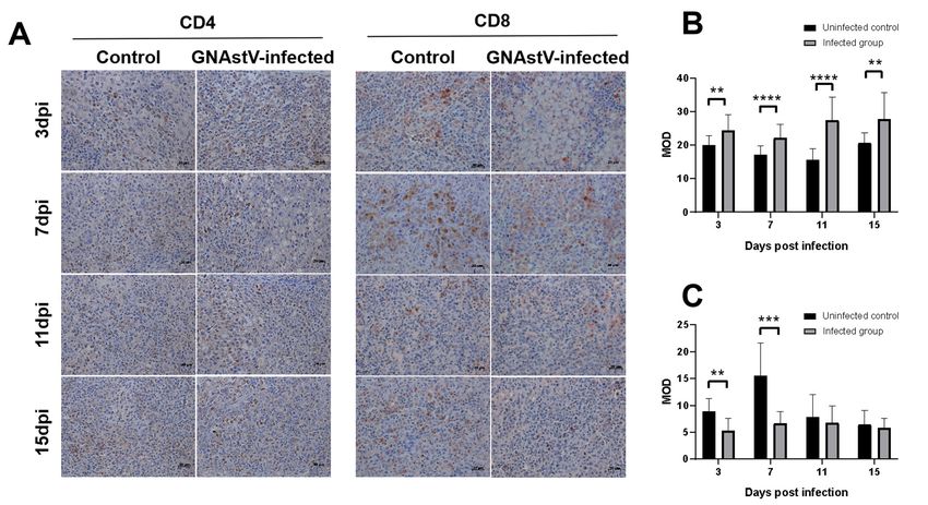
Viruses 2021, 13, 1108                                                                                                   5 of 13

                         Figure 1. Gross anatomical changes in goslings associated with GNAstV-mediated mortality. No
                         changes were observed in the liver (A), heart (A), kidney (B), spleen (C), articular cavity (D) and
                         claw (E) in the control goslings. Urate deposition on the surface of the liver and heart (a), and in
                         the ureter (b), articular cavity (d) and claw (e), swelling and pale coloration of the kidneys (b), and
                         enlargement and necrosis of spleens (c) were observed in the infected goslings. Black arrow: urate.

                         3.2. Gross Morphological Changes in Spleen Tissue
                              Enlargement of the spleen and diffuse multifocal whitish necrotic spots in severely
                         damaged spleens were observed in the infected group at 3 to 7 dpi compared to the
                         control group (Figures 1c and 2A). No obvious pathological changes were found at 11 and
                         15 dpi. The relative weight of the spleen at 3 and 7 dpi was significantly higher in the
                         GNAstV-infected goslings than in the negative control group (Figure 2B).

                         Figure 2. Infection with GNAstV induces gross morphological changes and increase in spleen weight
                         in infected goslings. The spleens of the infected goslings were larger than in the control at 5 dpi
                         (A). The relative weight of spleens was measured at 3, 7, 11, and 15 dpi (B). Values are expressed as
                         mean ± SD, n = 5. * p < 0.05; ** p < 0.01.

                         3.3. Histopathological Changes in the Spleen
                              On histopathological examination, the spleens from the control goslings appeared
                         histologically normal. However, necrosis of splenic lymphocytes and a starry sky pattern
                         were found in the spleens of the infected goslings, especially at the area between the white
                         pulp and red pulp. Serious pathological damage was found at 3 and 7 dpi; this damage
                         became alleviated after 7 dpi (Figure 3). Silver staining showed that the reticular fibers
                         in the infected group were broken (Figure 4A), and the number of reticular fibers in the
                         infected group was significantly lower than in the control at 3, 7, and 11 days (Figure 4B).
Viruses 2021, 13, 1108 6 of 13
Figure 3. Histopathological changes in the spleen of goslings infected with GNAstV. Scale bar: 50 µm.
Figure 4. Changes in fibers associated with GNAstV infection. Spleen tissue was stained with the
Gordon and Sweet method. The reticular fibers are indicated by black signals (A). The mean optical
density (MOD) of reticular fibers was calculated by Image-Pro Plus (B). Scale bar: 50 µm. Values are
expressed as mean ± SD, n = 5. * p < 0.05; ** p < 0.01.
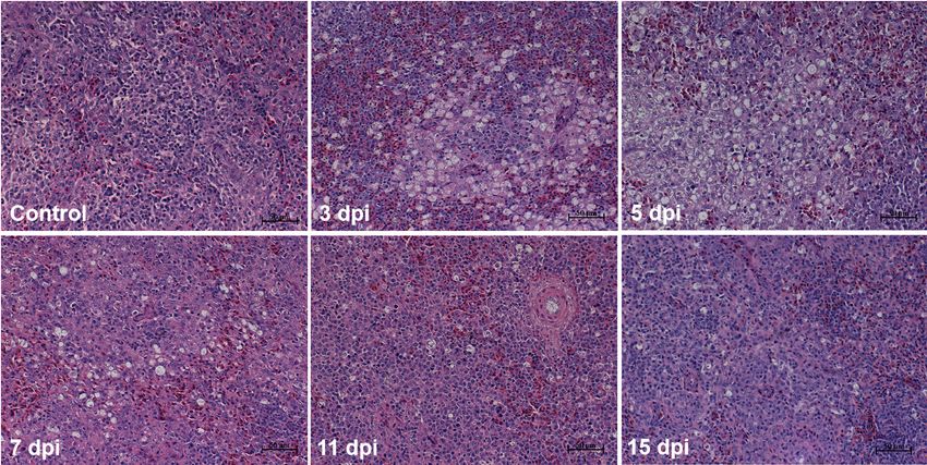
3.4. Virus Location and Viral Load in Spleen Tissue
Immunohistochemistry (IHC) was performed to detect the distribution of GNAstV
in the spleen. GNAstV was observed in the cytoplasm of splenic lymphocytes and
macrophages. Moreover, intense staining for the virus was found at the area between
the white pulp and red pulp; this is consistent with the pathological damage observed
in HE staining (Figure 5A). qPCR was performed to measure the viral load in the spleen,
and the viral load was found to begin to increase at 2 dpi, and reached the peak at 7 dpi,
then began to decrease (Figure 5B). The vial load at 19 dpi was almost the same as at the
beginning of the infection (12 h). Similar results in viral load were observed in the IHC
assays (Figure 5C,D).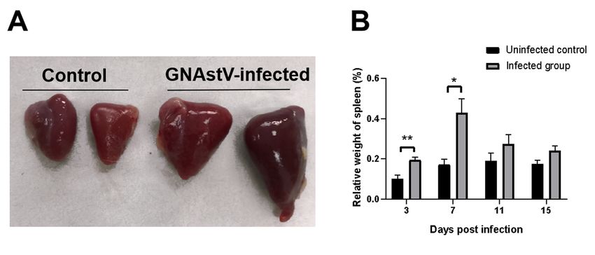
Viruses 2021, 13, 1108                                                                                                        7 of 13

      Figure 5. Virus location and viral load in the spleen. Spleen tissue was stained with hematoxylin and eosin (HE) (A), and
      the virus location was detected by IHC (B). Viral copy numbers in spleens in goslings infected with GNAstV were evaluated
      using real-time PCR (C). The virus location was detected by IHC (D), and the mean optical density (MOD) was calculated
      by Image-Pro Plus (E). Scale bar: 20 µm. Values are expressed as mean ± SD, n = 5.

                                 3.5. Ultrahistopathological Changes in Spleen Tissue
                                      The splenic lymphocytes and reticular cells from the control goslings are normal, and
                                 no obvious pathological changes were observed. However, some apoptotic cells were
                                 found among splenic lymphocytes of infected goslings, and these cells were characterized
                                 by sharply delineated masses of condensed chromatin and formation of apoptotic bodies
                                 (Figure 6, arrow). In addition, loss of plasma membrane integrity and swollen mitochondria
                                 were also observed in the reticular cells of infected goslings (Figure 6, triangle).

      Figure 6. Ultrahistopathological changes in spleen induced by GNAstV infection. Arrow: Condensed chromatin and
      apoptotic cells among lymphocytes. Triangle: swollen mitochondria and impaired plasma membrane integrity in reticular
      cells. Scale bar: 10 µm.
Viruses 2021, 13, 1108 8 of 13
3.6. Effects of GNAstV Infection on Apoptosis of Splenic Lymphocytes
TUNEL staining was used to evaluate apoptosis in splenic cells. As shown in
Figure 7A, the positive signals (brown points) were observed in the splenic lymphocytes of
control and infected goslings, and more positive signals were found in the infected goslings
compared to the control. The percentage of apoptosis was significantly increased in spleens
from the infected group at 3 and 7 dpi compared to the control group (p < 0.05), while there
was no significant difference in apoptosis between the two groups at 11 dpi (Figure 7B;
p > 0.05). The mRNA levels of Fas, FasL, cyt C, Bax, and Bcl-2 were determined by qPCR.
Expression of Fas mRNA at 3, 7, 11, and 15 dpi and expression of FasL mRNA at 3 and 7
were higher in the infected group than in the control group (Figure 7C, D; p < 0.05). No
differences were found in cyt C mRNA expression or in the ratio of Bax and Bcl-2 between
the two groups (Figure 7E, F; p > 0.05).
Figure 7. Effects of GNAstV infection on apoptosis of splenic cells. Paraffin-embedded sections of spleen tissue were stained
by the TUNEL method (A). The apoptotic index was calculated as the percentage of TUNEL-positive cells in spleen cells (B).
Changes in the mRNA levels of Fas (C), FasL (D), and cyt C (E), and the ratio of Bcl-2/Bax (F) in the spleen at 3, 7, 11, and
15 dpi in goslings infected with GNAstV were determined. White arrow: apoptosis of splenic cells. Scale bar: 20 µm. Values
are expressed as mean ± SD, n = 5. * p < 0.05; ** p < 0.01; **** p < 0.0001.
3.7. Effects of GNAstV Infection on Autophagy of Splenic Cells
To determine whether GNAstV infection influenced autophagy in the spleen, the
mRNA expression of autophagy-related genes, including ATG5 and Beclin1, and levels of
proteins including ATG5 and LC3II were measured by qPCR and Western blot, respectively.
No differences in ATG5 and Beclin1 mRNA levels in the spleen were observed betweenViruses 2021, 13, 1108 9 of 13
the infected and negative control groups (Figure 8A, p > 0.05). There were no differences
found in ATG5 and LC3II protein levels between the two groups (Figure 8B, p > 0.05).
Figure 8. Effects of GNAstV infection on autophagy of splenic cells. Changes in the mRNA levels of ATG5 and Beclin1 in
spleen tissue at 3, 7, 11, and 15 dpi after infection with GNAstV (A). Changes in the protein levels of ATG5 and LC3-II in
spleen tissue at 3, 7, 11, and 15 dpi after infection with GNAstV (B). C: uninfected control group; V: GNAstV-infected group.
Values are expressed as mean ± SD, n = 5.
3.8. Effects of GNAstV Infection on CD4 and CD8 Expression in the Spleen
The expression of genes and proteins associated with antigen presentation molecules
(CD4 and CD8) in the spleens was determined by IHC and Western blot. Both the control
group and the infected group had positive signals for CD4 expression (Figure 9A), and
statistical analysis showed that CD4 was significantly increased in the infected group at
3, 7, 11, and 15 dpi compared with the negative control group (Figure 9B; p < 0.05), while
CD8 was significantly decreased in the infected group at 3 and 7 dpi compared with the
control group (Figure 9C, p < 0.5). Similar results were obtained for CD4 and CD8 protein
expression (Figure 10), and the CD4 protein levels were higher in the infected group at 3, 7,
11, and 15 dpi than that of the control group (p < 0.5), while expression of the CD8 protein
was lower in the infected group at 3 and 7 dpi than that of the control (p < 0.5).Viruses 2021, 13, 1108 10 of 13
Figure 9. Immunohistochemical examination of the spleen using CD4 and CD8 antibodies. Changes in positive signals
of CD4 and CD8 in the spleen at 3, 7, 11, and 15 dpi after infection with GNAstV (A). The mean optical density (MOD)
of a CD4 positive signal (B) and CD8 positive signal (C) was calculated by Image-Pro Plus. Scale bar: 20 µm. Values are
expressed as mean ± SD, n = 5. ** p < 0.01; *** p < 0.001; **** p < 0.0001.
Figure 10. Changes in the protein levels of CD4 and CD8 in the spleen determined by
Western blot at 3, 7, 11, and 15 dpi after infection with GNAstV. C: uninfected control
group; V: GNAstV-infected group. Values are expressed as mean ± SD, n = 5. * p < 0.05;
** p < 0.01.Viruses 2021, 13, 1108 11 of 13
4. Discussion
The spleen is an important immune organ, and plays a vital role in host immune
responses against invading pathogens in geese. In the study, GNAstV infection was
shown to cause lymphocyte depletion in the spleen, especially during early stages of
infection, indicating that GNAstV induces immune suppression in goslings. The depletion
of lymphocytes was alleviated in later stages of GNAstV infection, which could be related
to improvement of host immune function, as the goslings grew older. Young goslings are
more easily infected with GNAstV than older goslings, and goslings at 25 and 35 days of
age only exhibit mild symptoms following GNAstV infection [20]. In addition, lymphocyte
depletion was mainly observed around the white pulp of the spleen in GNAstV-infected
goslings, which may be associated with the blood-spleen barrier (BSB) of the spleen. The
BSB is a filter bed for red blood cells that protects against microorganisms, and is located in
the ellipsoid and periellipsoidal lymphatic sheath (PELS) in avians, which are composed
of the white pulp [21]. These data suggest that GNAstV in the blood may accumulate
in the BSB, and subsequently cause damage to lymphocytes around the BSB. It has been
reported that duck Tembusu virus particles are primarily distributed in the PELS within the
spleen’s white pulp, and can cause vacuolar degeneration in the PELS [22]. Reticular cells
are stromal cells that play an important role in the migration and positioning of lymphoid
cells [23]. The pathological changes of reticular cells observed in this study indicate
that GNAstV infection may damage lymphocyte development and immune function.
Moreover, reticular fibers are important components of the supporting framework in the
spleen that provide channels and specific microenvironments for the migration of T and
B lymphocytes [24,25]. In the study, GNAstV infection caused reduction and rupture of
reticular fibers, which may also contribute to spleen damage.
Apoptosis is important in the defense against viral infection. HosT-cells quickly
initiate apoptotic processes following stimulation by viruses, and restrict viral replication
by clearing the infected cells [26]. In this study, GNAstV infection induced lymphocyte
apoptosis in the spleen, suggesting that apoptosis in splenic lymphocytes may be a primary
mechanism to combat GNAstV infection in goslings. Apoptosis can be triggered by
diverse cellular signals, such as the death receptor-mediated extrinsic pathway, the intrinsic
mitochondrial pathway, and the endoplasmic reticulum stress-mediated pathway. The
Fas/FasL signaling pathway is one of the major death receptor-mediated extrinsic apoptosis
pathways. In this study, GNAstV infection induced the mRNA up-regulation of Fas and
FasL, indicating that the death receptor-mediated extrinsic apoptosis pathway is activated
following GNAstV infection. GNAstV was found in lymphocytes and macrophages in this
study, and lymphocyte apoptosis may be directly caused by GNAstV. However, indirect
effects that induce apoptosis should not be excluded. The mechanism of apoptosis in
splenic lymphocytes induced by GNAstV needs to be further investigated.
CD4 T-cells can recognize the MHC-II antigen, and CD8 T-cells can recognize the
MHC-I antigen. The interaction between CD4 and CD8 T-cells and MHC molecules can
enhance the binding of MHC molecules with T-cell receptors, which is important for
immune defenses in the avian species. In this study, GNAstV infection increased levels of
CD4 and decreased levels of CD8 at the early stages of infection. CD8 plays an important
role in clearing cells infected with a virus. The reduction of CD8 induced by GNAstV
may indicate a reduction in the host immunity of goslings, which would facilitate virus
proliferation at the beginning of infection. The decrease of CD8 numbers may be related to
the apoptosis observed in splenic lymphocytes, although this would need to be confirmed
in future studies. In addition, we previously reported that the mRNA expression level
of CD8 T-cells is increased in spleens of goslings infected with GNAstV [11]; the mRNA
expression level of CD8 T-cells was also increased in the present study (data not shown). It
is interesting that the mRNA results are opposite to the protein results; this may be related
to the post-transcriptional mechanism, such as protein translation and post-translational
modification and degradation, but the detailed mechanisms need to be further investigated.Viruses 2021, 13, 1108 12 of 13
In conclusion, GNAstV localizes to splenic lymphocytes and macrophages in the
spleens in infected goslings. In the spleen, GNAstV infection causes lymphocyte depletion,
reticular cell necrosis, reticular fiber destruction, lymphocyte apoptosis, and CD8 T-cell
reduction. These pathological changes result in significant injury to the spleen. Our results
also suggest that GNAstV can induce immunosuppression, which may facilitate infection
by other pathogens.
Author Contributions: Conceptualization, Y.L.; methodology, R.D., H.H.; validation, R.D., H.H.,
H.W.; formal analysis, R.D., H.H., H.W., S.Q.; investigation, R.D., H.H., Z.Y.; resources, Y.L.; writ-
ing—original draft preparation, R.D., Y.L.; writing—review and editing, R.D., Y.L., E.B.; visualization,
R.D., H.H.; supervision, Y.L.; project administration, Y.L.; funding acquisition, Y.L., E.B. All authors
have read and agreed to the published version of the manuscript.
Funding: This research was funded by Priority Academic Program Development of Jiangsu Higher
Education Institutions.
Institutional Review Board Statement: Animal experiments were performed in compliance with
Chinese legislation, and were approved by the Institutional Animal Care and Use Committee (IACUC)
of Nanjing Agricultural University (protocol code PTA2020013, 9 July 2020).
Informed Consent Statement: Not applicable.
Data Availability Statement: The data that support the findings of this study are available from the
corresponding author upon reasonable request.
Conflicts of Interest: None of the authors has any conflict of interest.
References
1. Zhang, Q.; Cao, Y.; Wang, J.; Fu, G.; Sun, M.; Zhang, L.; Meng, L.; Cui, G.; Huang, Y.; Hu, X.; et al. Isolation and characterization
of an astrovirus causing fatal visceral gout in domestic goslings. Emerg. Microbes. Infect. 2018, 7, 1–11. [PubMed] [CrossRef]
2. Chen, H.; Zhang, B.; Yan, M.; Diao, Y.; Tang, Y. First report of a novel goose astrovirus outbreak in Cherry Valley ducklings in
China. Transbound. Emerg. Dis. 2020, 67, 1019–1024. [CrossRef] [PubMed]
3. Wei, F.; Yang, J.; Wang, Y.; Chen, H.; Diao, Y.; Tang, Y. Isolation and characterization of an astrovirus causing fatal visceral gout in
domestic goslings. Emerg. Microbes. Infec. 2020, 9, 1046–1054. [CrossRef] [PubMed]
4. Li, J.; Hu, W.; Liu, T.; Zhang, H.; Opriessnig, T.; Xiao, C. Isolation and evolutionary analyses of gout-associated goose astrovirus
causing disease in experimentally infected chickens. Poult. Sci. 2021, 100, 543–552. [PubMed] [CrossRef]
5. He, D.; Yang, J.; Jiang, X.; Lin, Y.; Chen, H.; Tang, Y.; Diao, Y. A quantitative loop-mediated isothermal amplification assay for
detecting a novel goose astrovirus. Poult. Sci. 2020, 99, 6586–6592. [PubMed] [CrossRef]
6. Chen, Q.; Xu, X.; Yu, Z.; Sui, C.; Zuo, K.; Zhi, G.; Ji, J.; Yao, L.; Kan, Y.; Bi, Y.; et al. Characterization and genomic analysis of
emerging astroviruses causing fatal gout in goslings. Transbound. Emerg. Dis. 2020, 67, 865–876. [PubMed] [CrossRef]
7. Yin, D.; Yang, J.; Tian, J.; He, D.; Tang, Y.; Diao, Y. Establishment and application of a TaqMan-based one-step real-time RT-PCR
for the detection of novel goose-origin astrovirus. J. Virol. Methods 2020, 275, 113757. [PubMed] [CrossRef]
8. Zhang, X.; Ren, D.; Li, T.; Zhou, H.; Liu, X.; Wang, X.; Lu, H.; Gao, W.; Wang, Y.; Zou, X.; et al. An emerging novel goose astrovirus
associated with gosling gout disease. Emerg. Microbes. Infec. 2018, 7, 1–8. [CrossRef] [PubMed]
9. Niu, X.; Tian, J.; Yang, J.; Jiang, X.; Wang, H.; Chen, H.; Yi, T.; Diao, Y. Novel Goose Astrovirus Associated Gout in Gosling. Vet.
Microbiol. 2018, 220, 53–56. [CrossRef] [PubMed]
10. Wang, Z.; Li, L.; Liu, P.; Wang, C.; Lu, Q.; Liu, L.; Yang, Y.; Luo, Q.; Shao, H. Host innate immune responses of geese infected with
goose origin nephrotic astrovirus. Microb. Pathog. 2021, 152, 104753. [CrossRef] [PubMed]
11. Wu, W.; Qiu, S.; Huang, H.; Xu, R.; Bao, E.; Lv, Y. Immune-related gene expression in the kidneys and spleens of goslings infected
with goose nephritic astrovirus. Poult. Sci. 2021, 100, 100990. [PubMed] [CrossRef]
12. Liu, H.; Hu, D.; Zhu, Y.; Xiong, H.; Lv, X.; Wei, C.; Liu, M.; Yin, D.; He, C.; Qi, K., et al. Coinfection of parvovirus and astrovirus in
gout-affected goslings. Transbound. Emerg. Dis. 2020, 67, 2830–2838. [PubMed] [CrossRef]
13. Wu, W.; Xu, R.; Lv, Y.; Bao, E. Goose astrovirus infection affects uric acid production and excretion in goslings. Poult. Sci. 2020, 99,
1967–1974. [CrossRef] [PubMed]
14. Fischer, A.H.; Jacobson, K.A.; Rose, J.; Zeller, R. Hematoxylin and Eosin Staining of Tissue and Cell Sections. Cold Spring Harb.
Protoc. 2008, t4986. [PubMed] [CrossRef]
15. McNeilly, F.; Allan, G.M.; Moffett, D.A.; McNulty, M.S. Detection of chicken anaemia agent in chickens by immunofluorescence
and immunoperoxidase staining. Avian Pathol. 1991, 20, 125-132. [CrossRef] [PubMed]
16. Qiu, S.; Xu, R.; Guo, Y.; Lv, Y. Establishment of a SYBR Green I-based real-time PCR for goose nephritic astrovirus. Chin. Vet. Sci.
2020, 3, 300–306. (In Chinese)Viruses 2021, 13, 1108 13 of 13
17. Sun, J.; Zhang, X.; Cao, Y.; Zhao, Q.; Bao, E.; Lv, Y. Ovarian Toxicity in Female Rats after Oral Administration of Melamine or
Melamine and Cyanuric Acid. PLoS ONE 2016, 11, e149063. [PubMed] [CrossRef]
18. Lou, Y. Study on Follicular Granulisa Cell Autophay Induced by Oxidative Stress in Goose. Master’s Thesis, Zhejiang A&F
University, Hangzhou, China, 2015. (In Chinese)
19. Huang, B.; Li, J.; Zhang, X.; Zhao, Q.; Lu, M.; Lv, Y. RIG-1 and MDA-5 signaling pathways contribute to IFN-β production
and viral replication in porcine circovirus virus type 2-infected PK-15 cells in vitro. Vet. Microbiol. 2017, 211, 36–42. [PubMed]
[CrossRef]
20. An, D.; Zhang, J.; Yang, J.; Tang, Y.; Diao, Y. Novel goose-origin astrovirus infection in geese: the effect of age at infection. Poult.
Sci. [CrossRef] [PubMed]
21. Xu, M.; Li, W.; Yang, S.; Sun, X.; Tarique, I.; Yang, P.; Chen, Q. Morphological characterization of postembryonic development of
blood–spleen barrier in duck. Poult. Sci. 2020, 99, 3823–3830. [PubMed] [CrossRef]
22. Sun, X.; Li, W.; Liu, E.; Huang, H.; Wang, T.; Wang, X.; Shi, Y.; Yang, P.; Chen, Q. In vivo cellular and molecular study on duck
spleen infected by duck Tembusu virus. Vet. Microbiol. 2019, 230, 32–44. [CrossRef] [PubMed]
23. Perez Shibayama, C.; Gil Cruz, C.; Ludewig, B. Fibroblastic reticular cells at the nexus of innate and adaptive immune responses.
Immunol. Rev. 2019, 289, 31–41. [PubMed] [CrossRef]
24. Pellas, T.C.; Weiss, L. Migration pathways of recirculating murine B cells and CD4+ and CD8+ T lymphocytes. Am. J. Anat. 1990,
187, 355–373. [CrossRef] [PubMed]
25. Fukuta, K.; Mochizuki, K. Formation of reticular fibers in the developing spleen of the chick embryo. Arch. Histol. Jpn. (Nihon
Soshikigaku Kiroku) 1982, 45, 181–189. [CrossRef] [PubMed]
26. Carruthers, V.B.; Cotter, P.A.; Kumamoto, C.A. Microbial Pathogenesis: Mechanisms of Infectious Disease. Cell Host Microbe 2007,
2, 214–219. [PubMed] [CrossRef]You can also read