Golden ratio and fractals in mitral valve geometry: Potential implications for valve imaging assessment - Open ...
←
→
Page content transcription
If your browser does not render page correctly, please read the page content below
Interventional
    Review Article

                                                                                                                   Cardiology
  Golden ratio and fractals in mitral valve
  geometry: Potential implications for valve
  imaging assessment
Abstract                                                                                      Luca Deorsola1*, Alessandra Bellone2
Nowadays, the procedure of choice to manage a diseased Mitral Valve is represented by         1
                                                                                               Pediatric Cardiac Surgery, Regina Margherita Children’s Hospital,
conservative treatment. Geometrical references are fundamental to perform a correct           Turin, Italy
                                                                                              2
                                                                                               Department of Adult Cardiology, San Giovanni Bosco Hospital,
evaluation on a diseased valve, to plan the best repair strategy and to assess its results,   Turin, Italy
both during the procedure and at follow-up. In the last decades, the number and
the precision of these references have widely grown, due to a significant amount of           *Author for correspondence:
                                                                                              Luca Deorsola, Pediatric Cardiac Surgery, Regina Margherita
research, as well as to a rapid empowering of imaging software and devices. Therefore,        Children’s Hospital, Turin, Italy, E-mail: deorsola@libero.it
the routine measurements now obtainable from the different imaging techniques
allow an extensive and deep spatial evaluation of valve geometry. Similarly, both repair      Received date: June 11, 2021
                                                                                              Accepted date: June 25, 2021
techniques and devices have undergone a consistent improvement in the last years,
                                                                                              Published date: JuLY 02, 2021
adding to traditional surgery a wide series of percutaneous and hybrid procedures. In
this scenario, an accurate geometrical analysis of the whole valve is actually mandatory,
particularly when a hybrid or percutaneous approach is chosen and imaging is the
only available eye. In two recently published papers, we hypothesized that the healthy
Mitral Valve could have a geometrical structure based on Golden Ratio, Fibonacci
Series and Fractals, a scalar 3D model where all components are related one another by
defined proportions and fit together like the pieces of a puzzle. Such a model, with the
use of very simple calculations, can describe every geometrical reference of the Mitral
Valve and seems to predict their expected normal values.
After a brief summary about the results of our previous research, we have reviewed
literature concerning the most common geometrical references retrievable from imaging
and currently employed to evaluate the Mitral Valve. Published data and normality
ranges have been compared with the values obtained from the 3D model, showing how
it seems to produce the same results and give them a logical interpretation.
                              .            .                   .         .
Keywords: Mitral valve Golden ratio Fibonacci series Fractals Interventional
procedures
Introduction
Conservative treatment is nowadays the routine choice to approach a diseased Mitral
Valve. Originally based on conventional on-pump surgery, in the last years it has been
integrated by several either off-pump or trans-catheter procedures. Particularly, this
new kind of interventions is rapidly growing, due to the ever better knowledge of
valve anatomy and function, as well as to the impressive improvement occurred in the
last years to medical devices, software, materials and engineering techniques. When
planning mitral repair, a deep and extremely accurate valve analysis is mandatory,
particularly when an interventional technique is chosen, where a direct anatomical
evaluation is impossible. Imaging techniques, mainly echocardiography, are the best
available tool for assessing both the structure and the function of the Mitral Valve.

 Interv. Cardiol. (2021) 13(4), 332-340                         ISSN 1755-5302                                                                              332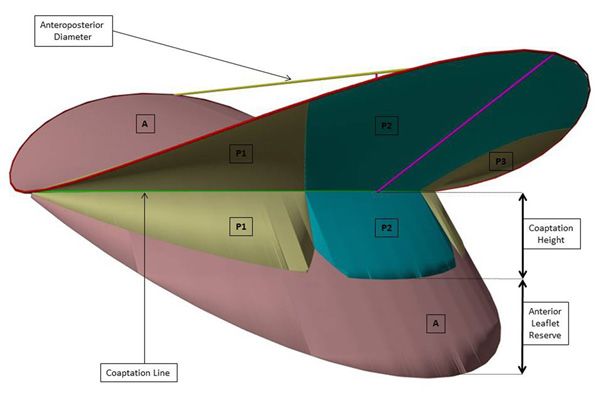
Review Article

Evaluating valve disease, planning a repair strategy, monitoring the     squared is equal to itself plus 1 (1.618²=1.618+1=2.618), while 1
effects, during of immediately after the procedure, and carrying         divided by 1.618 gives 0.618.
out the follow-up are the routine taken steps. For this purpose, the
                                                                         Fibonacci series
most important information obtainable from imaging techniques
consists of a huge amount of measurements, which allow us to             The Fibonacci Series is a famous sequence of numbers, named
describe and quantify with precision structure and function of the       after its inventor: The mathematician Leonardo Fibonacci. It is an
valve. In the last decades, medical imaging devices, computers and       infinite progression of integer values, starting from 0 and growing
software power have undergone an extremely fast and impressive           up with a simple mathematical rule: Each value is the sum of the
improvement, making this amount of measurements ever bigger              two previous ones. Thus, after 0 and 1, it goes on with 1, 2, 3, 5,
and more accurate.                                                       8, 13, 21, etc. Fibonacci published this series around 1200 A.D.,
                                                                         in his “Liber Abbaci” (Book of Calculus), as an example to show
In two recent studies we focused on the Coaptation Triangle and
                                                                         the positional numbering system he had learnt from the Arabic
on the Mitral Scallops, describing how their geometrical structure,
                                                                         people of Northern Africa. However, in time, it became ever more
in normal valves, could be based on Golden Proportion, Fractals
                                                                         evident that such a numerical pattern is one of the strongest rules
and Fibonacci Series [1,2].
                                                                         of Nature, mainly when dealing with growth and reproduction
This review summarizes the geometrical 3D model of the Mitral            processes [15]. Moreover, around 1600, the astronomer Johannes
Valve partially presented in our previous research and compares          Kepler realized there is a strong connection between Fibonacci
its geometrical references with the corresponding values described       Series and Golden Ratio: The ratio between two consecutive values
in literature and routinely employed when planning, performing           is extremely close to the Golden Number and the more we go
and evaluating a conservative valve treatment. The aim is to show        down the sequence the closer it gets [17].
how this 3D model describes the whole valve geometry and how,
                                                                         Fractals
employing very easy calculations only, it seems to produce the same
results published in literature and to allow a logical explanation or    The term “Fractal” indicates a specific geometrical structure based
normality ranges.                                                        on self-recurrence and self-nidification. At first sight, fractal
                                                                         objects appear hardly defined and chaotic, not complying with
Proportional Geometry
                                                                         conventional geometry. However, at a careful look, it is evident
Golden ratio                                                             that they can be broken down into smaller pieces, different in
                                                                         dimension, but identical in shape: The same of the original
The term “Golden Section”, “Golden Proportion” or “Golden
                                                                         object. In turn, these pieces can be broken down into ever
Ratio” refers to a way of dividing a segment into two different
                                                                         smaller parts, with the same result, and so on infinitely. Fractals
parts, so that the shorter is to the longer as the longer is to the
                                                                         are a quite early discovery, made between the 70s and 80s by the
whole segment. This ratio has an irrational value known as “Golden
                                                                         Polish mathematician Benoit Mandelbrot, who defined the inner
Number” and usually rounded to 0.618 or 1.618, depending on
                                                                         structure of these elements as “self-similar” [18]. Again, Fractal
whether it is obtained dividing the shorter by the longer part or
                                                                         objects are another basic rule in Nature and fractal growth is the
vice versa.
                                                                         most diffused pattern in the universe. Mountains, trees, leaves,
The knowledge of this ratio is not datable. It was documented for        river deltas are only a few examples [19-21]. Moreover, fractal
the first time by Euclid and Pythagoras in the 4th century B.C [3],      growth is often based on Golden Ratio, since it allows the most
and deeply analyzed by the mathematician Luca Pacioli during the         efficient space arrangement, avoiding both overlaps and empty
Renaissance [4]. However, the most important fact is that it has         areas at the same time [22].
been observed in a consistent amount of natural settings, such as
                                                                         Coaptation triangle
physics, botanics, biology and even human anatomy and physiology
[5-12]. Recently, it has also been observed in the human heart,          The “Coaptation Triangle” or “Tenting Area”, identifiable in
even if available studies are limited to a gross description only [13-   Parasternal Long Axis view during systole, is an upside-down
16]. Nowadays, the Golden Number is an important mathematical            triangle, where the base is the Mitral Anteroposterior Diameter
constant, represented by the Greek letter Φ (phi). Particularly, the     (APD) and the two sides are the Anterior Scallop and the P2
lower case (ϕ) stands for 0.618…, while the upper case (Φ) for           Scallop (Figure 1). Many useful measurements can be retrieved
1.618…. Moreover, this is the only known value to show a couple          from this triangle, which are nowadays routinely employed to
of important and unique mathematical characteristics: 1.618              analyze valve geometry [23-27].
333               Interv. Cardiol. (2021) 13(4), 332-340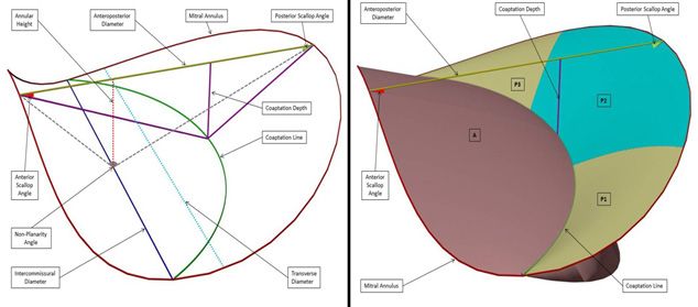
Review Article

                                                                                  the same shape but different dimensions (Figure 3).

                                                                                  Figure 3: Anatomical drawing by Henry Gray, representing the whole valve tissue
Figure 1: The Coaptation Triangle drawn on echocardiography imaging.              flattened on a plane after cutting the Mitral Annulus at the level of the Anterior
Parasternal Long Axis view (PLAX).                                                Commissure. The characteristic scalloped pattern is clearly visible.
                                                                                  In our last research, we found that also the Scallop geometry looks
                                                                                  to be based on Golden Proportion and have a fractal structure
In a previous research, we found that Golden Proportion looks
                                                                                  [2]. Again, the Scallops appear to show a fractal and scalar pattern
to define the geometrical structure of the Coaptation Triangle in
                                                                                  based on Golden Ratio, which determines both their aspect and
the healthy mitral valve [1]. This peculiar scalar structure follows
                                                                                  their dimensional relationships. Details are shown in Figure 4.
a fractal pattern and involves the Anteroposterior Diameter, the
Anterior Scallop and P2 Scallop Cords, as well as the Coaptation
Depth. A detailed description is presented in Figure 2.

                                                                                  Figure 4: Diagram of the whole valve tissue presented in the previous image
                                                                                  (Figure 3), according to the Proportional Geometry model. The four scallops are
                                                                                  framed within four Golden Rectangles and their dimensional relationships are
                                                                                  indicated, showing the continuous inner recurrence of the Golden Number and
                                                                                  the self-similar aspect typical of Fractals. The base of the P1/P3 Scallop and the
                                                                                  height of the P2 Scallop are the most critical elements, representing the radius of
                                                                                  the whole Mitral Annulus..

Figure 2: Diagram of the Coaptation Triangle with a detailed description of the   Final 3D model
possible references and measurements. An indication of the dimensional ratios
                                                                                  The Mitral Valve is well known to have a complex 3D geometry,
according to Proportional Geometry is also provided, where Φ represents the
                                                                                  with a saddle shaped annulus and a specific interaction among
Golden Number (1.618...).
                                                                                  its scallops. Integrating the fractal scalar patterns of both the
Mitral scallops
                                                                                  Coaptation Triangle and the scallop geometry we described
The Mitral Valve is commonly considered to have two Leaflets:                     previously, a complete 3D model can be obtained which describes
One anterior and one posterior, the latter divided into three                     the whole structure of the Mitral Valve in term of Fractals and
Scallops (P1, P2 and P3). However, all anatomical studies state                   Golden Proportion. In summary, the whole valve can be imagined
there is only one leaflet, a continuous veil of tissue running along              as a set of components interacting like the pieces of a puzzle;
the Annulus [28-32]. This veil is divided into four Scallops, with                components whose dimensional aspect and relationships are

 334                 Interv. Cardiol. (2021) 13(4), 332-340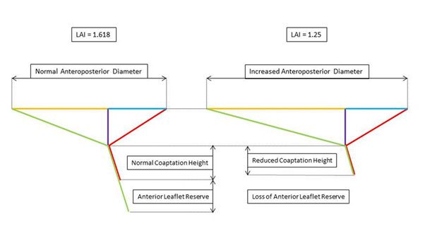
Review Article
strictly related one another by Golden Ratio and fractal pattern, represents its height: The distance of the Coaptation Point below
in an arrangement we could define “Proportional Geometry”. A the anteroposterior diameter (Figure 2 and Figure 5). Originally
complete description of this peculiar 3D geometrical structure is born for surgical mitral repair, it has progressively been extended
presented in Figure 5. to interventional procedures, mainly percutaneous edge-to-edge
repair. An increase in its value is an important sign of leaflet
tethering, while a reduction indicates either chordal elongation or
annular flattening. In literature, most authors report normal values
between 6 and 8 mm [25,36-48]. Only Zhang found slightly lower
values, around 4.2 mm [24].
Considering Proportional Geometry 3D model, the Coaptation
Depth is given by APD/Φ3=APD/4.236 (Table 1) and the
average individual previously cited has a Coaptation Depth of
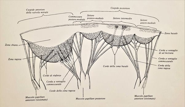
Figure 5: The complete Mitral Valve created with Proportional Geometry. The left
29/4.236=6.85 mm, perfectly within the normality range.
image represents a diagram of the Mitral Annulus with the Coaptation Triangle and The Coaptation Height represents the length of the coapting
all the possible geometrical references. The right image is actually the same diagram,
portion of both leaflets below the Coaptation Point and gives an
after adding the four scallops as they appear in systole. Some important geometrical
references are also indicated.
idea of how much valve tissue is involved in coaptation (Figure 2
Such a 3D model could have important implications for valve and Figure 6). A reduction in this value indicates either annular
assessment: Once identified a healthy component or a normal dilatation or leaflet tethering and implies an unstable coapting
dimension to start from, we could be able to predict the whole surface. As for the Coaptation Depth, most of authors agree in
normal valve geometry using a single number (the Golden considering normal and reliable values around 5-7 mm [48,49]. As
Number) and performing very easy calculations only. Alternatively, we will see later, when dealing with the “Anterior Leaflet Reserve”,
should a starting measurement not be identifiable, we could use the Coaptation Height is, in fact, represented only by the lenght of
for this purpose the expected normal valve diameters given by the the P2 Scallop below the Coaptation Point.
many existing normograms, which correlate valve dimensions with
patient’s BSA [33-35].
Table 1 is an example of how calculations can be made, starting
from the Anteroposterior Diameter, to obtain the most significant
geometrical references of the whole Mitral Valve.
Imaging Measurements and References
As mentioned before, we have thereafter reviewed the geometrical
references routinely employed for the whole diagnosis and
conservative treatment process, comparing them with the
corresponding ones obtained from the 3D geometrical model
based on the Golden Ratio and the set of calculations used to
describe it.
To transform these calculations into concrete numbers and to
use them for explanatory examples, we can consider an average
individual, 175 cm tall. According to Lorentz, Robinson and
Devine formulas, this leads to an ideal weight of 70 Kg and a BSA Figure 6: Diagram of the Mitral valve in systole created with Proportional
of 1.85 m2. According to the previously cited normograms [33- Geometry and seen as it appears from a posterolateral view. The coapting portion of
both leaflets is clearly visible and the Anterior Leaflet Reserve can be appreciated..
35], his average mitral Anteroposterior Diameter results around
29 mm. In Proportional Geometry 3D model, this value is again given by
Coaptation depth and height APD/Φ3=APD/4.236, the same identical calculation used for the
Coaptation Depth (Table 1).
Coaptation Depth is retrieved from the Coaptation Triangle and
335 Interv. Cardiol. (2021) 13(4), 332-340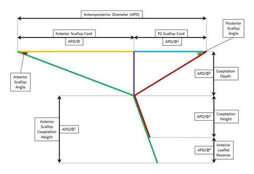
Review Article

  Table 1: Possible calculations using the anteroposterior diameter           Scallop slides anteriorly and uses its reserve to keep a normal
                         as starting reference.                               Coaptation Surface.
                                      Formula (with           Formula (with
             Dimensions                                                       The Proportional Geometry 3D model produces exactly the
                                        symbols)                 values)
          Anterior Scallop Width         APD × Φ               APD × 1.618    same result: The Anterior Scallop Coaptation Height is APD/
         Anterior Scallop Height           APD                    APD
             P2 Scallop Width              APD                    APD         Φ2=APD/2.618, while the Posterior Scallop Coaptation Height
            P2 Scallop Height             APD/Φ                 APD/1.618     is APD/Φ3= APD/4.236. The Anterior Leaflet Reserve is then
           P1/P3 Scallop Width            APD/Φ                 APD/1.618
                                                                              the difference between the two measurements, thus APD/2.618-
           P1/P3 Scallop Height          APD/Φ2                 APD/2.618
        Annular Perimeter Radius          APD/Φ                 APD/1.618     APD/4.236=APD × 0.145. Interestingly, this corresponds to
           Transverse Diameter          APD × 2/Φ              APD × 1.236    APD/Φ4=APD/6.854 (Table 1). The average individual we cited
       Intercommissural Diameter     APD × 2 × 0.966/Φ         APD × 1.194
         Anterior Annular Length         APD × Φ               APD × 1.618    previously has an Anterior Leaflet Reserve of 29 × 0.145=4.2 mm.
        Posterior Annular Length      APD × (2+Φ)/Φ            APD × 2,236
          Anterior Scallop Cord           APD/Φ                 APD/1.618     This information could potentially have an impact on
          Posterior Scallop Cord         APD/Φ2                 APD/2.618     percutaneous Neochord implantation, particularly when treating
            Coaptation Depth             APD/Φ3                 APD/4.236
                                                                              the Anterior Leaflet [54,55]. Repair could potentially result more
      Anterior Scallop Coaptation
                                        APD/Φ2            APD/2.618           reliable and durable if chordal adjustment considers not only the
                  Height
      Posterior Scallop Coaptation                                            disappearance of regurgitation at real time echocardiography, but
                                        APD/Φ3            APD/4.236
                  Height
         Anterior Leaflet Reserve       APD/Φ4
                                                          APD/6.854           also the recreation of an Anterior Leaflet Reserve. Colli et al. state
      Abbreviations: APD=Anteroposterior Diameter; Φ= 1.618.... (Golden       that after chordal adjustment under echo guidance a slight over-
                                   Number)
                                                                              tension should be applied to prevent recurrent regurgitation when
 Concerning percutaneous edge-to-edge, literature is generally                the ventricle undergoes reverse remodeling [56].
 oriented to consider this procedure safely feasible when the
                                                                              Tenting area
 Coaptation Depth is 2 mm
 [50], even if some recent studies have hypothesized good result              This is another important measurement and corresponds to
 extending indications beyond these cutoffs [51-53]. These limits             the area of the Coaptation Triangle (Figure 2 and Figure 5). An
 are deeply related one another and easily explainable considering            increase in this area is important indicator of leaflet tethering or
 the average patient described previously, with both the Coaptation           annular dilatation [23,26,27]. Most authors report normal values
 Depth and the Coaptation Height of 7 mm. A simple leaflet                    to be around 100 mm2 [40,48,57,58], even if some authors found
 tethering (without annular dilatation) leading to a Coaptation               values of about 66 mm2 [24, 25].
 Depth of 11 mm means a 4 mm displacement of the Coaptation                   Since in Proportional Geometry 3D model the Coaptation Depth
 Point towards the ventricular apex and an identical reduction of             corresponds to APD/Φ3=APD/4.236, the Tenting Area is given by
 the Coaptation Height, which becomes 3 mm long: Just a little bit            APD × APD/Φ3/2=APD2/2Φ3=APD2/8.472 (Table 1).
 >2 mm, the shortest allowed dimension for a safe clip positioning.
                                                                              In our average individual the Tenting Area results 292/8.472=99.268
 Anterior leaflet reserve                                                     mm2, which is again in accordance with literature [25, 40, 57, 58].
 In 2010, Gogoladze et al. [41] revealed an interesting characteristic        AL/PL ratio
 of the Coaptation Height. The portion of Anterior Scallop below
                                                                              This ratio is a recent index and deserves another bit of explanation.
 the Coaptation Point is longer than the corresponding one of
                                                                              The base of the Coaptation Triangle is divided by its height into
 P2 Scallop. In other words, there is a short terminal portion
                                                                              two different segments, named “leaflet cords”, since they represent
 of Anterior Scallop which does not participate in coaptation.
                                                                              the geometrical projections of the Anterior Scallop and the P2
 Therefore, the true Coaptation Height depends only on P2
                                                                              Scallop on the Anteroposterior Diameter (Figure 2).
 Scallop (Figure 2 and Figure 6). Imaging techniques easily show
 the thicker tissue portion where the two Scallops pair together,             The ratio between these two values is called “Anterior Leaflet to
 but poorly evidence the lonely terminal part of Anterior Scallop,            Posterior Leaflet Ratio” and estimates the horizontal position of
 hardly distinguishable from the chordal apparatus. The authors               the Coaptation Point. An increased ratio indicates a posterior
 define this excess of Anterior Scallop “Anterior Leaflet Reserve”            displacement in the Coaptation Point, most commonly given
 and state that this element allows the valve to partially tolerate           by annular dilatation. On the contrary, a reduction in this ratio
 annular dilatation: In the early phases of this process, the Anterior        suggests an anterior shift of the Coaptation Point, which is a

336                  Interv. Cardiol. (2021) 13(4), 332-340
Review Article
risk factor for Systolic Anterior Movement (SAM) development the Transverse Diameter: While the latter represents the maximum
[27]. Recent investigations after valve repair concluded that an annular dimension (twice the radius), the former is a bit shorter,
AL/PL Ratio below 1.5 significantly increases the risk for SAM since located in a more anterior position (Figure 5).
[27,59,60]. Particularly, Maslow et al found that repaired valves
Several studies state that normal valves show an AHCWR around
had a tendency to SAM depending on the AL/PL Ratio, being
20%, with ranges between 15% and 30% (27, 36, 63, 64). A value
maximum with values about 0.69 and completely absent when
158 degrees [48,66,67].
evidenced that a posterior leaflet angle >45 degrees is an index of
severe leaflet tethering and a risk factor for failure after surgery. As we said previously, in Proportional Geometry 3D model all
angles are constant, regardless of valve dimension. Anyway, in this
In Proportional Geometry 3D model, angles are actually constant,
3D model, the Non-Planarity angle results to be 121.7 degrees
regardless of valve dimensions. These two values result 21 degrees
wide, again in accordance with literature.
for the Anterior Scallop and 31.8 degrees for the Posterior Scallop,
very close to literature data. Leaflet-to-Annulus Index (LAI)
Annular saddling (AHCWR and non-planarity angle) Leaflet-to-Annulus Index or “LAI” is a quite recent reference and is
defined as the sum of the heights of the Anterior Scallop and the P2
As already demonstrated, Mitral Annulus is not a flat structure,
Scallop divided by the Anteroposterior Diameter in systole. Actually,
but has a characteristic 3D saddled shape. Because of this geometry
it estimates how much valve tissue is available to create the coapting
the Intercommissural Diameter and the Anteroposterior Diameter
leaflet surface in systole. It has nowadays become extremely popular
lie on two different planes. The vertical gap between these two
for interventional procedures, such as percutaneous Edge-to-Edge or
planes is called Annular Height and, interestingly, corresponds to
Transapical Off-Pump Neochord implantation. A consistent number
the Coaptaton Depth (Figure 5).
of studies states that the best procedural results occur when its value is
One of the most important indexes of annular saddling is the ratio >1.25 and, in accordance with these results, many researchers suggest
between the Annular Height and the Intercommissural Diameter, an additional annuloplasty when this value is below this limit [68, 69].
often expressed as a percentage. This ratio is called “Annular Colli et al. agree with these results, even if report a good feasibility of
Height to Commissural Width Ratio” or “AHCWR”. However, percutaneous Neochord implantation with LAI lower than this cutoff
to better understand this ratio, a base concept must be taken into [56,70].
account. The Intercommissural Diameter does not correspond to
337 Interv. Cardiol. (2021) 13(4), 332-340Review Article
Once more, applying the calculations of Proportional Geometry 3D 2.236 (Table 1). This value represents the posterior annular length
model, LAI results to be (APD+APD/Φ)/APD = (APD+APD/1.618)/ from Commissure to Commissure and, in our average patient, it
APD=APD/APD+1/1.618=1+0.618=1.618 (Table 1): Well above the results 29 × 2.236=64.844 mm, being again in accordance with
limit of 1.25. Additionally, using the same calculations, we can also literature [38]. The ability to predict the estimated normal length
hypothesize an explanation of this cutoff. In our average individual, of the posterior Mitral Annulus could then result an important
the Anteroposterior Diameter is 29 mm long, the height of the reference to guide annuloplasty.
Anterior Scallop (equal to APD) again 29 mm and the height of the
Conclusion
P2 Scallop (APD/1.618) 29/1.618=18 mm. In systole, the Anterior
Scallop Coaptation Height is APD/2.618=11 mm and the P2 PProportional Geometry 3D Mitral model appears to represent
Scallop Coaptation Height APD/4.236=7 mm. In such a setting, the the whole structure of the healthy Mitral Valve in a reliable way.
Coaptation Height is 7 mm, the Anterior Leaflet Reserve 4 mm and Starting from a single reference measurement and employing very
LAI results exactly 1.618. Considering the valve scallops unchanged, easy calculations, it seems able to predict the expected normal
a LAI reduction to 1.25 means an increase in the Anteroposterior dimensions and shape of every valve component, leading to results
Diameter to about 37 mm, 8 more than the expected. A valve with similar or almost identical to those observed in literature.
such a modification, actually, has completely run out of its Anterior Another important aspect of this model is that it is entirely based
Leaflet Reserve (4 mm) and has also reduced its Coaptation Height to on proportions rather that absolute values, tailoring the valve on
only 5 mm, 2 mm less per each scallop, critically impairing its own patient’s size. Commonly, when dealing with valve measurements,
coaptation stability (Figure 7). values are given in term of normality ranges, which include the
vast majority of adult individuals. However, part of the world
population, such as children or people with extremely small or big
body sizes, could produce both false positives and false negatives.
In these specific subgroups, calculations based on Proportional
Geometry rules could provide more significant values, leading
to identify with more reliability both healthy and pathological
aspects.
Even if further studies are needed to give it a stronger statistical
significance, this model could result helpful for performing both
surgical and interventional procedures, allowing operators to
Figure 7: Schematic diagram showing Leaflet-to-Annulus Index (LAI) in two be more accurate in their actions and leading them to tend to a
different settings. On the left the healthy valve, with a normal Anteroposterior precise final target.
Diameter and scallops coapting properly: LAI is 1.618. On the right an altered
Mitral Valve, with a dilated Anteroposterior Diameter and LAI reduced to 1.25: Additionally, since in such a 3D model every geometrical aspect
note the disappearance of the Anterior Leaflet Reserve and the significantly reduced can be studied and calculated, it could also be helpful for
Coaptation Height. investigating measurements and geometrical references, which
Annuloplasty could be discovered in the future.
Reduction and stabilization of Mitral Annulus is mandatory when Funding
repair is performed surgically. However, even during percutaneous None.
interventions, annuloplasty might be necessary either to
accomplish the procedure or to improve its result. Nowadays, Conflict of Interest
several percutaneous devices are available, which act either directly The author declares that there is no conflict of interest regarding
or indirectly on the posterior Mitral Annulus, and literature on the publication of this article.
their employment is constantly growing [71-77]. However, References
no specific geometrical reference actually exists to estimate the
1. Deorsola L, Bellone A. Coaptation triangle and golden proportion in mitral
amount of annular reduction and real-time echo guidance is the valve anatomy. Does Nature play with geometry? Echocardiography. 35(1):
only reference used to decide and evaluate annuloplasty entity. 30-38 (2018).
Once more, Proportional Geometry 3D model could give some 2. Deorsola L, Bellone A. The Golden Proportion in the scallop geometry
of normal mitral valves. When Nature plays with jigsaw puzzles.
help in this direction. In this model, the Posterior Annulus is APD
Echocardiography. 36(6): 1-7 (2019).
× (2+Φ)/Φ=APD × (2+1.618)/1.618 APD × 3.618/1.618=APD ×
338 Interv. Cardiol. (2021) 13(4), 332-340Review Article
3. Euclid. Elements. Book 2, Propositions 11; Book 4, Propositions 10-11; Book Heart J. 56(4): 408-414 (2015).
6, Propositions 3‐30; Book 13, Propositions 1-6, 8-11, 16-18.
25. Dudzinski DM, Hung J. Echocardiographic assessment of ischemic mitral
4. Pacioli L. De Divina Proportione. Venice, Italy: Paganinus de Paganinus. 1509. regurgitation. Cardiovasc Ultrasound. 12: 46-61 (2014).
5. Livio M. The Golden Ratio: The story of phi, the world’s most astonishing 26. Agricola E, Oppizzi M, Pisani M, et al. Ischemic mitral regurgitation:
number. New York, NY: Broadway Books; (2002). Mechanisms and echocardiographic classification. Eur J Echocardiography.
9(2): 207-221 (2008).
6. Coldea R, Tennant DA, Wheeler EM, et al. Quantum criticality in an ising
chain: Experimental evidence for emergent E8 symmetry. Science. 5962: 177- 27. Mahmood F, Matyal R. A quantitative approach to the intraoperative
180 (2010). echocardiographic assessment of the mitral valve for repair. Anesth Analg. 121:
34-58 (2015).
7. Yamagishi MEB, Shimabukuro AI. Nucleotide frequencies in human genome
and Fibonacci numbers. Bull Math Biol. 70(3): 643-53 (2008). 28. Rusted IE, Scheifley CH, Edwards JE. Studies of the mitral valve. I. Anatomic
features of the normal mitral valve and associated structures. Circulation. 6(6):
8. Losa M, Fusco A, Marchetti F, et al. The Golden Ratio of gait harmony:
825-831 (1952).
Repetitive proportions of repetitive gait phases. Biomed Res Int. 1: 1-7 (2013).
29. Morris EWT. Some features of the mitral valve. Thorax. 15(1): 70-73 (1960).
9. Okabe T. Physical phenomenology of phyllotaxis. J Theor Biol. 280: 63-75
(2011). 30. Du Plessis LA, Marchand P. The anatomy of the mitral valve and its associated
structures. Thorax. 19(3): 221-227 (1964).
10. Ferring V, Pancherz H. Divine Proportions in the growing face. Am J Orthod
Dentofacial Orthop. 134(4): 472-479 (2008). 31. Ranganathan N, Lam JHC, Wigle ED, et al. Morphology of the human mitral
valve: II. The valve leaflets. Circulation. 41(3): 459-467 (1970).
11. Ricketts RM. Divine Proportion in facial esthetics. Clin Plast Surg. 9(4): 401-
422 (1982). 32. Walmsley R. Anatomy of human mitral valve in adult cadaver and comparative
anatomy of the valve. Br Heart J. 40(4): 351-366 (1978).
12. Russell PA. The aesthetics of rectangle proportion: Effects of judgment scale
and context. Am J Psychol. 131(1): 27-42 (2000). 33. Rowlatt JF, Rimoldi JHA, Lev M. The quantitative anatomy of the normal
child’s heart. Pediatr Clin North Am. 10(2): 499-506 (1963).
13. Henein MY, Zhao Y, Nicoll R, et al. The human heart: Application of the
Golden Ratio and angle. Int J Cardiol. 150(3): 239-242 (2011). 34. Westaby S, Karp RB, Blackstone EH, et al. Adult human valve dimensions and
their surgical significance. Am J Cardiol. 53(4): 552-558 (1984).
14. Sharif RAL. Golden Ratio in architecture and the human heart. IJSER. 5:
1529-1541 (2014). 35. Sonne C, Sugeng L, Watanabe N, et al. Age and body surface area dependency
of mitral valve and papillary apparatus parameters: Assessment by real-time
15. Persuad‐Sharma D, O’Leary JP. Fibonacci Series, Golden Proportions, and the
three-dimensional echocardiography. Eur J Echocardiography. 10(2): 287-294
human biology. Austin J Surg. 2: 1-6 (2015).
(2009).
16. Chan JY, Chang GH. The Golden Ratio optimizes cardiomelic form and
36. Lee APW, Hsiung MC, Salgo IS, et al. Quantitative analysis of mitral
function. Irn J Med Hypotheses Ideas. 3: 2-6 (2009).
valve morphology in mitral valve prolapse with real-time 3-dimensional
17. Guinnes GI. Companion encyclopedia of the history and philosophy of the echocardiography. Importance of annular saddle shape in the pathogenesis of
mathematical sciences. Baltimore, MD: The Johns Hopkins University Press; mitral regurgitation. Circulation. 127(7): 832-841 (2013).
2003.
37. Ryan LP, Jackson BM, Enomoto Y, et al. Description of regional mitral
18. Mandelbrot B. How long is the coast of Britain? Statistical self-similarity and annular nonplanarity in healthy human subjects: A novel methodology. J
fractional dimension. Science. 156(3): 636-638 (1967). Thorac Cardiovasc Surg. 134(3): 644-648 (2007).
19. Meyer HV, Dawes TJW, Serrani M, et al. Genetic and functional insights into 38. Pouch AM, Vergnat M, McGarvey JR, et al. Statistical assessment of normal
the fractal structure of the heart. Nature. 584(7822): 589-594 (2020). mitral annular geometry using automated three-dimensional echocardiographic
analysis. Ann Thorac Surg. 97(1): 71-77 (2014).
20. Captur G, Karperien AL, Hughes AD, et al. The fractal heart-Embracing
mathematics in the cardiology clinic. Nat Rev Cardiol. 14(1): 56-64 (2017). 39. Grewal J, Suri R, Mankad S, et al. Mitral annular dynamics in myxomatous
valve disease: New insights with real-time 3-dimensional echocardiography.
21. Brown JH, Gupta VK, Li BL, et al. The fractal nature of Nature: Power laws,
Circulation. 121(12): 1423-1431 (2010).
ecological complexity and biodiversity. Philos Trans R Soc Lond B Biol Sci.
357(1421): 619-26 (2002). 40. Magne J, Pibarot P, Dagenais F, et al. Preoperative posterior leaflet angle
accurately predicts outcome after restrictive mitral valve annuloplasty for
22. Ramírez JL, Rubiano GN, De Castro R. A generalization of the Fibonacci
ischemic mitral regurgitation. Circulation. 115(6): 782-791 (2007).
word fractal and the Fibonacci snowflake. Theor Comput Sci. 528: 40-56
(2014). 41. Gogoladze G, Dellis SL, Donnino R, et al. Analysis of the mitral coaptation
zone in normal and functional regurgitant valves. Ann Thorac Surg. 89(4):
23. Karaca O, Avci A, Guler GB, et al. Tenting area reflects disease severity
1158-1161 (2010).
and prognosis in patients with non-ischaemic dilated cardiomyopathy and
functional mitral regurgitation. Eur J Heart Fail. 13: 284-291 (2011). 42. Ryan LP, Jackson BM, Eperjesi TJ, et al. Quantitative description of mitral
valve geometry using real-time three-dimensional echocardiography.
24. Zhang L, Qiu J, Yu L, et al. Quantitative assessment of mitral apparatus
geometry using dual-source computed tomography in mitral regurgitation. Int
339 Interv. Cardiol. (2021) 13(4), 332-340Review Article
Innovations. 2(5): 237-244 (2007). 61. Maslow AD, Regan MM, Haering JM, et al. Echocardiographic predictors of
left ventricular outflow tract obstruction and systolic anterior motion of the
43. Calafiore AM, Gallina S, Di Mauro M, et al. Mitral valve procedure in dilated
mitral valve after mitral valve reconstruction for myxomatous valve disease. J
cardiomyopathy: Repair or replacement? Ann Thorac Surg. 71(4): 1146-1152
Am Coll Cardiol. 34(7): 2096-2104 (1999).
(2001).
62. Lee AP, Acker M, Kubo SH, et al. Mechanisms of recurrent functional mitral
44. Donal E, Levy F, Tribouilloy C. Chronic ischemic mitral regurgitation. J Heart
regurgitation after mitral valve repair in nonischemic dilated cardiomyopathy.
Valve Dis. 15: 149-157 (2006).
Importance of distal anterior leaflet tethering. Circulation. 119(19): 2606-
45. Hayashi T, Inuzuka R, Shindo T, et al. Clinical implications of mitral valve 2614 (2009).
geometric alterations in children with dilated cardiomyopathy. Cardiol Young.
63. Salgo IS, Gorman III JH, Gorman RC, et al. Effect of annular shape on
26(7): 1365-1372 (2016).
leaflet curvature in reducing mitral leaflet stress. Circulation. 106(6): 711-717
46. Lim E, Ali ZA, Barlow CW, et al. Determinants and assessment of regurgitation (2002).
after mitral valve repair. J Thorac Cardiovasc Surg. 124: 911-917 (2002).
64. Mahmood F, Subramaniam B, Gorman III JH, et al. Three-dimensional
47. Adams DH, Rosenhek R, Falk V. Degenerative mitral valve regurgitation: Best echocardiographic assessment of changes in mitral valve geometry after valve
practice revolution. Eur Heart J. 31(16): 1958-1966 (2010). repair. Ann Thorac Surg. 88(6): 1838-1844 (2009).
48. Oliveira D, Srinivasan J, Espino D, et al. Geometric description for the 65. Saracino G, Daimon M, Greenberg NL, et al. A novel system for the assessment
anatomy of the mitral valve: A review. J Anat. 237(2): 209-224 (2020). of mitral annular geometry and analysis of 3D motion of the mitral annulus
from 3D echocardiography. Comput Cardiol 31: 69-72 (2004).
49. Wei D, Han J, Zhang H, et al. The correlation between the coaptation height
of mitral valve and mitral regurgitation after mitral valve repair. J Cardiothorac 66. Sun X, Jiang Y, Huang G, et al. Three-dimensional mitral valve structure in
Surg. 12: 120-124 (2017). predicting moderate ischemic mitral regurgitation improvement after coronary
artery bypass grafting. J Thorac Cardiovasc Surg. 157(5): 1795-1803 (2019).
50. Lesevic H, Karl M, Braun D, et al. Long-term outcomes after MitraClip
implantation according to the presence or absence of EVEREST inclusion 67. Bouma W, Gorman RC. Commentary: Three-dimensional P3 tethering angle
criteria. Am J Cardiol. 119(8): 1255-1261 (2017). at the heart of future surgical decision making in ischemic mitral regurgitation.
J Thorac Cardiovasc Surg. 157(5): 1806-1807 (2019).
51. Gössl M, Sorajja P. MitraClip patient selection: Inclusion and exclusion
criteria for optimal outcomes. Ann Cardiothorac Surg. 7(6): 771-775 (2018). 68. Tabata N, Weber M, Sugiura A, et al. Impact of the leaflet-to-annulus index on
residual mitral regurgitation in patients undergoing edge-to-edge mitral repair.
52. Attizzani GF, Ohno Y, Capodanno D, et al. Extended use of percutaneous
JACC: Cardiovasc Interv. 12(24): 2462-2472 (2019).
edge-to-edge mitral valve repair beyond EVEREST (Endovascular Valve Edge-
to-Edge Repair) criteria: 30-day and 12-month clinical and echocardiographic 69. Colli A, Besola L, Montagner M, et al. Prognostic impact of leaflet-to-annulus
outcomes from the GRASP (Getting Reduction of Mitral Insufficiency by index in patients treated with transapical off-pump echo-guided mitral valve
Percutaneous Clip Implantation) registry. JACC: Cardiovasc Interv. 8(1): 74- repair with NeoChord implantation. Int J Cardiol. 257: 235-237 (2018).
82 (2015).
70. Colli A, Bizzotto E, Manzan E, et al. Patient-specific ventricular access site
53. Shah M, Jorde UP. Percutaneous mitral valve interventions (repair): Current selection for the NeoChord mitral valve repair procedure. Ann Thorac Surg.
indications and future perspectives. Front Cardiovasc Med. 6: 1-18 (2019). 104(2): 199-202 (2017).
54. Colli A, Manzan E, Zucchetta F, et al. Feasibility of anterior mitral leaflet 71. Gasior T, Gavazzoni M, Taramasso M, et al. Direct percutaneous mitral
flail repair with transapical beating-heart NeoChord implantation. JACC: annuloplasty in patients with functional mitral regurgitation: When and how.
Cardiovasc Interv. 7(11): 1320-1321 (2014). Front Cardiovasc Med. 6: 2-13 (2019).
55. Ahmed A, Abdel-Aziz TA, Al Asaad MMR, et al. Transapical off-pump mitral 72. Nickenig G, Hammerstingl C, Schueler R, et al. Transcatheter mitral
valve repair with NeoChord implantation: A systematic review. J Card Surg. annuloplasty in chronic functional mitral regurgitation 6-month results with
36(4):1492-1498 (2021). the cardioband percutaneous mitral repair system. IACC: Cardiovasc Interv.
9(19): 2039-2047 (2016).
56. Colli A, Adams D, Fiocco A, et al. Transapical NeoChord mitral valve repair.
Ann Cardiothorac Surg. 7(6): 812-820 (2018). 73. Krishnaswamy A, Kapadia SR. Indirect mitral annuloplasty using the carillon
device. Front Cardiovasc Med. 7: 1-6 (2020).
57. Kongsaerepong V, Shiota M, Gillinov AM, et al. Echocardiographic predictors
of successful versus unsuccessful mitral valve repair in ischemic mitral 74. Messika-Zeitoun D, Nickenig G, Latib A, et al. Transcatheter mitral valve
regurgitation. Am J Cardiol. 98(4): 504-508 (2006). repair for functional mitral regurgitation using the Cardioband system: 1 year
outcomes. Eur Heart J. 40(5): 466-472 (2019).
58. Cummisford KM, Manning W, Karthik S, et al. 3D TEE and systolic anterior
motion in hypertrophic cardiomyopathy. JACC Cardiovasc Imaging. 3(10): 75. Nickenig G, Hammerstingl C. The Mitralign transcatheter direct mitral valve
1083-1084 (2010). annuloplasty system. Euro Interv. 11: 62-63 (2015).
59. Ragab A, Mahfouz MD. Utility of the posterior to anterior mitral valve 76. Schueler R, Nickenig G. The Mitralign: Strategies for optimal patient selection
leaflets length ratio in prediction of outcome of percutaneous balloon mitral and optimised results. Euro Interv. 12: 67-69 (2016).
valvuloplasty. Echocardiography. 28(10): 1068-1073 (2011).
77. Rogers JH, Boyd WD, Smith TW. Early experience with Millipede IRIS
60. Yoshimura M, Kunisawa T, Iida T, et al. Preoperative morphological analysis transcatheter mitral annuloplasty. Ann Cardiothorac Surg. 7(6): 780-786
by transesophageal echocardiography and predictive value of plasma landiolol (2018).
concentration during systolic anterior motion mitral valve repair: a report of
three cases. J Anesth. 28(3): 452-455 (2014).
340 Interv. Cardiol. (2021) 13(4), 332-340You can also read