Functional and Pathological Influence of Hypothermia on Spike Activity of Cortical Neurons - Scientific Research Publishing
←
→
Page content transcription
If your browser does not render page correctly, please read the page content below
Journal of Behavioral and Brain Science, 2021, 11, 107-130
                                                                                               https://www.scirp.org/journal/jbbs
                                                                                                           ISSN Online: 2160-5874
                                                                                                             ISSN Print: 2160-5866

Functional and Pathological Influence of
Hypothermia on Spike Activity of Cortical
Neurons

Yulia S. Mednikova1* , Nadezhda M. Zakharova2 , Natalia V. Pasikova1
1
 Institute of Higher Nervous Activity and Neurophysiology of RAS, Moscow, Russia
2
 ICB RAS, Federal Research Center “Pushchino Scientific Center for Biological Research of the Russian Academy of Sciences,
Pushchino, Russia

How to cite this paper: Mednikova, Yu.S.,     Abstract
Zakharova, N.M. and Pasikova, N.V. (2021)
Functional and Pathological Influence of      In sensorimotor cortical slices of guinea pig using local iontophoretic appli-
Hypothermia on Spike Activity of Cortical     cation of glutamate to the soma and dendrites it was found that a decrease of
Neurons. Journal of Behavioral and Brain
                                              temperature of incubating fluid from 34 to 21˚C - 22˚C changes the somatic
Science, 11, 107-130.
https://doi.org/10.4236/jbbs.2021.115009
                                              responses to the local injection of glutamate to the dendritic loci, while the
                                              responses to iontophoretic application of glutamate to the soma remain un-
Received: April 2, 2021                       changed. Hypothermic changes in reactivity to dendritic stimulation start
Accepted: May 25, 2021
                                              below 30˚C and coincide with changes in the spontaneous activity of neurons,
Published: May 28, 2021
                                              both in the direction of increasing and decreasing the frequency of firing in
Copyright © 2021 by author(s) and             different nerve cells. On hypothermic decrease of spontaneous activity, the
Scientific Research Publishing Inc.           latencies of evoked dendritic responses on the soma became more longer,
This work is licensed under the Creative
                                              while on hypothermic increase of firing level, somatic spike responses to ion-
Commons Attribution International
License (CC BY 4.0).
                                              tophoretic application of glutamate to dendritic loci appeared with shorter
http://creativecommons.org/licenses/by/4.0/   latencies. Hypothermic changes in the physiological parameters of neurons
               Open Access                    were accompanied by a drop in spike amplitude at the same temperature and
                                              with its further decrease. At the same time, there was a decrease of spike reac-
                                              tion to iontophoretic application of acetylcholine below 30˚. It is proposed
                                              that the reason for hypothermic changes of neuronal activity is decreasing
                                              rate of M-cholinergic process at 27˚C - 29˚C which leads to opening K+
                                              channels of neuronal membranes and hence to attenuation of conductive
                                              function of dendrites and to imbalance of K+ ion homeostasis. Peculiarities of
                                              hypothermic regulation of neuronal spike activity depend on individual func-
                                              tional properties of cortical neurons.

                                              Keywords
                                              Cortical Slices, Spike Activity, Acetylcholine, Glutamate, Hypothermia,

DOI: 10.4236/jbbs.2021.115009 May 28, 2021                      107                        Journal of Behavioral and Brain Science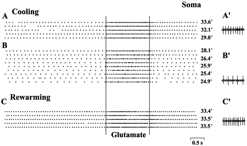
Yu. S. Mednikova et al.

                                  Dendro-Somatic Propagation

                                1. Introduction
                                When applying hypothermia and hypoxia exert almost identical disorders in
                                nerve tissues and that is why hypothermic and hypoxic changes in neuronal ac-
                                tivity are often considered together [1] [2] [3]. During long periods of even slight
                                and moderate hypothermia (t = 32˚C - 34˚C and 28˚C), as well as during hy-
                                poxic periods of any degree, the membrane permeability to ions, supporting the
                                stability of nerve membrane potential, increases, resulting in a flow of Na+ and
                                K+ in the direction of their thermodynamic gradients. The occurring decrease of
                                membrane potential opens potential-dependent Са2+ channels and leads to pa-
                                thologic entry of Ca2+ into cytosol, activation of phospholipases and proteases,
                                disintegration of neuronal membranes and necrotic death of the neurons [2] [3]
                                [4]. Such a dramatic chain of events describes the hypoxia-related disorders
                                quite well, because the first sign of hypoxic condition of the brain is a blockade
                                of Na+-K+-ATPase activity caused by deficiency of energy supply [5]. But under
                                hypothermic condition the reason for imbalance of ionic processes remains un-
                                clear: the activity of Na+-K+-ATPase even slightly increases with the fall of tem-
                                perature [6]; membrane potential and spike amplitude in neurons of frogs living
                                at low temperatures (which are mortal for the brains of homoeothermic animals)
                                have the same values as in mammalian neurons [7], while the energetic demands
                                of cold-blooded brain are significantly less [3] [8]. That is why neither tempera-
                                ture-dependence nor energetic limitations of Na+-K+-ATPase activity should be
                                considered as the reasons for the imbalance of ionic homeostasis in the cases of
                                hypothermia. Noting this contradiction, P. W. Hochachka had supposed that
                                there should be an additional mechanism sensitive to hypothermia, coupling
                                membrane functions and cellular metabolism, which had not been taken into
                                account earlier [3]. The evidence for such a mechanism follows from the fact
                                that a lot of hypothermic alterations of brain activity are linked to the same
                                temperature point: below 28˚C the mammals lose consciousness [9] [10], below
                                28˚C inflation of cortical EEG occurs [11], below 27˚C - 29˚C the frequency of
                                spontaneous neuronal activity decreases together with a sharp temperature de-
                                cline of the activation spike reaction to acetylcholine [12] [13].
                                   Muscarinic (M) cholinergic process is one of the main reactions of the brain.
                                It is involved in maintaining consciousness [14], providing active perception
                                [15], attention, memory and learning [16] [17]. Numerous functions of acetyl-
                                choline are associated with only M-cholinergic effect—regulation of membrane
                                properties of neurons via metabolic process limiting K+ permeability of cellular
                                membranes, thus increasing their specific resistance [18] [19] [20]. Functional
                                role of M-cholinergic process is related to regulation of conductive function of
                                dendrites, providing the decrease of excitation decrement when propagating

DOI: 10.4236/jbbs.2021.115009                        108                       Journal of Behavioral and Brain Science
Yu. S. Mednikova et al.
from dendrites to soma [21] and thus resulting in the increase of the level of
spontaneous neuronal firing [22]. High sensitivity of M-cholinergic reaction to
temperature [23] [13] and its dependence on the energy supply [24] were estab-
lished.
Thus, the revealed fact of decreasing the rate of M-cholinergic reaction in hy-
pothermia may be the reason of temperature-dependent ionic disbalance in the
neurons, which had been proposed by P. W. Hochachka [3]. The main parame-
ter regulated by hypothermia is the spontaneous activity of neurons [12]. The
relationship of this parameter with the cholinergic reaction of the brain is traced
by occasional change in the level of spontaneous firing [22]. The reason for the
change in the spontaneous activity became clear when analyzing the process of
dendrosomatic conduction of glutamatergic excitation, which turned out to be
associated with a cholinergic effect that block K+ channels on the neuronal
membranes. Therefore, it was suggested that hypothermia can have a triple effect
on the state of spike activity: ionic homeostasis can be disturbed [3], the rate of
the M-cholinergic reaction can be changed [13] [23] together with the efficiency
of dendrosomatic conduction [22]. Changes in all these parameters under con-
ditions of hypothermia were supposed to be studied on the cortical slices with
registration of spike activity. We used local delivery of mediators (glutamate and
acetylcholine) to different points on the neuronal membrane in order to estab-
lish the factor of variability of dendrosomatic conductance and expression of the
M-cholinergic reaction. The variability of studied parameters was analyzed in
different groups of neurons, differing in the rate of spontaneous activity. The
pathological effect of hypothermia was analyzed by the change in the amplitude
of neuronal spikes. It is quite possible that all hypothermic manifestations in the
activity of nerve cells are united by a single cause.
2. Methods
2.1. Slice Preparation and Incubation Conditions
Animals were treated with observance of recommendation on ethics of work
with animals offered by European Communities Council Direction (86/609 EEC)
and experimental protocols approved by ethics committees of Institute of Higher
Nervous Activity Russian Academy of Sciences. The experiments were carried
out on slices of sensorimotor cortex of guinea pigs (weight 200 - 250 g). After
fast decapitation of the animals by guillotine and scull break-up, the brain sur-
face was sprinkled by ice-cold aerated Ringer-Krebs solution. 500 μm thick slices
were prepared from longitudinal cortex block on VSL vibratome (World Preci-
sion Instruments, USA). Incubation chamber, where the slices were placed, was
consisted of two cells (reserve and experimental) with independent flow of
Ringer-Krebs solution. Incubation solution saturated by gas mixture (95%О2
+5%СО2) consisted of the following components (mM): 124-NaCl; 5-KCl;
1.24-KH2PO4; 1.3-MgSO4; 2.4-CaCl2; 26-NaHCO3 and 10-glucose (pH 7.4). Flow
rate of incubating solution was 1.5 - 3 ml/min. The slices were incubated at
DOI: 10.4236/jbbs.2021.115009 109 Journal of Behavioral and Brain ScienceYu. S. Mednikova et al.
room temperature for half an hour after preparation. Then, the temperature was
increased to 32 ºC - 34ºC. The slices were incubated at this temperature for 1.5 - 2
hours before the beginning of spike activity registration. The pre-heating of in-
cubation medium was performed by U1 thermostat (VEB, Germany). For finer
temperature regulation, a Peltier element-based thermostating device was used
(NPO “Biopribor”, Russia). The temperature in experimental cell was controlled
continuously by an electronic thermometer (NTC “NIKAS”, Russia).
2.2. Electrodes and Recording Equipment
Three-channel glass microelectrodes (diameter 7.4 - 8 μm) were used for extra-
cellular registration of neuronal spike activity and for iontophoretic transmitter
application. The channel for registration and channel for controlling iontopho-
retic current were filled with 3 M NaCl. The third channel was filled with 1 M
sodium glutamate (pH 7.5; Sigma Chemical Co., USA) or 2 M acetylcholine
chloride (pH 4.0; Sigma Chemical Co., USA). The channel for controlling ion-
tophoretic current was often replaced by a second channel for transmitter pho-
resis, because the action of currents chosen for transmitter phoresis but being
passed through current controlling channel did not evoke spike responses of
recorded neurons. After amplification (DAM 80, World Precision Instruments,
USA) and digitalization (E14-440, L-Card, Russia) the spike activity was input-
ted into computer (Intel (R) Core (TM) Duo) for storage, reproduction and
processing of registered signals.
2.3. Registration Area and Iontophoretic Current Parameters
A three-barrel microelectrode containing channel for recording was moved
along V layer of the cortex (1.2 - 1.6 mm from pial surface) in order to find spike
activity of neurons and transmitter applications to the soma of registered nerve
cells. After the detection of spike activity of a neuron, the independent micropi-
pette, containing only phoretic channel, was placed at different points of pre-
sumable dendritic tree (Figure 1). Short-time bursts of spike activity registered
by “somatic” electrode during glutamate application through “dendritic” elec-
trode served as indicators of its location near dendritic surface [25]. The distance
between somatic and dendritic micropipettes was measured by an eyepiece pro-
vided with micrometric scale. Glutamate was applied to dendrites and soma by
60 - 80 nA current (negative pole inside the electrode); acetylcholine was applied
to any membrane locus by 70 nA current (positive pole inside the electrode).
The duration of iontophoretic current for glutamate application was from 1 to
4.5 s, whereas for acetylcholine application it was always 4.5 s. Throughout an
intervals between iontophoretic ejections the retaining 3 - 5 nA current of oppo-
site direction was set in each of phoresis channel. Iontophoretic glutamate ap-
plication to local dendritic points in the course of gradual withdrawal of micro-
pipette from the point of maximal effect allowed to find out that the effective
dose of glutamate near dendritic surface could be achieved at the distance not
DOI: 10.4236/jbbs.2021.115009 110 Journal of Behavioral and Brain ScienceYu. S. Mednikova et al.
Figure 1. Scheme for recording spike activity and application of mediators to different
loci of neuronal membrane. The spike activity was recorded from the neurons of layer V
of guinea pig sensorimotor cortex in the soma region (1). The mediators were injected to
soma region and dendritic loci: 2—channel for acetylcholine phoresis; 3—channel for
glutamate phoresis.
exceeding 20 μm, which implies a high locality of iontophoretic action.
2.4. Experimental Protocols
Standard temperature, 32˚C - 34˚C, was maintained constantly in the reserve
cell of incubating chamber, whereas in the experimental cell it was maintained
during the procedure of searching for spike activity of the neurons and its con-
trol testing. Cooling the solution in the experimental cell to 21˚C - 24˚C was
performed with the rate of two degrees per minute. The rate of temperature res-
toration was the same. Thus, the whole cycle of hypothermic action took not
longer than 10 minutes. During cooling, the neurons were always tested by a
certain application manner: glutamate application to soma or dendrites every 12
s or acetylcholine application to soma every 24 s. If registration conditions al-
lowed, the neuronal activity was observed during an hour after the episode of
hypothermia. Every hypothermic action was carried out usually on a fresh slice
in order to avoid post-hypothermic events.
2.5. Data Analysis
The parameters of spike activity were analyzed by Power-Graph 3.3 computer
program (PO “Power-Graph”, Russia). An average level of spontaneous activity
in 3-second interval before every iontophoretic application was calculated for
pre-hypothermic, hypothermic and post-hypothermic periods. Spike amplitudes
were also determined in these periods. Spike reactions of the neurons to trans-
mitter application were estimated by the duration of latency period of the re-
sponse and its intensity. Difference between maximal current average of spike
DOI: 10.4236/jbbs.2021.115009 111 Journal of Behavioral and Brain ScienceYu. S. Mednikova et al.
frequency in the response and in preceding background activity served as esti-
mates of the intensity of the reaction. The significance of the parameters’ altera-
tions was determined by non-parametric statistical methods [26].
3. Results
In the experiments on the layer V of sensorimotor cortex slices, the activity of
111 neurons was registered. The level of spontaneous activity varied from 0 to 25
impulses per second in different neurons. The distribution of the neurons in ac-
cordance with the level of spontaneous activity is presented on Figure 2. As can
be seen, a significant majority of the neurons have no background activity.
Presence of such neurons was detected by arising of spike activity in response to
glutamate applied to the soma by short pulses of current. Among 111 nerve cells,
37.8% (42 neurons) had no spontaneous activity, 27 neurons (24.3%) had spon-
taneous activity frequency up to 4 impulses per second, whereas high-frequency
neurons (8 - 25 impulses per second) comprised 18.9 % of the population
(Figure 2).
The results of previous experiments [13] [22] and data obtained by other au-
thors [27] gave evidence that alteration degree of spontaneous activity of the
neurons significantly depends on their initial firing level. That’s why the analysis
of spontaneous activity frequency under short-time hypothermia was performed
separately for the neurons divided in groups depending on the background ac-
tivity level. Group I consists of neurons without background activity; group II
included neurons with activity below 4 impulses per second; group III consists of
neurons with activity from 4 to 8 impulses per second; group IV consists of
Figure 2. Distribution of neurons of the sensorimotor cortex according to the frequency
of spontaneous activity. The abscissa shows the frequency of spontaneous activity
(imp/s); The ordinate axis is the percentage of neurons that have a given level of sponta-
neous firing. At the top right is the number of registered neurons in the sensorimotor
cortex.
DOI: 10.4236/jbbs.2021.115009 112 Journal of Behavioral and Brain ScienceYu. S. Mednikova et al.
high-frequency neurons (from 8 to 25 impulses per second)
Overall, 35 neurons were registered during hypothermia procedure. Figure 3
shows most typical cases of changing spontaneous activity level in the neurons of
Figure 3. Frequency of spontaneous activity of sensorimotor cortex neurons in the course
of decreasing temperature from 34˚C to 21˚C and after restoration of the initial tempera-
ture values. Each graph illustrates the dynamics of single neuron spontaneous activity
frequency during cooling from 32˚C - 34˚C to 21˚C - 22˚C (A) and after restoration of
the initial temperature (B). I, II, III, IV—four groups of nerve cells with different levels of
spontaneous activity before cooling: I—neurons without spontaneous activity (0 imp/s);
II—neurons with spontaneous activity frequency up to 4 imp/s; III—neurons with spon-
taneous activity frequency from 4 to 8 imp/s; IV—neurons with spontaneous activity
frequency above 8 imp/s. A: abscissa—temperature, ordinate—spontaneous activity fre-
quency, (imp/s). B: horizontal axis - 1 and 2, - the same temperature value (32˚C - 34˚C)
before (1) and after (2) exposure to hypothermia; vertical axis – spontaneous activity fre-
quency, (imp/s).
DOI: 10.4236/jbbs.2021.115009 113 Journal of Behavioral and Brain ScienceYu. S. Mednikova et al.
all four groups during decrease of incubation liquid temperature in the experi-
mental cell from 32˚C - 34˚C to 21˚C - 22˚C (Figure 3(A)) and further restora-
tion of the temperature to the initial values (Figure 3(B)).
The specific feature for neurons of all four groups was the lack of significant
difference in spontaneous activity level at 30˚C compared to 34˚C (Wilcoxon
matched pairs signed rank test, α >> 5%). Systematic changes of neuronal activ-
ity frequency began to appear by cooling incubation medium below 30˚C, hav-
ing different rate of firing change in different neurons and as it is quite interest-
ing, these changes may be either increase or decrease of spike activity in different
neurons. The neurons with the same level of spontaneous activity under cooling
and at t = 32˚C - 34˚C were also present. Despite all the diversity of observed al-
terations, their dependence from the initial spontaneous activity level before
cooling can be seen. Spontaneously inactive neurons almost in all cases of tem-
perature fall below 30˚C displayed the increase of spontaneous activity frequency
the more significant the deeper cooling (Figure 3(A)-I). Among the neurons
with spontaneous activity up to 4 impulses per second, the increase of activity
rate during cooling also prevailed, but the decrease of spontaneous activity could
also be found in certain neurons (Figure 3(A)-II). In the group of neurons hav-
ing the activity of 4-8 impulses per second, the majority of registered cells dis-
played the decrease of spontaneous activity below 30˚C, but one neuron showed
growth of frequency (Figure 3(A)-III). Finally, high-frequency neurons showed
only decrease of spontaneous activity under hypothermia (Figure 3(A)-IV). The
neurons from groups III and IV showed significant decrease of spontaneous ac-
tivity under cooling revealed at t = 26˚C compared to the level at 30˚C (Wilcox-
on matched pairs signed rank test, α < 1%). The significant increase of activity
rate under hypothermia in groups I and II was achieved at t = 24˚C compared to
30˚C (Wilcoxon matched pairs signed rank test, α < 1%).
Restoration of initial temperature values (32˚C - 34˚C) after episodes of hy-
pothermia lead to the increase of spike activity compared to the level before
cooling in almost all neurons for any character of activity changes under cooling
(Figure 3(B); Wilcoxon matched pairs signed rank test, α < 0.1%). Hyperactivity
of low-frequency neurons (groups I and II) was characterized by an increase of
frequency by 231.4% ± 40% compared to the level before cooling, and for the
neurons with activity above 4 impulses per second (groups III and IV) it was
only by 34.9% ± 5% of the initial level. The hypothermia-induced increase of
spike activity was maintained during long time after restoration of initial tem-
perature: in one third of the neurons recovery period lasted as long as the
post-hypothermic time of observation (1 hour). Fast restoration of activity fre-
quency was detected mainly just for high-frequency neurons from group IV,
which displayed full restoration of activity in 10 - 20 min after episodes of hy-
pothermia.
Cooling-related alterations of spontaneous activity level in nerve cells were
accompanied by decrease of spike amplitude observed in all neurons without
DOI: 10.4236/jbbs.2021.115009 114 Journal of Behavioral and Brain ScienceYu. S. Mednikova et al.
dependence on the direction of change of neuronal activity level under hypo-
thermia (Figure 4(A)). The decrease of spike amplitude during cooling incuba-
tion medium from 34˚C to 30˚C was very slight: 6.3 ± 0.5 μV in average (Wil-
coxon matched pairs signed rank test, α < 5%). A dramatic drop of spike ampli-
tude, by 34.5 ± 4 μV on average, occurred when temperature had fallen from
30˚C to 26˚C (Wilcoxon matched pairs signed rank test, α < 0.1%). Further
cooling led to even more significant decrease of spike amplitude down to com-
plete spike elimination in the noise of certain neurons (Figure 4(A)). Restora-
tion of initial temperature in overwhelming majority of cases did not lead to to-
tal restoration of spike amplitude (Figure 4(B)). The decrease of spike ampli-
tude compared to the value before cooling was 15.5% on average (Wilcoxon
matched pairs signed rank test, α < 0.1%). Half of the neurons did not display
full restoration of spike amplitude even in hour of observation after hypothermic
episode. Only in the group of high-frequency neurons (group IV) the majority of
nerve cells showed restoration of initial spike amplitude right after restoration of
initial temperature of incubation medium (Figure 4(B)-IV).
Thus, the level of spontaneous activity and spike amplitude of the neurons
undergo the most significant alterations in the temperature range 27˚C - 29˚C.
As shown earlier, a dramatic decrease of activation response to acetylcholine ap-
plied iontophoretically to nerve cells takes place in the same temperature range
[13]. The reaction to acetylcholine comprises slow increase in spike activity fre-
quency and a long lasting period of activation (till 10 s and more) after termina-
tion of phoretic current (Figure 5(I)-1 and Figure 5(II)-1), which shows that its
development results from muscarinic mechanism [19] [20]. Nine neurons tested
by acetylcholine applications during hypothermic action showed no significant
difference in spike rate increments over background activity in response to ace-
tylcholine at 34˚C and 30˚C (Wilcoxon matched pairs signed rank test, α>>5%).
Meanwhile, at t = 26˚C - 27˚C acetylcholine applications exerted either no in-
crease of spike activity above background level (Figure 5(I)-2) or the increase of
firing was much weaker and had a longer latency period (Figure 5(II)-2). The
reaction to acetylcholine estimated by increment of spike rate over the back-
ground level was significantly lower for all 9 neurons at t = 26˚C than at t = 30˚C
(Wilcoxon matched pairs signed rank test, α < 1%). It is also clearly shown on
Figure 5(II) that spontaneous activity of the neuron may disappeared simulta-
neously with the weakening of the reaction to acetylcholine at 27˚C (Figure
5(II)-1-2), which indicates on common temperature dependence of cholinergic
process and the process of regulating spontaneous activity.
Figure 6 graphically demonstrates the changes of spontaneous activity level
and increment of firing value under acetylcholine action during the decrease of
temperature from 30˚C - 31˚C to 26˚C - 27˚C. The temperature-related differ-
ences are presented for 9 neurons in percentage to the level of spontaneous ac-
tivity (Figure 6(A)) and the expression of response to acetylcholine (Figure 6(B))
at t = 30˚C - 31˚C. As it was shown on Figure 3, the level of spontaneous activity
DOI: 10.4236/jbbs.2021.115009 115 Journal of Behavioral and Brain ScienceYu. S. Mednikova et al.
in different neurons was either decreasing (5 neurons), or increasing (4 neurons)
at 26˚C - 27˚C (Figure 6(A)). At the same time, the response to acetylcholine
during cooling was decreasing for any character of spontaneous activity changes;
Figure 4. Spike amplitude of sensorimotor cortex neurons during decrease of tempera-
ture from 34˚C to 21˚C and further restoration of initial temperature values. Each graph
illustrates the dynamics of single neuron spike amplitude changes during cooling from
32˚C - 34˚C to 21˚C - 22˚C (A) and during restoration of the initial temperature (B).
Temperature-related changes of spike amplitude presented for the neurons which spon-
taneous activity is indicated on Figure 3(I-IV)—four groups of nerve cells with different
levels of spontaneous activity before cooling (as shown on Figure 3). А: abscis-
sa—temperature, ordinate—spike amplitude, μV. B: horizontal axis—1 and 2, the same
temperature value (32˚C - 34˚C) before (1) and after (2) exposure to hypothermia; vertic-
al axis—spike amplitude, μV. The line parallel to abscissa axis on each fragment desig-
nates noise level of the recording equipment.
DOI: 10.4236/jbbs.2021.115009 116 Journal of Behavioral and Brain ScienceYu. S. Mednikova et al.
Figure 5. Responses of the sensorimotor cortex neurons to iontophoretic acetylcholine
application during cooling of incubating medium below 30˚C. Represented two different
neurons (I and II) of layer V of sensorimotor cortex. Acetylcholine was applied to the
soma by 70 nA current (positive pole inside the electrode) simultaneously with recording
spike activity of the neurons. Neuronal spike activity was recorded at following tempera-
tures: I—28.1˚C (1); 27.3˚C (2); II—30˚C (1) and 27˚C (2). Duration of acetylcholine
electrophoresis current is shown as a line under each record of neuronal activity.
Figure 6. Changes in spontaneous activity level and level of the response to iontophoretic
acetylcholine application during hypothermia. The plots schematically represent changes
in activity of 9 sensorimotor cortical neurons tested by iontophoretic acetylcholine appli-
cation during cooling the incubation medium. Electrophoretic acetylcholine current equals
70 nA everywhere (positive pole inside the electrode). (A) Average level of spontaneous
activity for each neuron. Horizontal axis: temperature values: 1) t = 26˚C - 27˚C; 2) t =
30˚C - 31˚C; vertical axis—spontaneous firing rate of each neuron as percentage of 30˚C -
31˚C level taken as 100%. (В) Level of response of each neuron to iontophoretic acetyl-
choline application as a difference between maximal average spike frequency in response
periods and in preceding background activity. Horizontal axis: temperature values like on
A; vertical axis: level of response of each neuron to acetylcholine as percentage of re-
sponse level at t = 30˚C - 31˚C taken as 100%.
for 7 of 9 neurons, the decline of the reaction was more than 60% (Figure 6(B)).
Thus, the decrease of M-cholinergic reaction in temperature range 27˚C - 29˚C
DOI: 10.4236/jbbs.2021.115009 117 Journal of Behavioral and Brain ScienceYu. S. Mednikova et al.
causes both increase and decrease of spontaneous activity frequency in different
neurons. This means that temperature-dependent decrease of cholinergic reac-
tion rate may act in two opposite directions on mechanisms regulating sponta-
neous activity level.
The presence of two mechanisms regulating the level of spontaneous activity
can be revealed by comparison of spontaneous change of activity level under
standard temperature, 32˚C - 34˚C, and under the same temperature before
and after hypothermic action. The shift to higher activity level at standard
temperature conditions did not lead to significant changes of spike amplitude in
cortical neurons (Wilcoxon matched pairs signed rank test, α >> 5%). At the
same time, rising hyperactivity in nerve cells, registered after short episodes of
hypothermia (Figure 3(B)) almost ever was accompanied by decrease of spike
amplitude (Figure 4(B); Wilcoxon matched pairs signed rank test, α < 1%) at
the same temperature 32˚C - 34˚C before and after cooling. For comparison,
Figure 7 shows two cases of increase of spike activity in different neurons:
during hypothermic increase of firing activity (Figure 7(A)) and spontaneous
change of activity under standard temperature conditions (Figure 7(B)). In
the first case the increase of firing rate, started at 30.1˚C and more expressed at
lower temperature, was accompanied by significant (Wilcoxon-Mann-Whitney
Figure 7. Hypothermia-induced and spontaneous increase of neuronal firing rate. The
activity of two different neurons (A and B) is presented. (A) Increase of spike activity as a
result of hypothermia. (В) Spontaneous increase of spike activity. I—Relations between
firing rate (spikes/s) and spike amplitude (µV). II—Records of spike activity: in A—1)
before cooling (33.5˚C), 2-4) in the course of cooling (from 31.8˚C to 26.6˚C); in B—each
record made with 10 min intervals at temperature 34.2˚C - 34.5˚C.
DOI: 10.4236/jbbs.2021.115009 118 Journal of Behavioral and Brain ScienceYu. S. Mednikova et al.
test, α < 0.005) fall of spike amplitude (Figure 7(A)-I and Figure 7(A)-II,1-4).
In the second case the increase of discharge rate at nearly constant temperature
had occurred without changes in spike amplitude (Figure 7(B)-I and Figure
7(B)-II,1-3).
Thus, the increase of activity frequency actually depends on two mechanisms,
one of them being related to the drop of spike amplitude and another being not
related to spike amplitude alterations. Both opportunities for increasing level of
spontaneous activity, however, should depend on a single mechanism of its reg-
ulation. Earlier it was found that the neuronal spontaneous firing is as higher as
shorter the latency period of somatic response to dendritic glutamate application
[22] [28]. The same parameter was the most variable during changes of sponta-
neous activity level whatever the cause of these changes. Figure 8 shows the
example of two cases of increasing duration of latency period of response to glu-
tamate applied to dendritic point (100 μm from soma) of the same neuron, when
spontaneous activity decreases to zero level.
Figure 8. Hypothermia-induced and spontaneous decrease of spike activity of the senso-
rimotor cortical neuron during simultaneous recording of spike response to iontophoret-
ic glutamate application to dendritic locus. Glutamate was applied to dendritic locus at
100 μm from soma by 80 nA current (negative pole inside the electrode). (А) activity of
the neuron during cooling the incubation medium from 34˚C to 23.2˚C (1-5). (В) activity
of the neuron in post-hypothermic period at different time moments after restoration of
initial temperature: 1) 10 min; 2) 15 min; 3) 1 hour; 4) 1 hour 15 min. Time of glutamate
iontophoretic current is shown as a line under each record.
DOI: 10.4236/jbbs.2021.115009 119 Journal of Behavioral and Brain ScienceYu. S. Mednikova et al.
In the first case (Figure 8(A)), the decrease of frequency took place under in-
fluence of cooling the incubation solution to 26˚C and lower (Figure 8(A)-4-5)
and it was accompanied by a slight decrease of spike amplitude. In the second
case, the frequency dropped spontaneously while registering the activity at
constant temperature (near 34˚C) 1 hour after hypothermic action (Figure
8(B)-3-4) and almost full restoration of spike activity changed during hypo-
thermia (Figure 8(B)-1-2). In both cases, the decrease of spontaneous activity
frequency occurred simultaneously with increase of latency period of the re-
sponse to local glutamate application to dendritic point: from 150 to 400 ms in
the first case and from 140 ms to 350 and 700 ms in the second one. Thus, both
hypothermia-induced and spontaneous drops of frequency are related to wea-
kening of dendritic impacts onto the neuronal soma.
Figure 9 illustrates similar changes under hypothermic influence observed af-
ter the decrease of temperature to 29˚C and below. Spontaneous activity disap-
pears at 29˚C (Figure 9(A)-4) and increase of latency period of the response to
glutamate application to dendritic locus occurs at the same temperature. The la-
tency period of the reaction reaches the value of 2500 ms at t = 28˚C (Figure
9(A)-6), whereas at t = 27˚C a reverse process is beginning: the latency period of
the response to local dendritic depolarization decreases to 400 ms (Figure
9(B)-1), followed by the restoration of spontaneous activity with higher frequency
Figure 9. Responses of sensorimotor cortical neuron to iontophoretic glutamate applica-
tion to dendritic locus during hypothermic decrease of spike frequency and its posthypo-
thermic increase. Glutamate was applied to dendritic locus at 150 μm from soma (A)
when cooling the incubating medium from 32˚C to 28˚C (1-6) and (B) during tempera-
ture restoration (1-3). Iontophoretic current and duration of its action are the same as for
Figure 8.
DOI: 10.4236/jbbs.2021.115009 120 Journal of Behavioral and Brain ScienceYu. S. Mednikova et al.
at t = 28.5˚C. Restoration of spontaneous activity is accompanied by decrease of
spike amplitude, increase of spike burst activity and masking the activation
reaction to glutamate application by intensified spontaneous activity (Figure
9(B)-2-3).
Figure 10 shows a case of appearance of spontaneous activity under hypo-
thermia after decrease of temperature below 26.9˚C for the neuron, which was
inactive at higher temperatures. The lifting of spontaneous activity is accompa-
nied by decrease of spike response latency to local dendritic application of glu-
tamate from 650 ms at t = 32.8˚C - 26.9˚C (Figure 10(A)-1-3) to 350 ms at t =
23˚C (Figure 10(A)-5). These changes occur along with the decrease of spike
amplitude. The following temperature restoration up to initial values demon-
strates the hyperactivity phenomenon, i.e. maintenance of spontaneous activity
in 10 minutes of incubation above 33˚C with simultaneous short-latency reac-
tion to local depolarization of a dendritic point (Figure 10(B)-1). After 15 mi-
nutes of restoration period the spontaneous activity disappears and the spike re-
sponse to dendritic glutamate application simultaneously with spike amplitude
are restored (Figure 10(B)-2).
Stimulation of dendritic points (distance from soma ranging from 50 to 250
μm) under hypothermic conditions was carried out on 20 neurons. As well as
spontaneous activity level, the responses induced from dendrites also changed
during cooling. Decrease of spontaneous activity level (6 neurons) was always
accompanied by increase of latency periods or even disappearing of the re-
sponse, independent on distance from the soma. Increase of spike frequency
Figure 10. Increase of spontaneous activity of the neuron during hypothermia and res-
ponses to glutamate application to dendritic locus. Glutamate was applied to dendritic
locus at 100 μm from soma. (А) Records of neuronal activity during cooling the incuba-
tion medium from 32.8˚C to 23˚C (1-5); (В) Records of neuronal activity during restora-
tion of incubation medium temperature: 1) 5 min after restoration, 2) 15 min after resto-
ration. Iontophoretic current and duration of its action are the same as for Figure 8.
DOI: 10.4236/jbbs.2021.115009 121 Journal of Behavioral and Brain ScienceYu. S. Mednikova et al.
when cooling or during post-hypothermic period took place either along with
decrease of latency period of the response to dendritic stimulation (6 neurons),
or (in the cases of markedly increase of spike frequency) along with masking of
the response (3 neurons). If hypothermia did not cause changes in spontaneous
activity, the changes in response expression to glutamate applied to dendrites
also were not observed (5 neurons).
On the contrary, spike responses to glutamate applied to soma (6 neurons)
were always stable in hypothermic conditions and did not change together with
changing spontaneous activity. Figure 11 illustrates the stable spike reactions to
glutamate injected to neuronal soma in wide temperature zone (from 33.6˚C to
24.9˚C) while spontaneous activity is constant only from 33.6˚C to 29˚C (9
imp/s—Figure 11(A)), decreases below 29˚C (7.4 imp/s at 28.1˚ and 4.6 imp/s at
24.9˚—Figure 11(B)) and increases after restoration of the initial temperature
from the hypothermic episode (12.6 imp/s—Figure 11(C)).
Thus, despite all the diversity of symptoms, the analysis of the effects of hy-
pothermia on the activity of cortical neurons revealed the following:
1) A correlation between changing spontaneous activity caused by hypother-
mia and conductive efficiency of glutamatergic excitation along the dendrites
was found.
2) In most of the cases, the decrease of spike amplitude had occurred when the
temperature was decreased.
3) The revealed changes in the activity of cortical nerve cells occurred in tem-
perature interval from 27˚C to 29˚C, where excitatory spike reactions to acetyl-
choline sharply attenuates.
4) The neuronal spike reactions to glutamate, applied to soma are stable in the
Figure 11. Changes of spontaneous activity of neuron during hypothermia and responses
to somatic application of glutamate. Rasters spike density histograms during cooling the
incubation medium from 33.6˚C to 24.9˚C (A) and (B) and after rewarming to initial
temperature (C). The time of glutamate injection to soma (60 nA, negative pole inside the
electrode) is restricted by vertical lines. (A') Spontaneous spike activity at temperature
33.6˚C; (B') Spontaneous spike activity at temperature 24.9˚C. (C') Spontaneous spike ac-
tivity after restoration of initial temperature (33.5˚C).
DOI: 10.4236/jbbs.2021.115009 122 Journal of Behavioral and Brain ScienceYu. S. Mednikova et al.
course of hypothermia.
4. Discussion
4.1. The Main Reason of Hypothermic Effect in the Brain is the
Temperature Related Attenuation of M-Cholinergic Reaction
The obtained data give evidence that hypothermic action leads to serious dis-
orders in the activity of cortical nerve cells. Small changes are occurring even in
34˚C - 30˚C temperature range, comprising slight decrease of spike amplitudes
of cortical neurons, but global problems begin to take place when cooling below
30˚C. As shown earlier [13] and confirmed by the current study (Figure 5 and
Figure 6), below this point there is the first transition zone for M-cholinergic
brain reaction, where (27˚C - 29˚C) the decrease of its rate occurs. M-cholinergic
process blocks any types of K+ channels on neuronal membranes [18] [19] [20]
and sharp temperature related attenuation of this process provides additional
growth of K+ permeability and facilitates K+ efflux from nerve cells in the direc-
tion of concentration gradient. Such ionic flow below 27˚C is so significant in
mammals that the capabilities of Na+, K+—ATPase to restore ionic homeostasis
cannot fully compensate the disorder. The membrane potential begins to de-
crease, which leads to significant decrease of spike amplitude even under short
hypothermia action (Figure 4). All other hypothermia-induced disorders are the
consequences of these two processes, i.e. temperature limitation of M-cholinergic
reaction rate and progressive K+ ion accumulation in extracellular space.
4.2. Mechanism of Formation and Regulation
of Spontaneous Activity
The most varying parameter during cooling is the frequency of spontaneous
spike activity in nerve cells. The basic reason for formation of spontaneous ac-
tivity is the constant flow of miniature dendritic glutamatergic EPSPs [29],
which can have an amplitude of several tens mV at the origin points on dendrite
loci [30], but are rapidly attenuated during motion towards the soma [29]. The
conditions of more effective excitation moving along dendrites are determined
by the geometry of neurons and specific resistance of their membranes [31].
That is why acetylcholine, blocking K+ permeability of the neuronal membranes
when interacting with M-cholinoreceptors [18] [19] [20], leads to both more ef-
fective excitation conduction along the dendrites [21] and increase of spontane-
ous activity level [22].
Limitation of K+ permeability of the neuronal membranes could also be
achieved via increase of extracellular K+ concentration, which takes place at
temperature below 27˚C, when the rate of M-cholinergic process decreases. As a
result, the specific resistance of cell membranes is increasing, which has been
recorded as increase of input resistance below 27˚C [32] [33] and has as conse-
quence more effective conducting the flow of miniature excitatory postsynaptic
potentials from dendrites to soma for its transformation into spike sequence.
DOI: 10.4236/jbbs.2021.115009 123 Journal of Behavioral and Brain ScienceYu. S. Mednikova et al.
The proof for large increase of extracellular concentration of K+ ions below 27˚C
is the inhibition of A-type K+ current [34], the increase of spike width and slow-
ing the rates of spike rise and fall recorded intracellularly [32] [33], as well as
bursts formation in activity of some neurons after cooling and further tempera-
ture restoration (Figure 9). The adduced data point on a decrease of repolariza-
tion rate of neuronal membranes after depolarizing potential shifts, which is the
consequence of increased K+ concentration in extracellular medium below 27˚C.
Therefore we conclude that two processes influence on spontaneous firing under
hypothermia: from one side, the increase of membrane K+ permeability via de-
crease of M-cholinergic reaction rate at 27˚C - 29˚C, and from another side, de-
crease of K+ permeability due to progressive growth of K+ concentration in
extracellular space. Prevailing of one or another process should depend on struc-
tural and membrane properties of the neurons, which would determine the dy-
namics of spontaneous spike activity changes of cortical neurons under cooling
(Figure 3).
4.3. Diversity of K+ Membrane Conductance is Crucial for
Peculiarities of Neuronal Activity during Hypothermia
Density of K+ channels on the membranes of mammalian cortical and hippo-
campal neurons is differently expressed on various nerve cells [35] [36]. K+ con-
ductance is increasing with age ever since 7-th day of life, and in mature cells
becomes fully developed. Mature low-frequency hippocampal granule cells have
input resistance lower than 200 MΩ, suggesting that high density of K+ channels
is crucial for sparse firing (King et al., 2020). Due to this, one can imply that
neurons having low [36] initial spontaneous activity level (up to 4 impulses per
second, i. e. experimental groups I and II—Figure 3(A)) have a large average
density of K+ channels on the membranes. It may determine their low activity
level as a result of a large decrement of EPSP amplitude when moving along the
dendrites. For the same reason, hypothermic increasing K+ permeability of the
membranes below 27˚C leads to rapid increase of K+ concentration outside of
these cells. That is why fast decrease of spike amplitude occurs, whereas sponta-
neous activity was increasing with further accumulation of K+ ions in extracellu-
lar environment as a result of concentration-dependent increase of membrane
resistance (Figure 3(A)-I-II; Figure 7(A)).
The opposite effect caused by hypothermia mainly in neurons with activity
above 4 spikes per second (experimental groups III, IV—Figure 3(A)) is appar-
ently linked to relatively low content of K+ channels in their membranes. As a
result, under 27˚C the prevailing influence on spontaneous activity level is con-
nected with opening of K+ channels, leading to drop of conductive function of
the dendrites and decrease of activity rate (Figure 3(A)-III-IV). Increase of
extracellular K+ concentration under these conditions is slow, appearing as a
small decrease of spike amplitude and a slight hyperactivity after temperature
restoration.
DOI: 10.4236/jbbs.2021.115009 124 Journal of Behavioral and Brain ScienceYu. S. Mednikova et al.
4.4. Hypothermic Pathology Related
to Evolutionary Development
Efflux of K+ ions from neurons beginning at 27˚C does not end further and can
lead to a significant decrease of membrane potential level under further cooling
or just in a certain time course [32]. Together with the depolarization shift, the
drop of spike amplitude takes place (Figure 4), which is confirmed by earlier
investigations [12] [37] [38], and the destructive intracellular processes are trig-
gered [3] [8]. But even a small stable depolarization for potential-dependent K+
channels is quite sufficient to open ever more [39], causing further increase of K+
efflux from the cell and further depolarization shift [6]. Thus a self-maintained
pathological process appears, resulting in increased extracellular K+ concentra-
tion maintained for a long time after restoration of initial brain temperature.
Experimentally it was observed as a long period of increased posthypothermic
spontaneous activity frequency and long period of the decrease of spike ampli-
tude.
The lack of change in activation spike reactions during glutamate application
to soma on cooling (Figure 11) supports the results of previous studies [12] and
evidences that glutamate sensitivity is not changing under hypothermia. This
fact allows us to draw a conclusion that whatever the reason of change of neu-
ronal spontaneous activity would be, its drop is finally related to the decrement
of glutamatergic EPSP amplitude moving along the dendrites. Figure 8 and
Figure 9 show that the disappearing of spontaneous activity below 27˚C - 29˚C
is coupled to prolongation of latency periods of responses to iontophoretic glu-
tamate application to dendritic loci (Figure 8(A)-4-5 and Figure 9(A)-4-6).
This means that under hypothermia the stationary excitation flow passing along
the dendrites is so strongly attenuated that it can hardly penetrate the soma for
overcoming its shunting properties. The same event can happen spontaneously
(Figure 8(B)-3-4), without altering temperature, but due to spontaneous limita-
tion of acetylcholine efflux from cholinergic synapses in the cortex [40] in regis-
tered zone. During the decrease of spontaneous activity under stationary condi-
tions the prolongation of latency period of the response to glutamatergic stimu-
lation of the dendrite appears to be even more significant than under action of
hypothermia (Figure 8(B)-4 and Figure 8(A)-5), because it is regulated only by
attenuation of influence from cholinergic system and is not interfered by the in-
crease of membrane resistance related to the increase of extracellular K+ content.
Thus, the dendrites should be considered as a special machinery for spontaneous
activity formation, which is finely regulated via alteration of K+ permeability of
neuronal membranes. At the temperatures specific for homoeothermic animals
in stationary conditions it occurs mainly due to the regulating effect of brain
cholinergic system.
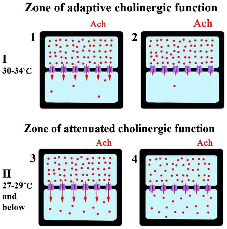
The scheme on the Figure 12 demonstrates the changes in the cholinergic
reaction during hypothermia in the temperature range of 27˚C - 29˚C. Above
this range M-cholinergic reaction performs an adaptive function, blocking K+
DOI: 10.4236/jbbs.2021.115009 125 Journal of Behavioral and Brain ScienceYu. S. Mednikova et al.
channels of neuronal membranes and increasing their resistance (Figure 12-1
and Figure 12-2). An increase in membrane resistance promotes more efficient
conduction of excitation from dendrites and creates an adaptive growth of
spontaneous activity. Below 27˚C, the rate of the M-cholinergic reaction de-
creases, which leads to opening the K+ channels and the gradual leakage of K+
ions from the cell (Figure 12-3). This process is opposite to that which take
place in the adaptive zone of cholinergic action – membrane resistance and
spontaneous activity are falling. In this case the adaptive function of acetylcho-
line weakens, but it is replaced by a passive process of reducing the K+ trans-
membrane current due to concentration accumulation of K+ ions on the outer
side of the membrane (Figure 12-4). Membrane resistance increases and con-
duction of excitation from dendrites improves. The resulting passive growth of
spontaneous activity is not adaptive. It occurs as a consequence of a growing
impairment of ionic homeostasis and coincided with the fall of spike amplitude.
This state, named delirium, with further equalization of the K+ ions concentra-
tion on the both sides of the membranes can lead to cold death.
The described functional and pathological changes in the nervous system
during hypothermia have another feature—they are characteristic only for
warm-blooded neuronal content. The rate of M-cholinergic reaction in temper-
ature range 27˚C - 36˚C is sharply increasing twice. The first temperature de-
pendent shift at 27˚C - 29˚C is a reason for hypothermic disorder of activity of
the cortical neurons. The second temperature-dependent shift occurs at 34˚C -
Figure 12. Scheme of hypothermic effect on the cholinergic reaction and K+ transmem-
brane current. Each fragment of the figure (1 - 4) depicts the neuronal inside (top) and
outside environment which are separated by a membrane with built-in K+ channels. The
circles represent potassium ions, arrows indicate K+ transmembrane current under dif-
ferent functional conditions. The rate of M-cholinergic reaction (concentration-depended
– I and temperature-depended – II) is indicated by different font sizes depicting the effect
of acetylcholine (Ach). 1 and 2 – state of K+ permeability with different release of acetyl-
choline (Ach) in zone I; 3 and 4 – different state of K+ permeability during hypothermia
(zone II) and accumulation of K+ ions in the outer side of the membrane over time.
DOI: 10.4236/jbbs.2021.115009 126 Journal of Behavioral and Brain ScienceYu. S. Mednikova et al.
36˚C and is characterized by significant increase of the rate of the process [13].
The high rate of cholinergic reaction regulating the permeability state of К+-
channels of neuronal membranes at brain temperatures normal for homoeo-
thermic animals requires great energetic support. The energetic demands of this
process can be fully provided only by perfecting of circulatory, respiratory, di-
gestive and other organs, which makes the difference between the any of ho-
moeothermic animals and their poikilothermic evolutionary precursors [8].
Thermodynamically almost impossible affords to regulate specific for homoeo-
thermic animals low-ohm neuronal membranes lead to high dependence of their
brain from energetic supply, the lack of which leads straight to hypoxic disorders
[3] [5] [8] despite the additional existence of glial cells that contribute to effi-
cient energy supply of neurons and maintain ionic constancy of the extracellular
compound [41] [42]. The pathological alterations caused by hypoxia are abso-
lutely identical to pathological processes occurring under hypothermia because
the first sign of hypoxia is limitation of Na+, K+-ATPase activity followed by ac-
cumulation of K+ ions in extracellular space [5]. That is why hypoxia and hypo-
thermia are equally destructive for mammals, and the Nature leaves us only
1.5˚C temperature interval for normal functional working of our perfect brain
[8]. And what a high price had to be paid for this perfection.
5. Conclusion
The main reason of hypothermic disorders in functional activity of cortical neu-
rons is temperature-dependent limitation of the rate of M-cholinergic reaction
of the brain, which is a regulator of membrane properties of nerve cells by clos-
ing K+ channels. The decrease of this process rate below 27˚C reduces the influ-
ence of excitation, permanent arising in dendrites, on the soma, resulting fall of
spontaneous activity, and in the case of homoeothermic animals leads to neu-
ronal homeostasis impairment and K+ accumulation in extracellular space. This
is the reason of spike amplitude attenuation and pathological rise of spontaneous
firing in some cortical neurons under hypothermic conditions. Different func-
tional properties of cortical neurons, providing the different efficiency of den-
dro-somatic propagation, determine the peculiarities of neuronal response to
hypothermia. Spike reactions to glutamate applied to soma are stable in the course
of hypothermia.
Acknowledgements
Research was supported by the Russian Academy of Sciences.
Conflicts of Interest
The authors declare no conflicts of interest regarding the publication of this paper.
References
[1] Alkan, T. and Korfali, E. (2000) Hypothermia in Neuronal Protection. Turkish Neu-
DOI: 10.4236/jbbs.2021.115009 127 Journal of Behavioral and Brain ScienceYu. S. Mednikova et al.
rosurgery, 10, 1-13.
[2] Boutilier, R.G. (2001) Mechanisms of Cell Survival in Hypoxia and Hypothermia.
Journal of Experimental Biology, 204, 3171-3181.
https://doi.org/10.1242/jeb.204.18.3171
[3] Hochachka, P.W. (1986) Defense Strategies against Hypoxia and Hypothermia. Science,
231, 234-241. https://doi.org/10.1126/science.2417316
[4] Warren, D.E., Bickler, P.E., Clark, J.P., Gregersen, M., Brosnan, H., McCleroy, W.
and Gabatto, P. (2012) Hypothermia and Rewarming Injury in Hippocampal Neu-
rons Involve Intracellular Ca2+ and Glutamate Excitotoxicity. Neuroscience, 207,
316-325. https://doi.org/10.1016/j.neuroscience.2011.12.034
[5] Lipton, P. (1999) Ischemic Cell Death in Brain Neurons. Physiological Reviews, 79,
1431-1568. https://doi.org/10.1152/physrev.1999.79.4.1431
[6] Aslanidi, K.B., Aslanidi, G.V., Varchadze, D.M., Zinchenko, V.P. and Labas, Yu.A.
(1997) Possible Participation of Ionic Stress in Cells’ Cold Death. Biologicheskie
Membrany, 14, 50-65. (In Russian)
[7] Eccles, J.C. (1957) The Physiology of Nerve Cells. J. Hopkins Press, Baltimore.
[8] Ivanov, K.P. (2004) Principles of Energetics in an Organism: Theoretical and Ap-
plied Aspects. Vol. 4. Energy Resources of Organism and Physiology of Survival.
Nauka, St. Petersburg. (In Russian).
[9] Ivanov, K.P. (1996) Changes of Physiological Functions, Mechanisms of Their Res-
toration and Temperature Limitations of Life Under Hypothermia. Uspekhi Fizi-
ologicheskikh Nauk, 27, 84-105. (In Russian)
[10] Prosser, C.L. (1973) Temperature. In: Prosser, C.L., Ed., Comparative Animal Phy-
siology, WB Saunders Company, Philadelphia, London, Toronto, 362-428.
[11] Ignatev, D.A., Gordon, R.Ya., Vorobev, V.V. and Rogachevsky, V.V. (2005) Evolu-
tion of Electroencephalographic and Protein-Synthesizing Activities of the Neocor-
tex and Hippocampus During Rewarming after Hypothermia in Hibernating
(Ground Squirrel) and Nonhibernating (Rat) Animals. Biophysics, 50, 132-142.
[12] Mednikova, Y.S., Pasikova, N.V. and Kopytova, F.V. (2004) Effects of Temperature
on the Spike Activity of cortical Neurons in Guinea Pigs. Neuroscience and Beha-
vioral Physiology, 34, 459-465.
https://doi.org/10.1023/B:NEAB.0000022630.53594.99
[13] Mednikova, Y.S., Pasikova, N.V., Isakova, A.V. and Kopytova, F.V. (2008) Choli-
nergic Process and Functional State of Cortical Neurons under Conditions of Ar-
tificial Incubation. Neurochemical Journal, 2, 115-119.
[14] Magoun, H.W. (1958) The Waking Brain. Charles Thomas Publisher, Springfield.
https://doi.org/10.1037/11149-000
[15] Acquas, E., Wilson, C. and Fibiger, H.C. (1996) Conditioned and Unconditioned
Stimuli Increase Frontal Cortical and Hippocampal Acetylcholine Release: Effects of
Novelty, Habituation, and Fear. Journal of Neuroscience, 16, 3089-3096.
https://doi.org/10.1523/JNEUROSCI.16-09-03089.1996
[16] Bentley, P., Driver, J. and Dolan, R.J. (2011) Cholinergic Modulation of Cognition:
Insights from Human Pharmacological Functional Neuroimaging. Progress in Neuro-
biology, 94, 360-388. https://doi.org/10.1016/j.pneurobio.2011.06.002
[17] Hattori, R., Kuchibhotla, K.V., Froemke, R.C. and Komiyama, T. (2017) Functions
and Dysfunctions of Neocortical Inhibitory Neuron Subtypes. Nature Neuroscience,
20, 1199-1208. https://doi.org/10.1038/nn.4619
[18] Brown, D.A., Abogadie, F.C., Allen, T.G.J., Buckley, N.J., Caulfield, M.P., Delmas,
DOI: 10.4236/jbbs.2021.115009 128 Journal of Behavioral and Brain ScienceYou can also read