Frontier of Dynamic Materials Using Ultrafast X-ray Radiography - A. Gleason, SLAC/Stanford University July 1, 2021
←
→
Page content transcription
If your browser does not render page correctly, please read the page content below
Frontier of Dynamic Materials Using
Ultrafast X-ray Radiography
A. Gleason, SLAC/Stanford University

HEDS Seminar Series at LLNL
July 1, 2021
Outline

 •   Introduce X-ray imaging platforms for high-pressure
     science

 •   Case-studies
      • ICF ablator materials
      • Geomaterials & possibilities in astrobiology

 •   Outlook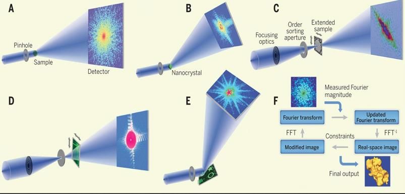
Dynamic X-ray Imaging Is the Key to Solving
Structure-Properties-Performance Challenges

                                  Materials Genome   BESAC 2012   NAS 2015

                                    BESAC 2015       NAS 2017     NAS 2019

                                                                         3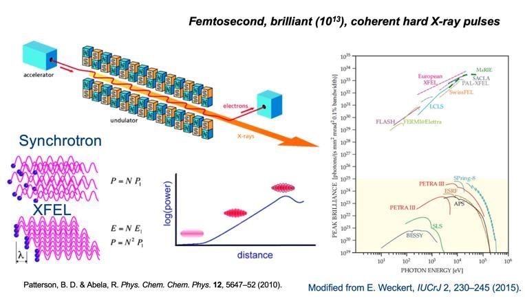
Frontier in Condensed Matter, Materials Science and
  Plasma Physics
To understand the fundamental physics that govern atomic interactions in High Energy
Density (HED) materials, measurements are required at relevant temporal- and
spatial-scales.

→ dynamic-loading + ultrafast X-ray techniques (XRD, imaging, spectroscopy)

                                                    Good news: temporal- and spatial-
                                                    fidelity of experiments now rival the best
                                                    simulations

                                                    Bad news: cannot accurately model the
                                                    behavior of materials at extreme
                                                    conditions without including the
                                                    microphysics
                             NIF: lasers.llnl.gov
                                                                                             4
        Kadau et al., 2007
Why make a bright, coherent source?
Why make a bright, coherent source?
Moving toward the far-field in dynamic hard X-ray
          imaging: coherent X-ray diffractive imaging (CXDI)
             ◼   Traditional imaging (think lens)
                 •      Fresnel, Kirz, Laue, Kinoform lenses
                        for hard x-rays (up to 0.5 MeV)
                 •      Resolution: down to ~100s nms
             ◼   Radiography approaches (line-projection, tomography,
                 phase contrast imaging (PCI) and divergent beam PCI)
                 •      Resolution typically scales with pixel size (max r ~ pixel/100 > 100 nm,
                        typically 1 mm)
             ◼   Coherent approaches (holography, PCI, coherent diffraction imaging (CDI))
                 •      Resolution: Few nanometer resolution possible
                     Shadow/contact imaging Phase                 Holography/CXDI
                                            contrast
Slide 7                                                                             CXDI can provide
                                                                                    3D information, but
                                                                                    has only been
                                                                                    demonstrated for
                                                                                    softer x-rays (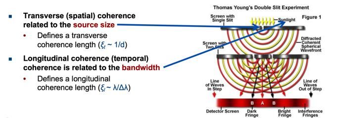
CXDI can provide 3D information
           Plane wave CDI   Bragg-CDI           Ptychographic-CDI

           Fresnel-CDI

                             Reflection-CDI
                                              Iterative phase retrieval; 100s-
  Miao et al., 2015                           1000s iterations can find the
                                              correct phase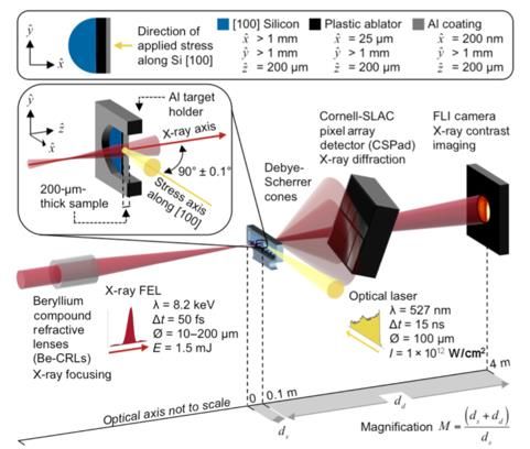
Linac Coherent Light Source
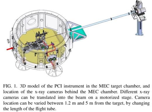
MEC instrument optics and diagnostics
Target Chamber

 Item        Purpose                    Specifications
         TW-class short      Pulsewidth: ≤40 fs
Short       pulse laser      Energy: ≥150 mJ per pulse
Pulse       for target
Laser       driver and       Repetition Rate: 10 Hz
System      short pulse      Vacuum optical transport to target
            diagnostics          chamber
                             Wavelength: 527 nm
         Multi-Joule high-
Long                         Pulsewidth: Variable 2-200 ns
             intensity
Pulse
             shock driver    Variable Temporal Pulse Shape
Laser
             for target
System                       Energy: ≥ 50J per pulse              https://portal.slac.stanford.edu/sites/lcls_public/Instruments/mec/Pages/S
             interactions
                             Repetition Rate: 1-shot per 10 min   pecifications.aspx

The Matter in Extreme Conditions (MEC) instrument combines the unique LCLS beam with high power optical
laser beams, and a suite of dedicated diagnostics tailored for the study of Warm Dense Matter, High Pressure
Physics, Shock Physics, and High Energy Density Physics.
LCLS has a wide range of dual-pulse / dual-color modes
available & ability to select Dt, DE, seeding, polarization
• Double Slotted Foil
• Split Undulator
• Injector laser pulse splitting
• Multiple laser pulses at cathode (dual lasers)
• Fresh Slice Technique
Fresh Slice (HXR)
Slotted Foil
ns – Double Bunch
fs – Double Bunch
X-ray Split and Delay
-10 ps 0 10 fs 37 fs 82 fs 135 fs 0.35 ns 0.7 ns 1.05 ns 1.4 ns … > 100 ns
Multi-bunch (2, 4, 8) Operation
11hCMOS camera developed by SNL/LLNL has been tested* using LCLS
femtosecond x-ray pulses: linearity, gate profile, QE…
LCLS : Two x-ray pulses, 7.2keV, 4.2ns apart
Pulse 1
hCMOS Frame 1
(T0)
Cold diffraction
Pulse 2
Frame 2
To +4.2ns
Cu target
LCLS x-ray New peak
camera
Diffraction angle (2q)
4.2ns
hCMOS
Pulse 2 Pulse 1
Hart et al., 2019
12
* Experiment led by P. Hart (SLAC)4-frame + 4 pulse test at XCS hutch, LCLS: camera characterization
+ optical laser pump experiment
Icarus hCMOS Images
4 x-ray pulses, 7.2keV, 2.1 + 3.8 + 2.1 ns apart; 1 ns gate
X-ray damage/heating expansion
1 2 4
Diffraction angle (2q)
3
Au 111
Au 200
Laser ablation/heating expansion
Au 111
800 nm optical
pump:
~1-3 mJ
hCMOS 200 um spot on
target Au 200
1 ps pulse duration
2.1 2.1 Op. Run 83
3.8 Au target pump
ns ns
ns • 3-4 fold more expansion recorded with
hCMOS
the optical pump
Pulse 4 Pulse 3 Pulse 2 Pulse 1 • Dynamic recrystallization
* Experiment led by P. Hart (SLAC), Dan Damiani, Arianna Gleason, Phil Heimann, Silke Nelson, Emma McBride, Sanghoon Song,
13
Diling Zhu, Mike Glownia, XCS staff, ; LLNL: Arthur Carpenter, Matthew Dayton, Emily Hurd; SNL: Marcos Sanchez 13Laser driven shock compression + X-ray techniques
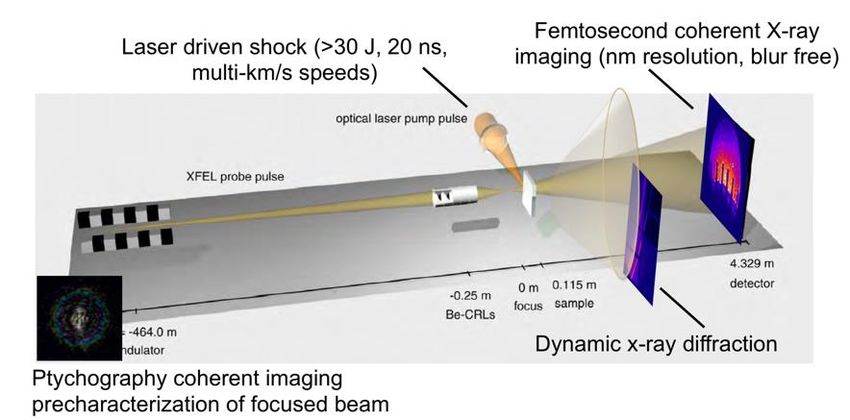
DRIVE DIAGNOSTICS
X-ray Techniques
Streaked
-Diffraction: Optical
Records Pyrometer
lattice-level
structural
Recordsinformation from sample
TARGET thermal emission from sample
for calculation of absolute temperature
-Emission Spectroscopy: Measure spin
Focused Laser transition
Profile Reflectivity Monitor
-Imaging: reconstruct 2D density
~1013 W/cm2 Observes
distribution via PCIchanges in sample optical properties
532nm Probe Beam
Delay Element
Interferometer
VISAR (Velocity Interferometry System for Any Reflector)
Measures Doppler shift from moving surfaces to
determine shock and particle velocitiesVoid Collapse Physics:
Here is Mesoscale
a sample Materials Properties
image (unfortunately in inversed colo
Control Functionality at Extreme Conditions
Fusion energy materials face harsh environments:
-structural materials in a reactor
-plasma facing materials / first walls of a tokamak
-ICF materials
3D simulation of ICF implosion X-ray radiograph of diamond-ablator
showing voids
Clark et al., 2015
Spiked perturbations due to hydrodynamic instability growth 1
seeded by defects in the ablator. Courtesy T. Doeppner, 2019Void Collapse Physics:
Here is Mesoscale
a sample Materials Properties
image (unfortunately in inversed colo
Control Functionality at Extreme Conditions
Fusion energy materials face harsh environments:
-structural materials in a reactor
-plasma facing materials / first walls of a tokamak
-ICF materials
Key questions: X-ray radiograph of diamond-ablator
• How do we mitigate hydrodynamic showing voids
instabilities plaguing ICF?
• What is the relationship between
void size and collapse rate and
interaction with the shock front?
• What are the plasma properties
inside the void and what is the
extent of jetting and how does that
modify the surrounding material? 1
Courtesy T. Doeppner, 2019Revolution in X-ray sources is enabling a revolution in
High Energy Density (HED) Science
modified from Schropp et al., 2015
1Phase contrast imaging (PCI) for 2D density distribution @
MEC, LCLS
Nagler et al., 2016PCI + shock compression @ MEC in diamond
Caveats/Notes:
→ phase-contrast images measured at
different time delays on different
samples
→ recording the intensity only in the
detector plane, so the phases of the x-
ray wave field are lost.
→ do ptychography of incident beam
enables calculation of transmission
function via iterative phase-retrieval
techniques
→ compression of the material by the shock
wave introduces an additional phase shift
in the x-ray wave field behind the sample
→ reconstructed phase change corresponds
to an integrated value accumulated along
the path of the x rays through the sample
→ assume spherical symmetry use
tomography to reconstruct local phase
Schropp et al., 2015 change per voxelDynamic process timeseries in a single sample
Ultrafast movie of a shock
front traversing a void
Void collapse during a shock at MEC
Static
Shocked
shock direction
14 ns 16 ns 16.5 ns 17 ns
Sandberg et al.
2Dynamic process timeseries in a single sample
Ultrafast movie of a shock front
traversing a void for 2-D & 3-D image
reconstruction
• ICF ablator materials with natural or
synthetic voids:
-Be & diamond (with LLNL)
-polystyrene (with LLE)
• Robust image algorithm
deployment/development
• X-ray optics design/testing for multi-
zone plate or design + split and
delay 2Silica
Fabricating synthetic voids
Diamond shell
Silica
Laser Milled:
Femtosecond laser milled voids:
- in polystyrene (10-20 um diameter)
- in silica glass (20 um diameter)
Silica
PS
Photoresist spin + hollow shell :
- in SU-8 + hollow glass shell (40 um
diameter) SU-8
SU-8
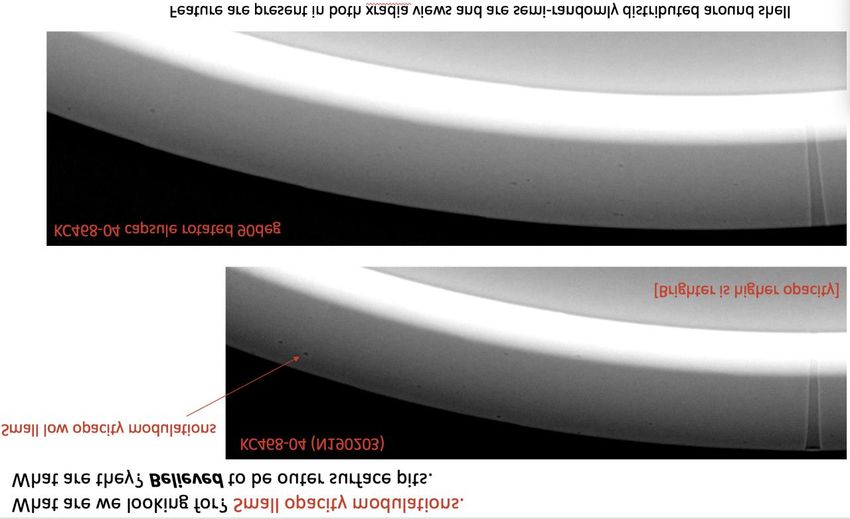
These are radiographs at ~ 8 keV, looking at the limb o
thickness is ~ 70 um, the thickness of the bright (W-dop
on the top left two dark spots can be seen (interpreted
SU-8 (since capsule o
also could be pits on the outer surface
An additional observation from simulations: there appe
cause damage to the implosions.Void collapse in energetic materials at HP-CAT & DCS, APS
Armstrong et al., 2019PCI + shock compression @ MEC in Si
5
4
3
2
1
1 2 3 4 5
Pinpoint phase, density and microstructure in a single-shot
Brown et al., 2019 down to sub um resolutionPCI + shock compression @ MEC in Si
5
4
3
2
1
1 2 3 4 5
Pinpoint phase, density and microstructure in a single-shot
Brown et al., 2019 down to 500 nm resolutionStereo PCI + shock compression @ MEC via multi-angle
imaging
Si (400)
*Split and delay line -spatially split and steer the beam
benchmarked at -Be CRL stack for each beam
HPCAT
-3 beams cross at sample
target
Si (220)Stereo PCI + shock compression @ MEC via multi-angle
imaging
Si (400)
*Split and delay line -spatially split and steer the beam
benchmarked at -Be CRL stack for each beam
HPCAT
-3 beams cross at sample * 4 to 8 pulses per beam
target
Si (220)Evolution of prebiotic to biotic materials via shock
wave interaction
X-ray imaging
-visualize phase
transformations in
presence of trapped
volatiles
X-ray absorption
spectroscopy Figure 5. Previously reported kinetics data for simulated
-visualize impacts with formamide showing the growth and decay of
chemical various intermediates, both from theoretical (a) and
https://www.nasa.gov/images/content/107500main_panel1_m.jpg experimental (b) data. Adapted from Ferus et al. 2014.
dynamicsCharacterization of Materials with xFEL Ptychography Demonstration of 2D & 3D ptycho-tomography with fly scanning at an XFEL -high resolution (20-50 nm) -operated in fly scan mode -evaluated reconstruction quality using ePix10k and JungFrau detectors
Single shot CXDI
                 Shadow/contact imaging   Phase contrast        Holography/CXDI

                                                                                  CXDI can provide
                                                                                  3D information, but has
                                                                                  only been demonstrated
                                                                                  for softer x-rays (
Single shot CXDI
- so far only test with
time resolved ptychography optical light & soft
X-rays
Seaberg et al, 2015
random mask for each xFEL beam
- so far only soft X-
time resolved ptychotomography rays
Duarte et al, 2019
computed stereo lensless x-ray imaging
- so far only soft X-rays
- maintain know (unchanging
time resolved in-flight holography
or predictable) reference
which must scatter more or
equivalent to the sample
Gorkhover et al, 2018Exp. techniques and sample environments for single
pulse dynamic imaging
1. Biggest, current challenges: (technical) Detectors, X-ray beam conditioning,
experimental platform synergy; (logistical) beamtime and workforce
2. Current state-of-the-art methods & limitations: single-shot coherent X-ray
diffractive imaging (CXDI) methods with hard (>25 keV) X-rays for 2-D
reconstructions are maturing; 3-D is more nascent; streamlining concurrent data
collection – analysis/algorithms – reconstruction is paramount
3. Most promising current/new methods: single-shot CXDI, e.g. phase contrast
imaging (PCI), ptychography + multi-dimensional X-ray pulse train and gated
detectors
4. Beneficial methods not yet under development: hard X-ray single-shot
holography; grating interferometry + dynamic compression
→ All of the above can be achieved in the next 5 years. This is not a one size fits all –
different science scopes, with different spatial resolution requirements, will dictate
which X-ray imaging methodology to adopt.Thank you for your attention!!
Collaborators:
LANL: Cindy Bolme, Richard Sandberg* now at BYU, Don Brown, Pawel Kozlowski, Kyle
Ramos, Michael Powell, David Montgomery
Stanford University: Wendy Mao, Silvia Pandolfi
SLAC: Hae Ja Lee, Bob Nagler, Eric Galtier, Eric Cunningham, Yanwei Liu, Kenan Li,
Anne Sakdinawat, Philip Hart, Roberto Colina, Richard Walroth, Chandra
Curry
DESY: Andreas Schropp, Frank Seiboth, Christian Schroer
LLNL: Suzanne Ali, Peter Celliers, Amy Lazicki, Richard Kraus, Dayne Fratanduono, Jon
Eggert, Rip Collins, Matthew Dayton, Nir Goldman, Leora Dresselhaus-
Marais, Tilo Doeppner
Geophysical Lab: Andrew Steele
- many more!!
Funding: DOE, FES; NSF, LANL LDRD
33You can also read


