Femtosecond diode-based time lens laser for multiphoton microscopy
←
→
Page content transcription
If your browser does not render page correctly, please read the page content below
Research Article                                Vol. 12, No. 10 / 1 Oct 2021 / Biomedical Optics Express   6269

Femtosecond diode-based time lens laser for
multiphoton microscopy
Y. L ANGE S IMMONS , 1 K ENNETH J. U NDERWOOD , 1 O MKAR D.
S UPEKAR , 2,3 B RENDAN M. H EFFERNAN , 1 TARAH A. W ELTON , 4
E MILY A. G IBSON , 4 AND J ULIET T. G OPINATH 1,3,*
1 Department of Physics, University of Colorado, Boulder, CO 80309, USA
2 Department of Mechanical Engineering, University of Colorado, Boulder, CO 80309, USA
3 Department of Electrical, Computer, and Energy Engineering, University of Colorado, Boulder, CO
80309, USA
4 Department of Bioengineering, University of Colorado Anschutz Medical Campus, Aurora, CO 80045,
USA
* Juliet.Gopinath@colorado.edu

Abstract: We demonstrate a near-infrared, femtosecond, diode laser-based source with kW
peak power for two-photon microscopy. At a wavelength of 976 nm, the system produces sub-ps
pulses operating at a repetition rate of 10 MHz with kilowatt class peak powers suitable for deep
tissue two-photon microscopy. The system, integrated with a laser-scanning microscope, images
to a depth of 900 µm in a fixed sample of PLP-eGFP labeled mouse brain tissue. This represents
a significant development that will lead to more efficient, compact, and accessible laser sources
for biomedical imaging.

© 2021 Optical Society of America under the terms of the OSA Open Access Publishing Agreement

1.   Introduction
Pulsed lasers in the near-infrared find use in groundbreaking scientific and medical research,
including biological imaging [1,2], laser surgery [3], and the study of Alzheimer’s and Parkinson’s
disease [4–6]. Chief among these is their ability to probe the structure, function, and activity
of neurons through multiphoton microscopy, including real-time monitoring and control of
neuronal activity [1,2]. In conjunction with genetically encoded indicators such as GCaMP and
GFP (two-photon absorption peaking around 976 nm), these lasers enable imaging of biological
tissue up to millimeter depths [7–9]. Multiphoton imaging possesses distinct advantages over
its single-photon excitation counterpart. The square dependence (two-photon) on intensity
for excitation provides intrinsic axial sectioning [7,10]. The longer wavelength light reduces
Rayleigh scattering, allowing greater imaging depths, and is spectrally separated from the
emission light [10,11]. Sources suitable for multiphoton imaging must produce a high photon
flux for multiphoton excitation, achieved with high peak power, sub-ps pulses [7]. With the
numerous, impactful applications of multiphoton microscopy, we seek to improve near-infrared
pulsed sources to create wider adoption and lower the barrier for use of multiphoton technologies.
   Ultrafast pulsed lasers in the near-infrared (near-IR), have numerous drawbacks that complicate
their use. The most popular sources, Ti:Sapphire lasers, often in conjunction with optical
parametric oscillators (OPO), are prohibitively expensive, inefficient, and have large footprints.
There are also mode-locked fiber ring oscillators that operate at 920 nm and 976 nm; however these
have fixed repetition rates and require pulse picking for further adjustment [12,13]. In contrast,
diode lasers provide an attractive solution. They are one of the most widespread and successful
laser technologies, and are compact, inexpensive and power efficient. We demonstrate a viable
path forward with a diode-based source that has comparable performance to popular sources for
multiphoton imaging with a design including pulse shaping and enhancement techniques [14].

#430549                                                                      https://doi.org/10.1364/BOE.430549
Journal © 2021           Received 5 May 2021; revised 30 Jul 2021; accepted 20 Aug 2021; published 14 Sep 2021
Research Article                          Vol. 12, No. 10 / 1 Oct 2021 / Biomedical Optics Express   6270

   While diode lasers are low cost, compact and robust, there are issues that arise in pulsed opera-
tion. The fundamental physics—short upper-state lifetime and large two-photon absorption—of
the semiconductor gain media limits achievable pulse energy and peak power [15–19]. We set
out to overcome these limitations with a novel approach combining external pulse shaping, fiber
amplification, and nonlinear pulse compression to greatly outperform other diode-based systems.
   We chose to employ several pulse enhancement techniques in concert to achieve excellent
performance from a pulsed laser diode. This novel approach to pulse generation allows us to
maintain the benefits of each individual approach and achieve better results than with any one
alone. We use a “time lens” paired with fiber amplification, similar to chirped-pulse amplification,
to increase pulse energy and decrease pulse duration in tandem [20–22]. For further enhancement,
double clad fiber amplification and compression of chirped pulses are used to further increase
peak pulse power [23]. Research on ultrafast diode lasers report impressive results [24–26], but
have not covered the wavelength range needed for two-photon microscopy with GFP [27]. The
resulting performance of system presented here offers a path towards low cost, miniaturizable,
and robust systems for biomedical research and clinical applications.
   Our laser produces kilowatt peak power femtosecond pulses in the near-infrared, a critical
region for multiphoton microscopy, by overcoming drawbacks in diode lasers that traditionally
prohibit high peak powers. In this work, we report on a diode-based laser system with a tunable
repetition rate. The source has an optimized repetition rate and wavelength for multiphoton
imaging of the brain and peripheral nerves. The wavelength of the laser, 976 nm, is near
the peak for two-photon response of eGFP, GCaMP, and YFP (requiring labeling or genetic
modification) [27]. Additionally, it is also a suitable pump wavelength for coherent anti-Stokes
Raman Scattering (CARS) microscopy (a label-free process) [28,29]. Further, we validate the
laser for multiphoton microscopy by imaging fixed brain samples of PLP-eGFP mice [30]. We
demonstrate fluorescent imaging at depths of 900 µm in the neo-cortex.

2.   Laser design
Our laser consists of five stages: a pulsed diode seed, amplitude and phase control, amplification,
spectral broadening, and a compressor (Fig. 1). Generation of clean seed pulses is critical to
avoid amplifying a continuous wave (CW) signal. To achieve a high extinction ratio between
the pulse and background, a combination of diode gain switching and amplitude modulation is
used. A 976 nm distributed Bragg reflector (DBR) laser diode (Photodigm PH976DBR180BF) is
electronically pumped with a high repetition rate, short pulse laser driver (Highland Technology
T165), producing optical pulses with a full-width at half-maximum (FWHM) of approximately
70 ps and 200 mW peak power. A distributed Bragg reflector diode was chosen for its ability to
produce short, stable pulses in this regime, enabled by the shorter cavity length when compared
to other diode lasers [31]. The repetition rate is electronically controlled and can be adjusted
from 1 MHz to 200 MHz, which allows for balancing peak and average powers for optimal
imaging conditions [32,33]. To avoid photobleaching of samples and to maximize fluorescence
signal while maintaining high frame acquisition rates, an operating repetition rate of 10 MHz
was chosen [33].
   In the second of the four stages, as seen in Fig. 1, the seed pulses are amplitude modulated
with an electro-optic modulator (EOM, EOSPACE AZ-0S5-20-PFA-PFA-980) to shorten the
width of gain switched pulse and match the profile of the phase modulation in the next step. The
EOM is operated at 18 GHz to carve out an approximately 20 ps from the pulse envelope, with
∼20 dB of extinction. This results in a small satellite pulse as the gain switched pulse width is
greater than the wavelength of the 18 GHz modulation signal. These pulses are phase modulated
with an electro-optic phase modulator (EOSPACE, PM-DS5-20-PFA-PFA-980-LV) also driven
at 18 GHz with 30 dBm (Vπ < 3 V, ∼10 radians maximum). This phase modulation is the first
step of the time lens system, adding a quadratic phase to the pulse.
Research Article Vol. 12, No. 10 / 1 Oct 2021 / Biomedical Optics Express 6271
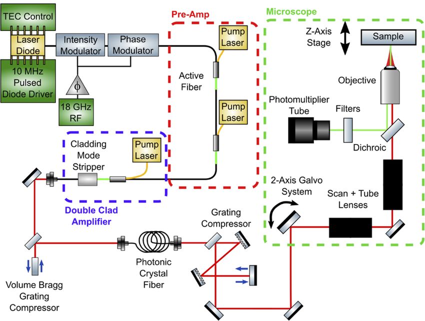
Fig. 1. Diagram of laser system. Pulses from a gain-switched diode are sent through
amplitude and phase modulators, which have a controllable delay offset. Two stages of
single mode fiber amplification with 976 nm notch filters to remove pump light make up the
pre-amplification. The power amplification stage consists of a double-clad amplifier and a
cladding mode stripper. A chirped volume Bragg grating (VBG) compresses the time lens
pulses. Pulses are further compressed through self-phase modulation in photonic crystal
fiber and a grating pair compressor.
After modulation, the pulses go through a multi-stage amplifier, consisting of two stages
of Yb-doped single mode fiber amplification to scale the system average power followed by a
Yb-doped double-clad fiber final amplification stage. The single mode amplifiers use 180 mW and
300 mW of 915 nm light to pump 10 cm and 16 cm fiber lengths (CorActive Yb401), respectively,
achieving 56 mW average power [21]. Further amplification to 250 mW is achieved with double
clad Yb-doped fiber (46 cm, LIEKKI Yb1200-10/125DC) cladding pumped at 40 W. This fiber
length and large pump power are to help overcome the overlap of the absorption and emission
cross sections at 976 nm and strong ASE that hinders Yb fiber amplification around 980 nm
[34,35]. Excess pump light is removed with a cladding mode stripper (DK Photonics CPS-30-50-
10/125/008/046-10). Pulses are coupled out of the fiber and compressed using a chirped volume
Bragg grating (OptiGrate CBG-976-85, AR coated, 85% diffraction efficiency), which induces
17 ps/nm of dispersion, completing the time lens. Autocorrelations after compression show the
pulses to be 2 ps FWHM with an average power of 180 mW, which match simulated results (Fig.
S1).
These pulses are further temporally compressed through self-phase modulation in fiber and
dispersion compensation. The pulses are coupled into single mode silica fiber (Corning HI1060)
from free space after the volume Bragg grating. The silica fiber is spliced to photonic crystal
fiber (PCF) (NKT SC-5.0-1040, mode field diameter 4 µm, -1.2 dB splice loss) which offers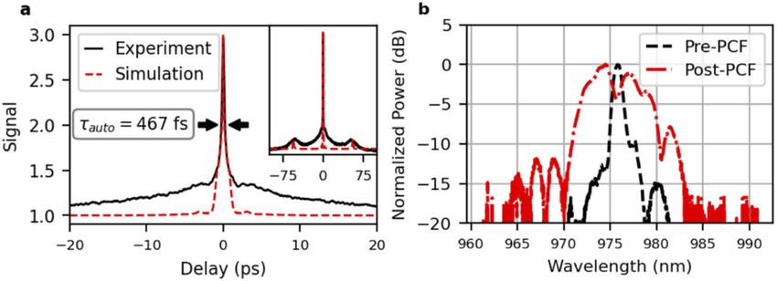
Research Article                             Vol. 12, No. 10 / 1 Oct 2021 / Biomedical Optics Express   6272

 an increased effective nonlinear coefficient (11 W−1 km−1 vs. 5 W−1 km−1 ) due to its smaller
 mode field diameter (4 µm) compared to other single mode fibers (Corning HI 1060, mode field
 diameter 6 µm). Combined input and output coupling losses as well as splice loss total -2.2 dB.
The spectrally broadened pulses are collimated and compressed with a grating pair, with a loss of
-1.3 dB resulting in 90 mW average power.

3.   Pulse characterization
Pulse spectra were measured before and after the PCF to characterize the effect of the self-phase
modulation. The pulse spectrum shows power-dependent broadening after the PCF in Fig. 2(b),
as would be expected in the case of self-phase modulation.

       Fig. 2. a Autocorrelation of the output pulses and the autocorrelation of a simulated pulse
       from the system. Experiment autocorrelation full-width at half-max is labeled. Insert
       shows full extent of pulse, from autocorrelation peak to background levels. b Laser spectra
       before and after photonic crystal fiber (PCF), showing spectral broadening from self-phase
       modulation.

   Intensity autocorrelations used to characterize the pulse are shown in Fig. 2(a). The autocorre-
lation shows a central peak with a width of 467 fs with a pedestal extending from the base of
the peak. A side pulse is seen at 50 ps from the center, corresponding to the 18 GHz amplitude
modulation. The pedestal is caused by two effects. The first is uncompressed nonlinear phase
from the self-phase modulation in the photonic crystal fiber [36–38]. The second is incoherent
artifacts from the uncorrelated spectral phase of the gain-switched seed [39]. This jitter brings
the modulation away from the ideal, purely quadratic modulation of a perfect time lens pulse.
   The intensity autocorrelation alone does not provide a determination of the electric field, but
supports that the laser pulses are ps-scale with peak power in the kW range when viewed with the
imaging results from Section 4. Uncompressed nonlinear phase, timing jitter in the seed pulses
(∼10 ps) [40], and the strength of phase modulation from the electro-optical modulator, limit
the achievable pulse width. Pulse power is limited by the losses in the cladding mode stripper,
PCF coupling, and grating compressor (total ∼-3.5 dB) as well as gain saturation of Yb fiber
amplification at 976 nm, most of which can be improved by removing free space losses. If greater
pulse energies are desired, the repetition rate may be reduced electronically, although dispersion
compensation would need to be adjusted for the increase in self-phase modulation from increased
power [21].

4.   Two photon microscopy demonstration
To prove our system is viable for use in two photon microscopy and can achieve results comparable
to Ti:sapphire lasers in terms of tissue penetration depth, the system was integrated with a two-
photon microscope. Imaging of eGFP-labeled oligodendrocytes was chosen for a demonstration
Research Article Vol. 12, No. 10 / 1 Oct 2021 / Biomedical Optics Express 6273
as the potential size and cost reduction our system offers is well suited to expanding recording of
neural activity of freely moving animals in real time. Our microscope (Fig. 1(b)) consists of
a galvanometer mirror system which sends the pulses through a scan and tube lens system. A
dichroic is placed before the objective and filters out the fluorescence signal which is sent to a
photo-multiplier tube (PMT). The sample is mounted above the objective on a z-axis stepper
motor for axial scanning. A dichroic mirror (Semrock FF740-Di01) and notch filters (Thorlabs
MF525-39, Chroma ET520/20 m) provide ∼90 dB of excitation source isolation to the PMT.
During image collection, incident power was controlled using a half-wave plate mounted on a
piezo rotation stage and a polarizer.
A 1 mm thick, coronal slice of fixed, PLP-eGFP mouse brain tissue was mounted on a
microscope slide in VECTASHIELD Plus Antifade Mounting Media (H-1900). All animal
procedures were performed in accordance with protocols approved by the Institutional Animal
Care and Use Committee of the University of Colorado Anschutz Medical Campus. The
neo-cortex region of the sample was imaged using a water immersion microscope objective
(Olympus, 20x 1 NA, XLUMPLFLN20XW). An average power of 28 mW (maximum used at
depth) was measured at the back aperture of the objective during tissue imaging. The spot size
calculated on the focal plane was 600 nm with a 1.6 µm depth of focus. To collect a volumetric
image, samples were scanned with a 260 × 260 µm2 field of view going from top to bottom in 5
µm steps with power and pixel dwell time increased with imaging depth into the sample. For the
first layers, pixel dwell time was 24 µs for the images (400 × 400 pixels) for a total acquisition
time of 3.9 seconds per frame. For depths greater than 800 µm, the dwell time was 64 µs for a
total time of 11 seconds per frame. This acquisition time is over twice as fast than previous time
lens systems for similar samples and penetration depths [21]. Sample bleaching was not observed
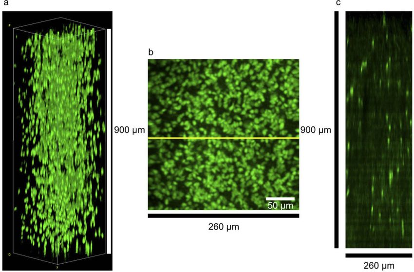
Fig. 3. Deep tissue, two-photon microscope images of oligodendrocyte cell bodies in the
neocortex region of fixed, PLP-eGFP mouse brain slices. Images were acquired at 5-micron
intervals in the z direction for 900 microns. a 3D stack of the slices (see Visualization 1). b
Projection in the z direction (maximum intensity). c Slice through the image stacks with the
slice position denoted by the yellow line in b.Research Article Vol. 12, No. 10 / 1 Oct 2021 / Biomedical Optics Express 6274
after this acquisition. Resulting volume images for a depth of 900 µm are shown in Fig. 3 with
a projection and vertical slice (see Visualization 1). The decreased resolution in the z-axis is
expected as the laser is focused at greater depths through index of refraction heterogeneities in
the tissue itself and between the tissue sample and the mounting media, as these aberrations were
not measured or corrected in the system [7,11]. Higher resolution images (1024 × 1024 pixels)
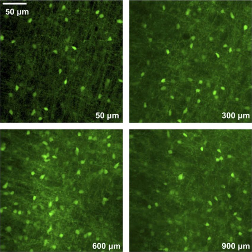
were acquired at 100 µm steps with a selection shown in Fig. 4 to emphasize fine details in the
sample. Individual soma and processes are resolvable, even at depths of 900 µm into the sample,
and imaging depth is only limited by the thickness of the sample. Although the signal to noise
ratio is limited by peak power and repetition rate in comparison to a Ti:Sapphire, images taken at
depth in tissue are comparable to those acquired with a Ti:Sapphire system (Fig. S2) and clearly
demonstrate our system’s utility for multiphoton microscopy.
Fig. 4. Two-photon microscope images of oligodendrocyte cell bodies and processes in
the neocortex region of fixed, PLP-eGFP mouse brain slices. These images were taken at
a selection of depths into a 1 mm thick coronal slice of brain tissue and have their depths
denoted in the lower left corners.
5. Conclusion
We have demonstrated a 976 nm diode-based laser source with ps-scale pulses and kW class
peak power suitable for two-photon microscopy based on time-lens pulse generation and pulse
compression. Our system was successfully utilized for two-photon microscopic imaging of eGFP
labeled mouse oligodendrocytes at depths up to 900 µm.
When looking towards the future of multiphoton microscopy, it is clear that new laser sources
in the near-IR are required to expand research applications and for eventual commercial and
medical use. Diode-based designs create a clear path forward for reduction in size and cost and
lends itself to robust operation in a variety of environments. Our design validates the feasibility
of a compact two photon laser microscopy source with these benefits which will increase access
to and widen the application off multiphoton microscopy in research and clinical settings.Research Article Vol. 12, No. 10 / 1 Oct 2021 / Biomedical Optics Express 6275
Funding. National Institute of Neurological Disorders and Stroke (1R01NS118188); State of Colorado Advanced
Industry Accelerator (CTGG1 2017-1874); National Institutes of Health SPARC (1OT2OD023852).
Acknowledgements. The authors would like to thank Professor Diego Restrepo and Gabriel Martinez for supplying
the samples for imaging. Thanks also goes to Megan Kelleher for assistance with the laser system and Dr. Scott Diddams
and Gabriel Ycas for technical discussions. The content of this publication is solely the responsibility of the authors and
does not necessarily represent the official views of the National Institutes of Health.
Disclosures. The authors declare no conflicts of interest.
Data availability. Data presented in this paper may be obtained from the authors upon reasonable request.
Supplemental document. See Supplement 1 for supporting content.
References
1. B. N. Ozbay, G. L. Futia, M. Ma, V. M. Bright, J. T. Gopinath, E. G. Hughes, D. Restrepo, and E. A. Gibson, “Three
dimensional two-photon brain imaging in freely moving mice using a miniature fiber coupled microscope with active
axial-scanning,” Sci. Rep. 8(1), 8108–8114 (2018).
2. M. R. Warden, J. A. Cardin, and K. Deisseroth, “Optical Neural Interfaces,” Annu. Rev. Biomed. Eng. 16(1), 103–129
(2014).
3. V. Sriramoju and R. R. Alfano, “In vivo studies of ultrafast near-infrared laser tissue bonding and wound healing,” J.
Biomed. Opt. 20(10), 108001 (2015).
4. A. Burgess, T. Nhan, C. Moffatt, A. L. Klibanov, and K. Hynynen, “Analysis of focused ultrasound-induced blood-
brain barrier permeability in a mouse model of Alzheimer’s disease using two-photon microscopy,” J. Controlled
Release 192, 243–248 (2014).
5. S. Liebscher and M. Meyer-Luehmann, “A peephole into the brain: Neuropathological features of Alzheimer’s
disease revealed by in vivo two-photon imaging,” Front. Psychiatry 3, 1–11 (2012).
6. L. Li, C.-W. Zhang, G. Y. J. Chen, B. Zhu, C. Chai, Q.-H. Xu, E.-K. Tan, Q. Zhu, K.-L. Lim, and S. Q. Yao, “A
sensitive two-photon probe to selectively detect monoamine oxidase B activity in Parkinson’s disease models,” Nat.
Commun. 5(1), 3276 (2014).
7. F. Helmchen and W. Denk, “Deep tissue two-photon microscopy,” Nat. Methods 2(12), 932–940 (2005).
8. K. Svoboda and R. Yasuda, “Principles of two-photon excitation microscopy and its applications to neuroscience,”
Neuron 50(6), 823–839 (2006).
9. P. Theer, M. T. Hasan, and W. Denk, “Two-photon imaging to a depth of 1000 µm in living brains by use of a
Ti:Al2 O3 regenerative amplifier,” Opt. Lett. 28(12), 1022 (2003).
10. E. E. Hoover and J. A. Squier, “Advances in multiphoton microscopy technology,” Nat. Photonics 7(2), 93–101
(2013).
11. W. R. Zipfel, R. M. Williams, and W. W. Webb, “Nonlinear magic: multiphoton microscopy in the biosciences,” Nat.
Biotechnol. 21(11), 1369–1377 (2003).
12. T. Hellerer, C. Polzer, A. Friedenauer, I. Greisberger, B. Wolfring, and C. Skrobol, “920 nm fiber laser delivering 100
fs pulses for nonlinear microscopy,” in Multiphoton Microscopy in the Biomedical Sciences XIX, A. Periasamy, P. T.
So, and K. König, eds. (SPIE, 2019), p. 29.
13. J. Lhermite, C. Lecaplain, G. Machinet, R. Royon, A. Hideur, and E. Cormier, “Mode-locked 05 µJ fiber laser at
976 nm,” Opt. Lett. 36(19), 3819 (2011).
14. C. Lefort, “Laser sources in multiphoton microscopy: overview and optimization,” in Unconventional Optical
Imaging, C. Fournier, M. P. Georges, and G. Popescu, eds. (SPIE, 2018), p. 30.
15. S. Kono, H. Watanabe, R. Koda, N. Fuutagawa, and H. Narui, “140-fs duration and 60-W peak power blue-violet
optical pulses generated by a dispersion-compensated GaInN mode-locked semiconductor laser diode using a
nonlinear pulse compressor,” Opt. Express 23(25), 31766 (2015).
16. F. R. Ahmad, Y. W. Tseng, M. A. Kats, and F. Rana, “Energy limits imposed by two-photon absorption for pulse
amplification in high-power semiconductor optical amplifiers,” Opt. Lett. 33(10), 1041 (2008).
17. A. Galvanauskas, P. Blixt, and J. A. Tellefsen, “Generation of femtosecond optical pulses with nanojoule energy from
a diode laser and fiber based system,” Appl. Phys. Lett. 63(13), 1742–1744 (1993).
18. J. T. Gopinath, B. Chann, R. K. Huang, C. Harris, J. J. Plant, L. Missaggia, J. P. Donnelly, P. W. Juodawlkis, and D. J.
Ripin, “980-nm monolithic passively mode-locked diode lasers With 62 pJ of pulse energy,” IEEE Photonics Technol.
Lett. 19(12), 937–939 (2007).
19. J. J. Plant, J. T. Gopinath, B. Chann, D. J. Ripin, R. K. Huang, and P. W. Juodawlkis, “250 mW, 15 µm monolithic
passively mode-locked slab-coupled optical waveguide laser,” Opt. Lett. 31(2), 223 (2006).
20. B. H. Kolner and M. Nazarathy, “Temporal imaging with a time lens,” Opt. Lett. 14(12), 630 (1989).
21. R. D. Niederriter, B. N. Ozbay, G. L. Futia, E. A. Gibson, and J. T. Gopinath, “Compact diode laser source for
multiphoton biological imaging,” Biomed. Opt. Express 8(1), 315 (2017).
22. D. Strickland and G. Mourou, “Compression of amplified chirped optical pulses,” Opt. Commun. 56(3), 219–221
(1985).
23. W. J. Tomlinson, R. H. Stolen, and C. V. Shank, “Compression of optical pulses chirped by self-phase modulation in
fibers,” J. Opt. Soc. Am. B 1(2), 139–149 (1984).Research Article Vol. 12, No. 10 / 1 Oct 2021 / Biomedical Optics Express 6276
24. K. Wang and C. Xu, “Wavelength-tunable high-energy soliton pulse generation from a large-mode-area fiber pumped
by a time-lens source,” Opt. Lett. 36(6), 942 (2011).
25. W. Fu, L. G. Wright, and F. W. Wise, “High-power femtosecond pulses without a modelocked laser,” Optica 4(7),
831 (2017).
26. J. van Howe, J. H. Lee, and C. Xu, “Generation of 35 nJ femtosecond pulses from a continuous-wave laser without
mode locking,” Opt. Lett. 32(11), 1408 (2007).
27. C. Xu and W. R. Zipfel, “Multiphoton excitation of fluorescent probes,” Cold Spring Harb. Protoc. 2015(3),
pdb.top086116 (2015).
28. C. L. Evans and X. S. Xie, “Coherent anti-Stokes Raman scattering microscopy: chemical imaging for biology and
medicine,” Annu. Rev. Anal. Chem. 1(1), 883–909 (2008).
29. H. Hajjar, H. Boukhaddaoui, A. Rizgui, C. Sar, J. Berthelot, C. Perrin-Tricaud, H. Rigneault, and N. Tricaud,
“Label-free non-linear microscopy to measure myelin outcome in a rodent model of Charcot-Marie-Tooth diseases,”
J. Biophotonics 11(12), e201800186 (2018).
30. B. S. Mallon, H. E. Shick, G. J. Kidd, and W. B. Macklin, “Proteolipid promoter activity distinguishes two populations
of NG2-positive cells throughout neonatal cortical development,” J. Neurosci. 22(3), 876–885 (2002).
31. J. K. O’Daniel and M. Achtenhagen, “High-power spectrally-stable DBR semiconductor lasers designed for pulsing
in the nanosecond regime,” in Novel In-Plane Semiconductor Lasers IX, A. A. Belyanin and P. M. Smowton, eds.
(2010), Vol. 7616, p. 76160W.
32. B. Resan, R. Aviles-Espinosa, S. Kurmulis, J. Licea-Rodriguez, F. Brunner, A. Rohrbacher, D. Artigas, P. Loza-
Alvarez, and K. J. Weingarten, “Two-photon fluorescence imaging with 30 fs laser system tunable around 1 micron,”
Opt. Express 22(13), 16456 (2014).
33. S. Mirkhanov, A. H. Quarterman, C. J. C. P. Smyth, B. B. Praveen, P. Appleton, C. Thomson, S. Swift, and K. G.
Wilcox, “Optimal repetition rate and pulse duration studies for two photon imaging,” in Multiphoton Microscopy
in the Biomedical Sciences XVII, A. Periasamy, P. T. C. So, K. König, and X. S. Xie, eds. (2017), Vol. 10069, p.
100692F.
34. F. Roeser, C. Jauregui, J. Limpert, and A. Tünnermann, “94 W 980 nm high brightness Yb-doped fiber laser,” Opt.
Express 16(22), 17310 (2008).
35. J. Wu, X. Zhu, H. Wei, K. Wiersma, M. Li, J. Zong, A. Chavez-Pirson, V. Temyanko, L. J. LaComb, R. A. Norwood,
and N. Peyghambarian, “Power scalable 10 W 976 nm single-frequency linearly polarized laser source,” Opt. Lett.
43(4), 951 (2018).
36. J. P. Heritage, R. N. Thurston, W. J. Tomlinson, A. M. Weiner, and R. H. Stolen, “Spectral windowing of
frequency-modulated optical pulses in a grating compressor,” Appl. Phys. Lett. 47(2), 87–89 (1985).
37. R. Thurston, J. Heritage, A. Weiner, and W. Tomlinson, “Analysis of picosecond pulse shape synthesis by spectral
masking in a grating pulse compressor,” IEEE J. Quantum Electron. 22(5), 682–696 (1986).
38. E. M. Dianov, L. M. Ivanov, P. V. Mamyshev, and A. M. Prokhorov, “Efficient compression of high-energy laser
pulses,” IEEE J. Quantum Electron. 25(4), 828–835 (1989).
39. J. Ratner, G. Steinmeyer, T. C. Wong, R. Bartels, and R. Trebino, “Coherent artifact in modern pulse measurements,”
Opt. Lett. 37(14), 2874 (2012).
40. “T165 picosecond to nanosecond laser diode pulser,” https://www.highlandtechnology.com/DSS/T165DS.shtml.You can also read