Extraction of Pulmonary Airway in CT Scans Using Deep Fully Convolutional Networks
←
→
Page content transcription
If your browser does not render page correctly, please read the page content below
Extraction of Pulmonary Airway in CT Scans
                                                Using Deep Fully Convolutional Networks

                                                                                Shaofeng Yuan

                                            School of Biomedical Engineering, Southern Medical University, Guangzhou, China
                                                                     shaofeng.yuan.smu@gmail.com
arXiv:2208.07202v1 [eess.IV] 12 Aug 2022

                                                   Abstract. Accurate, automatic and complete extraction of pulmonary
                                                   airway in medical images plays an important role in analyzing tho-
                                                   racic CT volumes such as lung cancer detection, chronic obstructive pul-
                                                   monary disease (COPD), and bronchoscopic-assisted surgery navigation.
                                                   However, this task remains challenges, due to the complex tree-like struc-
                                                   ture of the airways. In this technical report, we use two-stage fully con-
                                                   volutional networks (FCNs) to automatically segment pulmonary airway
                                                   in thoracic CT scans from multi-sites. Specifically, we firstly adopt a 3D
                                                   FCN with U-shape network architecture to segment pulmonary airway in
                                                   a coarse resolution in order to accelerate medical image analysis pipeline.
                                                   And then another one 3D FCN is trained to segment pulmonary airway
                                                   in a fine resolution. In the 2022 MICCAI Multi-site Multi-domain Airway
                                                   Tree Modeling (ATM) Challenge, the reported method was evaluated on
                                                   the public training set of 300 cases and independent private validation
                                                   set of 50 cases. The resulting Dice Similarity Coefficient (DSC) is 0.914
                                                   ± 0.040, False Negative Error (FNE) is 0.079 ± 0.042, and False Positive
                                                   Error (FPE) is 0.090 ± 0.066 on independent private validation set.

                                                   Keywords: Pulmonary Airway · Fully Convolutional Networks · Medi-
                                                   cal Image Segmentation · Multi-site · Multi-domain · Thoracic CT

                                           1     Introduction
                                           Recently, fully convolutional networks (FCNs) were increasingly used in medical
                                           image segmentation [1, 2], such as U-Net and V-Net. For extraction of pulmonary
                                           airway in CT scans, FCNs-based methods [3–24] were proposed and proved supe-
                                           rior to previous approaches in [25]. The organizers of the 2022 MICCAI Multi-site
                                           Multi-domain Airway Tree Modeling (ATM) Challenge collected 500 CT scans
                                           from multi-sites, i.e., the public LIDC-IDRI dataset and the Shanghai Chest
                                           hospital. In ATM 20221 , 300 CT volumes are used for training2 , 50 CT volumes
                                           are used for validation, and 150 private CT volumes are used for testing.

                                           2     Method
                                           The overview of the proposed AirwaySeg method is shown in Fig. 1.
                                           1
                                               https://atm22.grand-challenge.org
                                           2
                                               actually, 299 cases are valid, and case 164 is discarded.
2
       Fig.1
         F. Author et al.

                        Coarse Seg                               Crop

    Volumetric Image                       Coarse Seg Result                  Cropped Volumetric Image

                          Fine Seg                             Post-process

                                            Fine Seg Result                        Final Seg Result

                 Fig. 1. The overview of the proposed AirwaySeg method.

2.1      Coarse Segmentation
Coarse segmentation of pulmonary airway is in order to accelerate medical image
analysis pipeline. Thoracic CT volumes usually have very large image size. If 3D
FCNs are used directly on original resolution, the processing time of one case is
a few minutes. We adopt nnU-Net [26] as the coarse segmentation model.

2.2      Fine Segmentation
Fine segmentation of pulmonary airway in the extended bounding box from
coarse segmentation is in order to extract airway tree as complete as possible.
For convenience, we don’t use 3D FCNs along the centerlines of pulmonary
airway, but we use sliding window strategy. Also, We adopt nnU-Net [26] as the
fine segmentation model.

3      Result
Quantitative result in coarse segmentation stage Table 1 gives a quanti-
tative result of coarse segmentation model.

                       Table 1. Quantitative results of AirwaySeg.

                   Dataset      Scans   Dice           Jaccard       Recall          Precision
     Coarse Seg    validation   60      0.832±0.041    0.715±0.059   0.771±0.062     0.908±0.033
     Coarse Seg    train        239     0.838±0.037    0.722±0.055   0.779±0.062     0.909±0.029
     Fine Seg      validation   60      0.912±0.064    0.844±0.092   0.949±0.025     0.885±0.098
     Fine Seg      train        239     0.911±0.045    0.840±0.071   0.947±0.031     0.882±0.075
Title Suppressed Due to Excessive Length 3
Quantitative result in fine segmentation stage Table 1 gives a quantitative
result of fine segmentation model. Note that we don’t keep the largest connected
component in each case.
Quantitative result on independent private validation set Table 2 gives
a quantitative result of AirwaySeg on independent private validation set. Note
that we keep the largest connected component in each case.
Table 2. Quantitative results of AirwaySeg.
Dataset Scans Dice False Negative False Positive
AirwaySeg online validation 50 0.914±0.040 0.079±0.042 0.090±0.066
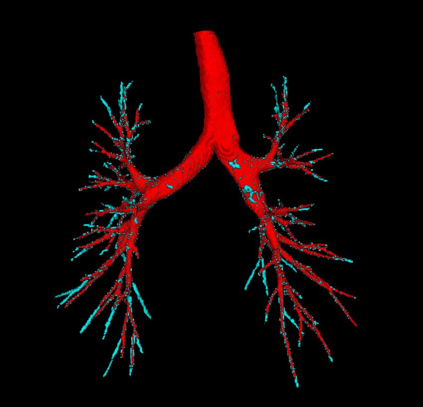
Qualitative result on independent private validation set Fig. 2 gives
a qualitative result of AirwaySeg on independent private validation set. We
observe that Fig. 2 (c) only has false positive voxels. It is because nnU-Net may
be over-fitting to the training set.
Fig.2
Dice=0.962 Dice=0.918 Dice=0.756
(a) atm_042 (b) atm_073 (c) atm_501
Fig. 2. Pulmonary airway segmentation of the proposed AirwaySeg method. (a) the
case with dice score higher than the average dice score. (b) the case with dice score
close to the average dice score. (c) the case with dice score lower than the average dice
score. Yellow is false negative. Cyan is false positive.Bibliography
[1] Ronneberger, O., Fischer, P., Brox, T.: U-net: Convolutional networks for
biomedical image segmentation. In: Navab, N., Hornegger, J., Wells, W.,
Frangi, A. (eds.) MICCAI 2015, LNCS, vol. 9351, pp. 234–241. Springer,
Cham (2015). https://doi.org/10.1007/978-3-319-24574-4 28
[2] Milletari, F., Navab, N., Ahmadi, S.-A.: V-net: Fully convolutional neural
networks for volumetric medical image segmentation. In: 2016 fourth inter-
national conference on 3D vision (3DV), pp. 565–571. IEEE, Stanford, CA,
USA (2016)
[3] Charbonnier, J.-P., Van Rikxoort, E. M., Setio, A. A. A., Schaefer-Prokop,
C. M., van Ginneken, B., Ciompi, F.: Improving airway segmentation in com-
puted tomography using leak detection with convolutional networks. Medical
image analysis 36, 52–60 (2017) https://doi.org/10.1016/j.media.2016.11.001
[4] Meng, Q., Roth, H. R., Kitasaka, T., Oda, M., Ueno, J., Mori, K.: Tracking
and segmentation of the airways in chest CT using a fully convolutional net-
work. In: Descoteaux, M., Maier-Hein, L., Franz, A., Jannin, P., Collins, D.,
Duchesne, S. (eds) MICCAI 2017, LNCS, vol. 10434, pp. 198–207. Springer,
Cham (2017). https://doi.org/10.1007/978-3-319-66185-8 23
[5] Nadeem, S. A., Hoffman, E. A., Saha, P. K.: A fully automated CT-
based airway segmentation algorithm using deep learning and topo-
logical leakage detection and branch augmentation approaches. Medical
Imaging 2019: Image Processing, vol. 10949, pp. 83–94. SPIE (2019).
https://doi.org/10.1117/12.2512286
[6] Qin, Y., Chen, M., Zheng, H., Gu, Y., Shen, M., Yang, J., Huang, X., Zhu,
Y.-M., Yang, G.-Z.: AirwayNet: A voxel-connectivity aware approach for
accurate airway segmentation using convolutional neural networks. In: , et
al. (eds) MICCAI 2019, LNCS, vol. 11769, pp. 348–356. Springer, Cham
(2019). https://doi.org/10.1007/978-3-030-32226-7 24
[7] Wang, C., Hayashi, Y., Oda, M., Itoh, H., Kitasaka, T., Frangi, A. F.,
Mori, K.: Tubular structure segmentation using spatial fully connected net-
work with radial distance loss for 3D medical images. In: , et al. (eds)
MICCAI 2019, LNCS, vol. 11769, pp. 348–356. Springer, Cham (2019).
https://doi.org/10.1007/978-3-030-32226-7 39
[8] Yun, J., Park, J., Yu, D., Yi, J., Lee, M., Park, H. J., Lee, J.-G.,
Seo, J. B., Kim, N.: Improvement of fully automated airway segmen-
tation on volumetric computed tomographic images using a 2.5 dimen-
sional convolutional neural net. Medical image analysis 51, 13–20 (2019)
https://doi.org/10.1016/j.media.2018.10.006
[9] Zhao, T., Yin, Z., Wang, J., Gao, D., Chen, Y., Mao, Y.: Bronchus segmen-
tation and classification by neural networks and linear programming. In: ,
et al. (eds) MICCAI 2019, LNCS, vol. 11769, pp. 230–239. Springer, Cham
(2019). https://doi.org/10.1007/978-3-030-32226-7 26Title Suppressed Due to Excessive Length 5
[10] Qin, Y., Gu, Y., Zheng, H., Chen, M., Yang, J., Zhu, Y.-.: AirwayNet-SE: A
simple-yet-effective approach to improve airway segmentation using context
scale fusion. In: 2020 IEEE 17th International Symposium on Biomedical
Imaging (ISBI), pp. 809–813. IEEE, Iowa City, IA, USA (2020)
[11] Selvan, R., Kipf, T., Welling, Max., Juarez, A. G.-U., Pedersen, J. H., Pe-
tersen, J., de Bruijne, M.: Graph refinement based airway extraction using
mean-field networks and graph neural networks. Medical image analysis 64,
101751 (2020) https://doi.org/10.1016/j.media.2020.101751
[12] Garcia-Uceda, A., Selvan, R., Saghir, Z., Tiddens, H. A. W. M., de Bruijne,
M.: Automatic airway segmentation from computed tomography using robust
and efficient 3-D convolutional neural networks. Scientific Reports 11(1), 1–
15 (2021) https://doi.org/10.1038/s41598-021-95364-1
[13] Nadeem, S. A., Hoffman, E. A., Sieren, J. C., Comellas, A. P., Bhatt, S.
P., Barjaktarevic, I. Z., Abtin, F., Saha, P. K.: A CT-based automated
algorithm for airway segmentation using freeze-and-grow propagation and
deep learning. IEEE transactions on medical imaging 40(1), 405–418 (2021)
https://doi.org/10.1109/TMI.2020.3029013
[14] Qin, Y., Zheng, H., Gu, Y., Huang, X., Yang, J., Wang, L., Yao, F., Zhu,
Y.-M., Yang, G.-Z.: Learning tubule-sensitive CNNs for pulmonary airway
and artery-vein segmentation in CT. IEEE transactions on medical imaging
40(6), 1603–1617 (2021) https://doi.org/10.1109/TMI.2021.3062280
[15] Tan, Z., Feng, J., Zhou, J.: SGNet: Structure-Aware Graph-Based
Network for Airway Semantic Segmentation. In: , et al. (eds) MIC-
CAI 2021, LNCS, vol. 12901, pp. 153–163. Springer, Cham (2021).
https://doi.org/10.1007/978-3-030-87193-2 15
[16] Zhou, K., Chen, N., Xu, X., Wang, Z., Guo, J., Liu, L., Zhang, Y.: Au-
tomatic airway tree segmentation based on multi-scale context information.
International journal of computer assisted radiology and surgery 16(2), 219–
230 (2021) https://doi.org/10.1007/s11548-020-02293-x
[17] Zhang, M., Yu, X., Zhang, H., Zheng, H., Yu, W., Pan, H., Cai, X., Gu,
Y.: FDA: Feature decomposition and aggregation for robust airway segmen-
tation. In: , et al. (eds) DART 2021, LNCS, vol. 12968, pp. 25–34. Springer,
Cham (2021). https://doi.org/10.1007/978-3-030-87722-4 3
[18] Zheng, H., Qin, Y., Gu, Y., Xie, F., Sun, J., Yang, J., Yang, G.-Z.: Re-
fined local-imbalance-based weight for airway segmentation in CT. In: , et
al. (eds) MICCAI 2021, LNCS, vol. 12901, pp. 410–419. Springer, Cham
(2021). https://doi.org/10.1007/978-3-030-87193-2 39
[19] Zheng, H., Qin, Y., Gu, Y., Xie, F., Yang, J., Sun, J., Yang, G.-Z.:
Alleviating class-wise gradient imbalance for pulmonary airway segmen-
tation. IEEE transactions on medical imaging 40(9), 2452–2462 (2021)
https://doi.org/10.1109/TMI.2021.3078828
[20] Guo, J., Fu, R., Pan, L., Zheng, S., Huang, L., Zheng, B., He, B.: Coarse-to-
fine airway segmentation using multi information fusion network and CNN-
based region growing. Computer Methods and Programs in Biomedicine 215,
106610 (2022) https://doi.org/10.1016/j.cmpb.2021.1066106 F. Author et al.
[21] Huang, W., Gong, H., Zhang, H., Wang, Y., Shen H., Li, G., Li,
H.: BronchusNet: Region and structure prior embedded representation
learning for bronchus segmentation and classification. arXiv preprint
arXiv:2205.06947 (2022)
[22] Wang, A., Tam, T. C. C., Poon, H. M., Yu, K.-C., Lee, W.-N.: NaviAirway:
A bronchiole-sensitive deep learning-based airway segmentation pipeline for
planning of navigation bronchoscopy. arXiv preprint arXiv:2203.04294 (2022)
[23] Wu, Y., Zhang, M., Yu, W., Zheng, H., Xu, J., Gu, Y.: LTSP: Long-term
slice propagation for accurate airway segmentation. International Journal of
Computer Assisted Radiology and Surgery 17(5), 857–865 (2022)
[24] Yu, W., Zheng, H., Zhang, M., Zhang, H., Sun, J., Yang, J.: BREAK:
Bronchi Reconstruction by gEodesic transformation And sKeleton embed-
ding. In: 2022 IEEE 19th International Symposium on Biomedical Imaging
(ISBI), pp. 1–5. IEEE, Kolkata, India (2022)
[25] Lo, P., Van Ginneken, B., Reinhardt, J. M., Yavarna, T., De Jong, P. A.,
Irving, B., Fetita, C., Ortner, M., Pinho, Rô., Sijbers, J., et al.: Extraction
of airways from CT (EXACT’09). IEEE transactions on medical imaging
31(11), 2093–2107 (2012) https://doi.org/10.1109/TMI.2012.2209674
[26] Isensee, F., Jaeger, P. F., Kohl, S. A. A., Petersen, J., Maier-Hein,
K. H.: nnU-Net: A self-configuring method for deep learning-based
biomedical image segmentation. Nature methods 18(2), 203–211 (2021)
https://doi.org/10.1038/s41592-020-01008-zYou can also read