Evaluation and Diagnosis of Tibial Bone Stress Injuries in Adolescents: Imaging and Nomenclature
←
→
Page content transcription
If your browser does not render page correctly, please read the page content below
Volume 4, Number 1, February 2022

                                            Current Concept Review

Evaluation and Diagnosis of Tibial Bone Stress
Injuries in Adolescents: Imaging and Nomenclature
Eric D. Nussbaum, Med, LAT, ATC1; Bryan Holtzman, BA2,3; Katherine H. Rizzone, MD, MPH4; Adam S. Tenforde, MD2,5,6;
Mark E. Halstead, MD7; Corinna C. Franklin MD8; Kathryn E. Ackerman, MD, MPH2,6,9
1Department   of Orthopaedic Surgery, Rutgers, Robert Wood Johnson Medical School, New Brunswick, NJ; 2Division of
Sports Medicine, Boston Children’s Hospital, Boston, MA; 3Perelman School of Medicine at the University of Pennsylvania,
Philadelphia, PA; 4Department of Orthopaedics, University of Rochester Medical Center, Rochester, NY; 5Spaulding
Rehabilitation Hospital, Charlestown, MA; 6Harvard Medical School, Boston, MA; 7Washington University School of
Medicine and St. Louis Children’s Hospital, St. Louis, MO; 8Shriners Hospital for Children–Philadelphia, Philadelphia, PA;
9Neuroendocrine Unit, Massachusetts General Hospital, Boston, MA

Correspondence to: Kathryn E. Ackerman, MD, MPH, Boston Children’s Hospital, Division of Sports Medicine, 319
Longwood Avenue–6th Floor, Boston, MA 02115, U.S, E-mail: kathryn.ackerman@childrens.harvard.edu
Received: October 29, 2021; Accepted: January 21, 2022; Published: February 1, 2022
DOI: 10.55275/JPOSNA-2022-0015

 Abstract:
 Tibial bone stress injuries (BSIs) are common injuries experienced by active adolescent athletes. The current literature
 lacks consensus of BSI nomenclature and appropriate use of imaging modalities. The purpose of this Current Concept
 Review is to identify existing classification of atraumatic tibial pain/BSI, propose unifying nomenclature, and review
 imaging tools available to clinicians caring for young athletes. Unified terminology and recommended imaging
 modalities for the adolescent athlete with these injuries would improve patient care and professional communication.
 We propose using “Bone Stress Injury” (BSI) to describe overuse injury of the tibia with associated physical
 examination findings. Radiography is recommended as the first imaging modality, and MRI should be considered to
 confirm diagnosis and determine grade of injury.

 Key Concepts:
 • Atraumatic tibial pain is a common injury experienced by pediatric and adolescent athletes, but no uniform
   diagnostic terminology currently exists for appropriate identification and management.
 • “Bone stress injury” should be used as a unifying term for “stress fracture,” “stress reaction,” and other phrases that
   are inconsistently used to define overuse injuries of the bone.
 • MRI is the gold-standard by which bone stress injuries of the tibia can be graded for severity.
 • Physicians are encouraged to maintain a broad differential diagnosis when treating tibial pain to avoid missing a
   catastrophic diagnosis (e.g., sarcoma) in a young athlete.

Copyright © 2022 JPOSNA®                                     1                                             www.jposna.org
Volume 4, Number 1, February 2022

Introduction
Bone stress injuries (BSIs) are estimated to affect up to           Bone Stress Injury of the Tibia
20% of adolescent athletes, with variability based on               Lower leg pain is common in athletes and has variable
primary sport.1 The tibia is the most common location               reported incidence, partially reflecting inconsistent
for injury, accounting for 19–54% of total injuries by              terminology to describe injury.13 During our review,
anatomical distribution,1-4 and the incidence of these              we identified 32 different diagnoses used to describe
injuries is rising: in 2005, the incidence in children 6–18         atraumatic, exercise-related shin pain. The terms stress
was 1.37 per 100,000 outpatients visits, and in 2015, the           fracture, fatigue fracture, stress reaction, shin splints,
incidence was 5.32 per 100,000.5 BSI may occur most                 periostitis, bone marrow edema, and medial tibial stress
frequently in athletes between the ages of 15 and 24,               syndrome (MTSS) are the most frequently—and often
corresponding with stage in development for reaching                interchangeably— used terms in the literature.13-21
peak bone mass.3,6,7 The proposed mechanism for injury              Inconsistency of terminology creates ambiguity among
is repetitive load exceeding bone strength, resulting in            clinicians, patients, coaches, and parents who are trying
micro damage that accumulates and may progress to                   to manage the problem. In the absence of advanced
complete fracture.8 BSIs occur when bone resorption                 imaging, however, many clinicians rely on clinical
exceeds ossification during normal remodeling.9                     examination and general characterizations of shin pain
                                                                    and may use outdated terminology.
Specifically, the posterior medial cortex (compression
side) of the tibial diaphysis is the most common anatomic           The term “shin splints” is frequently used to describe
region of injury. Anteromedial tibial diaphysis injury              non-descript shin pain related to physical activity. Shin
(tension side) is less common, though such an injury is             splints are reportedly associated with a theoretical
considered high risk to progress to complete fracture.10            pulling away of muscular attachments, causing an
The injury may isolate to one tibia or occur bilaterally.11         inflammation of the periosteum. They have long been
Athletes participating in land-based sports involving               considered the cause of MTSS.22 However, while
running, jumping, and repetitive stress are at higher risk          recent studies have suggested that shin splints cannot be
for BSI, and the incidence is higher in female athletes.7,12        imaged by MRI,23 there is ample evidence to warrant
                                                                    exclusion of the traction-based injury caused by the
Current literature lacks consensus of BSI classification
                                                                    tibialis posterior or flexor halluces longus attachment
and appropriate use of imaging. Consequently, the actual
                                                                    to the tibia as a possible mechanism of MTSS.22 MTSS
incidence of tibial BSI may be higher than currently
                                                                    may not represent inflammation of the periosteum but
noted. Clinical presentation and outcomes based on
                                                                    a progression to symptomatic BSI.24 Because of the
imaging of BSI may vary among adolescents. Injury
                                                                    routine occurrence and use of benign terminology like
classification, severity, or grade may differ substantially
                                                                    shin splints or MTSS, many athletes will overlook the
according to imaging modalities used and location
                                                                    significance of the bone injury and continue with their
of the injury. Appropriate diagnosis and management
                                                                    training; these injuries exist on the spectrum of BSI
of tibial BSIs is made more difficult by the lack of
                                                                    severity. Consequently, it is likely that tibial BSI are
common nomenclature and imaging consensus. With
                                                                    under-reported, as athletes may continue to train with
the frequency of various grades of BSI in adolescent
                                                                    ongoing pain and worsening injury.25
athletes, the effects of such injuries on return to play, and
the potential consequences radiation dosing can have on             Because BSI can occur as a spectrum of injury from
the growing skeleton, it is important to discuss unifying           periosteal irritation to complete fracture, often on a
nomenclature for BSI and to review imaging modalities               continuum known as a “stress reaction” prior to a visible
available to clinicians caring for young athletes.                  fracture line, not all injuries to bone should be referred

Copyright © 2022 JPOSNA®                                       2                                             www.jposna.org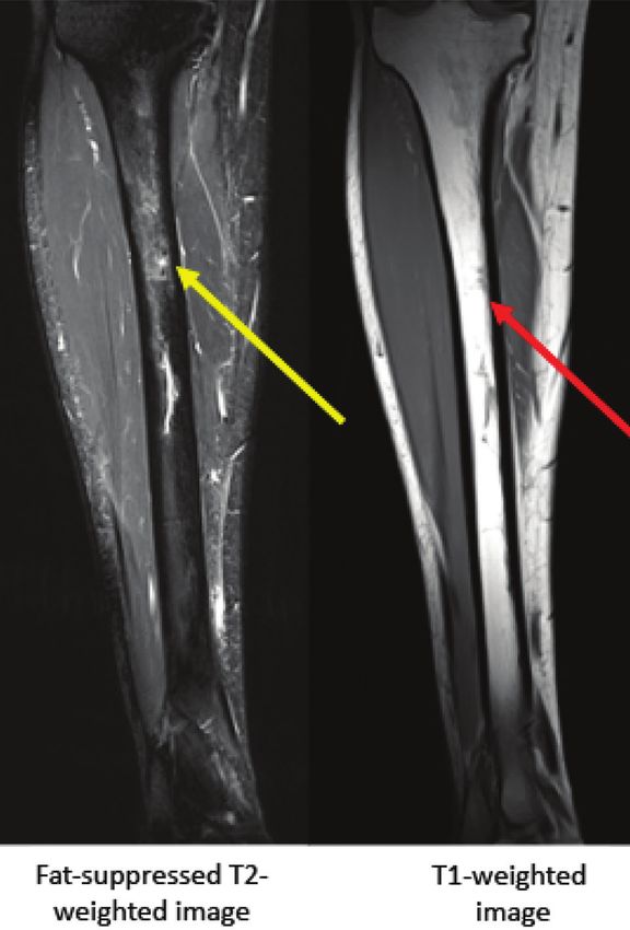
Volume 4, Number 1, February 2022

to as “stress fractures.” Misclassification is common:            of normal sun exposure.32 Most exercise-related tibial
most diagnosed “stress fractures” do not demonstrate              BSIs do not show evidence of a break in the bone cortex
the presence of fracture line on imaging.21 A fracture            on initial radiographs.27,36 Typically, radiographs will
line is apparent in only 1–4% of injuries localized to            show evidence of BSI once the healing response of
the tibia that present with exercise-induced shin pain.26         bone has begun,36 and the average/mean time interval
Using “stress fracture” terminology to denote any injury          between onset of symptoms and radiographic diagnosis
isolated to the bone may overestimate the incidence of            ranges from 2 to 6 weeks.37 Furthermore, up to 50%
true stress fractures.                                            of BSIs may remain radiographically occult.38 When
                                                                  BSI is detectable on radiograph, it is usually associated
Unified classification in terminology of BSI may
                                                                  with increased clinical significance and higher BSI
contribute to earlier, more efficient diagnosis, less time
                                                                  grade.18,39,40 In general, radiography has low initial
lost to injury, decreased patient confusion, and improved
                                                                  sensitivity for detecting tibial BSI, ranging from 3
patient care. Timely diagnosis can improve return to
                                                                  to 29%, and less than 50% of serial radiographs will
sport and reduce disruptions to an athletic career.27 Based
                                                                  demonstrate changes consistent with this diagnosis.34,41
on advances in our understanding of overuse injuries              Therefore, interpretation of radiographs should be in part
to bone, BSI is the preferred terminology to use in the           based on clinical suspicion for BSI, as adolescent athletes
diagnosis and management of this injury in adolescent             tend to continue training and propagate their injury if
athletes and may help improve our understanding for               they are reassured by a false negative radiograph. The
treatment and prevention.28                                       pathognomonic radiographic diagnosis of anterior tibial
                                                                  cortex stress injury is the presence of a “dreaded black
Imaging in the Diagnosis of Tibial BSI
                                                                  line” on lateral view,42 which is considered high risk for
Imaging is commonly used in the evaluation and
                                                                  development of frank fracture, though only a minority of
treatment of tibial BSI.29,30 For example, identifying            tibial BSIs will be in this category.
BSI at earlier stages may help facilitate an earlier return
to play.7 The benefits—manifest as improved patient               The first sign of BSI on radiograph may be reactive
outcomes—of any study should outweigh the costs and               periosteal inflammation and inflammation of the
risks levied on the patient. Given that pediatric tissue          overlying subcutaneous tissues. Both are difficult
is significantly more sensitive to ionizing radiation (in         to appreciate without comparative views. These
part due to increased mitotic rate and longer remaining           radiographic signs are not diagnostic of BSI; rather,
lifespans compared to adults), special considerations             they are suggestive of injury and indications for further
should be granted to limiting ionizing radiation when             imaging.43 Initial signs of bony injury on radiography
evaluating pediatric and adolescent athletes.31                   may include the appearance of “layers of eggs shell,”44
                                                                  periosteal or endosteal new bone,37,39,45 sclerosis,46 or
Plain Radiographs                                                 a “gray cortex sign” or “dreaded black line,” both of
Radiographs (x-rays) are the most common form of                  which represent a cortical fracture on radiograph.46-48
diagnostic imaging for the lower leg.32,33 Despite poor           However, Schilcher and colleagues performed biopsies
sensitivity early in the disease process, the American            on tibiae that demonstrated “dreaded black line”
College of Radiology recommends x-ray as the best                 and found that the radiographic fracture line is not a
initial imaging modality in the workup for tibial BSI             fracture on the microscopic level but instead represents
due to low cost and widespread availability.32,34,35 In           resorption cavities lined with active osteoblasts and
the pediatric patient, AP and lateral view radiographs            immature bone.42 This site is a weakened area of bone
of the tibia/fibula will emit approximately 0.002 mSv             that is histologically different from a frank fracture.42
of radiation combined, a dose equivalent to 6 hours               Other findings that can be visualized on radiography

Copyright © 2022 JPOSNA®                                     3                                             www.jposna.org
Volume 4, Number 1, February 2022

include endosteal thickening, roughening, or “fluffiness”         may be detected on fat-saturated T2 and STIR images
along the intramedullary canal.49 Radiographic osseous            with bone marrow or periosteal edema as well as cortical
changes that correspond with exam findings of pain                abnormalities.57 Kijowski and colleagues defined several
are significantly more likely to have features on MRI             MRI features of tibial BSI: periosteal edema, linear high
that would confirm diagnosis of BSI.39 Therefore, the             T2 signal immediately adjacent to the outer cortex; bone
clinical presentation of the patient remains a mainstay of        marrow edema, low T1 and high T2 within the canal;
assessment despite the radiographic findings.21                   cortical abnormality (Table 1).39
Bone lesions and infection are less common sources of             MRI may be useful in evaluating other causes of leg
leg pain in pediatric patients that can be visualized using       pain in athletes, such as chronic exertional compartment
plain radiographs. Primary bone tumors are the sixth              syndrome and peripheral neuropathy.55 Also, soft tissue
most common neoplasm in pediatric patients,50 although            injuries can be clearly differentiated using MRI. MRI has
most in this age population are benign.51 Consequently,           similar sensitivity to, but greater specificity than, bone
clinicians should not overlook the importance of imaging          scintigraphy to detect BSI.60
in the pediatric population who present with shin pain.
                                                                  Extrapolation of MRI findings in adults may not be
Magnetic Resonance Imaging                                        completely applicable to the adolescent population, but
When initial radiographs are negative and clinical                these grading scales serve as the best reference in the
suspicion for BSI is high, MRI without contrast is                absence of formal adolescent-focused guidelines. The
recommended by the American College of Radiology.32               adaptation of grading systems to help determine return
MRI may be the best imaging modality for visualizing              to play are helpful, but none of the studies are large or
BSI for the pediatric population because it uses non-             diverse enough to make convincing arguments for change
ionizing radiation, provides excellent imaging resolution,        in nomenclature from Fredericson, et al.18 Ultimately, the
and is both highly sensitive and specific.19,36,52 A 2016         scale proposed by Fredericson and colleagues was useful
systematic review found MRI to be the most sensitive              to bring some clarity to the 32 different terms used to
and specific imaging modality for BSI; the authors                describe BSI.18 See Figures 1 through 4 for examples of
also recommended MRI for patients with negative                   Fredericson, et al. grading of tibial BSI.
radiographs and a high clinical suspicion for a high-risk
BSI.27 MRI demonstrates stress abnormalities as early             Changes on MRI consistent with BSI may not
as bone scintigraphy53 and is more sensitive and specific         correspond to clinical symptoms.45,61 The findings of
than radiography, bone scintigraphy, and computed                 bone edema on MRI may represent osseous remodeling
tomography when used for BSI imaging. Furthermore,                or localized changes in bone mineral density.62,63 A study
the severity of change in MRI corresponds with the                of 55 asymptomatic Special Forces recruits found that
clinical severity.15                                              endosteal edema >100mm in length had a high likelihood
                                                                  of progressing over time.64 Those with edema measuring
When using MRI, fluid-sensitive sequences are typically
Volume 4, Number 1, February 2022
Table 1. Grading Scale for Categorizing BSI on MRI
MRI Fredericson, Arendt and Kiuru, Kijowski, Nattiv, Kaeding and Gmachowska,
Grade et al.18 * Griffiths14 et al.58 et al.39 et al.59 Miller10 et al.11
Grade 1 Mild to moderate Positive Endosteal Periosteal Mild marrow Imaging Periosteal
periosteal edema signal edema edema or periosteal evidence of edema
on T2 change on edema on T2 stress fracture,
Normal marrow STIR no fracture line,
on T1 & T2 no pain
Grade 2 Moderate to Positive Periosteal Periosteal Moderate Imaging Periosteal and
severe periosteal signal and endosteal and marrow marrow or evidence of marrow edema
edema on T2 change on edema edema periosteal stress fracture,
Marrow edema on STIR and edema and no fracture line,
T2 but not T1 positive T2 positive T2 pain
Grade 3 Moderate to Positive Muscle, Liner Severe Non-displaced Grade 2
severe periosteal STIR, T1, periosteal, intracortical marrow or fracture line plus cortical
edema on T2 and T2 and endosteal fracture line periosteal abnormalities
Marrow edema on edema edema on T1
T1 and T2 or T2
Grade 4 Moderate to Positive Fracture line Severe Displaced Grade 3 plus
severe periosteal fracture line marrow or fracture line >2 fracture line
edema on T2 on T1 and periosteal mm
Marrow edema on T2 edema on T1
T1 and T2 or T2 plus
Fracture line fracture line
apparent on T1 or T2
Grade 5 Callus at Non-union
endosteal and/
or periosteal
surface of
cortical bone
Multiple grading scales exist for categorizing bone stress injury (BSI) on Magnetic Resonance Imaging (MRI). All were established
primarily with adult patients.
*Short tau inversion recovery (STIR) and fat-suppressed T2 sequences may have higher sensitivity than T2.55
are the most useful.67 All patients in this case series In cases where there is strong clinical suspicion but
had either soft tissue edema or an eccentric periosteal normal MRI and radiographs, clinicians must weigh
reaction, and four of five had edema that began near the risk/benefit of additional imaging on the treatment
the entrance of the nutrient vessel into the medullary recommendation for their patient. In some cases,
cavity.67 Identification of longitudinal injury may require additional imaging may be important for the patient,
more than one imaging modality.68 In select cases of while in others, the additional imaging is not necessary.
symptomatic patients with normal MRI, CT may depict While MRI may be sufficient to detect most tibia BSI,
osteopenia representing early cortical stress injury.60 inconsistencies in the literature on imaging findings to
Copyright © 2022 JPOSNA® 5 www.jposna.orgVolume 4, Number 1, February 2022
Figure 1. MRI of Fredericson Grade 1 bone stress injury of
tibia. Arrow demonstrates mild to moderate periosteal edema
on fat-suppressed T2-weighted image (normal appearance on Figure 2. MRI of Fredericson Grade 2 bone stress injury of
T1-weighted image). tibia. Arrow demonstrates marrow edema on fat-suppressed
T2-weighted image (normal appearance on T1-weighted
image).
clinical features highlight the value in future research to
understand chronic leg pain.13 treatment. As such, MRI is not always approved by
insurance for acute use or before a course conservative
The use of MRI has greatly improved the ability treatment and rest.
of physicians to diagnose radiographically occult
BSI as a source of tibial pain in adolescents. The Bone Scintigraphy
benefits of the use of MRI are its high sensitivity and Bone scintigraphy (also known as bone scan) has long
specificity without exposing the patient to radiation. been used in the evaluation of BSIs. Bone scans will be
The drawbacks of MRI include its expense, scan time positive before radiographic changes are demonstrated,69
in a confined space, and justification for its use when with osteoblastic new bone formation represented
the results may not change the course of conservative by localized increased isotope uptake. Early studies
Copyright © 2022 JPOSNA® 6 www.jposna.orgVolume 4, Number 1, February 2022
on trabecular and cortical findings and compared with
radiography.71 The initial scintigraphic classification
by Chisin and colleagues did not identify differences
between classification and the clinical evolution of
patients when separated into four grades.72 A modified
version by Zwas and colleagues correlated recovery time
across four grades of bone involvement.25 Castropil and
colleagues compared bone scan with MRI and developed
a quantitative evaluation index (QEI) that compared
injured and non-injured legs, identifying a positive
correlation between QEI to time for recovery.73
Because many early studies looking at BSI used bone
scan as the gold standard, accurate representation
of sensitivity and specificity of the test is lacking. A
systematic review looking at the use of bone scan in
lower extremity stress fractures in patients of all ages
reported a sensitivity of 50–97%, with specificity ranging
from 33–98%.27 False positives are a concern with
bone scan. Any condition that causes increased bone
turnover may be positive on this imaging technique,
including infections, tumors, aseptic necrosis, trauma,
and complex regional pain syndrome/reflex sympathetic
dystrophy.58 Other disadvantages of bone scan include
the length of time to perform the test, difficulty in
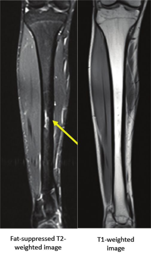
Figure 3. MRI of Fredericson Grade 3 bone stress injury of
determining the severity of BSI, and the high dose of
tibia. Arrows demonstrate marrow edema on fat-suppressed radiation. It is estimated that the radiation from a single
T2- and T1-weighted images. bone scan is equivalent to 60 two-view chest radiographs
(effective radiation dose: 6.3 mSv).74 There is also a
fairly high radiation load to the entire body from the
suggested a sensitivity of 100% by injecting a radiotracer radioactive isotope. It has been calculated that a 10-year-
of 99m-Tc-methylene diphosphonate intravenously.38 old patient undergoing a bone scan will receive 3.9
Increased radiotracer activity often can be detected mSv, theoretically inducing a 0.04% increased risk for
within 24 to 36 hours of the onset of fracture. The cancer.75
majority of the dose, however, is renally eliminated
There are limited data on the use of bone scintigraphy
within six hours of injection.70
for evaluating tibial BSI in pediatric and adolescent age
There have been several attempts to grade BSI and groups. Pediatric data for sensitivity and specificity are
project recovery time based on scintigraphy with mixed lacking. Since MRI scanners are readily available in most
results. Greaney and colleagues subjectively graded the areas, perform with excellent sensitivity and specificity,
spectrum of scintographic uptake into four levels graded and do not provide any radiation load concerns, the
as normal (0) to 3+, in an attempt to quantify the degree use of bone scan in the pediatric population should be
of bone involvement, then reviewed their findings based reserved for rare clinical scenarios.
Copyright © 2022 JPOSNA® 7 www.jposna.orgVolume 4, Number 1, February 2022
Figure 4. MRI of Fredericson Grade 4 bone stress injury of proximal tibia. Arrows
demonstrate edema on fat-suppressed T2 and T1-weighted images, and a horizontal
fracture line is apparent.
Ultrasound guidelines for MSK US diagnosis of metatarsal BSI
Musculoskeletal Ultrasound defined as meeting two or more criteria: hypoechoic
Musculoskeletal ultrasound (MSK US) is an inexpensive periosteal elevation above cortical bone, cortical
imaging technique that many sports medicine physicians disruption, or increased vascularity on power Doppler.81
now use in their clinical diagnosis of pathology. These criteria had reported sensitivity of 83% and
Periosteal reactions of BSI can sometimes be visualized specificity of 76%.81 Bianchi and Luong modified this
on ultrasound before bony callus is observed on plain algorithm and described five hallmarks of BSI: (1)
radiographs, aiding in earlier diagnosis.76 periosteal thickening, (2) a calcified bone callus, (3)
cortical irregularities, (4) subcutaneous edema, (5) hyper-
MSK US can identify periosteal thickening, vascular changes seen on color Doppler US.77 These
subcutaneous edema, calcified bone callus, hyper- studies, however, have not been reproduced with tibial
vascular changes, and cortical irregularities that are BSI but highlight the potential utility of MSK US for
associated with BSI.77 It has the ability to identify diagnosing BSI.
presence of hypoechoic calluses and buckling in the bony
cortex more than other imaging modalities.57 Diagnostic Because of its relative diagnostic accuracy when
compared with MRI, lack of exposure to ionizing
ultrasound has a sensitivity of 43–99% and specificity
radiation, portability, safety, point-of-care capability,
of 13–79%.27 The sensitivity of MSK US for identifying
and low cost, MSK US is recommended for clinical
BSI on the posterior cortex of the tibia is low.57
examination and should be considered an adjuvant
Additional extension of MSK US includes visualization imaging modality to utilize in addition to the traditional
of increased blood flow and vascularization related to initial plain film evaluation.57
fracture pathology using Doppler technology. Changes in
Limitations of MSK US include the inability to assess
vascularity using power Doppler imaging may be helpful
structures deep to the bone cortex such as marrow
for the early diagnosis of BSI.78 Bodner and colleagues
edema. However, many of the surface findings MSK
identified sonographic features of anatomy surrounding
US identifies may correlate with findings of bone
site of BSI, including presence of soft tissue edema, fluid
marrow edema.57 As an in-office imaging modality,
collection, and increased vascularity.79
perhaps the greatest limitation is that there is inter-
Imaging findings on MSK US frequently coincide with sonographer variability and a steep learning curve for
findings on MRI.79,80 Banal and colleagues established clinicians. In general, the literature associated with
Copyright © 2022 JPOSNA® 8 www.jposna.orgVolume 4, Number 1, February 2022
MSK US imaging of pediatric BSI is lacking, but CT provides diagnostic value in select cases of BSI or in
considering the safety and relative capabilities of MSK shin pain with a differential diagnosis expanding beyond
US, it is an imaging modality that should be included BSI, particularly when bone neoplasm is a possibility. In
in future BSI research. cases with negative MRI findings, CT can demonstrate
the earliest signs of cortical stress injury that are occult
Therapeutic Ultrasound on other imaging techniques.60 Furthermore, CT has
Many rehabilitation specialists (e.g., physical therapists, been shown to be particularly sensitive at diagnosing
athletic trainers) also have access to therapeutic longitudinal stress changes in the tibia. For example, one
ultrasound (TUS). TUS employs a low frequency range study87 reported the sensitivity of CT for longitudinal
of 1-3 mHz that is not used for visualization of anatomic BSI as 82% (versus 73% for MRI), and a recent review56
structures.82 Application of 1.5–2.0 W/cm2, 100% duty rated CT as the modality of choice for longitudinal
cycle for 1 minute over the suspected injury may create fracture lines. CT may also clarify lesion etiology
an increase in pain and heat in the damaged periosteum including osteomyelitis with a Brodie abscess and various
due to energy absorption; such increased pain and heat is neoplasms such as osteoid osteoma.55,88 Malignant tumors
considered a positive diagnostic test.83,84 TUS has been in young adults present similarly to BSI and, in situations
noted to provide positive findings 2–3 weeks prior to of adequate clinical suspicion, such a study can be
radiographic findings.85 In a comparative study, TUS was useful.89 While useful in the cases of a broad differential
less sensitive compared to bone scan, but more sensitive for shin pain, BSI severity as assessed by CT scan has not
been correlated with clinical severity or time to recovery.
than radiography at detecting tibial BSI (75% TUS,
When compared to other imaging, CT grades for the same
100% bone scan, 22% radiograph).37 However, TUS was
injury were lower than bone scans and MRI and higher
not as specific as bone scan or radiography (67%, 100%,
than x-ray.15 This poor clinical correlation suggests the
98%, respectively). TUS fails to reveal many low-grade
need for a new severity scale for CT scans of tibial BSI.
BSIs that can be identified by MRI and therefore is not
considered a definitive diagnostic tool for BSI.30 There CT scans of pediatric and adolescent patients carry
are no published BSI classification systems for TUS of significant concern for radiation load and have been
the tibia.86 noted to carry 200–300 times the radiation load of a
single radiograph. The effective radiation dose from a
Computed Tomography (CT) CT scan varies by area of the body studied but generally
CT scans are sometimes selected for imaging of a tibial ranges from 2–8 mSv for most body regions.74 With
BSI following radiographs. There are few reports of concerns about radiation load, modified protocols should
the sensitivity and specificity of CT for the assessment be implemented in the pediatric population. CT may be
of tibial BSIs in pediatric—particularly athletic— indicated for those who are unable to undergo an MRI
populations. Like other imaging modalities, a grading due to claustrophobia, implanted metal hardware or
system for assessment of BSI severity has been proposed. pacemaker, or an inability to remain still.
This grading system, however, was developed with a
non-pediatric sample (age range: 19–37 years; mean Summary
age ± standard deviation: 27.8 years ± 5.4), though Tibial bone stress injuries are common within the
all participants completed regular competitive or adolescent athletic population and can have a significant
recreational physical activity.60 CT has shown superior impact on athletic activity. Exercise-induced atraumatic
imaging of the cortex over MRI and bone scan. However, injury of the tibia has been referred to in the literature
MRI is more sensitive and specific than CT and bone by many different diagnostic terms, creating confusion
scan on imaging soft tissue and marrow edema.60 for clinicians and patients who attempt to appropriately
Copyright © 2022 JPOSNA® 9 www.jposna.orgVolume 4, Number 1, February 2022
Table 2. Grading Comparison of Imaging Modalities
X-ray MSK US Bone Scan MRI CT Scan Treatment
Grade 1 Normal Periosteal swelling Poorly defined Positive STIR Soft tissue mass Reduced
Hyperechoic area of increased Periosteal surface activity for 1–3
periosteal elevation activity weeks
Grade 2 Normal Periosteal elevation More intense Positive STIR Yellow 4–6 weeks rest
Periosteal Subcutaneous but still poorly Positive T2 attenuation
reaction edema defined activity
Grade 3 PNB*, ENB† Increased Areas of Positive T1 Osteopenia 8–12 weeks rest
Periosteal vascularity increased and T2 without Hypoattenuation
reaction Periosteal elevation activity cortical break
Cortical thickening Fusiform or
focal uptake
Grade 4 Sclerosis Callous Focal uptake Positive T1 Fracture line 4–6 months
“Black line” Increased and T2 rest vs. surgical
PNB, ENB vascularity on Fracture line fixation
doppler
Crack or fracture
line
*PNB – periosteal new bone; †ENB – endosteal new bone
manage the injury. Overuse injuries span a spectrum of should be used as the initial imaging modality. If
severity ranging from periosteal irritation through frank the initial radiographs are within normal limits, but
fracture that go by many names both in the scientific clinical suspicion is high for BSI, MRI is the preferred
discourse and clinic. Unification of terminology and next imaging choice, offering high sensitivity and
grading allows for improved diagnosis, treatment, patient specificity without radiation exposure. Radiation load
communication and return to play decision making. must be thoughtfully considered in the adolescent
Consequently, we propose the use of the term “bone population, and physicians should be mindful of the
stress injury” (BSI) to describe various degrees of bone potential cumulative effects when ordering additional
injury. Differences between imaging modalities exist. imaging. Ultimately, physicians must weigh the costs,
Based on review of available studies, we suggest using risks, and benefits of additional studies on patient
the Fredericson criteria for injury staging (Table 1),18 and outcomes.
fat-suppressed T2 sequences or STIR can be used in place
We suggest the following directions for future study
of the T2-weighted sequences, with same descriptive
of tibial BSI in adolescents: (1) validation of MRI for
findings applicable.55 A comparison of grading systems by
diagnosis of and return to play timelines following tibial
imaging modality is shown in Table 2.
BSI, (2) further study of MRI vs. US for diagnosis and
When evaluating patients with atraumatic tibial pain, prediction of return to play timelines after tibial BSI,
clinicians should maintain a high level of suspicion for and (3) development of best-practice imaging guidelines
BSI. Appropriate imaging can lead to earlier diagnosis, and nomenclature for common overuse injuries of other
which may result in shorter disability. Radiography bones.
Copyright © 2022 JPOSNA® 10 www.jposna.orgVolume 4, Number 1, February 2022
Additional Links 2012;263(3):81181-81188. doi: https://doi.org/10.1148/radiol.12102426
[published Online First: 2012/05/25].
• The AMSSM National Fellow Online Lecture Series: 16. Berger FH, de Jonge MC, Maas M. Stress fractures in the lower extremity:
the importance of increasing awareness amongst radiologists. Eur J
High-Grade Stress Fractures: https://www.youtube.com/ Radiol. 2007;62(1):16-26.
watch?v=F3IemapSJzk 17. Changstrom BG, Brou L, Khodaee M, et al. Epidemiology of stress
fracture injuries among US high school athletes, 2005-2006 through
• The Image Gently Alliance: https://www.imagegently. 2012-2013. Am J Sports Med. 2015;43(1):26-33. doi: https://doi.
org/10.1177/0363546514562739 [published Online First: 2014/12/07].
org/ 18. Fredericson M, Bergman AG, Hoffman KL, et al. Tibial stress reaction
in runners: correlation of clinical symptoms and scintigraphy with a
Acknowledgement new magnetic resonance imaging grading system. Am J Sports Med.
1995;23(4):472-481.
Thank you to radiologist, Dr. Richard Schwartz, for his 19. Heyworth BE, Green DW. Lower extremity stress fractures in pediatric
review of images and expertise. and adolescent athletes. Curr Opin Pediatr. 2008;20(1):58-61.
20. Johnell O, Rausing A, Wendeberg B, et al. Morphological bone changes
in shin splints. Clin Orthop Relat Res. 1982(167):180-184. [published
References Online First: 1982/07/01].
1. Tenforde AS, Kraus E, Fredericson M. Bone stress injuries in runners. 21. Jones BH. Overuse injuries of the lower extremities associated with
Phys Med Rehabil Clin. 2016;27(1):139-149. marching, jogging, and running: a review. Mil Med. 1983;148(10):
2. Iwamoto J, Sato Y, Takeda T, et al. Analysis of stress fractures in athletes 783-787. [published Online First: 1983/10/01].
based on our clinical experience. World J Orthop. 2011;2(1):7-12. 22. Stickley CD, Hetzler RK, Kimura IF, et al. Crural fascia and muscle
doi: https://doi.org/10.5312/wjo.v2.i1.7. origins related to medial tibial stress syndrome symptom location. Med
3. Snyder RA, Koester MC, Dunn WR. Epidemiology of stress fractures. Sci Sports Exerc. 2009;41(11):1991-1996. doi: https://doi.org/10.1249/
Clin Sports Med. 2006;25(1):37-52. MSS.0b013e3181a6519c [published Online First: 2009/10/09].
4. Ruddick GK, Lovell GA, Drew MK, et al. Epidemiology of bone 23. Ohnishi J. Differentiating tibial stress fracture from shin splints by using
stress injuries in Australian high performance athletes: a retrospective MRI. Sports Orthop Traumatol. 2015;31(3):188-194.
cohort study. J Sci Med Sport. 2019;22(10):1114-1118. doi: https://doi. 24. Gaeta M, Minutoli F, Mazziotti S, et al. Diagnostic imaging in athletes
org/10.1016/j.jsams.2019.06.008 [published Online First: 2019/07/17]. with chronic lower leg pain. AJR Am J Roentgenol. 2008;191(5):
5. Patel P, Wheatcroft R, Park RJ, et al. The children of mothers with eating 1412-1419. doi: https://doi.org/10.2214/ajr.07.3379 [published Online
disorders. Clin Child Fam Psychol Rev. 2002;5(1):1-19. [published Online First: 2008/10/23].
First: 2002/05/08]. 25. Zwas ST, Elkanovitch R, Frank G. Interpretation and classification
6. Ashe MC, Davis JC. Bone health across the lifespan: implications for of bone scintigraphic findings in stress fractures. J Nucl Med.
physical therapy practice. J Womens Health. 2005;29(3):13-18. 1987;28(4):452-457. [published Online First: 1987/04/01].
7. Ohta-Fukushima M, Mutoh Y, Takasugi S, et al. Characteristics of stress 26. Nussbaum ED, Gatt CJ, Jr., Epstein R, et al. Validation of the Shin
fractures in young athletes under 20 years. J Sports Med Phys Fitness. Pain Scoring System: a novel approach for determining tibial bone
2002;42(2):198-206. [published Online First: 2002/05/29]. stress injuries. Orthop J Sports Med. 2019;7(10):2325967119877803.
8. Saunier J, Chapurlat R. Stress fracture in athletes. Joint Bone Spine. doi: https://doi.org/10.1177/2325967119877803 [published Online First:
2018;85(3):307-310. 2019/11/07].
9. Boden BP, Osbahr DC. High-risk stress fractures: evaluation and 27. Wright AA, Hegedus EJ, Lenchik L, et al. Diagnostic accuracy of
treatment. J Am Acad Orthop Surg. 2000;8(6):344-353. doi: https:// various imaging modalities for suspected lower extremity stress
doi.org/10.5435/00124635-200011000-00002 [published Online First: fractures: a systematic review with evidence-based recommendations
2000/12/05]. for clinical practice. Am J Sports Med. 2016;44(1):255-263. doi:
10. Kaeding CC, Miller T. The comprehensive description of stress https://doi.org/10.1177/0363546515574066 [published Online First:
fractures: a new classification system. J Bone Joint Surg Am. 2015/03/26].
2013;95(13):1214-1220. 28. Niemeyer P, Weinberg A, Schmitt H, et al. Stress fractures in the juvenile
11. Gmachowska AM, Zabicka M, Pacho R, et al. Tibial stress injuries - skeletal system. Int J Sports Med. 2006;27(3):242-249. doi: https://doi.
location, severity, and classification in magnetic resonance imaging org/10.1055/s-2005-865649 [published Online First: 2006/03/17].
examination. Pol J Radiol. 2018;83:e471-e81. doi: https://doi.org/10.5114/ 29. Sallis RE, Jones K. Stress fractures in athletes: how to spot this
pjr.2018.80218 [published Online First: 2019/01/19]. underdiagnosed injury. Postgrad Med. 1991;89(6):185-192.
12. Sanderlin BW, Raspa RF. Common stress fractures. Am Fam Physician. 30. Shin AY, Morin WD, Gorman JD, et al. The superiority of magnetic
2003;68(8):1527-1532. [published Online First: 2003/11/05]. resonance imaging in differentiating the cause of hip pain in endurance
13. Batt ME, Ugalde V, Anderson MW, et al. A prospective controlled athletes. Am J Sports Med. 1996;24(2):168-176. doi: https://doi.
study of diagnostic imaging for acute shin splints. Med Sci Sports org/10.1177/036354659602400209 [published Online First: 1996/03/01].
Exerc. 1998;30(11):1564-1571. doi: https://doi.org/10.1097/00005768- 31. Icrp, Khong PL, Ringertz H, et al. ICRP publication 121: radiological
199811000-00002 [published Online First: 1998/11/14]. protection in paediatric diagnostic and interventional radiology. Ann
14. Arendt EA, Griffiths HJ. The use of MR imaging in the assessment and ICRP. 2013;42(2):1-63. doi: https://doi.org/10.1016/j.icrp.2012.10.001
clinical management of stress reactions of bone in high-performance [published Online First: 2012/12/12].
athletes. Clin Sports Med. 1997;16(2):291-306. doi: https://doi. 32. Bencardino JT, Stone TJ, Roberts CC, et al. ACR Appropriateness
org/10.1016/s0278-5919(05)70023-5 [published Online First: 1997/04/01]. Criteria((R)) Stress (Fatigue/Insufficiency) Fracture, Including Sacrum,
15. Beck BR, Bergman AG, Miner M, et al. Tibial stress injury: relationship Excluding Other Vertebrae. J Am Coll Radiol. 2017;14(5s):S293-S306.
of radiographic, nuclear medicine bone scanning, MR imaging, and doi: https://doi.org/10.1016/j.jacr.2017.02.035 [published Online First:
CT Severity grades to clinical severity and time to healing. Radiology. 2017/05/06].
Copyright © 2022 JPOSNA® 11 www.jposna.orgVolume 4, Number 1, February 2022
33. Patel NM, Mai DH, Ramme AJ, et al. Is the incidence of paediatric stress 55. Fredericson M, Jennings F, Beaulieu C, et al. Stress fractures in athletes.
fractures on the rise? Trends in New York State from 2000 to 2015. J Top Magn Reson Imaging. 2006;17(5):309-325.
Pediatr Orthop B. 2020;29(5):499-504. 56. Matcuk GR, Mahanty SR, Skalski MR, et al. Stress fractures:
34. Harmon KG. Lower extremity stress fractures. Clin J Sport Med. pathophysiology, clinical presentation, imaging features, and treatment
2003;13(6):358-364. options. Emerg Radiol. 2016;23(4):365-375.
35. Miller TL, Jamieson M, Everson S, et al. Expected time to return 57. Fukushima Y, Ray J, Kraus E, et al. A review and proposed rationale for
to athletic participation after stress fracture in division I Collegiate the use of ultrasonography as a diagnostic modality in the identification
athletes. Sports Health. 2018;10(4):340-344. doi: https://doi. of bone stress injuries. J Ultrasound Med. 2018;37(10):2297-2307.
org/10.1177/1941738117747868 [published Online First: 2017/12/15]. doi: https://doi.org/10.1002/jum.14588 [published Online First:
36. Bone marrow changes in stress injuries. Seminars in musculoskeletal 2018/04/15].
radiology; 2011. © Thieme Medical Publishers. 58. Kiuru MJ, Pihlajamäki H, Ahovuo J. Bone stress injuries. Acta Radiol.
37. Giladi M, Nili E, Ziv Y, et al. Comparison between radiography, bone 2004;45(3):317-326.
scan, and ultrasound in the diagnosis of stress fractures. Mil Med. 59. Nattiv A, Kennedy G, Barrack MT, et al. Correlation of MRI grading of
1984;149(8):459-461. [published Online First: 1984/08/01]. bone stress injuries with clinical risk factors and return to play: a 5-year
38. Meurman KO, Elfving S. Stress fracture in soldiers: a multifocal prospective study in collegiate track and field athletes. Am J Sports Med.
bone disorder. A comparative radiological and scintigraphic study. 2013;41(8):1930-1941. doi: https://doi.org/10.1177/0363546513490645
Radiology. 1980;134(2):483-487. doi: https://doi.org/10.1148/ [published Online First: 2013/07/05].
radiology.134.2.7352236 [published Online First: 1980/02/01]. 60. Gaeta M, Minutoli F, Scribano E, et al. CT and MR imaging findings
39. Kijowski R, Choi J, Shinki K, et al. Validation of MRI in athletes with early tibial stress injuries: comparison with bone
classification system for tibial stress injuries. AJR Am J Roentgenol. scintigraphy findings and emphasis on cortical abnormalities. Radiology.
2012;198(4):878-884. 2005;235(2):553-561. doi: https://doi.org/10.1148/radiol.2352040406
40. Kiuru MJ, Pihlajamaki H, Hietanen H, et al. MR imaging, bone [published Online First: 2005/04/29].
scintigraphy, and radiography in bone stress injuries of the pelvis and the 61. Lazzarini KM, Troiano RN, Smith RC. Can running cause the
lower extremity. Acta Radiol. 2002;43(2):207-212. appearance of marrow edema on MR images of the foot and ankle?
41. Markey KL. Stress fractures. Clin Sports Med. 1987;6(2):405-425. Radiology. 1997;202(2):540-542. doi: https://doi.org/10.1148/
[published Online First: 1987/04/01]. radiology.202.2.9015087 [published Online First: 1997/02/01].
42. Schilcher J, Bernhardsson M, Aspenberg P. Chronic anterior tibial stress 62. Magnusson HI, Ahlborg HG, Karlsson C, et al. Low regional tibial bone
fractures in athletes: No crack but intense remodeling. Scand J Med Sci density in athletes with medial tibial stress syndrome normalizes after
Sports. 2019;29(10):1521-1528. recovery from symptoms. Am J Sports Med. 2003;31(4):596-600.
43. Swischuk LE, Jadhav SP. Emergency Musculoskeletal Imaging in doi: https://doi.org/10.1177/03635465030310042001 [published Online
Children. Springer; 2013. First: 2003/07/16].
44. Papadimitriou NG, Christophorides J, Papadimitriou A, et al. Stress 63. Matheson GO, Clement DB, McKenzie DC, et al. Scintigraphic uptake
fractures in children: a review of 37 cases. Eur J Orthop Surg Traumatol. of 99mTc at non-painful sites in athletes with stress fractures. The
2007;17(2):131-137. concept of bone strain. Sports Med. 1987;4(1):65-75. doi: https://doi.
45. Bergman AG, Fredericson M, Ho C, et al. Asymptomatic tibial stress org/10.2165/00007256-198704010-00007 [published Online First:
reactions: MRI detection and clinical follow-up in distance runners. AJR 1987/01/01].
Am J Roentgenol. 2004;183(3):635-638. doi: https://doi.org/10.2214/ 64. Hadid A, Moran DS, Evans RK, et al. Tibial stress changes in new combat
ajr.183.3.1830635 [published Online First: 2004/08/31]. recruits for special forces: patterns and timing at MR imaging. Radiology.
46. Savoca CJ. Stress fractures. A classification of the earliest 2014;273(2):483-490. doi: https://doi.org/10.1148/radiol.14131882
radiographic signs. Radiology. 1971;100(3):519-524. doi: https://doi. [published Online First: 2014/07/16].
org/10.1148/100.3.519 [published Online First: 1971/09/01]. 65. Anderson MW, Ugalde V, Batt M, et al. Shin splints: MR appearance
47. Fottner A, Baur-Melnyk A, Birkenmaier C, et al. Stress fractures in a preliminary study. Radiology. 1997;204(1):177-180. doi: https://
presenting as tumours: a retrospective analysis of 22 cases. Int Orthop. doi.org/10.1148/radiology.204.1.9205242 [published Online First:
2009;33(2):489-492. doi: https://doi.org/10.1007/s00264-007-0488-5 1997/07/01].
[published Online First: 2007/12/18]. 66. Smith R, Moghal M, Newton J, et al. Negative magnetic resonance
48. Mulligan ME. The “gray cortex”: an early sign of stress fracture. Skeletal imaging in three cases of anterior tibial cortex stress fractures. Skeletal
Radiol. 1995;24(3):201-203. Radiol. 2017;46(12):1775-1782.
49. Hughes JM, Popp KL, Yanovich R, et al. The role of adaptive 67. Craig JG, Widman D, van Holsbeeck M. Longitudinal stress fracture:
bone formation in the etiology of stress fracture. Exp Biol Med. patterns of edema and the importance of the nutrient foramen. Skeletal
2017;242(9):897-906. Radiol. 2003;32(1):22-27. doi: https://doi.org/10.1007/s00256-002-0597-6
50. Gereige R, Kumar M. Bone lesions: benign and malignant. Pediatr Rev. [published Online First: 2003/01/15].
2010;31(9):355. 68. Jeske J, Lomasney L, Demos T, et al. Longitudinal tibial stress fracture.
51. Wyers MR. Evaluation of pediatric bone lesions. Pediatr Radiol. Orthopedics. 1996;19(3):263; 66; 68; 70.
2010;40(4):468-473. 69. Prather JL, Nusynowitz ML, Snowdy HA, et al. Scintigraphic findings in
52. Tins BJ, Garton M, Cassar-Pullicino VN, et al. Stress fracture of the pelvis stress fractures. J Bone Joint Surg Am. 1977;59(7):869-874. [published
and lower limbs including atypical femoral fractures—a review. Insights Online First: 1977/10/01].
Imaging. 2015;6(1):97-110. 70. Manzil FFP, Baldwin J, Bag AK. Pediatric skeletal scintigraphy:
53. Liong S, Whitehouse R. Lower extremity and pelvic stress fractures in what a general radiologist needs to know. Curr Probl Diagn Radiol.
athletes. Br J Radiol. 2012;85(1016):1148-1156. 2018;47(4):270-281.
54. Nachtrab O, Cassar-Pullicino VN, Lalam R, et al. Role of MRI in hip 71. Greaney RB, Gerber FH, Laughlin RL, et al. Distribution
fractures, including stress fractures, occult fractures, avulsion fractures. and natural history of stress fractures in U.S. Marine recruits.
Eur J Radiol. 2012;81(12):3813-3823. doi: https://doi.org/10.1016/j. Radiology. 1983;146(2):339-346. doi: https://doi.org/10.1148/
ejrad.2011.04.003 [published Online First: 2011/05/03]. radiology.146.2.6217486 [published Online First: 1983/02/01].
Copyright © 2022 JPOSNA® 12 www.jposna.orgVolume 4, Number 1, February 2022
72. Chisin R, Milgrom C, Giladi M, et al. Clinical significance of nonfocal 81. Banal F, Gandjbakhch F, Foltz V, et al. Sensitivity and specificity of
scintigraphic findings in suspected tibial stress fractures. Clin Orthop ultrasonography in early diagnosis of metatarsal bone stress fractures: a
Relat Res. 1987(220):200-205. [published Online First: 1987/07/01]. pilot study of 37 patients. J Rheumatol. 2009;36(8):1715-1719.
73. Castropil W, Guimaraes A, Buchpiguel CA. Prognostic value of focal 82. Hoglund LT, Silbernagel KG, Taweel NR. Distal fibular stress fracture
scintigraphic findings in clinically suspected cases of tibial stress fracture. in a female recreational athlete: a case report with musculoskeletal
Radiol Bras 2018;51(4):225-230. doi: https://doi.org/10.1590/0100- ultrasound imaging findings. Int J Sports Phys Ther. 2015;10(7):
3984.2017.0028 [published Online First: 2018/09/12]. 1050-1058. [published Online First: 2015/12/18].
74. Mettler Jr FA, Huda W, Yoshizumi TT, et al. Effective doses in radiology and 83. Boam WD, Miser WF, Yuill SC, et al. Comparison of ultrasound
diagnostic nuclear medicine: a catalog. Radiology. 2008;248(1):254-263. examination with bone scintiscan in the diagnosis of stress fractures.
75. Fahey FH, Treves ST, Adelstein SJ. Minimizing and communicating J Am Board Fam Pract. 1996;9(6):414-417.
radiation risk in pediatric nuclear medicine. J Nucl Med. 2011;52(8): 84. Papalada A, Malliaropoulos N, Tsitas K, et al. Ultrasound as a
1240-1251. doi: https://doi.org/10.2967/jnumed.109.069609 [published primary evaluation tool of bone stress injuries in elite track and field
Online First: 2011/07/19]. athletes. Am J Sports Med. 2012;40(4):915-919. doi: https://doi.
76. Moraux A, Gitto S, Bianchi S. Ultrasound features of the normal and org/10.1177/0363546512437334 [published Online First: 2012/
pathologic periosteum. J Ultrasound Med. 2019;38(3):775-784. doi: 03/01].
https://doi.org/10.1002/jum.14762 [published Online First: 2018/09/24]. 85. Nitz AJ, Scoville CR. Use of ultrasound in early detection of stress
77. Bianchi S, Luong DH. Stress fractures of the calcaneus diagnosed by fractures of the medial tibial plateau. Mil Med. 1980;145(12):844-846.
sonography: report of 8 cases. J Ultrasound Med. 2018;37(2):521-529. [published Online First: 1980/12/01].
doi: https://doi.org/10.1002/jum.14276 [published Online First: 86. Miller T, Kaeding CC, Flanigan D. The classification systems of stress
2017/06/13]. fractures: a systematic review. Phys Sportsmed. 2011;39(1):93-100.
78. Rawool NM, Goldberg BB, Forsberg F, et al. Power Doppler assessment doi: https://doi.org/10.3810/psm.2011.02.1866 [published Online First:
of vascular changes during fracture treatment with low-intensity 2011/03/08].
ultrasound. J Ultrasound Med. 2003;22(2):145-153. 87. Feydy A, Drapé J-L, Beret E, et al. Longitudinal stress fractures of
79. Bodner G, Stockl B, Fierlinger A, et al. Sonographic findings in the tibia: comparative study of CT and MR imaging. Eur Radiol.
stress fractures of the lower limb: preliminary findings. Eur Radiol. 1998;8(4):598-602.
2005;15(2):356-359. doi: https://doi.org/10.1007/s00330-004-2525-8 88. Swee RG, McLeod RA, Beabout JW. Osteoid osteoma: detection,
[published Online First: 2004/10/27]. diagnosis, and localization. Radiology. 1979;130(1):117-123.
80. Moran DS, Evans RK, Hadad E. Imaging of lower extremity stress 89. Somer K, Meurman K. Computed tomography of stress fractures.
fracture injuries. Sports Med. 2008;38(4):345-356. J Comput Assist Tomogr. 1982;6(1):109-115.
Copyright © 2022 JPOSNA® 13 www.jposna.orgYou can also read