Establishment of Mouse Primed Stem Cells by Combination of Activin and LIF Signaling
←
→
Page content transcription
If your browser does not render page correctly, please read the page content below
ORIGINAL RESEARCH
                                                                                                                                           published: 05 August 2021
                                                                                                                                      doi: 10.3389/fcell.2021.713503

                                            Establishment of Mouse Primed
                                            Stem Cells by Combination of Activin
                                            and LIF Signaling
                                            Mengyi Wei 1,2† , Yanglin Chen 1,2,3† , Chaoyue Zhao 1,2 , Li Zheng 1,2 , Baojiang Wu 1,2 ,
                                            Chen Chen 1,2 , Xihe Li 1,2,4 and Siqin Bao 1,2*
                                            1
                                             State Key Laboratory of Reproductive Regulation and Breeding of Grassland Livestock, Inner Mongolia University, Hohhot,
                                            China, 2 Institute of Animal Genetic Research of Mongolia Plateau, College of Life Sciences, Inner Mongolia University,
                                            Hohhot, China, 3 School of Basic Medical Sciences, Southern Medical University, Guangzhou, China, 4 Inner Mongolia
                                            Saikexing Institute of Breeding and Reproductive Biotechnology in Domestic Animal, Hohhot, China

                                            In mice, embryonic stem cells (ESCs) and epiblast stem cells (EpiSCs) are established
                                            from pre- and post-implantation embryos and represent the naive and primed state,
                                            respectively. Herein we used mouse leukemia inhibitory factor (LIF), which supports
                          Edited by:
                           Wei Jiang,       ESCs self-renewal and Activin A (Act A), which is the main factor in maintaining EpiSCs
               Wuhan University, China      in post-implantation epiblast cultures, to derive a primed stem cell line named ALSCs.
                          Reviewed by:      Like EpiSCs, ALSCs express key pluripotent genes Oct4, Sox2, and Nanog; one X
                      Tetsuya S. Tanaka,
Elixirgen Scientific, Inc., United States
                                            chromosome was inactivated; and the cells failed to contribute to chimera formation
                        Fabiana Passaro,    in vivo. Notably, compared to EpiSCs, ALSCs efficiently reversed to ESCs (rESCs)
  University of Naples Federico II, Italy
                                            on activation of Wnt signaling. Moreover, we also discovered that culturing EpiSCs in
                   *Correspondence:
                                            AL medium for several passages favored Wnt signaling-driven naive pluripotency. Our
                            Siqin Bao
                    baosq@imu.edu.cn        results show that ALSCs is a primed state stem cell and represents a simple model to
     † These  authors have contributed      study the control of pluripotency fate and conversion from the primed to the naive state.
    equally to this work and share first
                            authorship      Keywords: primed stem cells, embryonic stem cells, conversion, Activin A, LIF

                    Specialty section:
         This article was submitted to      INTRODUCTION
                  Stem Cell Research,
                a section of the journal    Embryonic stem cells (ESCs) are known for their potential of self-renewal and differentiating into
  Frontiers in Cell and Developmental       different embryonic tissues (Evans and Kaufman, 1981). Pluripotency is temporary and transient
                                Biology     in vivo, whereas in vitro many different states of pluripotent stem cells have been established to
           Received: 23 May 2021            model the embryonic stem cells of early embryos (Brons et al., 2007; Tesar et al., 2007; Ying et al.,
           Accepted: 09 July 2021           2008; Joo et al., 2014; Ohinata and Tsukiyama, 2014; Yang J. et al., 2017; Yang Y. et al., 2017).
         Published: 05 August 2021
                                            Using leukemia inhibitory factor (LIF) and two inhibitors, Ying et al. (2008) established mouse
                               Citation:    embryonic stem cells, which are defined as “naive.” These naive mESCs are in a ground state
    Wei M, Chen Y, Zhao C, Zheng L,         of pluripotency and display a distinct morphology and a more uniform gene expression profile
Wu B, Chen C, Li X and Bao S (2021)
                                            than conventional mESCs maintained in cultures supplemented with serum/LIF and, are capable
Establishment of Mouse Primed Stem
       Cells by Combination of Activin
                                            of producing chimera and germline offspring (Ying et al., 2008). In contrast, human pluripotent
                     and LIF Signaling.     stem cells or mouse epiblast stem cells, derived from culture medium containing Activin A (Act A)
       Front. Cell Dev. Biol. 9:713503.     and bFGF and defined as “primed,” fail to contribute to blastocyst chimera formation although they
      doi: 10.3389/fcell.2021.713503        have the ability to form teratoma (Brons et al., 2007; Tesar et al., 2007).

Frontiers in Cell and Developmental Biology | www.frontiersin.org                 1                                          August 2021 | Volume 9 | Article 713503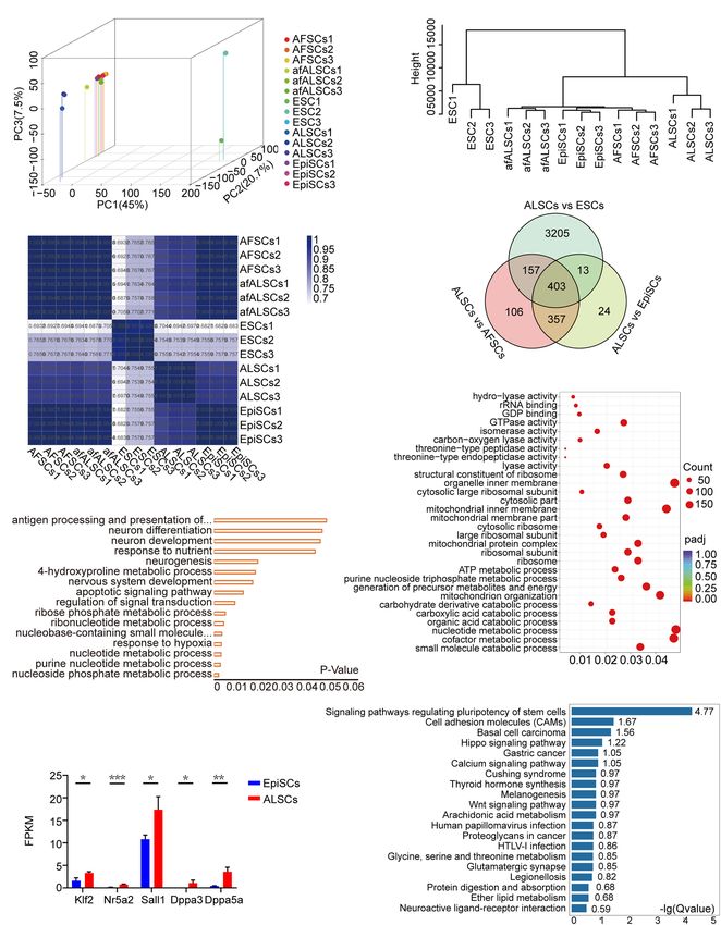
Wei et al.                                                                                                                  Mouse Primed Stem Cells

    Extensive efforts have been made to identify approaches able            AL medium consisted of Act A (20 ng/ml, R&D systems)
to reverse the two states of pluripotent stem cells and mainly              and LIF (1,000 U/ml, Millipore) added into a basic N2B27
involve either specific culture conditions with different factors           medium including 50% Neurobasal (Gibco), 50% DMEM/F12
or forced “naive” gene expression (Bao et al., 2009; Guo et al.,            (Gibco), 2 mM GlutaMax (Gibco), 1 × non-essential amino acids
2009; Guo and Smith, 2010; Okashita et al., 2016; Chen et al.,              (NEAA, Gibco), 1 × penicillin/streptomycin (Gibco), 0.1 mM
2018; Du et al., 2018; Pastor et al., 2018; Qiu et al., 2015;               β-mercaptoethanol (Gibco), and 0.005% (25 mg) bovine serum
Rathjen et al., 1999; Zhou et al., 2010; Tai and Ying, 2013; Yu             albumin (BSA; Gibco) supplemented with 0.5 × N2 (Gibco), and
S. et al., 2020). D’Aniello et al. (2017) proposed the pivotal              0.5 × B27 (Gibco). All culture dishes were coated with fibronectin
roles of vitamin C and L-proline in controlling the pluripotency            [1 mg/ml in phosphate-buffered saline (PBS), Millipore] for at
continuum from naive to primed states by affecting global DNA               least 0.5 hour (h) before use.
methylation, transcriptional profile, and energy metabolism.
Recently, Yu S. et al. (2020) proposed that BMP4 plays an
essential role in primed-to-naive transition (PNT) by opening
                                                                            Conversion of ALSCs to rESCs and
up chromatin loci to activate critical regulators of PNT. These             EpiSCs to epiALSCs
reports demonstrate that the state of pluripotent stem cells can            To convert ALSCs to rESCs, ALSCs were dissociated into single
be reversed to some extent by factors in their culture conditions.          cells using Accutase (Invitrogen) and were plated in CL medium
Two recent studies have suggested that there is a “formative” state         or 2iL medium. The CL medium consisted of N2B27 medium
in ESCs which is between the “naive” and “primed” state and                 supplemented with 3 µM CHIR99021 and LIF (1,000 U/ml),
also presents formative features of human stem cells and horse              while the 2iL medium consisted of N2B27 supplemented with
stem cells (Kinoshita et al., 2020; Yu L. et al., 2020). These two          3 µM CHIR99021, 1 µM PD0325901, and LIF (1,000 U/ml).
studies describe different culture systems. The first involves the          To transform EpiSCs to epiALSCs, EpiSCs were cultured in
inhibition of the Wnt signaling pathway and the cells named FS              AF medium supplemented with Act A (20 ng/ml) and bFGF
cells, while the other culture system is dependent on the Wnt               (12 ng/ml, R&D systems), then dissociated with Accutase, and
signaling pathway and the cells named XPSCs; however, both                  plated in AL medium. LDN193189 (100 nM, Selleckchem)
cell lines exhibit formative pluripotency features (Kinoshita et al.,       and SB431542 (10 µM, Selleckchem) were also added into the
2020; Yu L. et al., 2020). All intermediate stem cells, including the       AL medium and the CL medium to inhibit BMP4 and Act
formative stem cells (FS cells), XPSCs and rosette-like stem cells          A, respectively.
(RSCs), encode a higher pluripotent gene expression than EpiSCs
and contribute to chimera formation; however, different culture             AP Staining
conditions and the unique properties of stem cells still require            Before staining, cells were placed in four-well plates, washed
further exploration (Kinoshita et al., 2020; Neagu et al., 2020; Yu         with 1 × PBS, and then fixed in 4% paraformaldehyde at room
L. et al., 2020).                                                           temperature for 30 min. The cells were washed with 1 × PBS
    In this study, we investigated which factors play important             again, followed by the addition of AP staining solution. The
roles for establishing pluripotent stem cells from mouse post-              AP staining solution was prepared as follows: 50 µl sodium
implantation embryos. Using a chemically defined medium                     nitrite solution was gently mixed with 50 µl FRV-alkaline
N2B27 supplemented with Act A and LIF, we successfully derived              solution and incubated at 37◦ C for 3 min; next, 2.25 ml H2 O
primed pluripotent stem cells. These pluripotent stem cells were            and 50 µl naphthol-As-BI alkaline solution were added to the
named as ALSCs, like EpiSCs, which expressed pluripotent                    mixture. The staining solution with fixed cells was incubated in
genes Oct4, Sox2, Nanog, and one inactive X chromosome                      the dark overnight.
and contributed to multiple tissues in teratoma but failed to
contribute to chimera in vivo. The ALSCs were in a primed state,
closed to EpiSCs, and able to reverse to naive state with high              Karyotype
efficiency by activating Wnt signaling.                                     The tested cells were incubated with 0.2 µg/ml colchicine
                                                                            supplemented to the culture medium for 2 h, followed by cell
                                                                            dissociation using Accutase; the suspensions were centrifuged at
MATERIALS AND METHODS                                                       1,500 rpm for 5 min to collect the tested cells. The cell pellets
                                                                            were gently resuspended in 8 ml 0.075 mol/L KCL (Sigma) and
Derivation of ALSCs                                                         incubated at 37◦ C in a water bath for 40 min for hypotonic
Mouse gastrulas were collected from E6.5 pregnant female                    treatment. Fixative liquid (methanol/glacial acetic acid = 3:1) of
ICR mice mated with GOF/GFP transgenic male mice with a                     1 ml was subsequently added to the resuspended cells and mixed
mixed background. The isolate epiblasts (E6.5) were obtained                gently, and the solution was then centrifuged at 1,000 rpm for
from gastrulas using a glass needle and were cultured in AL                 10 min. After discarding the supernatant, the cells were mixed
medium. After 5–10 days, outgrowths were minced into several                gently in 8 ml fixative solution and incubated in 37◦ C water bath
smaller pieces using a glass needle and moved into a fresh AL               for 30 min for cell fixation, which was repeated twice. Then, the
medium. The colonies, named ALSCs, could stably propagate                   resuspended cells in 0.5 ml fixative liquid were dropped onto ice-
by Accutase (Life Technology) every 2 days at a ratio of                    cold glass slides, which were then dried for 1 h at 70◦ C in a drying
1:4–1:6, and fresh AL medium was provided every day. The                    oven. The glass slides were stained in Giemsa (Sigma) for 10 min,

Frontiers in Cell and Developmental Biology | www.frontiersin.org       2                                      August 2021 | Volume 9 | Article 713503
Wei et al.                                                                                                                  Mouse Primed Stem Cells

washed with distilled water, and then air-dried. The preparations       Matrigel (Corning) and 5 µM Y27632 (Selleckchem). A total of
were analyzed by LUCIA Cytogenetics.                                    5 × 10⁶ ALSCs were injected under the epithelium of NOD–
                                                                        SCID mice. The tumors were allowed to develop for 3–5 weeks,
Real-Time Quantitative Polymerase                                       then fixed in 4% paraformaldehyde, and processed for paraffin
Chain Reaction                                                          sectioning. Sections were then observed following hematoxylin
Total RNA was extracted by Rneasy Mini Kit (Qiagen),                    and eosin staining.
and cDNA was isolated by GoScript Reverse Transcription
System (Promega). Real-time quantitative polymerase chain               Transcriptome Analysis
reactions (RT-qPCR) were set up using the SYBR FAST                     The total RNA of EpiSCs and ALSCs was isolated using
Universal qPCR kit (KAPA). Relative expression values were              Rneasy Mini Kit (Qiagen). cDNA was synthesized from
normalized to Gapdh expression, and data was processed using            purified RNA templates. The cDNA libraries were sequenced
the comparative Ct method. Each experiment was performed                using Illumina HiSeq10 × platform. Paired-end reads were
with technical triplicates. The primers used are listed in              mapped to the mouse reference genome (GRCm38/mm10).
Supplementary Table 1. Significant differences between groups           The differentially expressed genes were compared between
were determined using t-test, and P-values < 0.05 were                  samples with the standard false discovery rate ≤0.005, fold
considered statistically significant.                                   change | log2Ratio| ≥1 by edgeR online.1 Principal component
                                                                        analysis, hierarchical clustering analysis, Venn diagram, Pearson’s
Immunofluorescence                                                      correlation analysis, and pathway enrichment analysis of
The cells for immunofluorescence assays were washed with                differential genes were performed using the OmicShare tools.2
PBS, fixed in 4% paraformaldehyde for 30 min at room                    Gene Ontology term enrichment analysis was achieved using
temperature, and then permeabilized with 0.1% Triton X-100              DAVID.3
(Sigma) and 1% BSA in PBS for 30 min. The cells were
then incubated with the appropriate primary antibody at 4◦ C
overnight. After the cells were washed three times in 1% BSA
                                                                        Western Blotting
and 0.1% Triton X-100 in PBS for 5 min per wash, they                   Cells were lysed in lysis buffer (Solarbio) with protease and
were incubated with secondary antibody for 1 h at room                  phosphatase inhibitors for 15 min on ice. The extracts were
temperature in the dark and then washed once for 5 min in               then centrifuged at 12,000 rpm for 10 min at 4◦ C. The
1% BSA and 0.1% Triton X-100 in PBS and twice for 5 min                 supernatants were denatured in loading buffer (95◦ C, 5 min) and
in PBS. The cells were then mounted in Vectashield with DAPI            collected for further analysis. A total of 25 mg of proteins was
(Vector Laboratories). The samples were observed with a laser           separated on 10% Protogel and transferred onto a polyvinylidene
microscope (Nikon, Tokyo, Japan). The antibodies used are listed        difluoride membrane. The membrane was blocked in blocking
in Supplementary Table 2.                                               solution (5% milk powder/TBST solution) for 1 h at 37◦ C.
                                                                        The membranes were washed three times in 0.01% Tween-
                                                                        20/TBS 1 × (TBST) and followed by incubation at 4◦ C
Flow Cytometry                                                          overnight with primary antibody (diluted in 5% milk powder
Cells were dissociated into single cells using Accutase and
                                                                        in TBST solution). The membranes were washed three times
collected using centrifugation. After washing once with PBS,
                                                                        in TBST solution and incubated with HRP-conjugated anti-
the cell pellet was resuspended with PBS containing 1–5% KSR,
                                                                        rabbit secondary antibodies (diluted in 2% milk powder in
followed by filtration using a cell strainer (FACSAria II, BD
                                                                        TBST solution) at room temperature for 1 h. Protein was then
Biosciences) to remove large clumps of cells. The cells were then
                                                                        visualized using X-ray films. The antibodies used are listed in
subjected to flow cytometry (FACSAria II, BD Company). The
                                                                        Supplementary Table 2.
GFP fluorescence intensity was detected in the FITC channel.
Data analysis was performed using FlowJo X.10.0.7.
                                                                        Formation of Embryonic Body
Generation of Chimera                                                   ALSCs and EpiSCs on fibronectin-coated plates were incubated
To generate chimeric embryos, donor cells (12–15                        with Accutase for 3 min at 37◦ C till the colonies were
cells/blastocyst) were microinjected into the ICR mice blastocoel       completely disaggregated. The colonies were resuspended
cavity using a piezo-assisted micromanipulator attached to an           in embryoid body (EB) medium: DMEM/F12 + 20%
inverted microscope. Following the recovery of the injected             KSR (Gibco) + 1% GlutaMax (Gibco) + 1% NEAA
blastocysts in KSOM medium (Millipore), the chimeric                    (Gibco) + 1% β-mercaptoethanol (Gibco). The colonies
blastocysts were transplanted into the uterus of pseudopregnant         were cultured in suspension for 3 days at a concentration of
ICR female mice at 2.5 days post-coitus (dpc). The chimeric             1 × 10⁵ cells/ml.
embryos were collected at E6.5–E13.5.

Generation of Teratoma                                                  1
                                                                          http:www.omicshare.com/tools/Home/Soft/diffanalysis
ALSCs were disaggregated using Accutase into small cell clusters        2
                                                                          www.omicshare.com/tools
and resuspended in PBS with 30% growth factor reduced                   3
                                                                          http://david.abcc.ncifcrf.gov/

Frontiers in Cell and Developmental Biology | www.frontiersin.org   3                                          August 2021 | Volume 9 | Article 713503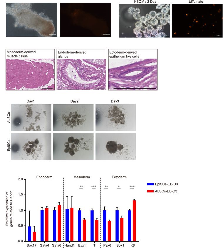
Wei et al.                                                                                                               Mouse Primed Stem Cells

RESULTS                                                                    (Figure 1G). Interestingly, the formative state markers, Etv5 and
                                                                           Tcf3 and Socs3, downstream target genes of LIF, showed a higher
Derivation of Primed Stem Cells From                                       expression in ALSCs than in EpiSCs, which implied that ALSCs
                                                                           were closer to a formative state than EpiSCs (Figure 1H; Kalkan
Post-implantation Embryos Supported
                                                                           et al., 2019). Immunofluorescence assays also confirmed the
by Act A and LIF                                                           expression of pluripotent markers OCT4, SOX2, and NANOG
Previously we reported that Act A is an essential factor for               in ALSCs and the presence of one inactive X chromosome,
maintaining the self-renewal of EpiSCs (Chen et al., 2020;                 which was in line with EpiSCs (Figure 1I and Supplementary
Wu et al., 2020). To test the role of Act A, CHIR99021                     Figure 1E). Western blotting analysis revealed that P-STAT3 was
(CHIR), and LIF in primed stem cells, we first cultured                    expressed in ALSCs at an intermediate level between that of ESCs
AFSCs, which are essentially a cell line similar to EpiSCs (Bao            and EpiSCs (Supplementary Figure 2A).
et al., 2018) in a chemically defined medium containing Act                   To further understand which factors were important for the
A with CHIR (AC medium) or LIF (AL medium). We found                       maintenance of ALSCs, we cultured ALSCs in medium with
that the AL medium was able to maintain the pluripotency,                  Act A or LIF alone (Supplementary Figure 2B). Unexpectedly,
while the AC medium failed (data not shown), which is in                   ALSCs were unable to sustain self-renewal in the medium
accordance with the notion that the AC medium supports                     supplemented with LIF alone and appeared apoptotic after three
primitive streak formation (Gadue et al., 2006; Turner et al.,             passages. In contrast, ALSC-like colonies were sustained in
2014; Tsakiridis et al., 2015; Morrison et al., 2016). Subsequently,       the presence of Act A alone (Supplementary Figure 2B) and
AFSCs were cultured in AL medium and designated afALSCs                    continued to self-renew up to passage 10. Moreover, the RT-
(Supplementary Figure 1A). We did not observe any distinctive              qPCR results showed that the pluripotent genes Oct4, Sox2, and
morphological change when AFSCs were placed into AL medium                 Nanog were significantly reduced in LIF or Act A-alone medium.
for more than 10 passages (Supplementary Figure 1B), and                   Furthermore, Socs3, a gene downstream of the LIF pathway,
afALSCs maintained the characteristics of pluripotency by                  endoderm genes Gata4 and Sox17, and ectoderm-related genes
AP staining and immunofluorescence of NANOG and OCT4                       Pax6 and Map2 were significantly increased in the LIF medium
(Supplementary Figures 1B,C). RT-qPCR analysis also revealed               than in control ALSCs (Supplementary Figures 2C,D). These
a higher expression of Nanog and Klf2 in afALSCs than AFSCs                data suggested that Act A was important but not sufficient to
(Supplementary Figure 1D).                                                 sustain ALSC growth and self-renewal, which is consistent with
   To further determine the effects of the combination of                  previous studies (Ashida et al., 2017; Mulas et al., 2017; Chen
Act A and LIF, we attempted to derive pluripotent stem cells               et al., 2020). We also show that the combination of Act A and
from mouse gastrulas in AL medium. We first collected E6.5                 LIF could support the pluripotency of ALSCs.
gastrulas from ICR female mice mated with Oct41PE-GFP                         Taken together, our results demonstrated that a chemically
reporter (GOF/GFP) male mice and cultured the epiblasts of                 defined medium with Act A and LIF was capable of establishing
E6.5 embryos in AL medium (Figures 1A,B). Flat and smooth                  ALSCs from E6.5 gastrulas. We showed that ALSCs were primed
colonies were selected with a glass needle to mechanically                 pluripotent stem cells that stably express pluripotent markers but
propagate and successfully develop into self-renewal stem cell             display molecular properties similar to those of EpiSCs.
lines in AL medium over 30 passages (Figure 1B). The stem
cells derived in AL medium from gastrulas were named ALSCs
and exhibited AP positive (Figure 1B), with normal karyotype               Developmental and Differentiated
(79.76%) (Figures 1C,D), and were GOF/GFP− . Furthermore, we               Potency of ALSCs in vivo
evaluated the derivation efficiency of ALSCs to be 27%, which              To further investigate the pluripotency of ALSCs, we performed
indicated a lower efficiency than 58% of EpiSCs (Figure 1E).               chimera tests in vivo. ALSCs were transfected with H2B
These ALSCs displayed the capacity to propagate as quickly as              tdTomato plasmid, and 12–15 ALSCs were injected into mouse
EpiSCs as shown in a growth curve analysis (Figure 1F).                    blastocysts. The chimeric embryos were transplanted in 2.5 dpc
                                                                           pseudopregnant female mice. Unexpectedly, we did not detect
ALSCs Display Similar Properties With                                      any ALSCs with tdTomato fluorescence in the E6.5 chimeras
EpiSCs                                                                     (Figure 2A and Table 1). As ALSCs did not contribute to chimera
RT-qPCR analysis in ALSCs revealed high levels of pluripotent              in vivo, we injected eight to 10 ALSCs with tdTomato reporter
gene expression, such as Oct4, Sox2, and Nanog, and low                    into eight-cell embryos and then cultured them in vitro for 48 h;
levels of primed pluripotent markers, such as Fgf5, compared               however, the ALSCs immediately became apoptotic, suggesting
to EpiSCs (Figure 1G). In addition, we also tested primordial              that ALSCs could not form chimeras or synchronize with
germ cell markers, such as Prdm14, Stella, and Vasa, which                 recipient embryo developmental stage (Figure 2B). Teratoma
were expressed at much lower levels in ALSCs than in ESCs                  formation was also performed and showed that ALSCs could
(Figure 1G). Furthermore, in ALSCs markers related to DNA                  differentiate to multiple tissue types in vivo (Figure 2C and
methylation, such as Dnmt3a and Dnmt3b, showed intermediate                Supplementary Figure 3A). These data suggest that ALSCs were
levels between those of EpiSCs and ESCs, while the genes related           similar to EpiSCs in chimera and teratoma formation (Brons
to differentiation, such as Eomes, were expressed at a similar             et al., 2007; Tesar et al., 2007). We also compared the embryoid
level with EpiSCs but were more highly expressed than ESCs                 body (EB) formation of ALSCs and EpiSCs for 3 days, and our

Frontiers in Cell and Developmental Biology | www.frontiersin.org      4                                    August 2021 | Volume 9 | Article 713503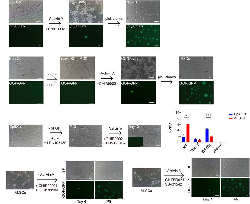
Wei et al.                                                                                                                                 Mouse Primed Stem Cells

  FIGURE 1 | Derivation and characterization of ALSCs. (A) Schematic of derivation of ALSCs. (B) Bright field image shows the derivation process and AP staining of
  ALSCs and EpiSCs. Scale bars, 100 µm. (C) Distribution of chromosome numbers of ALSCs. (D) Karyotype of ALSCs. (E) Efficiency of ALSCs and EpiSCs both
  derived from gastrulas. (F) Cell growth curve of EpiSCs and ALSCs. (G,H) RT-qPCR of the key genes in ALSCs, EpiSCs, and ESCs/2iL. Error bars indicate three
  independent biological replicates (mean ± SD). *P < 0.05, **P < 0.001, ***P < 0.0001. (I) Immunostaining for pluripotent markers in ALSCs, EpiSCs, and ESCs/2iL.
  Scale bars, 100 µm.

Frontiers in Cell and Developmental Biology | www.frontiersin.org                5                                           August 2021 | Volume 9 | Article 713503
Wei et al.                                                                                                                                  Mouse Primed Stem Cells

  FIGURE 2 | The pluripotency of ALSCs. (A) E6.5 chimera collected by injecting ALSCs in host blastocysts. Scale bars, 100 µm. (B) E3.5 chimeras generated by
  injecting ALSCs to eight-cell stage embryos followed culturing in KSOM for 2 days. Scale bars, 100 µm. (C) ALSCs contributed to the generation of multiple tissues
  of germ layer in vivo by the teratoma test. Scale bars, 100 µm. (D) Morphology of embryoid body induction by ALSCs and EpiSCs for 3 days. Scale bars, 100 µm.
  (E) RT-qPCR of germ layer markers in embryoid bodies of ALSCs and EpiSCs after 3 days of culture. Error bars indicate three independent biological replicates
  (mean ± SD). *P < 0.05, **P < 0.001, ***P < 0.0001.

results showed that the diameter of the EB spheres of ALSCs                           6 days by RT-qPCR and obtained similar results (Supplementary
was smaller than that of EpiSCs (Figure 2D). However, the RT-                         Figure 3B). Furthermore, we examined the response of ALSCs to
qPCR analysis showed no distinct difference between the two                           the cytokine cocktail for primordial germ cell induction (Ohinata
types of EB (Figure 2E). We further tested the gene expression                        et al., 2009), but we did not detect GOF/GFP and VASA double-
of the differentiated ALSCs and EpiSCs in N2B27 medium for                            positive cells (Supplementary Figure 3C).

Frontiers in Cell and Developmental Biology | www.frontiersin.org                 6                                           August 2021 | Volume 9 | Article 713503
Wei et al.                                                                                                                          Mouse Primed Stem Cells

TABLE 1 | ALSCs do not contribute to E6.5 chimera by injection to blastocysts.       to pathways regulating the pluripotency of stem cells such as
                                                                                     Klf2, Nr5a2, Sall1, and Dppa3 (Figures 3G,H). Indeed Klf2
Cell line      Stage of          No. of        No. of collected      No. of
               embryo        injected cells       embryos           chimeras         and Nr5a2 are known as naive pluripotency markers, thus our
                                                                                     data suggested that ALSCs were closer to the naive state than
ALSCs             E6.5            12–15               12               0             EpiSCs (Figure 3H).
                                                                                        A recent study suggested that inhibition of Wnt signaling
                                                                                     and retinoids by XAV939 and BMS493, respectively, with low
   Taken together, these data suggested that the ALSC cell line                      concentrations of Act A could capture mouse and human stem
was similar to EpiSCs for its developmental capacity in vivo and                     cells with formative pluripotent features, defined as FS cells
in vitro.                                                                            (Kinoshita et al., 2020). Similar to FS cells, ALSCs expressed
                                                                                     lower levels of somatic markers, such as Foxa2 and T, and
Molecular Features of ALSCs                                                          higher levels of pluripotent genes than EpiSCs (Figure 3H and
To better understand the molecular features of afALSCs and                           Supplementary Figure 3E). However, ALSCs did not reveal the
ALSCs freshly derived from embryos, we first performed                               expected generally low expression of neural lineage markers,
RNAseq to analyze the transcriptomes of afALSCs and AFSCs                            such as Sox11, Sox1, and Pax6, which differed from FS cells
(Supplementary Figure 3D). There were 430 differently                                (Supplementary Figure 3E). Moreover, ALSCs expressed active
expressed genes with 261 up- and 169 downregulated genes                             canonical Wnt signaling pathway genes with a high expression
in afALSCs. Among the upregulated genes, four distinct gene                          of β-catenin and Tcf3, and a low expression in Gsk3β and
modules were identified. Module I was related to cytokine-                           Axin1, molecules involved in the β-catenin destruction complex,
mediated signaling pathways (such as the JAK-STAT cascade),                          which is distinctly different with recent studies (Supplementary
which were stimulated by LIF. Similar to module I, genes from                        Figure 3E; Kim et al., 2013; Kinoshita et al., 2020; Neagu et al.,
module II were involved in growth hormone signaling pathways.                        2020). We also showed that Otx2 was expressed at a low level
By performing a similar analysis, we also identified two gene                        in ALSCs, which was also in contrast with previous studies
modules (modules III and IV) among the genes upregulated                             suggesting that Otx2 played a crucial role in pluripotent stem cells
in afALSCs. Significant numbers of genes of SMAD protein                             in the intermediate state; however, the specific impact of Otx2
signal transduction and protein phosphorylation were identified                      on ALSCs still needs to be investigated further (Supplementary
in the conversion of AFSCs to afALSCs. Collectively, these                           Figure 3E; Acampora et al., 2013; Kinoshita et al., 2020; Neagu
data suggested that the AFSCs are reset in AL medium and                             et al., 2020). Interestingly, we found that E-cadherin displayed a
present reinforced pluripotency, while the induced afALSCs                           lower expression in ALSCs than in EpiSCs, which may explain the
possess unique molecular features that are distinct from AFSCs                       different morphology of the embryoid body that ALSCs formed
(Supplementary Figure 3D).                                                           (Supplementary Figure 3E).
   To characterize the molecular features of ALSCs, we                                  These results indicated that the ALSCs were primed stem cells
assessed the transcriptomes of ALSCs, EpiSCs, and ESCs.                              with particular molecular features which were similar to EpiSCs.
Principal component analysis revealed that the global gene
expression patterns of ALSCs, afALSCs, AFSCs, and EpiSCs were                        Conversion of ALSCs to Naive ESCs
distinct from ESCs (Figure 3A). Hierarchical cluster analysis                        Multiple recent reports have focused on the conversion of primed
demonstrated that afALSCs, AFSCs, and EpiSCs were clustered                          ESCs to naive ESCs (Chen et al., 2018; Du et al., 2018; Pastor
closely and ALSCs were clustered in the second layer (Figure 3B).                    et al., 2018; Yu S. et al., 2020). CHIR is suggested to be a
This unique clustering of ALSCs was further confirmed by                             main factor for naive ESCs (Qiu et al., 2015; Choi et al., 2017)
constructing a correlation matrix of gene expression clustered                       as it inhibits the phosphorylation of β-catenin and simulates
using Pearson correlation coefficients (Figure 3C). We identified                    canonical Wnt signaling. To explore the possibility of conversion
403 genes which were uniquely presented in ALSCs, including                          from ALSCs to naive ESCs, we cultured ALSCs (GOF/GFP
Etv4, Lef1, Dusp9, and Sox3 (Figure 3D). It is interesting to                        reporter) in medium in which Act A was replaced with CHIR
note that the expression of formative markers Tcf3 in ALSCs                          (CL medium) (Figure 4A). After ALSCs were cultured in CL for
suggested that ALSCs were closer to the formative state than                         4 days, GOF/GFP+ colonies were induced, and the individual
EpiSCs, but the low expression of Otx2 demonstrated that ALSCs                       GOF/GFP+ colonies could be maintained as ESCs in morphology
did not reside in a formative state (Supplementary Figure 3E;                        over passage 30 (named rESCs) (Figure 4A). Interestingly, ALSCs
Kinoshita et al., 2020). These characteristic genes were obtained                    cultured in 2iL medium (PD0325901, CHIR, and LIF) showed a
from the intersection of differently expressed genes of ALSCs                        reduced efficiency of ALSCs converting to rESCs compared to
vs. ESCs, ALSCs vs. AFSCs, and ALSCs vs. EpiSCs. The gene                            those cultured in CL medium (Table 2). These data suggested
ontology analysis of most upregulated differently expressed genes                    that PD0325901 partially blocked the transition procedure, likely
showed an association with metabolic and neurogenesis processes                      because PD0325901 induced DNA damage and hypomethylation
(Figure 3E). In line with the gene ontology, we used gene set                        in ESCs (Choi et al., 2017). In contrast, EpiSCs began to
enrichment analysis to reveal the KEGG pathway enrichment                            differentiate or died in cultures containing 2iL or CL medium
for metabolic process (Figure 3F). Consistent with ALSCs being                       from AF medium (Act A and bFGF) and failed to convert to
in a state between naive and primed state, most ALSCs vs.                            rESCs (Table 2). Following the blastocyst injection of rESCs,
EpiSCs upregulated differentially expressed genes were related                       we could detect reporter expression in E12.5 chimeric embryos

Frontiers in Cell and Developmental Biology | www.frontiersin.org                7                                     August 2021 | Volume 9 | Article 713503
Wei et al. Mouse Primed Stem Cells FIGURE 3 | Transcriptomic characteristics of ALSCs. (A) Three-dimensional scatter plot based on the principal component analysis of RNA-seq data from three biological replicates. (B) Hierarchical clustering of RNA-seq data of ESCs, EpiSCs, ALSCs, afALSCs, and AFSCs. (C) The correlation matrix of gene expression was clustered using Pearson correlation. (D) Venn diagram showing overlaps of the differential expression gene of ALSCs vs. ESCs, ALSCs vs. EpiSCs, and ALSCs vs. AFSCs. (E) Gene Ontology categories significantly enriched in the biological process for 403 genes from the overlap in (D). (F) Kyoto Encyclopedia of Genes and Genomes (KEGG) pathway analysis of upregulated genes in 403 genes from the overlap in (D). (G) KEGG pathway analysis of upregulated genes in ALSCs vs. EpiSCs. (H) Pluripotent genes and naive genes were upregulated in ALSCs. Frontiers in Cell and Developmental Biology | www.frontiersin.org 8 August 2021 | Volume 9 | Article 713503
Wei et al.                                                                                                                                  Mouse Primed Stem Cells

  FIGURE 4 | Conversion of ALSCs to rESCs. (A) Morphological changes and rESC induction process of ALSCs. Scale bars, 100 µm. (B) Morphological changes
  and rESC induction process of EpiSCs. Scale bars, 100 µm. (C) Morphological changes and rESC induction process of EpiSCs supplemented with LDN193189
  (BMP4 inhibitor) in AL medium and CL medium. Scale bars, 100 µm. (D) The ALSCs maintenance was independent of BMP signaling. Error bars indicate three
  independent biological replicates (mean ± SD). *P < 0.05, **P < 0.001, ***P < 0.0001. (E) Morphological changes and rESC induction process of ALSCs
  supplemented with LDN193189 (BMP inhibitor) in CL medium. Scale bars, 100 µm. (F) Morphological changes and rESC induction process of ALSCs
  supplemented with SB431542 (Act A inhibitor) in CL medium. Scale bars, 100 µm.

and obtained several overt coat color chimeras (Supplementary                        (called epiALSCs) for several passages (at least 10 passages)
Figure 4A and Table 3).                                                              (Figure 4B). As expected, epiALSC colonies also developed to
   As ALSCs but not EpiSCs were converted to rESCs in                                become GOF/GFP+ when cultured in CL or 2iL for 4 days
CL medium, we hypothesized that EpiSCs may be capable of                             (Figure 4B), although the transition rate was lower than for
converting to rESC-like cells after culturing in AL medium                           ALSCs (Table 4). These results suggested that the successive
                                                                                     conversion from EpiSCs to rESCs was accomplished in two
                                                                                     stages, with the first step involving the replacement of bFGF in
TABLE 2 | Efficiency of rESCs conversed from ALSCs and EpiSCs in
                                                                                     AF medium to LIF and the second step involving the use of CHIR
CL and 2iL medium.                                                                   instead of Act A (stage one: AF-AL; stage two: AL-CL).

Cell line       Culture      Total cell lines   No. of rESCs        Percentage
                medium
                                                                                     TABLE 3 | rESCs contributed to chimeras by injection to blastocysts.
ALSCs              CL               6                  3               50%
                                                                                     Cell line      Stage of          No. of       No. of collected      No. of
                   2iL              6                  1              16.7%
                                                                                                    embryo        injected cells      embryos           chimeras
EpiSCs             CL               6                  0               0%
                   2iL              6                  0               0%            rESCs            E12.5           12–15               18                7

Frontiers in Cell and Developmental Biology | www.frontiersin.org                9                                            August 2021 | Volume 9 | Article 713503
Wei et al. Mouse Primed Stem Cells
TABLE 4 | Efficiency of rESCs conversed from epiALSCs in CL and 2iL medium. BMP4 or Act A, which are mechanisms that
require further study.
Cell line Culture Total cell lines No. of rESCs Percentage
medium
Epigenetic Changes in ALSCs
epiALSCs CL 5 2 40%
X chromosome inactivation (Xi) is a major epigenetic change
2iL 5 1 20%
occurring between rESCs and EpiSCs. We first analyzed
the X chromosome activation state of ALSCs using XiGFP
EpiSCs, in which the GFP transgene is located exclusively
Onishi et al. (2012) proposed that Act A, bFGF, and on the Xi, and the reactivation of Xi could be monitored
LIF could induce domed colonies like naive ESCs, but the by its GFP expression (Gillich et al., 2012). We observed
low cell density of transition cells led to a failed induction a stable Xi and did not detect any GFP+ cells in XiGFP
of domed colonies. Thus, we next attempted to test ALSC EpiSCs (Figure 5A). However, when we cultured these XiGFP
transition from AL to CL medium with low cell densities EpiSCs in AL medium, we observed a small number of
(Supplementary Figure 4B). The results showed that, when GFP+ cells, which was also confirmed by immunostaining
1,000, 2,000, and 5,000 ALSCs per well were placed into CL of H3K27me3 (Figure 5B). Flow cytometry analysis showed
medium in six-well plates, ALSCs could form both dome- 2.48% epiALSCs exhibited XaXa (Figure 5C). We next isolated
like and flat-like colonies in CL medium after 12 days. These GFP+ epiALSCs and GFP− epiALSCs by fluorescence-activated
colonies were all GOF/GFP− until propagated (Supplementary cell sorting to purify the XaXa epiALSCs subset. Unexpectedly,
Figure 4C). We next tested whether a single ALSC could both isolated GFP+ and GFP− epiALSCs propagated stably
convert to rESCs and found that four GOF/GFP+ rESCs and maintained XaXa and XaXi, respectively, in the first
were obtained (4/96), and similarly these cells did not 2 days but quickly reverted to the mixed state at day 4
develop GOF/GFP+ clones until propagated (Supplementary (Figure 5D), revealing that the AL medium promoted silenced
Figures 4D,E). These results demonstrated that ALSCs, even at X chromosome reactivation in a few of the epiALSCs, while
a low density or as single cells, were capable of transitioning to most sustained a stable XiGFP . Since Act A was capable
GOF/GFP+ clones in CL medium and that the transition cell of sustaining the ALSC pluripotency, we next focused on
density did not impact on the induction of naive-like colonies, whether Act A could maintain the reactivation of silenced X
which is different with the system proposed by Onishi et al. chromosome in epiALSCs. Subsequently, we cultured epiALSCs
(2012). in medium containing Act A alone and observed that, within
Yu S. et al. (2020) recently proposed that BMP4 was essential the first four passages, epiALSCs lost the ability to re-activate
for primed-to-naive transition (PNT). Thus, we investigated the silenced X chromosome (Figure 5E). It is worthy to
whether BMP4 was essential for our transition system. We note that a minority of cells with stable X reactivation was
first chose two EpiSC lines which successfully reversed to observed in AL medium, which suggested that the combination
rESCs by two steps and then cultured them in AL medium of Act A and LIF could have a positive effect on X
(stage one) supplemented with LDN193189, an inhibitor of chromosome reactivation.
BMP4. EpiSCs successfully reverted to epiALSCs but could Next, we examined the impact of CHIR and LIF on X
not induce GOF/GFP+ colonies in CL medium (stage two) chromosome reactivation during the reversion of ALSCs to
with the addition of BMP4 inhibitors (Figure 4C). This rESCs in CL medium. We found that GOF/GFP+ colonies
suggested that endogenous BMP4 was essential for the process started to emerge when ALSCs were cultured in CL medium
of reversing EpiSCs to rESCs, which was consistent with for 4 days. All GOF/GFP+ colonies exhibited XaXa, but
the studies of Yu S. et al. (2020). However, we focused some XaXa colonies did not activate GOF/GFP, suggesting
on the expression of BMP downstream genes identified by that silenced X chromosome reactivation occurred before
RNAseq and found that, except for Id1, BMP targets such as GOF/GFP activation (Figure 5F), which is in line with the
Tfap2c and Zbtb7 families were rarely expressed or steadily findings of Yu S. et al. (2020). Because DNA methylation is
expressed in ALSCs (Figure 4D). Thus, we directly cultured involved in X chromosome inactivation (Sado et al., 2004;
ALSCs, from AL medium to CL medium supplemented with Zvetkova et al., 2005; Schmidt et al., 2012; Auclair et al.,
LDN193189, and found that GOF/GFP+ colonies were induced 2014), we further explored whether DNA methylation levels
after 4 days and pure GOF/GFP+ colonies could be sustained were altered in ALSCs compared to EpiSCs. We examined
to passage19 (Figure 4E). Moreover, we also added SB431542 the protein expression of the DNA methyltransferase, Dnmt3a.
to CL medium to culture ALSCs in order to determine whether The protein expression of DNMT3A by immunofluorescence
Act A was essential for the conversion from ALSCs to rESCs. differed greatly in ALSCs and EpiSCs, which exhibited a sporadic
Interestingly, the GOF/GFP+ colonies were also produced in expression in ALSCs but was expressed almost throughout
4 days (Figure 4F). the nucleus in EpiSCs (Figure 5G). As expected, RNA-seq
Thus, our data showed that endogenous BMP4 analysis revealed that DNA methyltransferases Dnmt1, Dnmt3a,
was essential for conversion from EpiSCs to rESCs, Dnmt3b, and Dnmt3c were significantly downregulated in
as the inhibition of BMP4 resulted in the failure to ALSCs (Figure 5H).
convert to rESCs. However, our transition system Our data suggested that CHIR was critical for promoting
from ALSCs to rESCs does not require endogenous X chromosome reactivation, and in combination with LIF, it
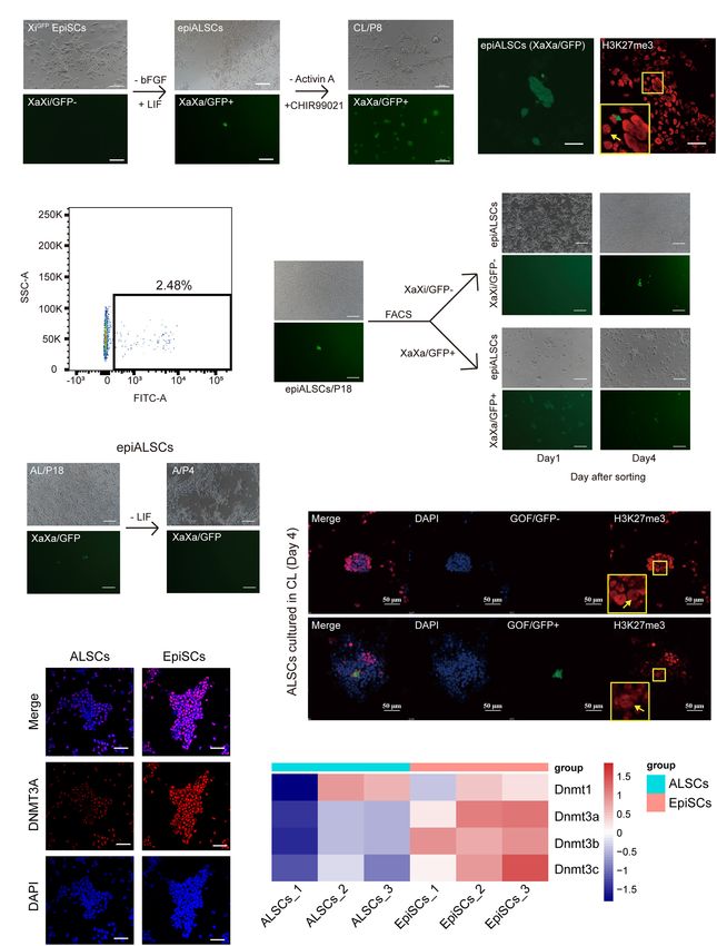
Frontiers in Cell and Developmental Biology | www.frontiersin.org 10 August 2021 | Volume 9 | Article 713503Wei et al. Mouse Primed Stem Cells FIGURE 5 | Conversion of XiGFP EpiSCs to rESCs. (A) Morphological changes and rESC induction process of XiGFP EpiSCs. Scale bars, 100 µm. (B) Immunostaining of H3K27me3 in epiALSCs. Scale bars, 50 µm. (C) Fluorescence-activated cell sorting analysis of epiALSCs, GFP+ cells were XaGFP epiALSCs. (D) Morphological changes and X chromosome change process from isolated XaXa-epiALSCs and XaXi-epiALSCs. Yellow arrow shows XaXi epiALSCs, green arrow shows XaXa epiALSCs. Scale bars, 100 µm. (E) Morphological changes and X chromosome inactivation process of epiALSCs placed in Act A-alone medium for four passages. Scale bars, 100 µm. (F) Immunostaining of H3K27me3 with GOF/GFP in ALSCs after having been placed in CL medium for 4 days. Yellow arrows show XaXa epiALSCs. Scale bars, 50 µm. (G) Immunostaining of DNMT3A in ALSCs and EpiSCs. Scale bars, 50 µm. (H) Heat map showing the scaled expression of DNA methylation-related genes. drives the conversion to rESCs from primed state stem cells. DISCUSSION Our data also indicate that ALSCs possess the plasticity of Xi reactivation, which was supported by the low expression of DNA Our findings showed that the AL medium could establish methyltransferases. primed pluripotent stem cells from mouse gastrulas and that Frontiers in Cell and Developmental Biology | www.frontiersin.org 11 August 2021 | Volume 9 | Article 713503
Wei et al.                                                                                                             Mouse Primed Stem Cells

ALSCs were similar to EpiSCs with unique molecular features.              with each other to sustain ALSC self-renewal still requires
We further demonstrated that ALSCs were able to convert to                more investigation.
naive ESCs by activating Wnt signaling, and the AL medium                    Compared to the transition system reported by Yu S. et al.
also prompts EpiSCs to transition to an intermediate stage                (2020), our system did not rely on BMP4, as the known
of the naive state from the primed state. Furthermore, our                targets of BMP4 were merely expressed independently of Id1,
findings showed Act A and LIF were both important for                     which was increased in ALSCs. Moreover, the inhibition of
maintaining the unique primed state, as the expression of                 BMP4 did not impact on the capacity of ALSCs to convert to
pluripotent genes decreased in the medium supplemented with               rESCs but influenced the conversion of EpiSCs to epiALSCs,
either Act A or LIF alone.                                                suggesting that endogenous BMP4 did play a vital role during
   In fact, post-implantation epiblasts (E5.0–E6.0) reside in             conversion to epiALSCs from EpiSCs. As the transition from
more states, and there is no clear common view on what                    the AF medium to the AL medium only required a change in
transcriptional and epigenetic features they may have. Recently,          bFGF supplementation to LIF, we considered that the inhibition
Neagu et al. (2020) showed that LIF could support intermediate            of BMP4 may be involved in LIF signaling and conversion
pluripotent stem cells for self-renewal. In here we thought               of EpiSCs to epiALSCs. In addition, the effects of LIF on
that ALSC is in an intermediate state between naive and                   ALSCs were confirmed by the higher expression of P-STAT3
primed states, LIF push epiblast cells to reversed naive state            detected in ALSCs than EpiSCs, illustrating that the Jak/Stat
(Bao et al., 2009), and Act A supports epiblast development               pathway was activated in ALSCs. The impact of LIF signaling
(Brons et al., 2007; Tesar et al., 2007). The combination of              in ALSCs could represent the focus of a future investigation.
LIF and Act A was applied to the epiblast to derive unique                These results imply that endogenous BMP4 was not essential
stem cells, unlike EpiSCs which are activated by Act A and                for our transition system and that distinct mechanisms may
bFGF, displaying a gene signature closer to that of the anterior          be involved in these two transition systems (Yu S. et al.,
epiblast of a late-gastrula stage embryo (E7.5) (Kojima et al.,           2020).
2014). Compared to the culture medium of FS cells and RSCs                   We showed that ALSCs could enter a primed-like state,
(Kinoshita et al., 2020; Neagu et al., 2020), the AL medium               which is different from the reported XPSCs, FS cells, and
does not contain inhibitors of the canonical Wnt signaling                RSCs (Kinoshita et al., 2020; Neagu et al., 2020; Yu L. et al.,
pathway. Our study showed that canonical Wnt signaling was                2020). We assumed that this intermediate state may represent
activated in ALSCs by endogenous stimuli, suggesting that                 a state of equilibrium between the two states as what occurs
ALSCs maintain a self-renewal capacity independently of the               in vivo, with the ephemeral state between E4.5 and E6.5.
inhibition of Wnt signals, which was different from FS cells              In addition, epiALSCs also exhibited several cells (2.48%)
and RSCs (Supplementary Figure 3E). However, Wnt signaling                presenting the reactivation of the X chromosome. Therefore,
plays a critical role in XPSCs having formative features (Yu              the AL medium for the middle state may include properties
L. et al., 2020). The effect of β-catenin on ALSCs needs to be            from both the primed state and the naive state. Furthermore,
further explored, as a recent study demonstrated that turning             ALSCs in the intermediate state may be heterogeneous and
Wnt signaling on or off could establish the same formative                maintain a balance between the naive and primed states.
features found in stem cells. FS cells and RSCs utilize a                 Based on our X chromosome reactivation data, we suggest
retinoid inhibitor and a MAPK inhibitor, respectively, to suppress        that X chromosome reactivation may not be the main
differentiation (Kinoshita et al., 2020; Neagu et al., 2020);             feature of this intermediate state, but rather the chromatin
however, this differs from the AL medium which requires Act A, a          accessibility in this stage may prompt the cells to convert to
component of Nodal signaling, and promotes the differentiation            a naive state more easily, a mechanism which still requires
of ESCs.                                                                  further investigation.
   Notably, neural lineage markers are generally highly expressed            Like EpiSCs, ALSCs failed to contribute to chimera formation
in ALSCs at the RNA level, but ALSCs still maintain EpiSC-                despite expressing pluripotent genes and alkaline phosphatase.
like colonies and obtain the ability of converting to rESCs,              We speculate that ALSCs are more similar to EpiSCs and exhibit
which may illustrate that the inhibition of neural ectoderm               a more compatible embryo stage similar to the post-implantation
differentiation is unnecessary in ALSCs. We hypothesized that             stage but unlike the cleavage stage of the embryo or blastocyst.
endogenous Wnt and MAPK signaling maintained a balanced                   Since we identified a higher expression of pluripotent genes
state in ALSCs, different with FS cells and RSCs, which inhibit           and formative genes than in EpiSCs, ALSCs may represent
Wnt signaling. Importantly, ALSCs efficiently reversed to rESCs           a primed state that can easily revert to ESCs. However,
when activated by Wnt signaling whereby FS cells failed to                germ layer genes like Eomes were also highly expressed in
reverse to ESCs (Kinoshita et al., 2020). Moreover, instead of            ALSCs, indicating that the detailed mechanisms involved warrant
producing PGCLCs by FS cells and XPSCs (Kinoshita et al.,                 further investigation.
2020; Yu L. et al., 2020), ALSCs only induced GOF/GFP+ cells;                In summary, the AL medium allows to establish ALSCs
this led us to speculate that ALSCs converted to rESCs                    from post-implantation embryos. ALSCs possess unique
rather than generated PGCLCs. From the discussion above,                  molecular properties, propagate robustly in long-term culture,
we propose that ALSCs are more similar to a primed state                  and share a status which could convert to naive state ESCs,
rather than a formative state; nonetheless, the mechanism                 thus offering new opportunities to study the control of
whereby the AL medium and Act A and LIF cooperate                         cell fate.

Frontiers in Cell and Developmental Biology | www.frontiersin.org    12                                   August 2021 | Volume 9 | Article 713503
Wei et al.                                                                                                                                                  Mouse Primed Stem Cells

DATA AVAILABILITY STATEMENT                                                                    FUNDING
All the sequencing data were deposited in the NCBI,                                            This work was supported by the Inner Mongolia Autonomous
Gene Expression Omnibus (GEO) under accession                                                  Region Science and Technology Plan of China (2020ZD0007) and
number GSE156673.                                                                              the National Natural Science Foundation of China (32060176).

ETHICS STATEMENT
                                                                                               ACKNOWLEDGMENTS
The animal study was reviewed and approved by Institutional
Animal Care and Use Committee at Inner Mongolia University.                                    We thank Juan Li for proofreading the manuscript. We also thank
                                                                                               Inner Mongolia Saikexing Institute of Breeding and Reproductive
                                                                                               Biotechnology in Domestic Animal for instrument support.
AUTHOR CONTRIBUTIONS
SB conceived the idea for this project and designed and
conducted the experiments. MW and YC wrote the manuscript                                      SUPPLEMENTARY MATERIAL
with help from all the other authors. MW, YC, CZ, and
LZ performed the experiments and analyzed data under the                                       The Supplementary Material for this article can be found
supervision of SB. All the authors read, accepted, and approved                                online at: https://www.frontiersin.org/articles/10.3389/fcell.2021.
the final manuscript.                                                                          713503/full#supplementary-material

REFERENCES                                                                                     Evans, M. J., and Kaufman, M. H. (1981). Establishment in culture of pluripotential
                                                                                                  cells from mouse embryos. Nature 292, 154–156. doi: 10.1038/292154a0
Acampora, D., Di Giovannantonio, L. G., and Simeone, A. (2013). Otx2 is an                     Gadue, P., Huber, T. L., Paddison, P. J., and Keller, G. M. (2006). Wnt and TGF-
   intrinsic determinant of the embryonic stem cell state and is required for                     beta signaling are required for the induction of an in vitro model of primitive
   transition to a stable epiblast stem cell condition. Development 140, 43–55.                   streak formation using embryonic stem cells. Proc. Natl. Acad. Sci. U. S. A. 103,
   doi: 10.1242/dev.085290                                                                        16806–16811. doi: 10.1073/pnas.0603916103
Ashida, Y., Nakajima-Koyama, M., Hirota, A., Yamamoto, T., and Nishida, E.                     Gillich, A., Bao, S., Grabole, N., Hayashi, K., Trotter, M. W., Pasque, V., et al. (2012).
   (2017). Activin A in combination with ERK1/2 MAPK pathway inhibition                           Epiblast stem cell-based system reveals reprogramming synergy of germline
   sustains propagation of mouse embryonic stem cells. Genes Cells 22, 189–202.                   factors. Cell Stem Cell 10, 425–439. doi: 10.1016/j.stem.2012.01.020
   doi: 10.1111/gtc.12467                                                                      Guo, G., and Smith, A. (2010). A genome-wide screen in EpiSCs identifies Nr5a
Auclair, G., Guibert, S., Bender, A., and Weber, M. (2014). Ontogeny of CpG island                nuclear receptors as potent inducers of ground state pluripotency. Development
   methylation and specificity of DNMT3 methyltransferases during embryonic                       137, 3185–3192. doi: 10.1242/dev.052753
   development in the mouse. Genome Biol. 15:545. doi: 10.1186/s13059-014-                     Guo, G., Yang, J., Nichols, J., Hall, J. S., Eyres, I., Mansfield, W., et al. (2009). Klf4
   0545-5                                                                                         reverts developmentally programmed restriction of ground state pluripotency.
Bao, S., Tang, F., Li, X., Hayashi, K., Gillich, A., Lao, K., et al. (2009). Epigenetic           Development 136, 1063–1069. doi: 10.1242/dev.030957
   reversion of post-implantation epiblast to pluripotent embryonic stem cells.                Joo, J. Y., Choi, H. W., Kim, M. J., Zaehres, H., Tapia, N., Stehling, M., et al.
   Nature 461, 1292–1295. doi: 10.1038/nature08534                                                (2014). Establishment of a primed pluripotent epiblast stem cell in FGF4-based
Bao, S., Tang, W. W., Wu, B., Kim, S., Li, J., Li, L., et al. (2018). Derivation of               conditions. Sci. Rep. 4:7477. doi: 10.1038/srep07477
   hypermethylated pluripotent embryonic stem cells with high potency. Cell Res.               Kalkan, T., Bornelov, S., Mulas, C., Diamanti, E., Lohoff, T., Ralser, M., et al.
   28, 22–34. doi: 10.1038/cr.2017.134                                                            (2019). Complementary Activity of ETV5, RBPJ, and TCF3 Drives Formative
Brons, I. G., Smithers, L. E., Trotter, M. W., Rugg-Gunn, P., Sun, B., Chuva de                   Transition from Naive Pluripotency. Cell Stem Cell 24, 785–801.e7. doi: 10.
   Sousa Lopes, S. M., et al. (2007). Derivation of pluripotent epiblast stem cells               1016/j.stem.2019.03.017
   from mammalian embryos. Nature 448, 191–195. doi: 10.1038/nature05950                       Kim, H., Wu, J., Ye, S., Tai, C. I., Zhou, X., Yan, H., et al. (2013). Modulation of beta-
Chen, A. F., Liu, A. J., Krishnakumar, R., Freimer, J. W., DeVeale, B., and Blelloch,             catenin function maintains mouse epiblast stem cell and human embryonic
   R. (2018). GRHL2-Dependent Enhancer Switching Maintains a Pluripotent                          stem cell self-renewal. Nat. Commun. 4:2403. doi: 10.1038/ncomms3403
   Stem Cell Transcriptional Subnetwork after Exit from Naive Pluripotency. Cell               Kinoshita, M., Barber, M., Mansfield, W., Cui, Y., Spindlow, D., Stirparo, G. G.,
   Stem Cell 23, 226–238.e4. doi: 10.1016/j.stem.2018.06.005                                      et al. (2020). Capture of Mouse and Human Stem Cells with Features of
Chen, Y., Wu, B., Zheng, L., Wu, C., Wei, M., Chen, C., et al. (2020). Induction                  Formative Pluripotency. Cell Stem Cell 28, 453–471.e8. doi: 10.1016/j.stem.
   and maintenance of specific multipotent progenitor stem cells synergistically                  2020.11.005
   mediated by Activin A and BMP4 signaling. J. Cell Physiol. 235, 8640–8652.                  Kojima, Y., Tam, O. H., and Tam, P. P. (2014). Timing of developmental events in
   doi: 10.1002/jcp.29708                                                                         the early mouse embryo. Semin. Cell Dev. Biol. 34, 65–75. doi: 10.1016/j.semcdb.
Choi, J., Huebner, A. J., Clement, K., Walsh, R. M., Savol, A., Lin, K., et al.                   2014.06.010
   (2017). Prolonged Mek1/2 suppression impairs the developmental potential of                 Morrison, G., Scognamiglio, R., Trumpp, A., and Smith, A. (2016). Convergence of
   embryonic stem cells. Nature 548, 219–223. doi: 10.1038/nature23274                            cMyc and beta-catenin on Tcf7l1 enables endoderm specification. EMBO J. 35,
D’Aniello, C., Habibi, E., Cermola, F., Paris, D., Russo, F., Fiorenzano, A., et al.              356–368. doi: 10.15252/embj.201592116
   (2017). Vitamin C and l-Proline Antagonistic Effects Capture Alternative States             Mulas, C., Kalkan, T., and Smith, A. (2017). NODAL Secures Pluripotency upon
   in the Pluripotency Continuum. Stem Cell Reports 8, 1–10. doi: 10.1016/j.                      Embryonic Stem Cell Progression from the Ground State. Stem Cell Reports 9,
   stemcr.2016.11.011                                                                             77–91. doi: 10.1016/j.stemcr.2017.05.033
Du, P., Pirouz, M., Choi, J., Huebner, A. J., Clement, K., Meissner, A., et al. (2018).        Neagu, A., van Genderen, E., Escudero, I., Verwegen, L., Kurek, D., Lehmann,
   An Intermediate Pluripotent State Controlled by MicroRNAs Is Required for                      J., et al. (2020). In vitro capture and characterization of embryonic rosette-
   the Naive-to-Primed Stem Cell Transition. Cell Stem Cell 22, 851–864.e5. doi:                  stage pluripotency between naive and primed states. Nat. Cell Biol. 22, 534–545.
   10.1016/j.stem.2018.04.021                                                                     doi: 10.1038/s41556-020-0508-x

Frontiers in Cell and Developmental Biology | www.frontiersin.org                         13                                                August 2021 | Volume 9 | Article 713503
Wei et al. Mouse Primed Stem Cells Ohinata, Y., Ohta, H., Shigeta, M., Yamanaka, K., Wakayama, T., and Saitou, M. Wu, B., Li, L., Li, B., Gao, J., Chen, Y., Wei, M., et al. (2020). Activin A and BMP4 (2009). A signaling principle for the specification of the germ cell lineage in Signaling Expands Potency of Mouse Embryonic Stem Cells in Serum-Free mice. Cell 137, 571–584. doi: 10.1016/j.cell.2009.03.014 Media. Stem Cell Reports 14, 241–255. doi: 10.1016/j.stemcr.2020.01.004 Ohinata, Y., and Tsukiyama, T. (2014). Establishment of trophoblast stem cells Yang, J., Ryan, D. J., Wang, W., Tsang, J. C., Lan, G., Masaki, H., et al. (2017). under defined culture conditions in mice. PLoS One 9:e107308. doi: 10.1371/ Establishment of mouse expanded potential stem cells. Nature 550, 393–397. journal.pone.0107308 doi: 10.1038/nature24052 Okashita, N., Suwa, Y., Nishimura, O., Sakashita, N., Kadota, M., Nagamatsu, G., Yang, Y., Liu, B., Xu, J., Wang, J., Wu, J., Shi, C., et al. (2017). Derivation of et al. (2016). PRDM14 Drives OCT3/4 Recruitment via Active Demethylation Pluripotent Stem Cells with In Vivo Embryonic and Extraembryonic Potency. in the Transition from Primed to Naive Pluripotency. Stem Cell Reports 7, Cell 169:e225. doi: 10.1016/j.cell.2017.02.005 1072–1086. doi: 10.1016/j.stemcr.2016.10.007 Ying, Q. L., Wray, J., Nichols, J., Batlle-Morera, L., Doble, B., Woodgett, J., et al. Onishi, K., Tonge, P. D., Nagy, A., and Zandstra, P. W. (2012). Microenvironment- (2008). The ground state of embryonic stem cell self-renewal. Nature 453, mediated reversion of epiblast stem cells by reactivation of repressed JAK-STAT 519–523. doi: 10.1038/nature06968 signaling. Integr. Biol. 4, 1367–1376. doi: 10.1039/c2ib20098h Yu, L., Wei, Y., Sun, H. X., Mahdi, A. K., Pinzon Arteaga, C. A., Sakurai, M., Pastor, W. A., Liu, W., Chen, D., Ho, J., Kim, R., Hunt, T. J., et al. (2018). TFAP2C et al. (2020). Derivation of Intermediate Pluripotent Stem Cells Amenable to regulates transcription in human naive pluripotency by opening enhancers. Nat. Primordial Germ Cell Specification. Cell Stem Cell 28, 550–567.e12. doi: 10. Cell Biol. 20, 553–564. doi: 10.1038/s41556-018-0089-0 1016/j.stem.2020.11.003 Qiu, D., Ye, S., Ruiz, B., Zhou, X., Liu, D., Zhang, Q., et al. (2015). Klf2 and Tfcp2l1, Yu, S., Zhou, C., Cao, S., He, J., Cai, B., Wu, K., et al. (2020). BMP4 resets Two Wnt/beta-Catenin Targets, Act Synergistically to Induce and Maintain mouse epiblast stem cells to naive pluripotency through ZBTB7A/B-mediated Naive Pluripotency. Stem Cell Reports 5, 314–322. doi: 10.1016/j.stemcr.2015. chromatin remodelling. Nat. Cell Biol. 22, 651–662. doi: 10.1038/s41556-020- 07.014 0516-x Rathjen, J., Lake, J. A., Bettess, M. D., Washington, J. M., Chapman, G., and Zhou, H., Li, W., Zhu, S., Joo, J. Y., Do, J. T., Xiong, W., et al. (2010). Conversion Rathjen, P. D. (1999). Formation of a primitive ectoderm like cell population, of mouse epiblast stem cells to an earlier pluripotency state by small molecules. EPL cells, from ES cells in response to biologically derived factors. J. Cell Sci. J. Biol. Chem. 285, 29676–29680. doi: 10.1074/jbc.C110.150599 112, 601–612. doi: 10.1242/jcs.112.5.601 Zvetkova, I., Apedaile, A., Ramsahoye, B., Mermoud, J. E., Crompton, L. A., John, Sado, T., Okano, M., Li, E., and Sasaki, H. (2004). De novo DNA methylation is R., et al. (2005). Global hypomethylation of the genome in XX embryonic stem dispensable for the initiation and propagation of X chromosome inactivation. cells. Nat. Genet. 37, 1274–1279. doi: 10.1038/ng1663 Development 131, 975–982. doi: 10.1242/dev.00995 Schmidt, C. S., Bultmann, S., Meilinger, D., Zacher, B., Tresch, A., Maier, K. C., et al. Conflict of Interest: The authors declare that the research was conducted in the (2012). Global DNA hypomethylation prevents consolidation of differentiation absence of any commercial or financial relationships that could be construed as a programs and allows reversion to the embryonic stem cell state. PLoS One potential conflict of interest. 7:e52629. doi: 10.1371/journal.pone.0052629 Tai, C. I., and Ying, Q. L. (2013). Gbx2, a LIF/Stat3 target, promotes Publisher’s Note: All claims expressed in this article are solely those of the authors reprogramming to and retention of the pluripotent ground state. J. Cell Sci. 126, and do not necessarily represent those of their affiliated organizations, or those of 1093–1098. doi: 10.1242/jcs.118273 the publisher, the editors and the reviewers. Any product that may be evaluated in Tesar, P. J., Chenoweth, J. G., Brook, F. A., Davies, T. J., Evans, E. P., Mack, D. L., this article, or claim that may be made by its manufacturer, is not guaranteed or et al. (2007). New cell lines from mouse epiblast share defining features with endorsed by the publisher. human embryonic stem cells. Nature 448, 196–199. doi: 10.1038/nature05972 Tsakiridis, A., Huang, Y., Blin, G., Skylaki, S., Wymeersch, F., Osorno, R., et al. Copyright © 2021 Wei, Chen, Zhao, Zheng, Wu, Chen, Li and Bao. This is an (2015). Distinct Wnt-driven primitive streak-like populations reflect in vivo open-access article distributed under the terms of the Creative Commons Attribution lineage precursors. Development 142:809. doi: 10.1242/dev.122093 License (CC BY). The use, distribution or reproduction in other forums is permitted, Turner, D. A., Trott, J., Hayward, P., Rue, P., and Martinez Arias, A. (2014). An provided the original author(s) and the copyright owner(s) are credited and that the interplay between extracellular signalling and the dynamics of the exit from original publication in this journal is cited, in accordance with accepted academic pluripotency drives cell fate decisions in mouse ES cells. Biol. Open 3, 614–626. practice. No use, distribution or reproduction is permitted which does not comply doi: 10.1242/bio.20148409 with these terms. Frontiers in Cell and Developmental Biology | www.frontiersin.org 14 August 2021 | Volume 9 | Article 713503
You can also read