Epithelial Protein Lost in Neoplasm, EPLIN, the Cellular and Molecular Prospects in Cancers
←
→
Page content transcription
If your browser does not render page correctly, please read the page content below
biomolecules

Review
Epithelial Protein Lost in Neoplasm, EPLIN, the Cellular and
Molecular Prospects in Cancers
Jianyuan Zeng             , Wen G. Jiang *        and Andrew J. Sanders *

                                          Cardiff China Medical Research Collaborative (CCMRC), Division of Cancer and Genetics (DCG),
                                          Cardiff University School of Medicine, Henry Wellcome Building, Cardiff CF14 4XN, UK; zengj8@cardiff.ac.uk
                                          * Correspondence: jiangw@cardiff.ac.uk (W.G.J.); sandersaj1@cardiff.ac.uk (A.J.S.)

                                          Abstract: Epithelial Protein Lost In Neoplasm (EPLIN), also known as LIMA1 (LIM Domain And
                                          Actin Binding 1), was first discovered as a protein differentially expressed in normal and cancerous
                                          cell lines. It is now known to be key to the progression and metastasis of certain solid tumours.
                                          Despite a slow pace in understanding the biological role in cells and body systems, as well as its
                                          clinical implications in the early years since its discovery, recent years have witnessed a rapid progress
                                          in understanding the mechanisms of this protein in cells, diseases and indeed the body. EPLIN
                                          has drawn more attention over the past few years with its roles expanding from cell migration and
                                          cytoskeletal dynamics, to cell cycle, gene regulation, angiogenesis/lymphangiogenesis and lipid
                                          metabolism. This concise review summarises and discusses the recent progress in understanding
                                          EPLIN in biological processes and its implications in cancer.

                                          Keywords: EPLIN; molecular signalling; interactive partners; cancer progression


Citation: Zeng, J.; Jiang, W.G.;          1. EPLIN, the Structure and Cellular Functions
Sanders, A.J. Epithelial Protein Lost           Epithelial Protein Lost In Neoplasm (EPLIN), also known as LIMA1 (LIM Domain
in Neoplasm, EPLIN, the Cellular and      And Actin Binding 1), was first reported some twenty years ago by Chang et al., as a
Molecular Prospects in Cancers.
                                          differentially expressed protein in normal and cancerous cell lines, including keratinocyte
Biomolecules 2021, 11, 1038. https://
                                          cell lines HNOK and HOK18L [1]. It has since been implicated in the control of cancer
doi.org/10.3390/biom11071038
                                          cells and the progression of certain solid tumours. Despite a slow pace in understanding
                                          the biological role in cells and body systems and clinical implications in the early years
Academic Editor: Q. Ping Dou
                                          following its discovery, more recently, rapid progress has been made in the mechanistic
                                          understanding of this protein in cells, the body and in a wider context of human tumours,
Received: 7 June 2021
Accepted: 13 July 2021
                                          as summarised in past review articles by Collins et al. and Wu et al. [2,3]. EPLIN has
Published: 16 July 2021
                                          drawn increased attention over the past few years, with its roles expanding from those
                                          initially indicated in cell migration and cytoskeletal dynamics to cell cycle, gene regulation,
Publisher’s Note: MDPI stays neutral
                                          angiogenesis/lymphangiogenesis and lipid metabolism. This concise review summarises
with regard to jurisdictional claims in
                                          and discusses the most recent progress in understanding EPLIN in biological processes
published maps and institutional affil-   and its implications in cancer.
iations.                                        EPLIN (Epithelial Protein Lost In Neoplasm) is present as two isoforms, a 600aa
                                          EPLIN-α and EPLIN-β. which has an additional 160aa at the amino terminus [4], and
                                          is generated from two distinct promoters [5]. A centrally located LIM Domain [4] and
                                          two actin binding sites, that flank on each side of it, allow EPLIN to gain the ability
Copyright: © 2021 by the authors.
                                          to bundle actin filaments [6] (Figure 1). EPLIN has previously been shown to inhibit
Licensee MDPI, Basel, Switzerland.
                                          branching nucleation through Actin-related protein 2/3 (Arp2/3), hence EPLIN is able
This article is an open access article
                                          to regulate actin dynamics [7]. Additionally, EPLIN was revealed to be a regulator to
distributed under the terms and           sustain adherens junctions (AJ), as it was directly linked to the cadherin–catenin complex
conditions of the Creative Commons        via α-catenin [6]. Downregulation or phosphorylation of EPLIN by extracellular signal-
Attribution (CC BY) license (https://     regulated kinase (ERK) [8] can lead to disorganisation of the cytoskeleton, disassembly
creativecommons.org/licenses/by/          of the cadherin–catenin complex, activation of the Wnt-catenin signalling pathway and
4.0/).                                    expressional alterations in a range of elements such as diminishment of E-cadherin and

Biomolecules 2021, 11, 1038. https://doi.org/10.3390/biom11071038                                     https://www.mdpi.com/journal/biomolecules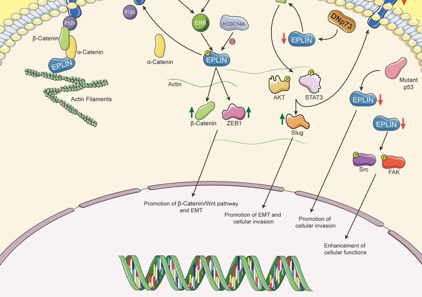
Biomolecules 2021, 11, x FOR PEER REVIEW 2 of 15
Biomolecules 2021, 11, 1038 2 of 15
the cadherin–catenin complex, activation of the Wnt-catenin signalling pathway and
expressional alterations in a range of elements such as diminishment of E-cadherin and
upregulation of
upregulation of ZEB1-promoting
ZEB1-promoting Epithelial-Mesenchymal
Epithelial-Mesenchymal Transition Transition (EMT) (EMT) [6,9].
[6,9]. This
This
allows epithelial cells to lose cell–cell adhesion and apical basal
allows epithelial cells to lose cell–cell adhesion and apical basal polarity, and hence reor- polarity, and hence
reorganise
ganise their their own structure
own structure leading leading to mesenchymal
to mesenchymal characteristics
characteristics in orderintoorder to gain
gain invasive
invasive potential. Zhitnyak et al. [10] elucidated that disorganisation
potential. Zhitnyak et al. [10] elucidated that disorganisation of the actin cytoskeleton of the actin
cytoskeleton and E-cadherin-based AJs occur during earlier stages
and E-cadherin-based AJs occur during earlier stages of Epidermal Growth Factor (EGF)- of Epidermal Growth
Factor (EGF)-induced
induced EMT in epithelial EMT in epithelial
IAR-20 cells. TheIAR-20 cells. The disorganisation
disorganisation is essential for is theessential for
entire EMT
the entire EMT process, which leads to loss of cell–cell contact. It is interesting
process, which leads to loss of cell–cell contact. It is interesting to note that the expression to note that
the
of expression
E-cadherin of E-cadherin
remains unchanged remains
despiteunchanged
the loss ofdespite
cell–cell the loss of cell–cell
adhesion. During theseadhesion.
early
During these early events of EGF-induced EMT, disruption
events of EGF-induced EMT, disruption of colocalisation between EPLIN and linear AJ of colocalisation between
EPLIN
and and linear AJ of
phosphorylation andEPLIN
phosphorylation
is observed,ofwhich EPLINremains
is observed,
in linewhichwith remains
earlier workin line
by
with earlier work by Zhang et al. [9–11]. These findings indicate
Zhang et al. [9–11]. These findings indicate that EPLIN contributes to the progression of that EPLIN contributes
to thebyprogression
EMT of EMT the
at least disrupting by at least disrupting
cell–cell the cell–cell
adhesion complex, whichadhesion
further complex,
induceswhichmetas-
tasis. Interestingly, EPLIN was found to be essential for cell division [12,13];cell
further induces metastasis. Interestingly, EPLIN was found to be essential for division
EPLIN was
[12,13]; EPLIN
detected to locate wasat detected
the cleavageto locate
furrow at and
the cleavage
to associate furrow
myosin andIItoand associate myosinthe
Sept2 during II
and Sept2 during the ingression period. Recruitment and accumulation
ingression period. Recruitment and accumulation of actin, myosin II, RhoA and Cdc42 of actin, myosin
II, RhoA
were impactedand during
Cdc42 late-stage
were impacted during
cytokinesis late-stage
following cytokinesis
EPLIN depletion. following
In addition,EPLINloss
depletion. In addition, loss of EPLIN led to multinucleation, which
of EPLIN led to multinucleation, which results in cytokinesis failure [12]. Further study by results in cytokinesis
failure [12].
Sundvold Further
et al., study by Sundvold
also emphasised et al., also
the role EPLIN playsemphasised
in this key eventthe role EPLIN
of cell plays in
proliferation,
this key event of cell proliferation, by showing that EPLIN recruits
by showing that EPLIN recruits Arv1 (ACAT-related protein required for viability 1), which Arv1 (ACAT-related
protein required
supports for viability
the efficient 1), which
progression of cellsupports
divisionthe [13].efficient progression
Collectively, of cell division
these studies suggest
that EPLIN is needed for cytokinesis and crucial for recruitment of a numberand
[13]. Collectively, these studies suggest that EPLIN is needed for cytokinesis crucial
of essential
for recruitment of a number of essential elements during this
elements during this process. The loss of EPLIN could lead to failure of this importantprocess. The loss of EPLIN
could enhancing
event, lead to failuretheof this important
possibility event,instability
of genetic enhancingand the contributing
possibility of to genetic instability
carcinogenesis.
and contributing to carcinogenesis.
Figure1.1.Schematic
Figure Schematicstructure
structureofofEPLIN.
EPLIN.Structural
Structuralinformation
informationisisadapted
adapted from
from [5,6]
[5,6] and
and figure
figure is is designed
designed and
and created
created by
by using Adobe Illustrator 2021Version 25.2.1 (Adobe Inc., San Jose, CA, USA).
using Adobe Illustrator 2021Version 25.2.1 (Adobe Inc., San Jose, CA, USA).
2. New Members of EPLIN’s Interacting/Regulatory Network
2. New Members of EPLIN’s Interacting/Regulatory Network
To achieve EPLIN’s distinct functions in cellular events and metastatic progression,
To achieve EPLIN’s distinct functions in cellular events and metastatic progression, a
a number of interacting/regulatory partners are involved. As discussed above and
number of interacting/regulatory partners are involved. As discussed above and illustrated
illustrated in Figure 1, EPLIN cross-links and bundles F-actin due to its two actin binding
in Figure 1, EPLIN cross-links and bundles F-actin due to its two actin binding sites and
sites and has the ability to inhibit the branching nucleation of actin filaments through
has the ability to inhibit the branching nucleation of actin filaments through Arp2/3 [7],
Arp2/3 [7], while ERK could phosphorylate EPLIN on Ser362 and Ser604 [11]. EPLIN is
while ERK could phosphorylate EPLIN on Ser362 and Ser604 [11]. EPLIN is also associated
also associated with the cadherin–catenin complex via α-catenin [6]. These characteristics
with the cadherin–catenin complex via α-catenin [6]. These characteristics not only allow
not only allow EPLIN to regulate and maintain the cytoskeleton and AJs, and hence affect
EPLIN to regulate and maintain the cytoskeleton and AJs, and hence affect cells’ motility,
but also contribute to EMT, enhancing the metastatic potential of tumour cells.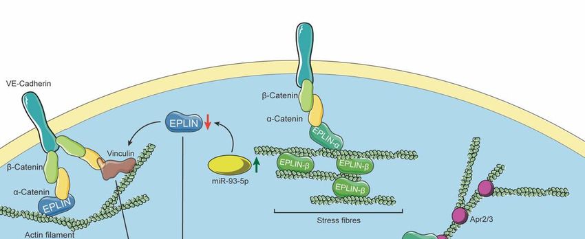
Biomolecules 2021, 11, 1038                                                                                            3 of 15

                                   Evidence has shown that, as a tumour suppressor and due to its unique role in sus-
                              taining the epithelial cytoskeleton, EPLIN is deeply involved in multiple cellular processes
                              associated with carcinogenesis and metastasis. To understand more about its role in
                              these processes, and others, increasing research has focused on elucidating interacting or
                              regulatory proteins of EPLIN, either upstream regulators or downstream participants.

                              2.1. p53, a Direct Regulator of EPLIN
                                    p53 is a universally known tumour suppressor in multiple human tumours, with
                              mutation or reduction of p53-promoting cancer progression [14]. Since EPLIN is also
                              characterised as a putative tumour suppressor, their relationship with each other seems
                              fascinating and worthy of investigation. DNp73, a mutant isoform of the p53 family capable
                              of inhibiting the expression of p73, was found to be involved in the regulation of EPLIN [15].
                              Steder et al. reported that DNp73 could induce downregulation of EPLIN, and this could
                              lead to disruption of AJs and allow insulin-like growth factor 1 receptor (IGF1R) to bind
                              its ligands. This would cause further phosphorylation of AKT and Signal Transducer and
                              Activator of Transcription-3 (STAT-3) to downregulate E-cadherin and upregulate Slug.
                              Morphological changes in melanoma cells are also observed upon regulation of EPLIN by
                              DNp73, which indicates progression of EMT and enhances metastatic potential [15]. Due to
                              the relationship between DNp73 and p53, it naturally draws attention to whether p53 also
                              takes part in the regulation of EPLIN. Indeed, later study by Ohashi et al. [16] uncovered
                              the regulatory association between these two tumour suppressors. The authors identified
                              that p53, p63γ and p73β overexpression, in H1299 lung cancer and Saos-2 osteosarcoma
                              cells, could enhance EPLIN mRNA expression. Furthermore, utilising a combination
                              of chromatin immunoprecipitation-sequencing (ChIP-seq [17]), ChIP-PCR and reporter
                              assays, the authors identified two p53 consensus motifs within EPLIN which facilitated
                              transactivation of EPLIN by p53 family members. Similarly, expression of p53, p63γ and
                              p73β was found to enhance EPLIN protein levels in H1299 cells. Interestingly, nutlin-3a
                              treatment of wild-type, but not p53-mutated cells, also enhanced EPLIN protein expression
                              without impacting on p63 or p73 expression in MCF7 breast cancer, LoVo colon cancer
                              and A549 lung cancer cells, implicating p53 in inducing EPLIN protein expression. The
                              authors went on to show a downregulation of EPLIN in breast and colorectal cancers
                              that had p53 mutations (TCGA database). Lower levels of EPLIN were associated with
                              significantly shorter survival periods in colorectal cancer, breast cancer and lung cancer.
                              The authors also demonstrated, utilising a combination of nutlin-3a, EPLIN knockdown
                              and p53 expression systems in A549 and Lu99 lung cancer cells, that p53 could suppress
                              cellular invasion and that this suppression was partially inhibited by knockdown of EPLIN.
                              Interestingly, in vivo administration of nutlin-3a, via the intra-peritoneal route, was able to
                              reduce the size of established tumours (derived from subcutaneously inoculated A549 cells)
                              but had a lesser impact on tumours from the EPLIN knockdown A549 cells. [16]. Hence,
                              EPLIN was reported to be a target of p53, with this relationship influencing metastatic
                              progression. These findings support EPLIN’s role as a tumour suppressor and clinical
                              prognosis indicator.

                              2.2. hCDC14A Dephosphorylates EPLIN
                                    EPLIN’s ability to sustain the cytoskeleton and regulate actin dynamics is attributed
                              to its capacity to directly bundle F-actin on two actin binding sites [7] and its link to the
                              cadherin–catenin complex to support AJs [6]. The ability of mouse EPLIN to bind and
                              regulate actin dynamics is reported to be weakened following ERK phosphorylation of
                              serine residues within EPLIN, induced by platelet-derived growth factor (PDGF) [8]. A
                              later study subsequently demonstrated the capacity of EGF-induced ERK to phosphorylate
                              human EPLIN in prostate cancer cells on Ser362 and Ser604, which pair to Ser360 and Ser602
                              in mouse EPLIN, leading to EPLIN ubiquitination and degradation and downregulation of
                              E-cadherin, an essential marker of EMT progression [11]. These findings indicate a link
                              between EPLIN, cell migration and cancer metastasis.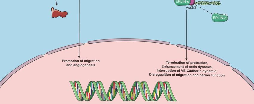
Biomolecules 2021, 11, 1038 4 of 15
Chen et al. [18] conducted phospho-proteome and Biotin identification (BioID) analy-
ses to identify that EPLIN is involved in the interacting network of human Cell Division
Cycle 14A (hCDC14A). Furthermore, hCDC14A was revealed to be able to dephosphorylate
EPLIN on Ser362 and Ser604 to counteract the phosphorylation induced by ERK, and influ-
ence F-actin stability via this particular feature in Hela cells [18]. Knocking down EPLIN or
reducing the activity of hCDC14A, through the generation of a phosphatase dead version
of hCDC14A, in the HCT-116 CRC cell line significantly decreased E-cadherin to allow
the cells to acquire mesenchymal characteristics [18]. The study also demonstrated that
downregulation of EPLIN and hCDC14A was associated with poor prognosis in colorectal
cancer, by exploring online databases. Thus, it would appear that hCDC14A acts as a vital
upstream player of EPLIN during cancerous development.
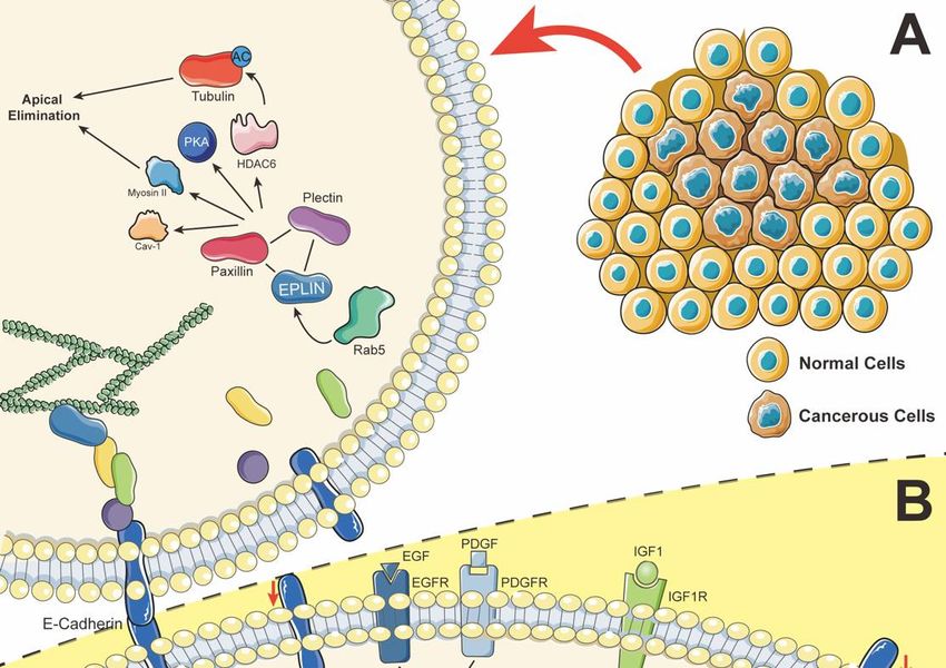
2.3. Paxillin–Plectin–EPLIN Complex Promotes Apical Extrusion
EPLIN has been indicated in the process of cytokinesis, in which loss of EPLIN and
its interacting partners could lead to multinucleation which, in turn, may contribute to
carcinogenesis [12,13]. During the early carcinogenesis period in epithelial tissue, mutations
of oncogenes are crucial contributors. Kajita et al. revealed, by using mammalian epithelial
cells, that when Ras-transformed cells are surrounded by normal cells, the latter will fight
to lift transformed cells from the monolayer in order to eliminate them. This competitive
self-defence process was described as epithelial defence against cancer (EDAC) [19]. EPLIN
has also been identified as playing a role in this process, contributing to apical extrusion of
RasV12-transformed Madin–Darby Canine Kidney (MDCK) cells, where it has been linked
to Caveolin-1 (Cav-1) [20]. In the study, Ohoka et al. utilised Cav-1 immunoprecipitation
under a number of cell culture conditions, namely, normal cell culture, RasV12-transformed
cell culture and combinations of the two cultures. The study has identified EPLIN as a
binding partner predominantly in the mixed culture setting and at apical and lateral
membrane domains, where it was observed to be partially co-localised with Cav-1 using
immunofluorescence. EPLIN was also seen in the cytosolic and RasV12 intracellular regions
where Cav-1 was absent. Knocking down EPLIN by short hairpin RNA in transformed cells
suppressed accumulation of Cav-1 and activation of myosin II and protein kinase A (PKA).
It also impacted on the apical extrusion process. Interestingly, Cav-1 knockdown did not
impact EPLIN accumulation or myosin II and PKA activity. It is interesting to note that
addition of the MAPK/ERK Kinase (MEK) inhibitor U10126, or the actin polymerisation
inhibitor cytochalasin D, could impact the accumulation, localisation or enrichment of Cav-
1 and EPLIN. The authors also demonstrated that the accumulation of filamin in normal
cells surrounding transformed cells is repressed following EPLIN or Cav-1 knockdown
in RasV12-transformed cells and, similarly, EPLIN and Cav-1 enrichment in transformed
cells is suppressed following filamin A knockdown in surrounding normal cells [20].
Hence, EPLIN has been implicated in this interesting, competitive process along with
a number of elements, with Mitogen Activated Protein Kinase (MAPK) pathways and
actin dynamics also reported to be involved. Subsequently, the mechanism behind apical
extrusion was explored. Ras-Associated Protein 5 (Rab5) regulates endocytosis, which
has been shown to be involved in cell migration and oncogenesis [21]. Rab5 induces
endocytosis of E-cadherin and mediates EPLIN to disconnect the E-cadherin complex,
potentially allowing EPLIN to subsequently interact with players such as myosin II and
PKA, to promote the EDAC elimination process, in RasV12-transformed cells surrounded
by normal cells [22]. It was further revealed that plectin and paxillin immunoprecipitated
and partly co-localised with EPLIN in Ras-transformed cells surrounded by normal cells.
Following knockdown of either of the molecules, using shRNA, apical extrusion activity
was repressed and accumulation of either of the proteins in this situation was also depleted
significantly [23,24]. Collectively, this would suggest that these molecules form a plectin–
paxillin–EPLIN complex to regulate apical extrusion. Digging deeper into the interaction
of this complex, α-tubulin is accumulated and regulated by the complex, and acetylated
tubulin is also upregulated in transformed cells, when they are surrounded by normal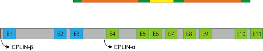
Biomolecules 2021, 11, 1038                                                                                           5 of 15

                              cells. This enhanced acetylation was found to be repressed by deacetylation caused by
                              Histone Deacetylase 6 (HDAC6), which could be regulated by paxillin [23,24]. Hence, the
                              picture of the mechanism behind this competitive process and EPLIN has become clearer.
                              EPLIN and its interacting molecules form a complex to support promotion of acetylation
                              of tubulin, by mediating HDAC6 to regulate microtubule filaments and cell–cell adhesion.
                              This would enhance apical elimination. Other molecules, such as PKA and myosin II, the
                              MAPK pathway and the actin cytoskeleton, also help regulate the elimination process.

                              2.4. EPLIN Regulates Cellular Functions Partly Through the FAK/Src Signalling Pathway
                                    A recent study by our laboratories [25] demonstrated a possible interaction between
                              paxillin, focal adhesion kinase (FAK) and proto-oncogene tyrosine protein kinase (Src).
                              Both kinases have been demonstrated to affect focal adhesion dynamics, cell migration and
                              cancer development via certain signalling pathways [26,27]. Collins et al. reported that
                              EPLIN expression is reduced in prostate cancer tissue when compared to normal paired
                              tissue from tissue microarrays. Overexpression of EPLIN-α in the PC-3 prostate cancer cell
                              line induced significant repression of cellular growth, invasion and migration. Knocking
                              down EPLIN in the CA-HPV-10 prostate cancer cell line, on the other hand, promoted
                              invasion and migration [25]. Such observations are in line with the findings from an earlier
                              study by Zhang et al. [9], supporting an argument that EPLIN influences cell function
                              by coordinating with FAK and Src. Direct evidence for this connection subsequently
                              came from protein microarray and Western blotting analysis, which showed significant
                              increases in expression of p-FAK Y925, p-Paxillin Y31, total Paxillin and p-Paxillin Y118,
                              following overexpression of EPLIN-α in the PC-3 cell line. EPLIN-α overexpressed LNCaP
                              cell lines, on the other hand, showed that p-FAK Y397 was upregulated and p-Paxillin
                              Y118 downregulated. Interestingly, in the PC-3 EPLIN-α overexpression cell model, an
                              activation site, p-Src Y419, was seen to be depleted and a regulatory site, p-Src Y530,
                              was increased. Knockdown of EPLIN in CA-HPV-10 cells using shRNA resulted in the
                              expression of total FAK being upregulated, while Src Y419 was depleted significantly [25].
                              Among these activation sites, FAK Y925/Y118, Paxillin Y118/Y31 and Src Y419 have been
                              identified to be key phosphorylation sites and have significant impacts on cellular functions
                              and cancer development [25,26,28].
                                    Furthermore, the study by Collins et al. explored the impact of the EPLIN, Src,
                              FAK relationship on cellular invasion and migration. Invasive capability in PC-3 control
                              groups was repressed when FAK and Src were inhibited, but no significant differences
                              were detected in EPLIN-α-overexpressed PC-3 cells lines. Migration was reduced at
                              some time periods following inhibition of FAK or Src in both the control and EPLIN-α-
                              manipulated groups in PC-3 cells, though the impact appeared generally reduced in the
                              EPLIN-α overexpression groups. In LNCaP models, migration was suppressed in EPLIN-
                              α-overexpressed cell lines when FAK was inhibited, whereas significant changes within
                              the control group following treatment were not observed, while the migration of both
                              the control and EPLIN-α-overexpressed cell lines were affected following Src inhibition.
                              The invasive capacity of LNCaP control cells was significantly reduced following Src
                              inhibition, though this trend was not found to be significant in the EPLIN-α overexpression
                              LNCaP line. On the contrary, invasion and migration were promoted significantly while
                              knocking down EPLIN in CA-HPV-10 cells compared to its control group. When Src
                              was inhibited, reduction of migrated and invasive cells was observed in both control and
                              EPLIN-knockdown cell lines, while invasion and migration abilities were only significantly
                              reduced in EPLIN-knockdown CA-HPV-10 cells following FAK inhibition [25]. Taken
                              together, EPLIN expression is downregulated in prostate cancer when compared to normal
                              tissues. Its impact on cellular functions in prostate cancer cells may be achieved through
                              regulation of FAK/Src signalling pathways, adding insights to the potential mechanism
                              behind this tumour suppressor.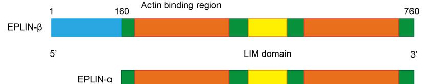
both control and EPLIN-knockdown cell lines, while invasion and migration abilities were
                    only significantly reduced in EPLIN-knockdown CA-HPV-10 cells following FAK
                    inhibition
Biomolecules 2021, 11, 1038    [25]. Taken together, EPLIN expression is downregulated in prostate cancer                 6 of 15
                    when compared to normal tissues. Its impact on cellular functions in prostate cancer cells
                    may be achieved through regulation of FAK/Src signalling pathways, adding insights to
                    the potential mechanism behind this tumour suppressor.
                                          Recent scientific focus on the tumour suppressor EPLIN has aided the understanding
                          Recent scientific focus on the tumour suppressor EPLIN has aided the understanding
                                    of this important molecule and its wider role and interactions in cancerous epithelial cells.
                    of this important molecule and its wider role and interactions in cancerous epithelial cells.
                                    Such hypothetical signalling pathways are summarised in Figure 2.
                    Such hypothetical signalling pathways are summarised in Figure 2.

                Figure 2. A. Role of EPLIN in apical extrusion. Rab5 allows disruption of the connection between
     Figure 2. A. Role of EPLIN in apical extrusion. Rab5 allows disruption of the connection between the cadherin–catenin
                the cadherin–catenin complex and EPLIN, allowing EPLIN to interact with paxillin and plectin to
     complex and   EPLIN, allowing
                recruit/activate    EPLIN
                                 Cav-1, PKAtoand
                                               interact
                                                  Myosinwith
                                                           II,paxillin and plectin
                                                               and acetylate        to recruit/activate
                                                                               tubulin                  Cav-1,
                                                                                        through paxillin’s     PKA and
                                                                                                           regulation of Myosin II,
     and acetylate
                HDAC6 activity. Hence, promoting apical elimination when Ras-transformed cells are surrounded when Ras-
                   tubulin  through paxillin’s regulation of  HDAC6     activity. Hence,  promoting  apical elimination
     transformed cells are surrounded by normal cells. B. EPLIN prospective pathways in epithelium. EPLIN stabilises AJs and
     actin dynamics by binding to the cadherin–catenin complex and actin directly. PDGF/EGF could induce phosphorylation of
     EPLIN via ERK signalling pathways and results in disorganisation of AJs and interruption of actin dynamics, which further
     upregulates expression/translocation of β-catenin and ZEB1, diminishes expression of E-cadherin leading to activation of
Biomolecules 2021, 11, 1038 7 of 15
the β-catenin/Wnt pathway and promotion of EMT, further impacting cellular functions. This phosphorylation of EPLIN
can be counteracted by hCDC14A. DNp73 induces downregulation of EPLIN, which allows IGF1R to interact with its
ligand, then phosphorylate AKT and STAT3, which increases expression of Slug and decreases expression of E-cadherin, to
contribute to promotion of the EMT process. p53 mutation can lead to downregulation of EPLIN expression, which results
in enhancement of cellular invasiveness. The downregulation of EPLIN has been reported to promote cellular functions,
which may be attributed to phosphorylation of FAK/Src and activation of FAK/Src pathways. Icons were obtained from
SMART-Servier Medical ART (https://smart.servier.com, accessed on 18 April 2021) and graphics of pathways were
designed and created using Adobe Illustrator 2021 Version 25.2.1 (Adobe Inc., San Jose, CA, USA).
2.5. MicroRNAs (miRs) as Regulators of EPLIN
Liang et al. reported a microRNA, miR-93-5p, as a novel upstream regulator of
EPLIN [29]. Inhibition of miR-93-5p significantly upregulates expression of EPLIN, whilst
enhancing the expression of miR-93-5p leads to downregulation of EPLIN in human
umbilical vascular endothelial cells (HUVECs), indicating a negative correlation between
the two elements. Similarly, luciferase reporters confirmed that miR-93-5p manipulates
expression of EPLIN by binding to its 30 -UTR sequence. This relationship was further
revealed to be associated with migration and angiogenesis in HUVECs (discussed later) [29].
More recently, work by Dart et al. has identified miR-221 as a potential regulator of EPLIN.
In their study, the authors generated miR-221-deleted PC-3 cells (PC3 miR-221 del) and
observed reductions in aggressive traits such as cellular migration and invasion in these
cells. Furthermore, characterisation of PC3 miR-221 del cells, for proteins associated with
processes such as motility, invasion and EMT, identified changes in EPLIN expression,
with enhanced expression of the alpha isoform noted. Taken together, this study supports
the association of EPLIN with these key processes and adds information regarding the
complex regulatory networks related to EPLIN [30].
2.6. EPLIN Interaction with LUZP1 and NPC1L1 and Implications in other Biological Processes
and Disease
From earlier studies, one of the most significant findings concerning EPLIN is that
it regulates actin dynamics by colocalising with actin filaments and other actin struc-
ture regulators and cross-linking actin filaments, inhibiting branched nucleation through
Arp2/3, further to affecting cells’ motility and migration in order to promote cancer de-
velopment [4,7]. Except for its notable impact on solid tumours, EPLIN was discovered
to be involved in cellular activities in noncancerous tissues. Tsurumi et al. reported
strong expression of EPLIN in mesangial cells and demonstrated EPLIN downregulation
in mesangial proliferative nephritis in vivo [31]. EPLIN colocalises at focal adhesions with
paxillin and their interaction takes part in stabilising focal adhesion in mesangial cells.
PDGF-induced MEK/ERK signalling is responsible for disruption of the EPLIN–paxillin
complex and translocation of EPLIN from focal adhesion sites to peripheral ruffles. In
addition, depletion of EPLIN results in disorganisation of focal adhesion and enhancement
of cells’ migration via PDGF [31].
A recent study by Goncalves et al. revealed that EPLIN is also involved in another
cellular activity, ciliation, by interacting with LUZP1 (Leucine Zipper Protein 1) [32].
Cilia are membranous protrusions which originate from centrosomes via complicated
mechanisms including the cytoskeleton, membrane traffic, etc. Cilia take part in certain
sensory and motional biological functions whose dysregulation could lead to ciliopathies,
including blindness, cystic kidneys, etc. [32–34]. LUZP1 has been reported as a negative
regulator of ciliogenesis and a positive regulator of actin dynamics [33,35].
Goncalves et al. identified EPLIN as a potential interacting partner of LUZP1 by
conducting BioID assays in cycling and serum-deprived HEK293 cells [32]. The relationship
was confirmed through conducting co-IP assays using GFP/FLAG vectors in RPE-1 cells
or HEK293 cells. Here, the authors revealed that LUZP1 and EPLIN interact with each
other via the C-terminal of LUZP1, as GFP-tagged LUZP1 pulled down both isoforms of
EPLIN in RPE-1 cells, GFP-EPLIN isoforms/FLAG-EPLIN-β pulled down LUZP1 in RPE-1Biomolecules 2021, 11, 1038 8 of 15
cells and HEK293 cells, respectively. GFP-EPLIN-β was able to pull down FLAG-tagged
full length and C-terminal LUZP1 in HEK293 cells. FLAG-EPLIN-β and FLAG-LUZP1 are
able to pull down actin. Intriguingly, immunofluorescence revealed that both EPLIN and
LUZP1 co-locate with actin filaments in RPE-1 cells. However, LUZP1 locates at centrosome
and basal regions, EPLIN-α locates mainly at the leading edge where membrane ruffles
occur, while EPLIN-β mainly locates along with actin filaments, indicating a possible
functional correlation between these proteins. Furthermore, accumulation of ciliated
cells and longer primary cilia were observed, following siRNA-mediated knockdown
of EPLIN/LUZP1 in RPE-1 cells, along with increased expression of myosin Va after
immunofluorescence analysis. Aberrant ciliation, caused by cytochalasin D, could be
counteracted by overexpressing EPLIN and LUZP1, while accumulation of Arp2 was
also observed. Hence, LUZP1 interaction with EPLIN contributed to ciliation regulation,
potentially partly through regulating actin structure [32].
Given that EPLIN participates in the regulation of ciliation progression, interacting
with LUZP1 [32], and that depletion and dysfunction of cilia can result in diseases such as
blindness, cystic kidneys, etc. [32,34], this demonstrates EPLIN’s implication and impor-
tance in other biological processes, not only in carcinogenesis and tumour development.
Indeed, another study by Zhang et al. [36] reported that EPLIN is associated with choles-
terol absorption in intestines. A Chinese Kazakh family, with inherited low levels of
low-density lipoprotein cholesterol (LDL-C) in plasma, has been established by the authors
as a study model. A mutation of EPLIN, LIMA1-K306fs, which includes a frameshift
variant on exon-7, was identified to be a potential candidate associated with LDL-C in the
family, by using whole-exome sequencing and sanger sequencing. The authors identified
that individuals who express LIMA1-K306fs have a significantly lower level of LDL-C and
campesterol:lathosterol ratio when compared to those who do not. Through analysis of a
larger cohort, the study identified that another mutation of EPLIN, LIMA1-L25I, also has a
similar effect, although the impact on LDL-C levels in these carriers was not as great as
in the K306fs groups. Moreover, the team developed a mouse experimental model and
discovered that silencing EPLIN in the intestines of the mice led to the downregulation
of cholesterol uptake, plasma cholesterol, liver 3 H-cholesterol and plasma 3 H-cholesterol,
when compared to control groups. Hence, these implicate EPLIN as a potential positive
regulator in LDL-C levels and intestinal cholesterol absorption in humans and mice [36].
To investigate the possible mechanism behind this interesting function, EPLIN was found
to bind and colocalise with myosin Vb and Niemann-Pick C1-Like 1 (NPC1L1), which
are known to be essential to cholesterol absorption [36,37] on the brush border in mice
intestines [36]. Furthermore, knocking down EPLIN in CRL1601 cells led to weakened
association between myosin Vb and NPC1L1. EPLIN is seen to interact with both proteins
on certain regions, namely the Q1277 KR residues of NPC1L1 and the C164 LG residues of
EPLIN being responsible for their interaction, while the 21 to 40 amino acid regions of
myosin Vb and the 491 to 511 amino acid regions of EPLIN contributed to the interaction of
these two molecules. Hence, the authors of the study propose that EPLIN might function
as a connecting bridge between myosin Vb and NPC1L1. Depletion of EPLIN or myosin
Vb, as well as mutation of EPLIN using CRISPR-Cas9 in CRL1601 cells, led to disruption of
NPC1L1 translocation from the endocytic recycling compartment to the plasma membrane.
Furthermore, the authors identified that the NPC1L1–EPLIN complex is needed for choles-
terol absorption, as disrupting the complex led to a weakened rate of transportation of
NPC1L1 in vitro and in vivo and attenuated liver cholesterol and plasma total cholesterol
levels in vivo [36]. Therefore, EPLIN was found to be associated with LDL-C plasma levels,
whose high concentration represents a significant risk factor for cardiovascular disease,
and to play a role in cholesterol absorption by interacting with NPC1L1 and myosin Vb.
Taken together, the above studies have increased our understanding of the inter-
acting and regulatory networks associated with EPLIN, which are outlined in Table 1.
This has added to previously established networks from earlier studies which have been
summarised and discussed in past reviews by Collins et al. [2] and Wu et al. [3]. Thus,Biomolecules 2021, 11, 1038 9 of 15
EPLIN has been linked with a wide range of partners to achieve multiple functions, not
only in carcinogenesis and tumour development, but also in maintenance of focal adhesion
in mesangial cells, cilia formation and cholesterol absorption. Interestingly, depleted or
mutant EPLIN impacts the interactions with NPC1L1 to weaken cholesterol absorption in
intestines, potentially decreasing the risk of high LDL-C-related diseases, which provides
a different picture of this tumour suppressor, as its downregulation in cancer cells often
leads to promotion of cancer developments.
Table 1. Novel Interacting and Regulatory Partners of EPLIN. For previously described interacting partners, please refer to
review papers from Collins et al. [2] and Wu et al. [3].
Interacting/Regulatory Partners Bio-Significances Ref.
p53 is a positive upstream regulator of EPLIN at the transcript level in
osteosarcoma and lung cancer cells and at the protein level in lung, colon
and breast cancer cells. p53 family transactivates the EPLIN gene on
p53 [16]
LIMA1-RE1 (AGGCAAGTTa tAACTgGCaT) and LIMA1-RE2
(GGACAgaaCT AGA-CAAGCCC). Depletion of EPLIN counteracts the
repressed cancer invasion induced by p53 in lung cancer cells.
Responsible for dephosphorylating EPLIN on Ser362 and Ser604. Knocking
hCDC14A down EPLIN or reducing the activity of hCDC14A in HCT-16 cell lines leads [18]
to promotion of the EMT process.
Cav-1
Knocking down EPLIN expression will inhibit the accumulation/activity of
Myosin II [20,23]
these proteins in Ras12-transformed cells when surrounded by normal cells.
PKA
Mutation of Rab5 could reduce accumulation of EPLIN in Ras-transformed
cells surrounded by normal cells, while Rab5 acts upstream of EPLIN,
Rab5 [22]
mediates endocytosis of E-cadherin and allows EPLIN disconnection with
the cadherin–catenin complex, leading to promotion of apical extrusion.
Plectin Plectin and paxillin colocalise with EPLIN in Ras-transformed cells and have
[23,24]
Paxillin a mutual positive correlation and regulate apical extrusion.
EPLIN regulates invasion and migration in prostate cancer cells through the
FAK/Src [25]
FAK/Src pathways.
miR-93-5p regulates EPLIN expression negatively by binding to its 30 -UTR
miR-93-5p [29]
sequence and associates with migration and angiogenesis in HUVECs.
Depletion of miR-221 in PC3 cells results in enhancement of EPLIN protein
miR-221 [30]
level in combination with reduction of cellular migration and invasion.
LUZP1 interacts and colocalises with both isoforms of EPLIN in RPE-1 cells
LUZP1 [32]
and regulates the ciliation process.
EPLIN colocalises with NPC1L1 and myosin Vb on the peripheral brush
border region of mouse small intestine. EPLIN interacts with NPC1L1 by
binding its C164LG residues to the Q1277KR residues of NPC1L1. EPLIN
NPC1L1 also interacts with myosin Vb due to the interaction between the aa 21 to 40 [36]
regions of myosin Vb and the aa 491 to 511 regions of EPLIN.
Mutant/depleted EPLIN has a positive effect on cholesterol absorption by
disrupting the transportation ability of NPC1L1 in vivo.
3. Role of EPLIN in Endothelial Cells, Angiogenesis and Lymphangiogenesis
EPLIN was first observed when looking at differential expression between cancer
and normal cells [1,4]. It had been largely investigated regarding its cellular function and
interacting/regulatory partners, mainly in epithelial-derived cancer cells, leading to its
labelling as a tumour suppressor. Angiogenesis is essential in cancer development and
progression, for its role in blood, nutrient and oxygen supply and for acting as an escape
route for disseminating cancer cells [38]. However, EPLIN’s role in angiogenesis wasBiomolecules 2021, 11, 1038 10 of 15
not in the spotlight in early studies. EPLIN bundles actin filaments and connects to the
cadherin–catenin complex via α-catenin. It inhibits branch nucleation through Arp2/3
contributing to sustaining the cytoskeleton and cell–cell adhesion in epithelial cells [6,7].
Similar cell junction activities in endothelial cells, which regulate endothelium integrity, are
crucial for angiogenesis [39,40]. Through overexpression of EPLIN-α in human endothelial
HECV cells and conducting wound-healing and Matrigel adhesion assays, Sanders et al.
reported that migration and adhesive ability were significantly downregulated when
compared to control groups. Furthermore, the impact of EPLIN-α overexpression in
HECV cells was also tested, using tubule formation assays and through the co-injection
of either overexpressed EPLIN-α or plasmid control HECV cells with MDA-MB-231 cells
in mice, demonstrating a role in inhibiting tubulelike structure formation in in vitro and
in vivo tumour development [41]. Hence, EPLIN has the potential to affect angiogenesis,
with a number of studies focused on EPLIN’s role and mechanism in endothelial cells
and angiogenesis.
An early study by Chevin-Petinot et al. [39], utilising a range of techniques, including
confocal microscopy and immunoprecipitation assays in HUVEC cells, demonstrated that
EPLIN co-locates, or is associated with, actin filaments, VE-cadherin, α/β -catenin and
vinculin at cell junction regions. Knocking down EPLIN leads to location changes of
vinculin, highlighted by the delocalisation of vinculin from cell–cell junctions in EPLIN
suppressed cells. A GST pull-down assay showed EPLIN links to the VE–cadherin–catenin
complex via α-catenin, although suppressing EPLIN did not affect adhesion, migration
and proliferation of HUVECs. The authors also conducted tubule formation assays to show
that suppression of EPLIN negatively impacted capillary network formation, enhancing
breakage events in comparison to controls [39]. Another study reported that miR-93-5p, a
microRNA which promotes migration, proliferation and angiogenesis in HUVECs, acts
upstream of EPLIN, demonstrating regulation of EPLIN by miR-93-5p, and that siRNA
suppression of EPLIN was able to negate the impact of miR-93-5p antisense oligos on
HUVEC migration and lumen formation [29]. This may imply that the role of EPLIN in
HUVECs cellular functions could be achieved through interaction with, or regulation by,
other elements. Taken together with other studies, such data suggest a complex and key
role for EPLIN in regulating angiogenesis.
EPLIN has two isoforms which generate from two distinct promoters [5], only differing
in the N-terminal region, in which EPLIN-β has an additional 160aa [4], and share a central-
located LIM domain and two actin binding sites [4,7], which are essential for EPLIN’s
function. Expression of EPLIN-α is frequently diminished in cancer, while EPLIN-β has
been reported to remain the same or slightly increase [5]. A recent study identified that
the two isoforms play different roles in endothelial cell dynamics [42]. In their study,
Taha et al. focused on the specific function of individual isoforms in endothelial cells.
The authors reported expression of both α and β isoforms in pig aorta and cava vein
endothelial cells, noting enhanced expression of EPLIN-β in aorta compared to cava vein
samples, with little change noted in EPLIN-α between samples. Interestingly, they reported
enhanced EPLIN-β expression in confluent HUVECs following sheer stress application,
whilst EPLIN-α levels were not significantly impacted. Additionally, EPLIN-α but not β
isoform expression was noted to correlate with HUVEC confluence, with its expression,
protrusion formation and migration velocity reducing in confluent compared to growing
cultures. EPLIN expression was observed to be localised to cell junctions and stress
fibres, with analysis of tagged isoform localisation also indicating their presence at cell
junctions and stress fibres. Importantly, the authors observed an isoform-specific role
for EPLIN-α, noting its presence within or in close proximity to branched actin filament
networks at membrane protrusions, such as classical lamellipodia (cLP) and junction-
associated intermittent lamellipodia (JAIL). However, EPLIN-β was mostly negative at
protrusions and was noted to be potentially involved in retraction. EPLIN-α was also
observed to be more dynamic than EPLIN-β, collectively supporting differential isoformBiomolecules 2021, 11, 1038 11 of 15
roles in endothelial cells and the proposal that EPLIN-α contributes to cell migration and
junction remodelling, whereas EPLIN-β contributes to filament stabilisation [42].
Subsequently, Taha et al. investigated the role of EPLIN-α at cLP and JAIL and its
relationship with the Arp2/3 complex [42]. JAIL is responsible for forming and develop-
ing endothelial cells’ junction sites, where vascular endothelial cadherin (VE-cadherin)
localises. This dynamic regulation by JAIL contributes to migration and junction dynamics
related to angiogenesis [43]. Spinning disc confocal microscopy (SpDM) demonstrated
that the distance between EPLIN-α and the Arp2/3 complex narrowed during protru-
sion extension, and that the eventual overlap of these proteins resulted in the halting
of protrusion movement [42]. This coincided with EPLIN-α and Arp2/3 disconnection
with, and loss of, actin filaments. Taken together with the findings that EPLIN isoforms
associated with the Arp2/3 complex during pull-down assays implicated a potential role
for EPLIN in protrusion termination via the Arp2/3 complex interaction. In support of
this, the authors demonstrated that CK666-mediated inhibition of Arp2/3 resulted in an
inhibition of protrusion formation and EPLIN-α’s materialisation at the membrane. In
keeping with this, targeting EPLIN using siRNA resulted in both increased protrusion
size and duration and an enhancement of migration velocity in normal medium. Further-
more, overexpression of EPLIN-α-EGFP resulted in EPLIN-α-EGFP appearance at, and
disruption of, JAIL-like structures, preventing protrusion expansion, and also enhanced
the formation of filopodia, indicating enhancement of actin dynamics. The authors sug-
gested that enhanced EPLIN-α results in elevated binding to the Arp2/3 complex and JAIL
formation termination, potentially impacting VE-cadherin dynamics. Consistent with this,
they demonstrated altered VE-cadherin dynamics in EPLIN-α overexpression HUVEC
cells, observing intracellular gaps and disrupted VE-cadherin at cell contacts, together with
decreased migration and barrier function [42]. Interestingly, the authors demonstrated a
prominent role of EPLIN-β in stress fibre induction and stabilisation, demonstrating that
stress fibre formation is enhanced substantially following EPLIN-β overexpression (and to
a lesser extent EPLIN-α overexpression) in endothelial cells. Furthermore, EPLIN-β was
found to protect stress fibres from depolymerisation resulting from Y27632 ROCK inhibitor
treatment, with minimal disassembly noted in EPLIN-β overexpression cells compared to
control, or EPLIN-α overexpression cells, following such treatment [42]. This important
study has shed light on the isoform specific role of EPLIN in endothelial cells, highlighting
EPLIN-α’s role in regulating protrusion progressions by interacting with Arp2/3 and
regulating JAIL formation, VE-cadherin dynamics and implication in migration and barrier
function, whereas EPLIN-β plays key roles in induction and stabilisation of stress fibres.
Currently, our understanding of the isoform specific role of EPLIN is limited. Further
investigations in this vital area are needed to enhance our understanding of this important
molecule across many biological areas including cancer development and progression.
Taken together, EPLIN takes part in the regulation of endothelial dynamics by bind-
ing to VE-Cadherin via α-catenin and actin filaments [39]. Depletion of EPLIN could be
induced by miR-93-5p, which contributes to elevation of cellular migration and lumen for-
mation [29], whereas overexpression of EPLIN-α has been shown to reduce cell migration
and tubule formation in HECV endothelial cells [41]. Hence, EPLIN appears to play key
roles in endothelial cells and associated processes. A summary outlining the potential role
of EPLIN in endothelial cells is outlined in Figure 3.Biomolecules 2021, 11, 1038 12 of 15
ecules 2021, 11, x FOR PEER REVIEW 12 of 15
Figure 3. EPLIN’s role in endothelium. EPLIN sustains cell junctions in ECs by linking to VE-
Figure 3. EPLIN’s role in endothelium. EPLIN sustains cell junctions in ECs by linking to VE-cadherin via α-catenin.
cadherin via α-catenin. Downregulation of EPLIN could be induced by upregulation of miR-93-5p,
Downregulation of EPLIN could be induced by upregulation of miR-93-5p, which may subsequently lead to translocation
which may subsequently lead to translocation of vinculin away from cell junctions. Downregulation
of vinculin away from also
of EPLIN cell junctions.
results inDownregulation of EPLIN
promotion of cellular also results
migration and in promotion
supports of cellular migration
angiogenesis. and supports
EPLIN-α has
angiogenesis. EPLIN-α has been reported to locate at JAIL and cLP and to regulate protrusion
been reported to locate at JAIL and cLP and to regulate protrusion progression via the Arp2/3 progression via the Arp2/3
complex, whilst EPLIN-β mainly locates at stress fibres to maintain its stability. Icons were obtained from
complex, whilst EPLIN-β mainly locates at stress fibres to maintain its stability. Icons were obtained SMART-Servier
Medical ARTfrom SMART-Servier Medical
(https://smart.servier.com, ART (https://smart.servier.com,
accessed on 18 April 2021) and graphics accessed on 18 April
of pathways 2021.) and
were designed and created
using Adobegraphics of 2021
Illustrator pathways
Versionwere
25.2.1designed and San
(Adobe Inc., created
Jose, using
CA, USA).Adobe Illustrator 2021 Version 25.2.1
(Adobe Inc., San Jose, CA, USA).
4. The Clinical Aspects of EPLIN in Human Cancers
4. The Clinical Aspects of EPLIN in Human Cancers
EPLIN has been implicated in playing a role in the development and progression of
EPLIN has been
solid implicated
cancers. in playing
Keeping in linea with
role inthethe development
findings that EPLIN and progression
contributesof to maintaining
solid cancers. Keeping in linecytoskeleton,
the epithelial with the findings that EPLIN
regulating contributes
metastatic to maintaining
progression the
and cell cytokinesis, there
epithelial cytoskeleton, regulating
has been strong metastatic
scientific focus on progression
EPLIN and andits cell cytokinesis,
implications there hastypes of cancer
in multiple
been strong scientific
includingfocus on EPLIN
oral cancer [1,4],and
breastits implications
cancer [4,44], in multiple
prostate types
cancer of cancer
[2,9], squamous cell carci-
including oralnomacancer [1,4], and
of head breast
neckcancer
(SCCHN) [4,44],
[9],prostate
lung cancercancer[45],[2,9], squamous
oesophageal cell [46], ovarian
cancer
carcinoma of cancer
head and[47],neck (SCCHN)
colorectal cancer [9],(CRC)
lung [9,16,18,48,49]
cancer [45], oesophageal
and most recentlycancer gastric
[46], cancer [50].
ovarian cancerEPLIN[47], has
colorectal
also beencancer (CRC)to[9,16,18,48,49]
revealed be associated with and most
multiplerecently gastric
cellular functions such as
cell motility,
cancer [50]. EPLIN has also migration,
been revealed invasion and proliferation,
to be associated with multiplein a number of different cancer cell
cellular functions
lines [2,9,25,44–47,51].
such as cell motility, migration, invasion Ourand lab,proliferation,
as a contributor in ato EPLINofresearch
number different for over a decade, has
cancer
demonstrated
cell lines [2,9,25,44–47,51]. downregulation
Our of EPLIN
lab, as a contributor in multiple
to EPLIN researchtumour tissues,
for over its association with
a decade,
has demonstratedcellular function and clinical
downregulation of EPLIN significance
in multiple [2,44–47,51].
tumour tissues, Furthermore, important work by
its association
Zhang et al. revealed that a lower level of EPLIN
with cellular function and clinical significance [2,44–47,51]. Furthermore, important workis related to poor prognosis and is
by Zhang et al. revealed that a lower level of EPLIN is related to poor prognosis and is that EPLIN
implicated in chemo drug resistance in prostate cancer [9]. The initial finding
may have
implicated in chemo drug a role to playin
resistance inprostate
drug resistance has The
cancer [9]. recently
initialbeen supported
finding by a more clinically
that EPLIN
may have a role oriented
to play in finding in gastric has
drug resistance cancer [50], been
recently in that higher expression
supported was generally seen in
by a more clinically
oriented findingcancers that responded
in gastric cancer [50], to in
neoadjuvant
that higherchemotherapy
expression was (NAC) compared
generally seen tointhose that did
cancers that responded to neoadjuvant chemotherapy (NAC) compared to those that diddistribution in
not, though this was not significant. Furthermore, a greater overall survival
not, though thispatients
was not displaying
significant.higher expressionaofgreater
Furthermore, EPLINoverall
was observed,
survivalbased on responsiveness to
distributionBiomolecules 2021, 11, 1038 13 of 15
NAC. Those patients with high levels of EPLIN, who responded to neoadjuvant therapies,
had a marked longer survival. Together, such work highlights the significant role played
by EPLIN in multiple aspects associated with cancer progression, further supporting its
identified role as a tumour and metastasis suppressor.
5. Conclusions
Focus on EPLIN, since its discovery several decades ago, has implicated it as a key
regulator of several important cellular processes and as a tumour suppressor. Furthermore,
research has identified a number of interacting partners or regulatory mechanisms asso-
ciated with EPLIN. In this review, we provide an update to previous review articles [2,3]
focused on this important molecule, summarising major findings related to EPLIN in recent
years. Such works have revealed that EPLIN contributes to not only metastasis-relevant
processes such as EMT with other novel interacting/regulatory proteins, but also apical
elimination, ciliation, cholesterol absorption, angiogenesis and endothelial cell dynamics,
providing additional routes to explore EPLIN’s implication and mechanisms in a wider
range of fields. Research into the field of EPLIN is intensifying to explore the full implica-
tions of EPLIN in multiple arenas. Widening our understanding of this important molecule
will enhance its therapeutic potential and aid in the design of novel therapeutic strategies.
Author Contributions: All authors have contributed to the conception, design, review and writing
of the manuscript. All authors have read and agreed to the published version of the manuscript.
Funding: This research received no external funding.
Institutional Review Board Statement: Not applicable.
Informed Consent Statement: Not applicable.
Acknowledgments: The authors are grateful to Jane Lane and Fiona Ruge for their assistance in
proofreading the manuscript.
Conflicts of Interest: The authors declare no conflict of interest.
References
1. Chang, D.D.; Park, N.H.; Denny, C.T.; Nelson, S.F.; Pe, M. Characterization of transformation related genes in oral cancer cells.
Oncogene 1998, 16, 1921–1930. [CrossRef] [PubMed]
2. Collins, R.J.; Jiang, W.G.; Hargest, R.; Mason, M.D.; Sanders, A.J. EPLIN: A fundamental actin regulator in cancer metastasis?
Cancer Metastasis Rev. 2015, 34, 753–764. [CrossRef] [PubMed]
3. Wu, D. Epithelial protein lost in neoplasm (EPLIN): Beyond a tumor suppressor. Genes Dis. 2017, 4, 100–107. [CrossRef] [PubMed]
4. Maul, R.S.; Chang, D.D. EPLIN, Epithelial protein lost in neoplasm. Oncogene 1999, 18, 7838–7841. [CrossRef] [PubMed]
5. Chen, S.; Maul, R.S.; Kim, H.R.; Chang, D.D. Characterization of the human EPLIN (Epithelial Protein Lost in Neoplasm) gene
reveals distinct promoters for the two EPLIN isoforms. Gene 2000, 248, 69–76. [CrossRef]
6. Abe, K.; Takeichi, M. EPLIN mediates linkage of the cadherin catenin complex to F-actin and stabilizes the circumferential actin
belt. Proc. Natl. Acad. Sci. USA 2007, 105, 13–19. [CrossRef]
7. Maul, R.S.; Song, Y.; Amann, K.J.; Gerbin, S.C.; Pollard, T.D.; Chang, D.D. EPLIN regulates actin dynamics by cross-linking and
stabilizing filaments. J. Cell Biol. 2003, 160, 399–407. [CrossRef]
8. Han, M.-Y.; Kosako, H.; Watanabe, T.; Hattori, S. Extracellular Signal-Regulated Kinase/Mitogen-Activated Protein Kinase
Regulates Actin Organization and Cell Motility by Phosphorylating the Actin Cross-Linking Protein EPLIN. Mol. Cell. Biol. 2007,
27, 8190–8204. [CrossRef]
9. Zhang, S.; Wang, X.; Osunkoya, A.O.; Iqbal, S.; Wang, Y.; Chen, Z.; Muller, S.; Chen, Z.; Josson, S.; Coleman, I.M.; et al. EPLIN
downregulation promotes epithelial–mesenchymal transition in prostate cancer cells and correlates with clinical lymph node
metastasis. Oncogene 2011, 30, 4941–4952. [CrossRef]
10. Zhitnyak, I.Y.; Rubtsova, S.N.; Litovka, N.I.; Gloushankova, N.A. Early Events in Actin Cytoskeleton Dynamics and E-Cadherin-
Mediated Cell-Cell Adhesion during Epithelial-Mesenchymal Transition. Cells 2020, 9, 578. [CrossRef]
11. Zhang, S.; Wang, X.; Iqbal, S.; Wang, Y.; Osunkoya, A.O.; Chen, Z.; Chen, Z.; Shin, D.M.; Yuan, H.; Wang, Y.A.; et al. Epidermal
Growth Factor Promotes Protein Degradation of Epithelial Protein Lost in Neoplasm (EPLIN), a Putative Metastasis Suppressor,
during Epithelial-mesenchymal Transition. J. Biol. Chem. 2013, 288, 1469–1479. [CrossRef]
12. Chircop, M.; Oakes, V.; Graham, M.E.; Ma, M.P.; Smith, C.M.; Robinson, P.J.; Khanna, K.K. The actin-binding and bundling
protein, EPLIN, is required for cytokinesis. Cell Cycle 2009, 8, 757–764. [CrossRef]You can also read