Endovascular Temporary Balloon Occlusion for Microsurgical Clipping of Posterior Circulation Aneurysms - MDPI
←
→
Page content transcription
If your browser does not render page correctly, please read the page content below
brain
           sciences
Article
Endovascular Temporary Balloon Occlusion for
Microsurgical Clipping of Posterior
Circulation Aneurysms
Jenny C. Kienzler 1 , Michael Diepers 2 , Serge Marbacher 1 , Luca Remonda 2 and
Javier Fandino 1, *
 1    Department of Neurosurgery, Kantonsspital Aarau, CH-5000 Aarau, Switzerland;
      jenny.kienzler@ksa.ch (J.C.K.); serge.Marbacher@ksa.ch (S.M.)
 2    Division of Neuroradiology, Department of Radiology, Kantonsspital Aarau, 5000 Aarau, Switzerland;
      michael.diepers@ksa.ch (M.D.); luca.remonda@ksa.ch (L.R.)
 *    Correspondence: fandino@neurochirurgie-ag.ch; Tel.: +41-62-838-6692; Fax: +41-62-838-6629

 Received: 5 April 2020; Accepted: 27 May 2020; Published: 30 May 2020

 Abstract: Based on the relationship between the posterior clinoid process and the basilar artery (BA)
 apex it may be difficult to obtain proximal control of the BA using temporary clips. Endovascular BA
 temporary balloon occlusion (TBO) can reduce aneurysm sac pressure, facilitate dissection/clipping,
 and finally lower the risk of intraoperative rupture. We present our experience with TBO during
 aneurysm clipping of posterior circulation aneurysms within the setting of a hybrid operating room
 (hOR). We report one case each of a basilar tip, posterior cerebral artery, and superior cerebellar
 artery aneurysm that underwent surgical occlusion under TBO within an hOR. Surgical exposure of
 the BA was achieved with a pterional approach and selective anterior and posterior clinoidectomy.
 Intraoperative digital subtraction angiography (iDSA) was performed prior, during, and after
 aneurysm occlusion. Two patients presented with subarachnoid hemorrhage and one patient
 presented with an unruptured aneurysm. The intraluminal balloon was inserted through the femoral
 artery and inflated in the BA after craniotomy to allow further dissection of the parent vessel and
 branches needed for the preparation of the aneurysm neck. No complications during balloon inflation
 and aneurysm dissection occurred. Intraoperative aneurysm rupture prior to clipping did not occur.
 The duration of TBO varied between 9 and 11 min. Small neck aneurysm remnants were present in
 two cases (BA and PCA). Two patients recovered well with a GOS 5 after surgery and one patient died
 due to complications unrelated to surgery. Intraoperative TBO within the hOR is a feasible and safe
 procedure with no additional morbidity when using a standardized protocol and setting. No relevant
 side effects or intraoperative complications were present in this series. In addition, iDSA in an hOR
 facilitates the evaluation of the surgical result and 3D reconstructions provide documentation of
 potential aneurysm remnants for future follow-up.

 Keywords: aneurysm clipping; posterior circulation aneurysm; temporary balloon occlusion;
 intraoperative digital subtraction angiography; hybrid operating room

1. Introduction
     Aneurysms of the posterior circulation, such as the basilar artery (BA), present a particular surgical
challenge [1,2]. They represent 5–8% of all intracranial aneurysms and more than 50% of those in the
posterior circulation [3,4]. Posterior circulation aneurysms are known to have a higher risk of rupture [5].
According to recently published scores such as PHASES [6] or UIATS [7], preventive endovascular
or surgical methods can be performed in patients at risk, to minimize the chance of aneurysm

Brain Sci. 2020, 10, 334; doi:10.3390/brainsci10060334                          www.mdpi.com/journal/brainsci
Brain Sci. 2020, 10, 334                                                                            2 of 20

rupture. The difficulties of microsurgical clipping are mainly caused by anatomical conditions and a
demanding approach [8]. Surgical complexity varies according to size, shape, and localization of the
aneurysm, degree of intraoperative brain swelling, and the microsurgical experience of the surgeon [9].
Moreover, standard clipping could fail due to insufficient proximal control and lead to incomplete
occlusion or intraoperative aneurysm rupture [10]. Nevertheless, microsurgical clipping is still more
accessible worldwide, especially in developing countries [11].
      The ISAT (International Subarachnoid Aneurysm Trial) reported a higher rupture rate for basilar
apex aneurysm in correlation with aneurysm size [12]. Increased morbidity and mortality, and worse
clinical outcome was also reported after surgical clipping compared to endovascular coiling of a posterior
circulation aneurysm [12]. These findings initiated the use of endovascular treatment for a basilar
apex region aneurysm [13]. The fact that fewer neurosurgeons are performing microsurgical clipping
of basilar apex aneurysm supports the trend for treating basilar artery aneurysms endovascularly
rather than surgically [14]. The safety of endovascular occlusion of BA aneurysms has been proven,
although long-term sustainability and efficacy remain unclear [13–17].
      Up to 50% recanalization and regrowth of a coiled aneurysm has been reported [17–20].
The annual risk of bleeding in a partially coiled or recanalized aneurysm is reported to range from
2.1–15% [17,18,21–23]. This is relatively high and similar to rates for an unruptured aneurysm [5,24–29].
      The exact location of the aneurysm is the key factor when deciding which surgical approach
to take. The prevention of any injury to the brainstem and its perforators is crucial [30].
Different approaches to BA aneurysms have been described including the pterional approach
introduced by G. Yasargil [31], the subtemporal approach pioneered by C. Drake [32], as well
as lateral supraorbital [33], orbitozygomatic [34,35], modified presigmoid [36], transpetrosal [37] or
transzygomatic transcavernous approaches, [13] and many others [38,39].
      Various methods of temporary vessel occlusion or local blood flow interruption have been
applied to facilitate a microsurgical approach to a large aneurysm in a narrow and deep location.
Also, adenosine-induced cardiac arrest [40], hypothermic circulatory arrest [41], temporary clip
placement [42], and temporary balloon occlusion [43] have been described. The relationship between the
posterior clinoid process and the BA apex may limit the access for temporary clips [39]. An endovascular
technique using balloon inflation in the parent vessel of the aneurysm can achieve proximal and distal
control during surgery and, therefore, eliminate the need for temporary clipping [43]. Intraoperative
temporary balloon occlusion (TBO) of the parent vessel might lower the risk of intraoperative rupture,
reduce pressure in the aneurysm sac, and facilitate dissection and microsurgical clipping. The aim
of this study is to describe the technical issues, setup, and experience of intraoperative TBO during
surgical occlusion of complex posterior circulation aneurysms within the hybrid operating room (hOR).

2. Materials and Methods
      We report three cases of intracranial aneurysms of the posterior circulation that underwent
clipping with the concurrent use of TBO in our department between 2013 and 2016. The first patient
suffered subarachnoid hemorrhage (SAH) after the rupture of a basilar tip aneurysm (16 × 16 × 15 mm).
Endovascular occlusion was not indicated due to the risk of occlusion of the posterior cerebral
artery (PCA) and the superior cerebellar artery (SCA). The second case presented with a ruptured,
partially thrombosed BA aneurysm (11 × 8 × 8 mm) with secondary wall hematoma and no SAH. As in
the previous case, endovascular occlusion was considered not possible due to the risk of SCA occlusion
caused by duplicate origin from the aneurysm fundus. The third patient had an incidental right
proximal PCA (P1) aneurysm (4 × 5 × 5 mm). Endovascular treatment was scheduled, but 3D digital
cerebral angiography (DSA) showed the SCA originating from the aneurysm sac and the treatment
strategy was changed to surgery occlusion. Complex anatomical vascular findings were considered
for the decision to choose a combined endovascular and microsurgical procedure within the (hOR) in
these three cases (Table 1).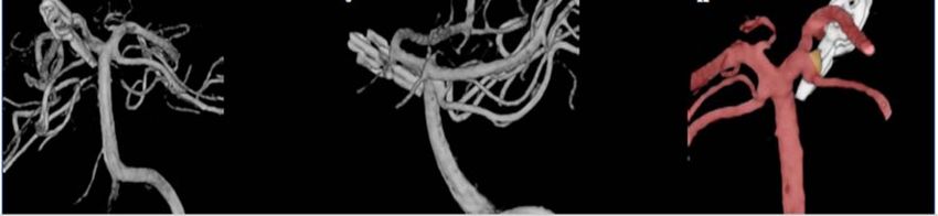
Brain Sci. 2020, 10, 334                                                                                                                                                             3 of 20

                                     Table 1. Demographic characteristics, clinical findings, aneurysm description, and outcome parameters.

                Age                      Aneurysm Location and              Aneurysm         Previous           Clinical           Fisher       H and H/
    Case                   Gender                                                                                                                                 GCS    mRS      GOS
               (Years)                    Anatomical Variation              Size (mm)       Treatment         Presentation         Grade       WFNS Grade
                                           Basilar tip aneurysm
                                                                             16 × 16 ×                      Dizziness attack
       1         53        Female       Bilateral PCA and SCA are                               no                                    4              5/5           3       6        1
                                                                                15                         and syncope, SAH
                                     leaving from the aneurysm base
                                      Partially thrombosed distal left
                                       side dissecting basilar artery
                                      aneurysm with secondary wall
                                                                                                              Thunderclap
       2         44         Male                 hematoma                    11 × 8 × 8         no                                  N/A             N/A            15      0        5
                                                                                                               headache
                                         Ampullary exit of the left
                                         double-laid SCA from the
                                             aneurysm fundus
                                                                                                           Incidental finding
                                         Proximal right side PCA
                                                                                                            Diagnostic in the
                                          aneurysm (P1 branch)
       3         48        Female                                            4×5×5              no            context of a          N/A             N/A            15      0        5
                                     Right SCA exits the P1 segment
                                                                                                               vestibular
                                      from the aneurysms side wall
                                                                                                               syndrome
      PCA = posterior cerebral artery, SCA = superior cerebellar artery, SAH = subarachnoid hemorrhage, N/A = not available, H and H = Hunt and Hess, GCS = Glasgow Comma Scale, mRS =
      modified Rankin Scale, GOS = Glasgow Outcome Scale.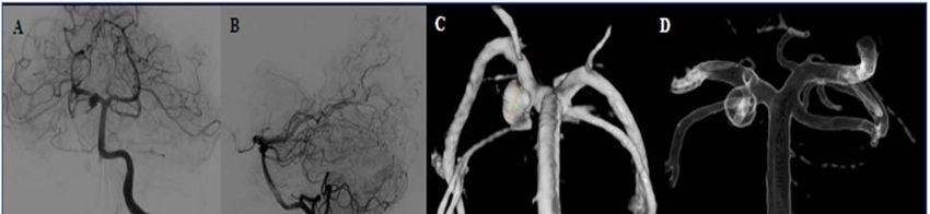
Brain Sci. 2020, 10, 334                                                                              4 of 20

     The technical aspects of performed combined approaches in the hOR have been described by our
group in an earlier publication [44]. The main unit consists of a 360◦ radiolucent carbon fiber table
(Alphamaquet 1150, Maquet AG, Switzerland) that is coupled with the C-arm angiography system
(Allura Xper FD20, Philips, Netherlands). A radiolucent head holder and pins are required for optimal
acquisition of angiograms and intraoperative CT scans (Mayfield, Integra GmbH, Ratingen, Germany).
A 7-Fr sheath is placed in the right or left femoral artery in preparation for intraoperative endovascular
balloon occlusion and control DSA. All cases underwent an intraoperative DSA (iDSA) and CT (iCT)
scan before they were transferred to the intensive care unit.

2.1. Illustrative Cases
      The surgical approach and endovascular techniques were similar in all three cases.

2.1.1. Surgical Procedure
     After the positioning of the patient0 s head in a carbon clamp in the hOR, a right fronto-temporal
craniotomy and selective extradural anterior clinoidectomy were performed. The proximal Sylvian
fissure was opened, and the chiasmatic cistern incised, followed by dissection of the optic and
oculomotor nerve, and carotid artery. Once the posterior clinoid process was exposed, a posterior
clinoidectomy was completed with a 2 mm drill. The afterward visible BA, PCA0 s, SCA0 s, and aneurysm
were inspected. After craniotomy, the first iDSA was performed by cannulation of the right femoral
artery with a 7-Fr sheath and inserting a 5F diagnostic catheter in one of the vertebral arteries. The first
iDSA showed the previously identified aneurysm and in Case 1 a progression of the dissecting basilar
tip aneurysm with a new bleb. The diagnostic catheter was exchanged for a soft guiding catheter
(Neuron 6F 058, Penumbra, Alameda CA, USA). An ASCENT® 4 × 7 balloon (DePuy Synthes) and was
then placed in the middle or distal segment of the BA and inflated under fluoroscopy to interrupt blood
flow. The dual lumen design of the ASCENT balloon allows the distal flushing of the occluded vessel
by saline. In the meantime, the BA or PCA aneurysm neck, which was significantly softened, as well
as PCA and SCA branches were further dissected. The aneurysms were occluded in a microtechnical
fashion with straight standard titanium 790-Yasargil-Clips (Aesculap, Tübingen, Germany) under
visualization of both SCA branches. The balloon was deflated after 9, 10, and 11 min of TBO. An iDSA
and intraoperative 3D-angiography in Case 1 showed complete occlusion of the BA aneurysm with
patent PCA and SCA branches (Figure 1). The iDSA in Cases 2 and 3, revealed a small remnant at the
aneurysm neck to preserve the SCA exit, no sign of aneurysm perfusion, and patent PCA and SCA
branches (Figures 2 and 3). The dura was sutured, the bone flap fixed, and the wound sutured using a
standard multilayered technique. The iCT scan documented no hemorrhage or midline shift.

Case 1

History
     This 53-year-old patient presented with a SAH after a sudden loss of consciousness at home.
The patient was intubated upon admission with a Glasgow Coma Scale (GCS) score of 3. The CT scan
showed a SAH caused by a ruptured basilar tip aneurysm (Fisher grade IV). A DSA was performed
after the patient improved to a GCS of 10 following two days of conservative treatment and CSF
drainage after ventriculostomy. A basilar tip aneurysm (16 × 16 × 15 mm) with the PCA and SCA
bilaterally arising from the aneurysm base was identified. Indication for surgical occlusion was decided
after interdisciplinary case discussion. The surgical procedure in the hOR had to be postponed for six
days due to severe vasospasms in the posterior circulation.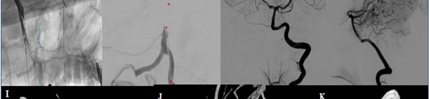
Brain Sci. 2019, 9, x FOR PEER REVIEW                                                                                   5 of 18

h after surgery. Despite of endovascular spasmolysis with Nimodipine was performed no clinical
Brain Sci. 2020, 10, 334                                                                      5 of 20
improvement could be observed. During the next days, the general condition of the patient
deteriorated due to pneumonia and respiratory failure leading to death eight days after surgery.

                              A                                    B

                              C                                D

                              E                                F

                              G                                H

                              I                                J

      Figure 1.
      Figure     Case 1.
              1. Case  1. Preoperative
                           Preoperative apap and
                                             and lateral
                                                  lateral DSA
                                                          DSA ofof the
                                                                   the basilar
                                                                       basilar tip
                                                                                tip aneurysm
                                                                                     aneurysm (A,B).
                                                                                                  (A,B). Preoperative
                                                                                                         Preoperative 3D3D
      angiography    of the   basilar aneurysm    presenting   the  bilateral  origin   of the PCA
      angiography of the basilar aneurysm presenting the bilateral origin of the PCA and SCA from the and  SCA   from  the
      aneurysm base
      aneurysm     base(C,D).
                         (C,D).Intraoperative
                                  Intraoperativeangiography
                                                    angiographyshowing    the endovascular
                                                                    showing    the endovascularplacement    of the balloon
                                                                                                       placement    of the
      and  occlusion  of  the basilar  artery (E,F). Intraoperative   DSA   after  clipping   and  closure  of
      balloon and occlusion of the basilar artery (E,F). Intraoperative DSA after clipping and closure of the  the balloon
      showingshowing
      balloon    complete    occlusion
                           complete      of the aneurysm
                                      occlusion             in ap and
                                                 of the aneurysm         lateral
                                                                     in ap        view with
                                                                            and lateral    viewallwith
                                                                                                   branches    open (G–I).
                                                                                                       all branches  open
      Intraoperative  picture   of the  opened  skull, placed  fish hooks,   spatula   and  clip
      (G–I). Intraoperative picture of the opened skull, placed fish hooks, spatula and clip (J).(J).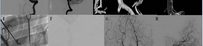
Brain Sci. 2020, 10, 334                                                                                                                                                                             6 of 20
          Brain Sci. 2019, 9, x FOR PEER REVIEW                                                                                                                                           7 of 18

             A                                    B                                 C                                 D                                E

             F                                        G                       H                     I                            J                              K

             L                                                     M                                 N                                       O
                                                      ICA
                                                                                                                             SCA                              SCA
                                   Pcom                                         A                                                                      SCA
                             III
                                                                                            PCA
                                                                                  PCA
                                                                                                                    PCA                                PCA

      Figure 2.Figure
                 Case2.2.Case  2. Preoperative
                          Preoperative     DSADSA     ap and
                                                 ap and       lateral
                                                           lateral    projectionsofofthe
                                                                   projections         theleft
                                                                                           left side
                                                                                                side dissecting
                                                                                                     dissectingbasilar
                                                                                                                 basilarartery aneurysm
                                                                                                                          artery        (A–C).
                                                                                                                                  aneurysm     Preoperative
                                                                                                                                            (A–C).          3D‐DSA
                                                                                                                                                   Preoperative     of the basilar
                                                                                                                                                                 3D-DSA     of theaneurysm,
                                                                                                                                                                                    basilar aneurysm,
              presenting   the ampullary   exit of the left double‐laid  SCA  from    the aneurysm     fundus  (D,E). Intraoperative DSA showing  the
      presenting the ampullary exit of the left double-laid SCA from the aneurysm fundus (D,E). Intraoperative DSA showing the endovascular placement endovascular placement   of the balloon
                                                                                                                                                                                        of the balloon
              and occlusion of the basilar artery through balloon inflation (F,G). Intraoperative angiography after clipping and deflation of the balloon demonstrating occlusion
      and occlusion of the basilar artery through balloon inflation (F,G). Intraoperative angiography after clipping and deflation of the balloon demonstrating occlusion of
              of the aneurysm in the ap and lateral view (H,I). Intraoperative 3D angiography in the ap and lateral view, showing a small remnant at the neck of the aneurysm to
      the aneurysm in the ap and lateral view (H,I). Intraoperative 3D angiography in the ap and lateral view, showing a small remnant at the neck of the aneurysm to
              preserve the SCA exit (J). Microsurgical view showing the aneurysm approach with mobilization of the ICA with hook retractor (K), BA aneurysm (L), and situs
      preserve the SCA exit (J). Microsurgical view showing the aneurysm approach with mobilization of the ICA with hook retractor (K), BA aneurysm (L), and situs after
              after clipping with patency of all branches and parent vessel (M–O). Abbreviations: DSA = digital subtraction angiography, SCA = superior cerebellar artery, ICA=
      clippinginternal
                with patency
                        carotid of  all branches
                                 artery,           andartery,
                                         BA= basilar    parentIIIvessel (M–O).nerve,
                                                                 = oculomotor     Abbreviations:
                                                                                          PCA = posterior    = digitalartery,
                                                                                                       DSA cerebral     subtraction  angiography,
                                                                                                                              A = aneurysm, Pcom =SCA   = superior
                                                                                                                                                   posterior       cerebellar
                                                                                                                                                             communicating      artery, ICA= internal
                                                                                                                                                                             artery.
      carotid artery, BA= basilar artery, III = oculomotor nerve, PCA = posterior cerebral artery, A = aneurysm, Pcom = posterior communicating artery.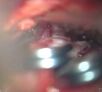
Brain Sci. 2020, 10, 334 7 of 20
Brain Sci. 2019, 9, x FOR PEER REVIEW 8 of 18
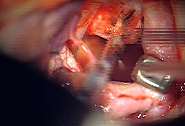
Figure Case
3. 3.
Figure 3. Preoperative
Case 3. PreoperativeDSA
DSA apap
andandlateral projections
lateral of of
projections thethe
proximal
proximalright sideside
right PCAPCAaneurysm
(P1aneurysm
branch) (P1
(A,B). Preoperative
branch) 3D-DSA of
(A,B). Preoperative the PCA
3D‐DSA aneurysm,
of the showing
PCA aneurysm, the right
showing SCA SCA
the right leaving the
P1leaving
segment thefrom the aneurysm
P1 segment sidewall
from the (C,D).
aneurysm Intraoperative
sidewall DSA presenting
(C,D). Intraoperative the placement
DSA presenting the of the
placement
balloon of the balloon
and occlusion of theand occlusion
BA after of the
balloon BA after
inflation balloon
(E,F). inflation angiography
Intraoperative (E,F). Intraoperative
after clipping
andangiography after clipping
balloon removal and balloon
demonstrating removal
occlusion demonstrating
of the aneurysm and occlusion
patencyof of
thethe
aneurysm
BA and andPCA (G–I).
patency of the BA and PCA (G–I). Intraoperative 3D‐DSA showing a small
Intraoperative 3D-DSA showing a small remnant at the aneurysm neck to preserve the remnant at the aneurysm
patency of the
neck to preserve the patency of the SCA (J,K). Abbreviations: DSA = digital subtraction angiography,
SCA (J,K). Abbreviations: DSA = digital subtraction angiography, PCA = posterior cerebral artery,
PCA = posterior cerebral artery, SCA = superior cerebellar artery.
SCA = superior cerebellar artery.
3. Results
Postoperative Course
All cases underwent combined surgical and endovascular procedures in our hOR. After
craniotomy and dissection
Further course on theofICU
the was
parent vessel and aneurysm
unsuccessful. the intraluminal
Consciousness balloon
persisted at awas
lowinserted
level of GCS
through the femoral artery and inflated in the BA. In all three cases, intraoperative
5 due to severe vasospasm and the deterioration of cerebral perfusion on CT was observed TBO was within
successfully performed without complications. No aneurysm rupture prior to clipping, or any other
48 h after surgery. Despite of endovascular spasmolysis with Nimodipine was performed no clinical
intra‐ or postoperative problems (necessary clip repositioning, parent vessel or branch occlusion,
improvement could be observed. During the next days, the general condition of the patient deteriorated
thromboembolic event, or re‐bleeding) occurred. The mean duration of TBO was 10 min (Table 2).
dueUpon
to pneumonia and respiratory failure leading to death eight days after surgery.
inflation of the balloon, the intraluminal pressure releases and the aneurysm softens rapidly,
which gives the surgeon more space and flexibility to explore the aneurysm and vessels branching
Case 2
out of the aneurysm base. Intraoperative DSA following clipping confirmed complete aneurysm
History
A 44-year-old patient was admitted to the emergency room with a thunderclap headache,
neck pain, vomiting, and paresthesia in the right arm. The patient had a GCS score of 15 without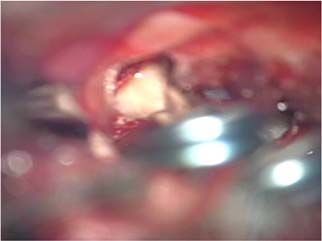
Brain Sci. 2020, 10, 334                                                                             8 of 20

meningism or any neurological deficits. Further investigations (CT, CTA, and DSA) excluded an
SAH but showed a dissecting, partially thrombosed BA aneurysm (11 × 8 × 8 mm) with a secondary
wall hematoma. Cerebral angiography revealed a duplicate origin of the SCA out of the aneurysm
fundus. Treatment options were discussed in the interdisciplinary neurovascular board. In principal,
endovascular occlusion by coiling or stent-assisted coiling was considered—with a high risk of SCA
occlusion and secondary cerebellar ischemia. Therefore, the group decided to recommend microsurgical
aneurysm clipping under endovascular TBO in the hOR.
Postoperative Course
     The patient was hospitalized for one more week. During this time, the patient suffered an epileptic
seizure. Apart from this, the patient could be rapidly mobilized with a GCS score of 15 and no new
neurological deficits.

Case 3

History
     After the occurrence of vertigo, this 48-year-old patient underwent an MRI scan which revealed
an incidental right PCA aneurysm. The patient was referred to our Institution, and the history and
clinical examination excluded any episodes of headache, epileptic seizure, or neurological deficits.
Further aneurysm imaging with DSA showed a saccular right BA aneurysm at the origin of the P1
segment of the PCA (4 × 5 × 5 mm). The pre-interventional 3D-DSA depicted the origin of the SCA
directly arising from the aneurysm fundus, as well as a hypoplastic bilateral posterior communicating
artery. After the case discussion with the neurovascular board, surgical occlusion with TBO in the hOR
was recommended.
Postoperative Course
     Postoperatively, the patient presented with a GCS score of 15, discrete ptosis, anisocoria, and double
vision. A further CT scan confirmed otherwise regular findings (the ophthalmological symptoms were
mainly caused by an impairment of oculomotor nerve function). The oculomotor nerve dysfunction
had resolved by itself by the three-month follow up.

3. Results
     All cases underwent combined surgical and endovascular procedures in our hOR. After craniotomy
and dissection of the parent vessel and aneurysm the intraluminal balloon was inserted through the
femoral artery and inflated in the BA. In all three cases, intraoperative TBO was successfully performed
without complications. No aneurysm rupture prior to clipping, or any other intra- or postoperative
problems (necessary clip repositioning, parent vessel or branch occlusion, thromboembolic event,
or re-bleeding) occurred. The mean duration of TBO was 10 min (Table 2). Upon inflation of the
balloon, the intraluminal pressure releases and the aneurysm softens rapidly, which gives the surgeon
more space and flexibility to explore the aneurysm and vessels branching out of the aneurysm base.
Intraoperative DSA following clipping confirmed complete aneurysm occlusion with patent parent and
branch vessels. In two cases, a small remnant at the aneurysm neck was visible in the intraoperative
3D angiography, which was necessary to preserve the branch origin. Two patients showed good
postoperative recovery with GOS 5 and one patient died due to severe vasospasm and pneumonia.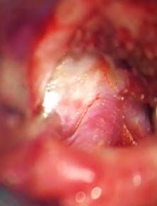
Brain Sci. 2020, 10, 334                                                                                      9 of 20

          Table 2. Details of the intraoperative balloon occlusion procedure, clipping, and iDSA findings.

                   Duration of   Number         Intraoperative DSA         Balloon Catheter
         Case                                                                                 Complications
                   TBO (Min)     of Clips            Findings                    Used
                                             Complete occlusion of the     ASCENT® 4 × 7
           1               9        1               aneurysm                   balloon            None
                                                All branches open          (DePuy Synthes)
                                            Small remnant at the neck to
                                                                           ASCENT® 4 × 7
                                               preserve the SCA exit
           2               10       2                                          balloon            None
                                              No aneurysm perfusion
                                                                           (DePuy Synthes)
                                                All branches open
                                            Small remnant at the neck to
                                                                           ASCENT® 4 × 7
                                               preserve the SCA exit
           3               11       3                                          balloon            None
                                              No aneurysm perfusion
                                                                           (DePuy Synthes)
                                                All branches open
      iDSA = intraoperative digital subtraction angiography, TBO = temporary balloon occlusion, SCA = superior
      cerebellar artery.

4. Discussion
      The findings of this technical note support the fact that TBO is a feasible, safe, and reliable method
for the clipping of posterior aneurysms that are technically demanding and complex due to size or
anatomy. In our institution, TBO with a combined endovascular and surgical approach is also used
for giant and complex recurrent middle cerebral artery (MCA) or anterior communicating artery
aneurysms. In cases with a ruptured MCA aneurysm and surrounding hematoma, endovascular TBO
facilitated clipping following hematoma evacuation and prevented an intraoperative rupture of fragile
high-risk aneurysms.
      In the case of intraoperative rupture, TBO could effectively control acute bleeding and increase
the safety and accuracy of clip placement. In one case report with the intraoperative rupture of a
paraclinoid aneurysm, TBO provided a salvage procedure for adequate hemostasis with additional
intraluminal support to preserve the parent artery during clip placement [43].
      In our opinion, microsurgery should still be viewed as a valuable option in the management
of posterior circulation aneurysms. Various authors have reported good radiological and clinical
outcomes for BA clipping with or without additional bypass [33,45–51]. Overall, the most common
complications in this location are perforator and branch ischemia-related events and cranial nerve
deficits, often involving oculomotor nerve palsies [47,52–54]. One case of transient oculomotor nerve
palsy occurred in our series.
      The angiographic obliteration rate of posterior circulation aneurysm has been reported with
a range from 91.9–98.1% [8,29,45,55]. However, other reports also cited 11.5% transient and 7.8%
permanent neurological deteriorations [45].
      Circumferential exposure of the aneurysm, including branches and perforators, is necessary prior
to a safe and efficacious clip application. Dissection and visualization of where they exit the aneurysm
can be very demanding. Various methods have been described to support the surgeon during the
clip application. Additional endovascular assistance can help prospective vascular neurosurgeons to
become more confident and proficient in these cases. Temporary parent vessel occlusion seems to be a
safe procedure. Interestingly, a study with a mean follow-up of 53 months showed that a temporary
artery occlusion time (mean 16.1 min) had no effect on overall long-term clinical outcomes [56].
      The “gold standard” for proximal vessel control is a temporary clip application, which is not
always feasible, especially in areas with limited access [57]. Proximal parent vessel ligation [58] can be
considered for treating giant aneurysms. Transient asystole with adenosine [40,59,60], deep hypothermic
circulatory arrest [61], or rapid ventricular pacing [62] are other techniques also described in the literature
but have higher risk profiles for side effects such as atrial or ventricular fibrillation, arrhythmias,
and prolonged hypotension [62]. In addition, the risk of stroke can increase after a circulatory arrest,
and the resulting need for a multidisciplinary team of surgeons and technologists is logistically
Brain Sci. 2020, 10, 334 10 of 20
challenging and expensive [63]. Perioperative morbidity and mortality for circulatory arrest has been
described as ranging from 8.3–17% [41,61]. The risk of side effects of these different techniques has to
be weighed against the significant chance of intraoperative aneurysm rupture, incomplete clipping, or
unintended branch occlusion due to poor visualization.
In comparison, TBO presents a simple, fast, and inexpensive technique with no need for special
anesthesiological monitoring or training and can be performed at any time in every center with
endovascular expertise. Although temporary clipping will remain the routinely used technique,
TBO may be more accurate for complex and large aneurysms, especially in the posterior circulation
and to prevent premature rupture. In posterior circulation aneurysm clipping, proximal control with
temporary clipping is often not possible due to the complex anatomy, skull base proximity, and location
near the brain stem and cranial nerves [64]. The temporary clip itself may hinder the placement of the
permanent one due to the limited surgical corridor [65]. In these cases, TBO can provide a reliable
alternative. Proximal control with TBO can be achieved before craniotomy with minimal obstruction
of the surgical field and less brain retraction.
Possible side effects of TBO and temporary clipping include wall injury of the parent vessel
or thromboembolic events causing postoperative ischemic deficits. MacDonald et al. compared
the degree of acute endothelial injury after temporary vessel occlusion with external clipping and
endovascular balloon occlusion in a pig model [66]. The results revealed that vessel injury worsened
with time and was more prevalent adjacent to the clip; as compared to the widespread pattern with
TBO [66]. There is the concern of a higher risk of ischemic complications with balloon occlusion in
perforator-rich vessels like MCA and BA, but neither MCA nor BA TBO interventions at our institution
led to perforator infarctions.
The TBO technique was first described in 1986 by Kinjo et al. [67], by Shucart et al. in 1990 [68],
and in many other case series since then. More recently, another group has reported on the use of TBO
in the hybrid OR [69]. Table 3 provides an overview of the literature to-date.
Most series included large or giant paraclinoid ICA aneurysms occluded with clip ligation after
balloon catheter placement in the ICA [69–71]. Intra-luminal pressure was decreased through the
additional placement of a temporary clip distal to the aneurysm on the posterior communicating artery
to reduce collateral blood flow, as well as the application of the retrograde decompression-suction
method in the ICA [69,71,72]. In these series, endovascular TBO eliminated the need for cervical ICA
dissection [68,69]. A review of the literature found a total of 188 aneurysms clipped with TBO. The largest
series, published by Fulkerson et al [73]. included 63 ophthalmic artery aneurysms. The description
of TBO in posterior circulation aneurysms, however, is less common with a total of only 20 cases.
Bailes et al. published the first series of TBO use in multiple basilar artery aneurysms [74]. Apart from
the recent study, eight other series have used TBO only for successful clipping [64,65,68,70,74–77].
Balloon placement in the aneurysm orifice or neck has only been described in two case series [65,77]
and was otherwise performed in the proximal parent vessel. TBO duration ranged from 1.5–3 min for
each balloon inflation and a total maximum of 50 min [43,63–65,68,71,75,77–81].
The overall reported complication rate for TBO is very low at 1.7–3.7% [72]. TBO procedure-related
thromboembolic events occurred in five patients (2.6%). This risk is increased in cases with pronounced
vessel wall sclerosis or prolonged temporary occlusion. One intraoperative balloon rupture and balloon
exchange led to a thrombus in the M1 segment, with subsequent intraoperative embolectomy and
postoperative transient hemiparesis. Symptoms such as dysphasia and hemiparesis were transient
in all other cases except one major MCA infarct, which lead to the death of the patient [71,80–82].
Further TBO-related complications included: ICA intima dissection with ICA occlusion at the neck
requiring medical treatment only (recovery was complete after four days) [81], as well as increases in
vasospasms due to mechanical wall stimulation with transient hemiparesis [77].Brain Sci. 2020, 10, 334 11 of 20
Table 3. Overview of all aneurysm cases including clipping with TBO in the literature. Information includes aneurysm characteristics, TBO duration, additional
techniques, aneurysm occlusion status and complications. All posterior circulation aneurysms and complications related to TBO are listed in bold type.
Author Aneurysm Location TBO Occlusion Time Additional Techniques Complete Occlusion Complications
Sendai cocktail
Kinjo T. et al. [41]
Giant BA aneurysm N/A Temporary clips on basilar N/A None
1986
artery and bilateral PCA
Transient oculomotor palsy
BA aneurysm 3 × optimal clip Oculomotor and abducens palsy
Shucart W.A. et al. 3–18 min (mean 12.5
3 × large paraclinoid ICA None placement Hemiparesis due to vasospasm
[71] 1990 min.)
aneurysms 1 × clip repositioning Aphasia due frontal infarct (Moya
Moya disease)
Temporary ICA clipping
Four large and giant Transient oculomotor palsy
Tamaki N. et al. distal to aneurysm
carotid-ophthalmic artery N/A Successful clipping Hemorrhagic infarction
[76] 1991 ICA blood aspiration
aneurysm Hydrocephalus (n = 2)
through second catheter
Temporary ICA clipping
distal to aneurysm
Scott J.A. et al. [68] Large ophthalmic artery 1.5 min. per balloon
Suction through distal Successful obliteration None
1991 aneurysm inflation
lumen of occlusion
balloon catheter
Intraoperative aneurysm rupture
Bailes J.E. et al. [4]
4 BA aneurysms N/A None Complete obliteration Transient oculomotor nerve palsy in 3
1992
patients
Albert F.K. et al. 2 proximal paraclinoid Suction-decompression
N/A N/A None
[2] 1993 aneurysm Temporary clip distal ICA
Intraoperative TBO balloon rupture →
3 × Clip repositioning surgery continued with new balloon
Retrograde (due to narrowing of →Embolus developed in M1 Segment
Mizoi K. et al. [51] 9 paraclinoid ICA 13–50 min. (mean 26.2
suction-decompression parent artery) → Embolectomy through M1 incisio,
1993 aneurysms min.)
Repeated TBO Successful obliteration in MCA occlusion for 20 min →
all cases aneurysm clipping followed → patient
with transient hemiparesis for 6 hBrain Sci. 2020, 10, 334 12 of 20
Table 3. Cont.
Author Aneurysm Location TBO Occlusion Time Additional Techniques Complete Occlusion Complications
3 cases successful obliteration
5 BA aneurysms
Mizoi K. et al. [52] 15–30 min. (mean 22 2 × clip repositioning: Finally one case Transient abducens nerve palsy
(including 1 large & 1 None
1994 min.) with remnant at the neck, one complete Transient hemiparesis
giant)
occlusion
Vasospasm and temporoparietal
infarction (hemiparesis and transient
Clip repositioning in all 3 cases
Fahlbusch R. et al. 3 giant paraclinoid ICA Retrograde dysphasia, deterioration of vision)
2–6 min. (stenosis of parent vessel or incomplete
[19] 1997 aneurysms suction-decompression 1 thromboembolic complication:
occlusion)
sensimotor dysphasia, infarct in
temporo-parietal region
Hacein-Bey L. et al. 1 large & 1 giant ICA
N/A None Complete obliteration None
[28] 1998 ophthalmic aneurysm
Transient oculomotor nerve palsy
2 temporal lobe infarction with new
neurological deficit
In 1 patient clipping was not possible Hydrocephalus and subdural hygroma
due to calcification of aneurysm neck Ipsilateral ICA intimal dissection and
Arnautovic K.I. et 8 giant and 8 large Distal temporary clip
mean 10.7 min. and wall, no aneurysm collapse occlusion at balloon side
al. [3] 1998 paraclinoid aneurysm Suction-decompression
achieved → Coiling Transient dysphasia (thromboembolic)
Successful clipping in 15 cases Cerebral abscess
Transient cerebral edema
One death due to intraventricular
hemorrhage
Clip adjustment in 7 cases (residual 1 major MCA infarct related to catheter
24 paraclinoid ICA filling of aneurysm and 4 ICA thromboembolism
Ng P-Y. et al. [60] 2–27 min. (mean 13 Suction-decompression
segment aneurysms (13 compromise) vasospasm in 3 cases with delayed
2000 min.) in 16 cases
large & 11 giant) Complete obliteration in 20 cases, ischemia
greater than 90% occlusion in 22 cases 2 deaths (one from fatal MCA infarct)
Lumbar drain due to subgaleal CSF
In 4 patients, navigation of the balloon
Balloon inflation time collection
Thorell W. et al. 6 paraclinoid giant or into intracranial circulation due to
less than 3 min. in all None Lacunar infarction ipsilateral 6 weeks
[80] 2004 complex ICA aneurysms carotid tortuosity was not possible
cases after surgery with mild transient
All aneurysms were complete occluded
hemiparesis
Case 1: Complete occlusion of the
intracranial aneurysm part at surgery
Acceleration of vasospasms 15 h after
and 5 months later, endovascular
Steiger H.J. et al. 2 giant carotid opthalmic 16 + 24 min. (mean 20 surgery and transient hemiparesis in
None occlusion of infraclinoid aspect of
[74] 2005 aneurysms min.) one case, resolved with hypertensive
aneurysm
therapy
Case 2: Small remnant at the neck for
patency of parent vesselBrain Sci. 2020, 10, 334 13 of 20
Table 3. Cont.
Author Aneurysm Location TBO Occlusion Time Additional Techniques Complete Occlusion Complications
5 giant paraclinoidal ICA
Ricci G. et al. [65] aneurysms Aneurysm obliteration achieved in all
N/A None None
2005 1 giant vertebrobasilar cases
junction aneurysm
Suction-decompression
Parkinson R.J. et al. Giant paraclinoid ICA Complete occlusion and reconstruction
Temporary clip distal to None
[63] 2006 aneurysm of parent vessel
aneurysm
All aneurysm excluded with parent
10 giant paraclinoid ICA
vessel patency
Petralia B. et al. aneurysm
15–20 min. None One balloon rupture None
[64] 2006 3 giant vertebrobasilar
One patient underwent coiling at
aneurysm
recurrence
Retrograde
Hoh D.J. et al. [32] Large paraclinoid suction-decompression
11 min. Aneurysm completely occluded None
2008 aneurysm Temporary clip distal to
aneurysm
Stroke
63 opthalmic artery
Temporary clip distal to Hematoma requiring treatment
Fulkerson D.H. et aneurysm
N/A aneurysm N/A Intraoperative aneurysm rupture
al. [23] 2008 (26 small, 23 large, 14
Suction-decompression New visual deficit
giant)
5 deaths
Prior attempt for with
temporary clipping, Hemiparesis due to stroke in the
Elhammady M.S. Large paraclinoid thereafter aneurysm inferior division of MCA
20 min. Complete obliteration
et al. [16] 2009 aneurysm rupture and switch to Vasospasm requiring intra-arterial
TBO as salvage nicardipine infusion
technique
One balloon below the
11 giant paraclinoid 15–20 min. (Average
aneurysm neck and One balloon rupture intraop
Skrap M. et al. [72] aneurysm 17 min.)
another in the PCA to One clip repositioning in BA None
2010 4 vertebrobasilar Max 5 min. per
stop retrograde flow Complete occlusion in all cases
aneurysm balloon inflation
from ICA
Temporary clip distal to
Dehdashti A.R [13] Large ophthalmic artery Complete occlusion and patent
N/A aneurysm (Pcomm and None
2015 aneurysm ophthalmic artery
ICA)Brain Sci. 2020, 10, 334 14 of 20
Table 3. Cont.
Author Aneurysm Location TBO Occlusion Time Additional Techniques Complete Occlusion Complications
STA-MCA Bypass prior
to clipping
Matano F. et al. Temporary clips on M1, Complete occlusion of aneurysms and
2 large ICPC aneurysms N/A None
[49] 2017 A1 and Pcomm patent parent vessel
Retrograde suction
decompression
Transient dysphonia
Capo G. et al. [11] Transient dysphagia
Giant VA aneurysm N/A Temporary clip on PICA Complete aneurysm occlusion
2018 Facial nerve palsy
PICA occlusion
Temporary clip on
Xu F. et al. [69] Large paraclinoid Pcomm
N/A Complete obliteration of aneurysm None
2018 aneurysm Retrograde
suction-decompression
TBO = temporary balloon occlusion, PCA =posterior cerebral artery, N/A = not available, BA = basilar artery, min = minutes, TBO = temporary balloon occlusion, ICA = internal carotid
artery, MCA = middle cerebral artery, CSF = cerebro-spinal fluid, ICPC = internal carotid posterior communicating artery aneurysms, Pcomm = posterior communicating artery aneurysm,
VA = vertebral artery, PICA = posterior inferior cerebellar artery.Brain Sci. 2020, 10, 334 15 of 20
Thromboembolic events may be reduced by limiting TBO duration and using double-lumen
balloon systems that allow for maintaining continuous heparinized saline catheter flush. In thrombosed
aneurysms or patients with severe atherosclerosis, the risk for thromboembolism might be increased.
No complications occurred in our series that had a mean TBO time of 10 min. Several series suggested
multiple short inflation times of 1.5–5 min to reduce thromboembolic event rate [63,65,78]. Some studies
used a preoperative TBO test [83] to investigate the capacity of collateral support. In view of the
short TBO time in our series, it is questionable if this is needed. The advantage of performing a TBO
procedure in the hybrid OR is that control angiography is possible immediately after clip placement.
We performed 2D and 3D intraoperative angiography in the hybrid OR and confirmed aneurysm
occlusion in all cases. This standardized protocol can achieve better outcomes [84,85].
The main limitation of our study is the small sample size, as we chose to report on posterior
circulation aneurysms only.
5. Conclusions
Intraoperative endovascular TBO is a feasible, safe, and valuable procedure for surgical treatment
of complex posterior circulation aneurysm undergoing clipping. In addition, intraoperative DSA and
3D-DSA in the hOR was confirmed as a valuable tool for the evaluation of aneurysm occlusion and
possible aneurysm remnants.
Author Contributions: J.F., J.C.K., J.F., L.R. methodology; Software, L.R., M.D.; Validation, J.F., L.R., J.C.K., Y.Y.
and M.D.; Formal analysis, J.F.; Writing—original draft preparation, J.C.K., J.F.; Writing—review and editing, J.F.,
S.M.; Supervision, J.F., L.R. All authors have read and agreed to the published version of the manuscript.
Funding: This research received no external funding.
Conflicts of Interest: The authors declare no conflict of interest.
References
1. Batjer, H.H.; Samson, D.S. Retrograde suction decompression of giant paraclinoidal aneurysms. Technical
note. J. Neurosurg. 1990, 73, 305–306. [CrossRef] [PubMed]
2. Batjer, H.H.; Kopitnik, T.A.; Giller, C.A.; Samson, D.S. Surgery for paraclinoidal carotid artery aneurysms.
J. Neurosurg. 1994, 80, 650–658. [CrossRef] [PubMed]
3. Vlak, M.H.; Rinkel, G.J.; Greebe, P.; van der Bom, J.G.; Algra, A. Trigger factors for rupture of intracranial
aneurysms in relation to patient and aneurysm characteristics. J. Neurol. 2012, 259, 1298–1302. [CrossRef]
4. Brisman, J.L.; Song, J.K.; Newell, D.W. Cerebral aneurysms. N. Engl. J. Med. 2006, 355, 928–939. [CrossRef]
[PubMed]
5. Molyneux, A.J.; Kerr, R.S.; Yu, L.M.; Clarke, M.; Sneade, M.; Yarnold, J.A.; Sandercock, P.; International
Subarachnoid Aneurysm Trial Collaborative Group. International subarachnoid aneurysm trial (ISAT) of
neurosurgical clipping versus endovascular coiling in 2143 patients with ruptured intracranial aneurysms:
A randomised comparison of effects on survival, dependency, seizures, rebleeding, subgroups, and aneurysm
occlusion. Lancet 2005, 366, 809–817. [PubMed]
6. Greving, J.P.; Wermer, M.J.; Brown, R.D., Jr.; Morita, A.; Juvela, S.; Yonekura, M.; Ishibashi, T.; Torner, J.C.;
Nakayama, T.; Rinkel, G.J.; et al. Development of the PHASES score for prediction of risk of rupture of
intracranial aneurysms: A pooled analysis of six prospective cohort studies. Lancet Neurol. 2014, 13, 59–66.
[CrossRef]
7. Etminan, N.; Brown, R.D., Jr.; Beseoglu, K.; Juvela, S.; Raymond, J.; Morita, A.; Torner, J.C.; Derdeyn, C.P.;
Raabe, A.; Mocco, J.; et al. The unruptured intracranial aneurysm treatment score: A multidisciplinary
consensus. Neurology 2015, 85, 881–889. [CrossRef]
8. Nanda, A.; Sonig, A.; Banerjee, A.D.; Javalkar, V.K. Microsurgical management of basilar artery apex
aneurysms: A single surgeon’s experience from Louisiana State University, Shreveport. World Neurosurg.
2014, 82, 118–129. [CrossRef]
9. Hernesniemi, J.; Goehre, F. Approaches to upper basilar artery aneurysms. World Neurosurg. 2014, 82, 1001–1002.
[CrossRef]Brain Sci. 2020, 10, 334 16 of 20
10. Batjer, H.; Samson, D. Intraoperative aneurysmal rupture: Incidence, outcome, and suggestions for surgical
management. Neurosurgery 1986, 18, 701–707. [CrossRef]
11. Molyneux, A.; Kerr, R.; Stratton, I.; Sandercock, P.; Clarke, M.; Shrimpton, J.; Holman, R.; International
Subarachnoid Aneurysm Trial Collaborative Group. International Subarachnoid Aneurysm Trial (ISAT) of
neurosurgical clipping versus endovascular coiling in 2143 patients with ruptured intracranial aneurysms:
A randomised trial. Lancet 2002, 360, 1267–1274. [CrossRef]
12. Ogilvy, C.S.; Hoh, B.L.; Singer, R.J.; Putman, C.M. Clinical and radiographic outcome in the management
of posterior circulation aneurysms by use of direct surgical or endovascular techniques. Neurosurgery
2002, 51, 14–21; discussion 21–22. [CrossRef] [PubMed]
13. Krisht, A.F.; Bikmaz, K.; Kadri, P.A.; Partington, S. Outcome of Surgical Clipping of 40 Complex Basilar
Aneurysms Using the Transcavernous Route: Paper 34. Neurosurgery 2006, 58, 407. [CrossRef]
14. Eskridge, J.M.; Song, J.K. Endovascular embolization of 150 basilar tip aneurysms with Guglielmi detachable
coils: Results of the Food and Drug Administration multicenter clinical trial. J. Neurosurg. 1998, 89, 81–86.
[CrossRef]
15. Bavinzski, G.; Killer, M.; Gruber, A.; Reinprecht, A.; Gross, C.E.; Richling, B. Treatment of basilar artery
bifurcation aneurysms by using Guglielmi detachable coils: A 6-year experience. J. Neurosurg. 1999, 90, 843–852.
[CrossRef]
16. Tateshima, S.; Murayama, Y.; Gobin, Y.P.; Duckwiler, G.R.; Guglielmi, G.; Vinuela, F. Endovascular treatment
of basilar tip aneurysms using Guglielmi detachable coils: Anatomic and clinical outcomes in 73 patients
from a single institution. Neurosurgery 2000, 47, 1332–1339; discussion 1339–1342. [CrossRef]
17. Henkes, H.; Fischer, S.; Mariushi, W.; Weber, W.; Liebig, T.; Miloslavski, E.; Brew, S.; Kuhne, D. Angiographic
and clinical results in 316 coil-treated basilar artery bifurcation aneurysms. J. Neurosurg. 2005, 103, 990–999.
[CrossRef]
18. Owen, C.M.; Montemurro, N.; Lawton, M.T. Microsurgical Management of Residual and Recurrent
Aneurysms After Coiling and Clipping: An Experience With 97 Patients. Neurosurgery 2015, 62 (Suppl. 1),
92–102. [CrossRef]
19. Spetzler, R.F.; McDougall, C.G.; Zabramski, J.M.; Albuquerque, F.C.; Hills, N.K.; Russin, J.J.; Partovi, S.;
Nakaji, P.; Wallace, R.C. The Barrow Ruptured Aneurysm Trial: 6-year results. J. Neurosurg. 2015, 123, 609–617.
[CrossRef]
20. Ferns, S.P.; Sprengers, M.E.; van Rooij, W.J.; Rinkel, G.J.; van Rijn, J.C.; Bipat, S.; Sluzewski, M.; Majoie, C.B.
Coiling of intracranial aneurysms: A systematic review on initial occlusion and reopening and retreatment
rates. Stroke 2009, 40, e523–e529. [CrossRef]
21. van Eijck, M.; Bechan, R.S.; Sluzewski, M.; Peluso, J.P.; Roks, G.; van Rooij, W.J. Clinical and Imaging
Follow-Up of Patients with Coiled Basilar Tip Aneurysms Up to 20 Years. AJNR Am. J. Neuroradiol.
2015, 36, 2108–2113. [CrossRef] [PubMed]
22. Molyneux, A.J.; Kerr, R.S.; Birks, J.; Ramzi, N.; Yarnold, J.; Sneade, M.; Rischmiller, J.; Collaborators, I. Risk of
recurrent subarachnoid haemorrhage, death, or dependence and standardised mortality ratios after clipping
or coiling of an intracranial aneurysm in the International Subarachnoid Aneurysm Trial (ISAT): Long-term
follow-up. Lancet Neurol. 2009, 8, 427–433. [CrossRef]
23. Ferns, S.P.; Sprengers, M.E.; van Rooij, W.J.; van Zwam, W.H.; de Kort, G.A.; Velthuis, B.K.; Schaafsma, J.D.;
van den Berg, R.; Sluzewski, M.; Brouwer, P.A.; et al. Late reopening of adequately coiled intracranial
aneurysms: Frequency and risk factors in 400 patients with 440 aneurysms. Stroke 2011, 42, 1331–1337.
[CrossRef] [PubMed]
24. Juvela, S.; Porras, M.; Heiskanen, O. Natural history of unruptured intracranial aneurysms: A long-term
follow-up study. J. Neurosurg. 1993, 79, 174–182. [CrossRef] [PubMed]
25. Juvela, S.; Porras, M.; Poussa, K. Natural history of unruptured intracranial aneurysms: Probability of and
risk factors for aneurysm rupture. J. Neurosurg. 2000, 93, 379–387. [CrossRef]
26. Wiebers, D.O.; Whisnant, J.P.; Huston, J., III; Meissner, I.; Brown, R.D., Jr.; Piepgras, D.G.; Forbes, G.S.;
Thielen, K.; Nichols, D.; O’Fallon, W.M.; et al. Unruptured intracranial aneurysms: Natural history,
clinical outcome, and risks of surgical and endovascular treatment. Lancet 2003, 362, 103–110. [CrossRef]Brain Sci. 2020, 10, 334 17 of 20
27. Molyneux, A.J.; Birks, J.; Clarke, A.; Sneade, M.; Kerr, R.S. The durability of endovascular coiling versus
neurosurgical clipping of ruptured cerebral aneurysms: 18 year follow-up of the UK cohort of the International
Subarachnoid Aneurysm Trial (ISAT). Lancet 2015, 385, 691–697. [CrossRef]
28. Investigators, U.J.; Morita, A.; Kirino, T.; Hashi, K.; Aoki, N.; Fukuhara, S.; Hashimoto, N.; Nakayama, T.;
Sakai, M.; Teramoto, A.; et al. The natural course of unruptured cerebral aneurysms in a Japanese cohort.
N. Engl. J. Med. 2012, 366, 2474–2482. [CrossRef]
29. Sekhar, L.N.; Tariq, F.; Morton, R.P.; Ghodke, B.; Hallam, D.K.; Barber, J.; Kim, L.J. Basilar tip aneurysms:
A microsurgical and endovascular contemporary series of 100 patients. Neurosurgery 2013, 72, 284–298;
discussion 298–299. [CrossRef]
30. Hernesniemi, J.; Korja, M. At the apex of cerebrovascular surgery–basilar tip aneurysms. World Neurosurg.
2014, 82, 37–39. [CrossRef]
31. Yasargil, M.G.; Antic, J.; Laciga, R.; Jain, K.K.; Hodosh, R.M.; Smith, R.D. Microsurgical pterional approach to
aneurysms of the basilar bifurcation. Surg. Neurol. 1976, 6, 83–91. [PubMed]
32. Drake, C.G. Bleeding aneurysms of the basilar artery. Direct surgical management in four cases. J. Neurosurg.
1961, 18, 230–238. [CrossRef] [PubMed]
33. Tjahjadi, M.; Kivelev, J.; Serrone, J.C.; Maekawa, H.; Kerro, O.; Jahromi, B.R.; Lehto, H.; Niemela, M.;
Hernesniemi, J.A. Factors Determining Surgical Approaches to Basilar Bifurcation Aneurysms and Its
Surgical Outcomes. Neurosurgery 2016, 78, 181–191. [CrossRef] [PubMed]
34. Day, J.D.; Fukushima, T.; Giannotta, S.L. Cranial base approaches to posterior circulation aneurysms.
J. Neurosurg. 1997, 87, 544–554. [CrossRef]
35. Hsu, F.P.; Clatterbuck, R.E.; Spetzler, R.F. Orbitozygomatic approach to basilar apex aneurysms. Neurosurgery
2005, 56 (Suppl. 1), 172–177. [CrossRef]
36. Gonzalez, L.F.; Amin-Hanjani, S.; Bambakidis, N.C.; Spetzler, R.F. Skull base approaches to the basilar artery.
Neurosurg. Focus 2005, 19, E3. [CrossRef]
37. Kawase, T.; Toya, S.; Shiobara, R.; Mine, T. Transpetrosal approach for aneurysms of the lower basilar artery.
J. Neurosurg. 1985, 63, 857–861. [CrossRef]
38. Kato, Y.; Sano, H.; Behari, S.; Kumar, S.; Nagahisa, S.; Iwata, S.; Kanno, T. Surgical clipping of basilar
aneurysms: Relationship between the different approaches and the surgical corridors. Minim. Invasive
Neurosurg. MIN 2002, 45, 142–145. [CrossRef]
39. Spiessberger, A.; Strange, F.; Fandino, J.; Marbacher, S. Microsurgical Clipping of Basilar Apex Aneurysms:
A Systematic Historical Review of Approaches and their Results. World Neurosurg. 2018, 114, 305–316.
[CrossRef]
40. Groff, M.W.; Adams, D.C.; Kahn, R.A.; Kumbar, U.M.; Yang, B.Y.; Bederson, J.B. Adenosine-induced transient
asystole for management of a basilar artery aneurysm. Case report. J. Neurosurg. 1999, 91, 687–690. [CrossRef]
41. Lawton, M.T.; Raudzens, P.A.; Zabramski, J.M.; Spetzler, R.F. Hypothermic circulatory arrest in neurovascular
surgery: Evolving indications and predictors of patient outcome. Neurosurgery 1998, 43, 10–20;
discussion 20–21. [CrossRef]
42. Samson, D.; Batjer, H.H.; Bowman, G.; Mootz, L.; Krippner, W.J., Jr.; Meyer, Y.J.; Allen, B.C. A clinical study
of the parameters and effects of temporary arterial occlusion in the management of intracranial aneurysms.
Neurosurgery 1994, 34, 22–28; discussion 28–29. [PubMed]
43. Elhammady, M.S.; Nakaji, P.; Farhat, H.; Morcos, J.J.; Aziz-Sultan, M.A. Balloon-assisted clipping of a
large paraclinoidal aneurysm: A salvage procedure. Neurosurgery 2009, 65, E1210–E1211; discussion E1211.
[CrossRef] [PubMed]
44. Fandino, J.; Taussky, P.; Marbacher, S.; Muroi, C.; Diepers, M.; Fathi, A.R.; Remonda, L. The concept of a
hybrid operating room: Applications in cerebrovascular surgery. Acta Neurochir. Suppl. 2013, 115, 113–117.
[PubMed]
45. Sanai, N.; Tarapore, P.; Lee, A.C.; Lawton, M.T. The current role of microsurgery for posterior circulation
aneurysms: A selective approach in the endovascular era. Neurosurgery 2008, 62, 1236–1249; discussion
1249–1253. [CrossRef] [PubMed]
46. Lawton, M.T.; Abla, A.A.; Rutledge, W.C.; Benet, A.; Zador, Z.; Rayz, V.L.; Saloner, D.; Halbach, V.V. Bypass
Surgery for the Treatment of Dolichoectatic Basilar Trunk Aneurysms: A Work in Progress. Neurosurgery
2016, 79, 83–99. [CrossRef]Brain Sci. 2020, 10, 334 18 of 20
47. Basma, J.; Ryttlefors, M.; Latini, F.; Pravdenkova, S.; Krisht, A. Mobilization of the transcavernous
oculomotor nerve during basilar aneurysm surgery: Biomechanical bases for better outcome. Neurosurgery
2014, 10 (Suppl. 1), 106–114; discussion 114–115. [CrossRef]
48. Shi, X.; Qian, H.; Singh, K.C.; Zhang, Y.; Zhou, Z.; Sun, Y.; Liu, F. Surgical management of vertebral and basilar
artery aneurysms: A single center experience in 41 patients. Acta Neurochir. (Wien) 2013, 155, 1087–1093.
[CrossRef]
49. Yanagisawa, T.; Kinouchi, H.; Sasajima, T.; Shimizu, H. Long-Term Follow-Up for a Giant Basilar Trunk
Aneurysm Surgically Treated by Proximal Occlusion and External Carotid Artery to Posterior Cerebral
Artery Bypass Using a Saphenous Vein Graft. J. Stroke Cerebrovasc. Dis. 2016, 25, e212–e213. [CrossRef]
50. Indo, M.; Oya, S.; Matsui, T. Ruptured Basilar Tip Aneurysm in a Patient with Bilateral Internal Carotid
Artery Occlusion Successfully Treated with Bilateral Superficial Temporal Artery-Middle Cerebral Artery
Anastomoses: Case Report. World Neurosurg. 2016, 86, 512.e5–512.e8. [CrossRef]
51. Kai, Y.; Hamada, J.; Morioka, M.; Yano, S.; Hamasaki, K.; Ushio, Y. Successful treatment of a ruptured
dissecting basilar artery aneurysm. Case report. J. Neurosurg. 2004, 100, 1072–1075. [CrossRef] [PubMed]
52. Krisht, A.F.; Krayenbuhl, N.; Sercl, D.; Bikmaz, K.; Kadri, P.A. Results of microsurgical clipping of 50
high complexity basilar apex aneurysms. Neurosurgery 2007, 60, 242–250; discussion 250–252. [CrossRef]
[PubMed]
53. Tanaka, Y.; Kobayashi, S.; Hongo, K.; Tada, T.; Nagashima, H.; Kakizawa, Y. Intentional body clipping of
wide-necked basilar artery bifurcation aneurysms. J. Neurosurg. 2000, 93, 169–174. [CrossRef] [PubMed]
54. Matsukawa, H.; Tanikawa, R.; Kamiyama, H.; Tsuboi, T.; Noda, K.; Ota, N.; Miyata, S.; Tokuda, S. Localization
in the Interpeduncular Cistern as Risk Factors for the Thalamoperforators’ Ischemia, Poor Outcome, and
Oculomotor Nerve Palsy in Patients with Complex Unruptured Basilar Apex Aneurysm Treated with Neck
Clipping. World Neurosurg. 2015, 84, 475–482. [CrossRef] [PubMed]
55. Mooney, M.A.; Kalani, M.Y.; Nakaji, P.; Albuquerque, F.C.; McDougall, C.G.; Spetzler, R.F.; Zabramski, J.M.
Long-term Patient Outcomes After Microsurgical Treatment of Blister-Like Aneurysms of the Basilar Artery.
Neurosurgery 2015, 11 (Suppl. 3), 387–393. [CrossRef]
56. Griessenauer, C.J.; Poston, T.L.; Shoja, M.M.; Mortazavi, M.M.; Falola, M.; Tubbs, R.S.; Fisher, W.S., III.
The impact of temporary artery occlusion during intracranial aneurysm surgery on long-term clinical
outcome: Part I. Patients with subarachnoid hemorrhage. World Neurosurg. 2014, 82, 140–148. [CrossRef]
57. Taylor, C.L.; Selman, W.R.; Kiefer, S.P.; Ratcheson, R.A. Temporary vessel occlusion during intracranial
aneurysm repair. Neurosurgery 1996, 39, 893–905; discussion 905–906.
58. Steinberg, G.K.; Drake, C.G.; Peerless, S.J. Deliberate basilar or vertebral artery occlusion in the treatment
of intracranial aneurysms. Immediate results and long-term outcome in 201 patients. J. Neurosurg.
1993, 79, 161–173. [CrossRef]
59. Al-Mousa, A.; Bose, G.; Hunt, K.; Toma, A.K. Adenosine-assisted neurovascular surgery: Initial case series
and review of literature. Neurosurg. Rev. 2019, 42, 15–22. [CrossRef]
60. Desai, V.R.; Rosas, A.L.; Britz, G.W. Adenosine to facilitate the clipping of cerebral aneurysms: Literature
review. Stroke Vasc. Neurol. 2017, 2, 204–209. [CrossRef]
61. Mack, W.J.; Ducruet, A.F.; Angevine, P.D.; Komotar, R.J.; Shrebnick, D.B.; Edwards, N.M.; Smith, C.R.;
Heyer, E.J.; Monyero, L.; Connolly, E.S., Jr.; et al. Deep hypothermic circulatory arrest for complex cerebral
aneurysms: Lessons learned. Neurosurgery 2007, 60, 815–827. [CrossRef] [PubMed]
62. Konczalla, J.; Platz, J.; Fichtlscherer, S.; Mutlak, H.; Strouhal, U.; Seifert, V. Rapid ventricular pacing for clip
reconstruction of complex unruptured intracranial aneurysms: Results of an interdisciplinary prospective
trial. J. Neurosurg. 2018, 128, 1741–1752. [CrossRef] [PubMed]
63. Skrap, M.; Petralia, B.; Toniato, G. Temporary balloon occlusion during the surgical treatment of giant
paraclinoid and vertebrobasilar aneurysms. Acta Neurochir. (Wien) 2010, 152, 435–442. [CrossRef] [PubMed]
64. Mizoi, K.; Yoshimoto, T.; Takahashi, A.; Ogawa, A. Direct clipping of basilar trunk aneurysms using
temporary balloon occlusion. J. Neurosurg. 1994, 80, 230–236. [CrossRef] [PubMed]
65. Thorell, W.; Rasmussen, P.; Perl, J.; Masaryk, T.; Mayberg, M. Balloon-assisted microvascular clipping of
paraclinoid aneurysms. Technical note. J. Neurosurg. 2004, 100, 713–716. [CrossRef]You can also read