Emerging Connections between Nuclear Pore Complex Homeostasis and ALS
←
→
Page content transcription
If your browser does not render page correctly, please read the page content below
International Journal of
               Molecular Sciences

Review
Emerging Connections between Nuclear Pore Complex
Homeostasis and ALS
Sunandini Chandra                      and C. Patrick Lusk *

                                            Department of Cell Biology, Yale School of Medicine, 295 Congress Ave, New Haven, CT 06520, USA;
                                            sunandini.chandra@yale.edu
                                            * Correspondence: patrick.lusk@yale.edu

                                            Abstract: Developing effective treatments for neurodegenerative diseases such as amyotrophic lateral
                                            sclerosis (ALS) requires understanding of the underlying pathomechanisms that contribute to the
                                            motor neuron loss that defines the disease. As it causes the largest fraction of familial ALS cases,
                                            considerable effort has focused on hexanucleotide repeat expansions in the C9ORF72 gene, which
                                            encode toxic repeat RNA and dipeptide repeat (DPR) proteins. Both the repeat RNA and DPRs
                                            interact with and perturb multiple elements of the nuclear transport machinery, including shuttling
                                            nuclear transport receptors, the Ran GTPase and the nucleoporin proteins (nups) that build the
                                            nuclear pore complex (NPC). Here, we consider recent work that describes changes to the molecular
                                            composition of the NPC in C9ORF72 model and patient neurons in the context of quality control
                                            mechanisms that function at the nuclear envelope (NE). For example, changes to NPC structure
                                            may be caused by the dysregulation of a conserved NE surveillance pathway mediated by the
                                            endosomal sorting complexes required for the transport protein, CHMP7. Thus, these studies are
                                            introducing NE and NPC quality control pathways as key elements in a pathological cascade that
                                            leads to C9ORF72 ALS, opening entirely new experimental avenues and possibilities for targeted
                                            therapeutic intervention.


                                            Keywords: C9ORF72 ALS; NPC injury; CHMP7; ESCRT; nuclear transport; POM121; nuclear quality
Citation: Chandra, S.; Lusk, C.P.
                                            control
Emerging Connections between
Nuclear Pore Complex Homeostasis
and ALS. Int. J. Mol. Sci. 2022, 23,
1329. https://doi.org/10.3390/
                                            1. Introduction to Amyotrophic Lateral Sclerosis: Background of Genotypic and
ijms23031329
                                            Pathological Features
Academic Editor: Patrick R. Onck                  Neurodegenerative diseases constitute a spectrum of pathologies that includes amy-
Received: 22 December 2021
                                            otrophic lateral sclerosis (ALS), frontotemporal dementia (FTD), Parkinson’s disease,
Accepted: 20 January 2022
                                            Alzheimer’s disease and Huntington’s disease, which affect millions of people world-
Published: 25 January 2022
                                            wide [1]. The most common motor neuron disease is ALS, also known as Lou Gherig’s
                                            disease in honor of the famous New York Yankees baseball player who succumbed to it.
Publisher’s Note: MDPI stays neutral
                                            ALS is characterized by a loss of motor neurons in the brain and spinal cord, which leads
with regard to jurisdictional claims in
                                            to progressive muscle weakness and atrophy, dysarthria, dysphagia and spasticity [2]. Al-
published maps and institutional affil-
                                            though there are only two FDA-approved drugs that can slow the progression of symptoms,
iations.
                                            like all neurodegenerative diseases, there is no cure [3]. Thus, there remains a critical need
                                            to understand the underlying pathomechanisms at the molecular level in order to inform
                                            new therapeutic strategies.
Copyright: © 2022 by the authors.
                                                  Efforts to define such mechanisms benefit from genetic studies that, beginning with
Licensee MDPI, Basel, Switzerland.          SOD1 28 years ago, have identified several causative genetic variants in a multitude of
This article is an open access article      genes, including C9ORF72, TARDBP, FUS, ANG, MATR3, OPTN, TBK1, NEK1, C21ORF2,
distributed under the terms and             CHCHD10, DCTN1, TUBA4A, PFN1, SQSTM1, VCP and UBQLN2 [4,5]. These genes were
conditions of the Creative Commons          identified because of their autosomal dominant pattern of inheritance within families.
Attribution (CC BY) license (https://       These “familial” ALS (fALS), however, only constitute ~10% of total ALS cases; the vast
creativecommons.org/licenses/by/            majority of ALS cases are sporadic (sALS), even if they sometimes share a known genetic
4.0/).                                      association with fALS [6]. For example, the most common genetic cause of ALS, responsible

Int. J. Mol. Sci. 2022, 23, 1329. https://doi.org/10.3390/ijms23031329                                           https://www.mdpi.com/journal/ijms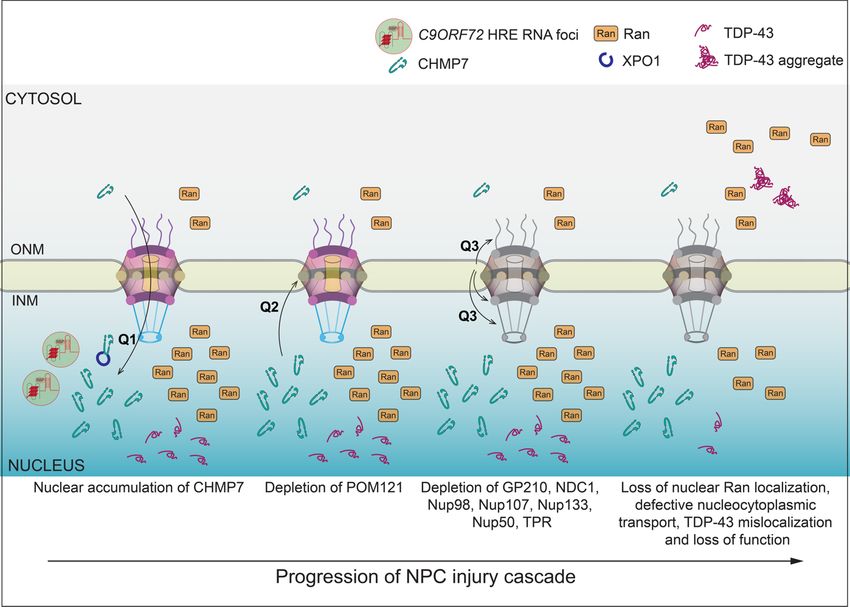
Int. J. Mol. Sci. 2022, 23, 1329 2 of 12
for 40% of fALS and 8% sALS, is a G4 C2 hexanucleotide repeat expansion (HRE) in the first
intron of the C9ORF72 gene (hereafter referred to as C9-ALS) [7]. This repeat expansion
is also common in FTD, the second leading cause of dementia after Alzheimer’s disease,
which shares clinical and pathological features with ALS [8]. Thus, because of its central
importance to ALS/FTD, there has been considerable interest in defining the underlying
mechanisms regarding how the expression of the HREs impact cellular physiology [9].
It is now known that G4 C2 repeats are bidirectionally transcribed to form sense and
anti-sense repeat RNA that can then undergo repeat-associated non-ATG translation (RANT)
into five dipeptide repeat (DPR) proteins, namely poly(GA), poly(GP), poly(GR), poly(PA)
and poly(PR). Both repeat RNAs and DPRs are thought to drive neurotoxicity [10,11]. In
fact, the expression of repeat RNAs and DPRs can inhibit cell growth in model organisms
such as flies and yeast, hinting that they may impact shared molecular processes between
all cells. Indeed, a confluence of evidence over the past few years has strongly implicated
nuclear transport machinery as a key target of both repeat RNAs and DPRs (reviewed
extensively in [12–16]). Most recently, even the nuclear pore complex (NPC) itself appears
to be susceptible to damage caused by expression of HRE repeat RNA [7]. These and
other data are raising questions as to the underlying mechanisms that could lead to the
removal and degradation of NPC components (nucleoporins or nups) and whether nuclear
envelope (NE)-specific quality control mechanisms may be at play. Here, we examine the
latest research implicating ALS-specific pathomechanisms that intersect with the nuclear
transport machinery in the context of emerging quality control mechanisms that function
at the NE. These pathways could provide a roadmap forward to identifying the underlying
causes of these diseases.
2. Nuclear Transport and NE Quality Control in ALS
2.1. The Soluble Nuclear Transport Machinery Is Impacted in ALS
NPCs are 100 MD protein assemblies that form selective transport channels that span
the double membraned NE; there are hundreds to thousands of NPCs that gate the nucleus
in a typical mammalian cell, including neurons [17–19]. Each NPC is built from ~30 nups
that are assembled into subcomplexes that form modular units repeated in 8-fold, radi-
ally symmetric, concentric ring assemblies to construct a stable core scaffold architecture
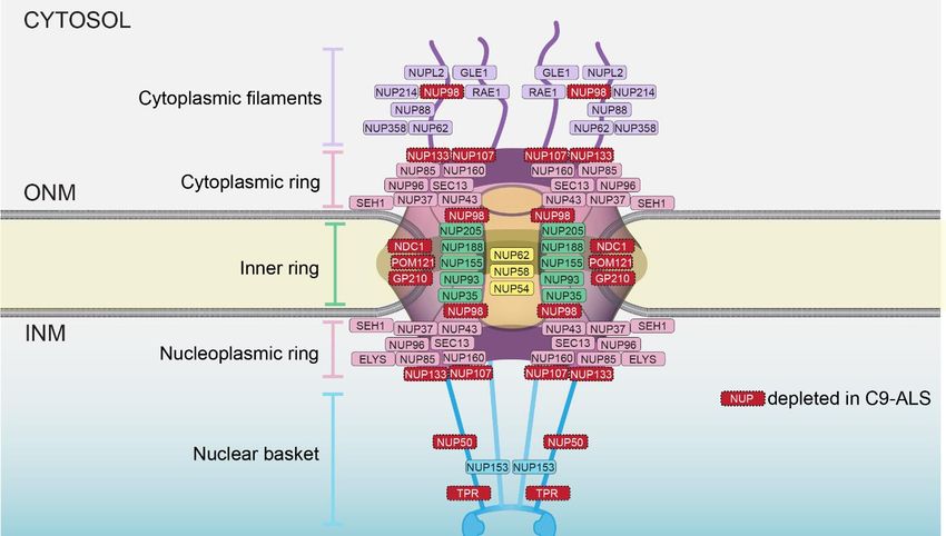
(Figure 1). The cytoplasmic ring and nucleoplasmic ring are largely compositionally similar,
with notable differences that allow for the anchorage of cytoplasmic filaments/an mRNA
export platform on the cytoplasmic side of the NPC and a nuclear basket structure that
extends into the nucleus [20]. The scaffold also provides anchor points for intrinsically
disordered proteins, rich in FG amino acid residues found in repetitive motifs (the FG-nups)
that fill the central transport channel. There are thousands of FG-repeats in the central chan-
nel that are principally responsible for nucleocytoplasmic compartmentalization through
two core mechanisms: they establish a diffusion barrier that impedes the passage of macro-
molecules, while also providing binding sites for shuttling nuclear transport receptors
(NTR; also known as karyopherins/importins/exportins) that carry signal-bearing cargo
through the NPC. Directionality and energy for multiple rounds of transport are provided
by the Ran GTPase, which itself is predominantly localized to the nucleus at steady state,
bound to GTP [21,22].Int.J.J.Mol.
Int. Mol.Sci.
Sci.2022, 23,x1329
2022,23, FOR PEER REVIEW 33ofof1212
Figure
Figure 1.
1. Schematic of NPC
Schematic of NPC embedded
embedded inin the
thenuclear
nuclearenvelope.
envelope.Major
Majorarchitectural
architecturalassemblies
assembliesand
and
relative
relative position of individual nups are indicated. Nups, in red, are depleted in C9-ALS. ONM isis
position of individual nups are indicated. Nups, in red, are depleted in C9-ALS. ONM
outer nuclear membrane; INM is inner nuclear membrane.
outer nuclear membrane; INM is inner nuclear membrane.
There
There is now an
is now an abundance
abundance of ofevidence
evidencedemonstrating
demonstratingthat thatessentially
essentiallyallallelements
elements
of
of nuclear
nuclear transport machinery,including
transport machinery, includingNTRs,
NTRs,nups
nupsand andRan,
Ran,arearemislocalized
mislocalizedinineither
either
nucleoplasmic or cytosolic aggregates in ALS [12,14,15,23,24]. The biogenesis
nucleoplasmic or cytosolic aggregates in ALS [12,14,15,23,24]. The biogenesis of these of these ag-
gregates is not always understood. Some, for
aggregates is not always understood. Some, for example, example, are stress granules, which can bebe
stress granules, which can
formed
formed by the DPRs themselves
themselves [25,26].
[25,26]. Others
Others might
mightreflect
reflectaaprotective
protectiveresponse
responseasasthe the
direct bindingofofNTRs
direct binding NTRs to to
thethe DPRs
DPRs can can suppress
suppress their their pathological
pathological interactions
interactions with RNA- with
binding proteins
RNA-binding such as
proteins TDP-43.
such Such aSuch
as TDP-43. mechanism might also
a mechanism be also
might related
be to the ability
related to the
of NTRs to promote the disaggregation of pathological FUS and TDP-43
ability of NTRs to promote the disaggregation of pathological FUS and TDP-43 aggregates aggregates [27].
This latter role speaks to a fundamental role of NTRs that can act as chaperones
[27]. This latter role speaks to a fundamental role of NTRs that can act as chaperones that that shield
binding
shield interfaces
binding that contribute
interfaces to both productive
that contribute and pathological
to both productive phase separation
and pathological phase [28].
sepa-
Indeed,
ration theIndeed,
[28]. relationship between the
the relationship phase-separation
between behavior of
the phase-separation pathological
behavior RNA
of pathologi-
binding
cal RNA proteins
binding such as FUS
proteins suchand TDP-43
as FUS and (which
TDP-43also bindalso
(which to and
bindimpact
to and the phase
impact the
separation properties of nups as well [29]) and NTRs is just beginning to
phase separation properties of nups as well [29]) and NTRs is just beginning to be unrav-be unraveled [28].
eled [28].
2.2. NPC Injury in C9-ALS
Although
2.2. NPC Injurythe
in presence
C9-ALS of nuclear and cytosolic aggregates containing nuclear transport
components has been well documented, whether the NPCs themselves were altered in
C9-ALSAlthough the presence
is just coming of nuclear
into focus. In fact,and
suchcytosolic
an idea may aggregates
not have containing nuclear
been prioritized
transport
as it is wellcomponents
establishedhas been
that the well documented,
scaffold of the NPC whether the NPCs
is extremely long-themselves were al-
lived in neurons,
tered in C9-ALS is just coming into focus. In fact, such an idea may not
suggesting that, once assembled, it is difficult to dislodge a nup from the NPC [18,30,31]. have been priori-
tized as it is well established that the scaffold of the NPC is extremely
Recent work is challenging this idea. Using an induced pluripotent stem-cell-derived long- lived in neu-
rons,
neuron suggesting
(iPSN) modelthat, ofonce assembled,
C9-ALS, Coyne itetisal.difficult to dislodge
(2020) purified a nup
nuclei from the NPC
and examined the
[18,30,31].
localizationRecent
of 23 work is challenging
nups using this idea. Using
immunofluorescence an induced
structured pluripotent
illumination stem-cell-
microscopy,
derived
which can,neuron (iPSN) model
in principle, resolveof C9-ALS,
individual Coyne
NPCs et [32,33].
al. (2020)Remarkably,
purified nuclei
8 ofand
the examined
23 nups
the
were found at lower levels in nuclear and NE pools (Figure 1). These nup lossesmicros-
localization of 23 nups using immunofluorescence structured illumination were
copy,
causedwhich
by thecan, in principle,
expression of theresolve
HRE RNA, individual
as theyNPCswere[32,33]. Remarkably,
not observed in C9ORF728 of the
null23
nups
iPSNs, were
nor found at loweroflevels
in the context DPRs.inMoreover,
nuclear and bothNEsense
pools(SOs)
(Figure
and1). These nup
antisense losses
oligos were
(ASOs)
caused by the RNAs
that target expression of thethe
prevented HREnupRNA, as they
loss [7]. were not
Curiously, observed
these in C9ORF72
nup depletions spannednull
iPSNs, nor in the context of DPRs. Moreover, both sense (SOs) and antisense oligos (ASOs)
that target the RNAs prevented the nup loss [7]. Curiously, these nup depletions spannedInt. J. Mol. Sci. 2022, 23, x FOR PEER REVIEW 4 of 12
Int. J. Mol. Sci. 2022, 23, 1329 4 of 12
all
all major
major architectural
architectural units
units of the NPC,
of the NPC, including
including the the nuclear
nuclear basket
basket(NUP50
(NUP50and andTPR),
TPR),
the central transport channel (NUP98), the cytoplasmic/nucleoplasmic
the central transport channel (NUP98), the cytoplasmic/nucleoplasmic rings (NUP107 rings (NUP107 and
NUP133) and all three of the pore membrane proteins
and NUP133) and all three of the pore membrane proteins (GP210/NUP210, NDC1 and(GP210/NUP210, NDC1 and
POM121) (Figure
POM121) (Figure 1). 1).
Consistent
Consistent with
with the
the relevance
relevance of of these
these reductions
reductionsin innup
nuplevels
levelstotothe
thedisease,
disease,similar
similar
phenotypes
phenotypes werewere observed
observed in in postmortem
postmortem C9ORF72C9ORF72patientpatientmotor
motorcortex
cortexandandspinal
spinalcord
cord
tissue
tissue samples [7]. Perhaps most remarkably, the reduction in nups was not restrictedto
samples [7]. Perhaps most remarkably, the reduction in nups was not restricted
the familial
to the C9-ALS,
familial C9-ALS, butbut
alsoalso
extended
extended to iPSNs
to iPSNs derived
derivedfrom frompatients withwith
patients sALS as well
sALS as
[34].
well The
[34].latter is strongly
The latter suggestive
is strongly that the
suggestive thatreduction in specific
the reduction nupsnups
in specific fromfrom
the NPC
the
may
NPCbe a foundational
may pathognomonic
be a foundational pathognomonic feature of ALS
feature moremore
of ALS generally. ThisThis
generally. selective loss
selective
loss
of of nups
nups was accompanied
was accompanied by a by a mislocalization
mislocalization of Ran
of the the Ran
GTPase,GTPase,
whichwhich resulted
resulted in
in dys-
dysfunctional active transport of reporter proteins—a common
functional active transport of reporter proteins—a common theme in several ALS/FTD theme in several ALS/FTD
studies [13–16,23,24,35].
studies [13–16,23,24,35].This Thisledledto to
a decreased
a decreased stress-induced
stress-induced neuronal cell viability,
neuronal along
cell viability,
with the
along withaberrant, cytoplasmic
the aberrant, cytoplasmicaccumulation
accumulationof TDP-43; the latter
of TDP-43; phenotype
the latter phenotypebeing an
being
established
an established pathogenic
pathogenic marker
marker in in
ALS/FTD.
ALS/FTD.Thus, Thus,there
thereisisa acascade
cascadeofofevents
eventsthat
thatlead
lead
toNPC
to NPC injury
injury and
and downstream
downstream consequences
consequences in in the
the context
contextof ofC9ORF72
C9ORF72HRE HREexpression
expression
(Figure 2).
(Figure 2).
Figure
Figure2. 2. Model
Model ofof the
the NPC
NPC injury
injury cascade observed in
cascade observed in C9-ALS
C9-ALSIPSNs
IPSNswith
withkey
keyunknown
unknownquestions
questions
(Q). NPC injury is thought to proceed in a stepwise process (left to right) beginning
(Q). NPC injury is thought to proceed in a stepwise process (left to right) beginning with anwith aninsult
insult
that leads to CHMP7 nuclear import or inhibition of its nuclear export by XPO1 (Q1). This aberrant
that leads to CHMP7 nuclear import or inhibition of its nuclear export by XPO1 (Q1). This aberrant
accumulation of CHMP7 leads to the loss of the linchpin POM121 through a mechanism that re-
accumulation of CHMP7 leads to the loss of the linchpin POM121 through a mechanism that remains
mains ill defined (Q2), which in turn results in the loss of additional nups (Q3). The overall nup loss
ill defined (Q2), which in turn results in the loss of additional nups (Q3). The overall nup loss burden
burden (depicted as graying of the NPC) is suggested to impact nuclear transport and disrupt Ran
(depicted
and TDP-43 aslocalization.
graying of the NPC) is suggested to impact nuclear transport and disrupt Ran and
TDP-43 localization.Int. J. Mol. Sci. 2022, 23, 1329 5 of 12
2.3. How Are Nups Selectively Removed from the NPC?
To answer this question, it is worth considering that the observed loss of nups across
multiple subcomplexes is unusual as it runs counter to what is typical, at least for the
experimental depletion of nups. In these cases, it is most common for a nup to be co-
depleted with its binding partners. Indeed, a drug-inducible, degron-mediated depletion
strategy of specific nups revealed that, not only can they be removed from NPCs, but the
degradation of a single component of a given subcomplex resulted in the co-depletion of
its binding partners. This led to the degradation of whole ring complexes while leaving
the other ring complexes intact [36–38]. That the observed reduction in a subset of nups
does not result in the co-depletion of their subcomplex partners in the C9-ALS scenario
is interesting, and one can consider several possibilities to explain this result. The most
obvious is that the experimental, degron-mediated degradation of nups is not an effective
proxy for C9-ALS-mediated nup loss and unique, yet to be discovered mechanisms are
involved. For example, it is possible that the removal of just a few copies of a given nup
from the NPC does not lead to a chain reaction that triggers the complete disassembly of
an entire ring assembly. Such an idea is in line with scanning-EM data of C9-ALS NPCs [7],
which suggest no gross morphological changes to the NPC structure (with the caveat
that this approach does not have sufficient resolution to observe subtle changes in NPC
structure). A better understanding of morphological and structural alterations of the NPC
would certainly benefit from future studies involving in situ cryo-focused-ion-beam milling
and electron tomography of ALS nuclei.
An alternative possibility to explain the unique pattern of nup depletion in C9-ALS
neurons is that there are, in fact, physical connections between these eight nups (Figure 1)
that are yet to be defined. Such an idea is supported by genetic evidence where the re-
introduction of overexpressed POM121, and only POM121, is sufficient to restore all eight
nups into the NPC in the C9-ALS iPSNs [7]. These data clearly implicate POM121 as a
linchpin and might predict physical interactions between POM121 and the eight nups that
span multiple subcomplexes (Figure 2, Q3). Consistent with this, POM121 has been shown
to physically interact mutually exclusively with nups in both the outer-ring (NUP107–160)
and inner-ring (NUP93–205) subcomplexes [39,40]. However, a comprehensive understand-
ing of the POM121 interactome is still needed as, being a membrane protein, it is refractory
to biochemical purification and reconstitution experiments that have been possible for
most other nups. Such an effort is essential, however, to fully understand why the loss
of POM121 is part of the C9-ALS pathomechanism. These efforts must also focus on the
human protein, as nup losses are not recapitulated in mouse models where the POM121 is
considerably diverged at the DNA sequence level [7].
Regardless of the nups that interact with POM121, there remains a critical miss-
ing molecular link between the repeat RNAs and the triggering of the nup loss cascade
(Figure 2, Q1). Further, the ultimate mechanism of POM121 degradation remains un-
known (Figure 2, Q2). We consider the latter problem first, which informs the former.
The clearance of a membrane protein such as POM121 almost certainly utilizes either an
ER-associated degradation (ERAD) or autophagy mechanism [41–43]. However, in general,
neurodegenerative diseases are most often associated with defects in these proteostasis
pathways, which leads to the stabilization (not the degradation) of proteins [24,44,45].
In fact, many of the genetic mutations associated with ALS include mutations in genes
involved in autophagy (C9ORF72, FIG4, OPTN and TBK1), or proteasomal degradation
(UBQLN2), or both (SQSTM and VCP) [4,5], and the upregulation of autophagy using the
mTOR inhibitor rapamycin is being used as a therapeutic intervention for ALS [46]. As
the C9-ALS-mediated nup loss appears to be an aberrant degradation event, this suggests
a unique mechanism that might better reflect the dysregulation, or gain of function, of a
proteostasis pathway. For further insight into what these pathways might be, we turn to
work in non-neuronal model systems, which is revealing that there may be NE-specific
factors that contribute to the ERAD and autophagy of NE components such as NPCs and
integral inner nuclear membrane (INM) proteins.Int. J. Mol. Sci. 2022, 23, 1329 6 of 12
2.4. NE-Specific Quality Control Mechanisms
In the case of INM-associated degradation (INMAD), there are dedicated E3 ubiquitin
ligases that have been discovered in budding yeast that are specific for ubiquitylating
integral INM proteins [47–50]. Whether these ligases are conserved in humans remains
to be determined, although there is evidence suggesting that some integral INM pro-
teins are targeted by ERAD mechanisms that release unfolded membrane proteins into
the nucleus in human cells, suggesting that INM-specific ERAD machineries are about
to be discovered [51]. Further, there are proteasomes attached to the INM in algae [52],
yeast [53–56] and mammalian cells [57]. Yeast systems have also been instrumental in iden-
tifying nuclear autophagy pathways that require outer nuclear membrane cargo adaptors
that remodel the nuclear membranes and capture INM proteins [58,59] within intralumenal
vesicles [58]. The latter mechanism provides a satisfying solution for how INM can be
captured by cytosolic autophagy machinery without a loss of nuclear integrity. Likewise,
mechanisms of NPC-phagy have been uncovered that also require elaborate membrane
remodeling to remove entire NPCs [60–62]. Again, whether such mechanisms have any
role in the context of ALS remains ill defined, but compelling data implicate that whole
NPC removal mechanisms, albeit outside of core autophagy factors, are likely to function
in mammalian cells as well [63].
2.5. CHMP7 as a Key Player in NPC Injury in ALS
If there is a common molecular thread between NPC removal mechanisms in yeast and
in human cells, it is the involvement of endosomal sorting complexes required for transport
(ESCRT). ESCRT proteins, in particular a class of ESCRT proteins called ESCRT-III, form
spiraling polymers that remodel many organelle membranes away from the cytosol (or
nucleoplasm) and drive membrane scission [64]. The ESCRT pathway has been implicated
in the removal and sealing-off of defective NPCs in budding yeast [60,65–67] and in the
turnover of NPCs in mammalian systems as well [63]. It was these connections that
prompted an investigation into whether ESCRT proteins, specifically an NE-specific ESCRT
called CHMP7, might play a role in nup degradation in C9-ALS [34]. Such an investigation
was further bolstered by evidence that several mutations in a core ESCRT-III component
gene, CHMP2B, have been discovered in ALS/FTD patient tissue samples [68–73]. Further,
transcriptomic profiling of motor neurons in mice models of spinal and bulbar muscular
atrophy (SBMA—another degenerative motor neuron disease) identified that the CHMP7
transcript was downregulated, potentially implicating CHMP7 function in the disease
pathogenesis [74].
CHMP7 is a principal component of an NE surveillance mechanism that monitors
the function of NPCs and the integrity of NE membranes [75]. The surveillance system
is established by preventing the nuclear accumulation of CHMP7. Indeed, although
CHMP7 can passively diffuse through NPCs to enter the nucleus, it is actively exported
by the NTR CRM1/XPO1. This export is necessary to prevent CHMP7’s untimely binding
and activation by an integral INM protein, LEM2 [67,76]. Thus, in the context of robust
nucleocytoplasmic compartmentalization, the surveillance system is found in a poised or
primed state, with CHMP7 and LEM2 physically segregated on either side of the NE. In
scenarios in which NPCs are defective or there are ruptures to the nuclear membranes, the
resulting loss of nucleocytoplasmic compartmentalization leads to the binding of CHMP7
to LEM2, which activates its polymerization [67,77] and membrane remodeling abilities that
help reseal the NE [75]. The first clue that this pathway might be compromised in C9-ALS
was the observation that CHMP7 is found localized in the nucleus of C9-ALS iPSNs [34]
(Figure 2, Q1); more recent evidence indicates that another ESCRT regulatory factor VPS4
is also in the nucleus [78]. Counterintuitively, however, this change in localization occurred
before, not after, any detectable nup loss, raising the possibility that CHMP7 was acting as
a dominant negative and the aberrant triggering of this surveillance pathway was an input
to nup degradation [34] (Figure 2).Int. J. Mol. Sci. 2022, 23, 1329 7 of 12
The idea that CHMP7 might act as a dominant negative already has precedent in
other model systems [66,67,76,79]. For example, by simply preventing its nuclear export
by chemically inhibiting XPO1/CRM1, CHMP7 inappropriately localizes to the nucleus,
where its binding and activation by LEM2 drives the formation of a fenestrated network of
proliferated INM in both yeast [67] and mammalian systems, which may even directly cause
DNA damage [76]. Therefore, to test if the aberrant nuclear accumulation of CHMP7 was
upstream, and perhaps causative of NPC injury, Coyne et al. overexpressed CHMP7 with
mutations in its nuclear export signal (NES) in otherwise normal neurons [34]. Strikingly,
the resulting nuclear accumulation of CHMP7 mimicked the pathogenic state as it led to
NPC injury and a specific reduction in the eight nups (Figures 1 and 2). In contrast to other
systems, however, there was no obvious accumulation of the CHMP7-NES mutant at the
INM. Furthermore, consistent with these data, a reduction in LEM2 levels did not lead to
NPC injury [34]. The latter results are suggestive of a potentially novel mode of CHMP7
activation, which is likely an early step in the NPC injury cascade (Figure 2). Indeed, using
ASOs against CHMP7 in IPSNs derived from both fALS and sALS patients mitigated all
aspects of NPC injury and its downstream impact on the Ran GTPase and nuclear transport.
Of additional significance, the CHMP7 ASOs also rescued TDP-43 mislocalization (Figure 2).
As TDP-43 mislocalization is observed in 97% of all ALS cases, in 50% of FTD cases and in
other neurodegenerative diseases [80], it will be important to understand how frequently
CHMP7 dysfunction is tied to TDP-43 mislocalization.
2.6. What Are the Mechanisms That Trigger CHMP7 Nuclear Accumulation and Ensuing
NPC Injury?
As the aberrant accumulation of CHMP7 in the nucleus is likely an early step in
C9-ALS pathogenesis, it is worth considering potential insults that could lead to this. One
likely possibility is that the C9ORF72 repeat RNAs prevent the nuclear export of CHMP7
by either directly or indirectly interfering with CHMP7’s interaction with XPO1/CRM1
(Figure 2, Q1). Such interference could be specific to a CHMP7–XPO1 interaction, or their
targeting of XPO1/CRM1 could result in the global inhibition of XPO1/CRM1-mediated
nuclear export, an analogy to how DPRs directly impede cargo loading of some NTRS [81].
Alternatively, repeat RNA might directly or indirectly potentiate the aberrant activation of
CHMP7 in the nucleus. Regardless of the ultimate mechanism, simultaneous efforts must
also be undertaken to understand how nuclear CHMP7 actually leads to nup degradation
(Figure 2, Q2).
It is most plausible that CHMP7 activity is tied in some way to the removal of POM121
from the NPC (Figure 2, Q2). Although one can imagine direct mechanisms, the lack of
any detectable NE accumulation of CHMP7 in the context of C9-ALS iPSNs disfavors
this possibility [34]. Thus, it may be more likely that aberrant CHMP7 activity in the
nucleus sequesters, or otherwise inhibits, factors that stabilize POM121 incorporation
into the NPC (Figure 2, Q2). NTRs may be the key players here as, in addition to its
FG-repeats that interact with NTRs, POM121 also has an NLS that binds specifically to the
NTR Karyopherin β1/Karyopherin α complex. This interaction is thought to be important
for POM121’s unique necessity to the interphase, as opposed to the post-mitotic, NPC
assembly mechanism [40,82,83]. Thus, perhaps repeat RNA targeting of Karyopherin
β1/Karyopherin α could result in less stable POM121 incorporation into NPCs. Such
putative destabilization might not be easily detectable by microscopy early in the NPC
injury cascade, but may, nonetheless, be sensed by endogenous cellular factors. For example,
recent evidence suggests that the LEM2 paralogue, MAN1, might directly assess the
compositional integrity of NPCs [84]. That budding yeast MAN1 also binds to CHMP7 [66]
suggests a potential link between an NPC integrity sensing mechanism and CHMP7 that
might be worth investigating.
Indeed, it is possible that NE protein(s) more generally serve as sensors of NE and
NPC function and couple this role with proteastasis pathways. For example, in Hutchinson–
Gilford Progeria Syndrome (HGPS), neuronal nuclei express a mutant lamin A proteinInt. J. Mol. Sci. 2022, 23, 1329 8 of 12
called progerin. The aggregation of progerin causes clustering of the INM protein SUN2.
As SUN2 reaches into the NE lumen, this clustering leads to the sequestration of lumenal
chaperones and the triggering of the unfolded protein response [85]. It is possible that this
protective pathway is abrogated as part of an ALS mechanism. For example, the expression
of a C-terminal fragment of TDP-43 (TDP-CTF—a major component of cytoplasmic TDP-
43 aggregates in ALS/FTD patient brain tissue [86,87]) resulted in the mislocalization of
SUN2 [88]. Thus, collectively, these data support that the INM may be a fertile area to
investigate ALS mechanisms.
3. Conclusions
In closing, defects in the nuclear transport machinery including the loss of nups within
the NPC itself are central to both C9-ALS, and likely some sALS, pathogenesis. That
CHMP7 has emerged as a potential driver of nup loss (Figure 2) suggests looking for
the upstream events that trigger the pathogenic cascade should be a priority for future
work. Likewise, ultimately understanding how nup loss leads to neuron dysfunction
must also be considered. Historically, the deletion of a small subset of nups without
compromising overall NPC structure would not have been predicted to have a profound
impact on nuclear transport. This idea, however, rests on the conceptualization of the NPC
as a static entity that is refractory to small perturbations, which is now being challenged
by a plethora of recent NPC structures that capture NPCs in their native cellular state
in algae [89], yeasts [62,90,91] as well as mammalian cells [38,90,92,93]. These structures
present compelling evidence that the NPC scaffold is capable of dilating and constricting in
response to NE tension [38] and/or the overall energy state of a cell [90]. As such changes
would require massive rearrangements within the NPC scaffold, it is easy to imagine how
a loss of just a subset of nups could lead to a “jamming” of these kinds of dynamics. Thus,
an exciting future for exploring how NPC dysfunction could contribute to ALS pathology
would be to more directly explore the impact of nup loss on such NPC dynamics. This work
might also inform the function of the NPC dynamics themselves, as it is not yet understood
how these structural changes impact the diffusion barrier and active transport properties
of the FG-nup collective.
Funding: This work and the APC was funded by the NIH R01 NS122236.
Conflicts of Interest: The authors declare no conflict of interest.
References
1. Feigin, V.L.; Nichols, E.; Alam, T.; Bannick, M.S.; Beghi, E.; Blake, N.; Culpepper, W.J.; Dorsey, E.R.; Elbaz, A.; Ellenbogen, R.G.;
et al. Global, regional, and national burden of neurological disorders, 1990–2016: A systematic analysis for the Global Burden of
Disease Study 2016. Lancet Neurol. 2019, 18, 459–480. [CrossRef]
2. Borasio, G.D.; Voltz, R.; Miller, R.G. Palliative Care in Amyotrophic Lateral Sclerosis. Neurol. Clin. 2001, 19, 829–847. [CrossRef]
3. Norris, S.P.; Likanje, M.N.; Andrews, J.A. Amyotrophic lateral sclerosis: Update on clinical management. Curr. Opin. Neurol. 2020,
33, 641–648. [CrossRef] [PubMed]
4. Mejzini, R.; Flynn, L.L.; Pitout, I.L.; Fletcher, S.; Wilton, S.D.; Akkari, P.A. ALS Genetics, Mechanisms, and Therapeutics: Where
Are We Now? Front. Neurosci. 2019, 13, 1310. [CrossRef] [PubMed]
5. Kim, G.; Gautier, O.; Tassoni-Tsuchida, E.; Ma, X.R.; Gitler, A.D. ALS Genetics: Gains, Losses, and Implications for Future
Therapies. Neuron 2020, 108, 822–842. [CrossRef] [PubMed]
6. Valdmanis, P.N.; Rouleau, G.A. Genetics of familial amyotrophic lateral sclerosis. Neurology 2008, 70, 144–152. [CrossRef]
[PubMed]
7. Coyne, A.N.; Zaepfel, B.L.; Hayes, L.; Fitchman, B.; Salzberg, Y.; Luo, E.C.; Bowen, K.; Trost, H.; Aigner, S.; Rigo, F.; et al. G4C2
Repeat RNA Initiates a POM121-Mediated Reduction in Specific Nucleoporins in C9orf72 ALS/FTD. Neuron 2020, 107, 1124–1140
e1111. [CrossRef] [PubMed]
8. DeJesus-Hernandez, M.; Mackenzie, I.R.; Boeve, B.F.; Boxer, A.L.; Baker, M.; Rutherford, N.J.; Nicholson, A.M.; Finch, N.A.;
Flynn, H.; Adamson, J.; et al. Expanded GGGGCC hexanucleotide repeat in noncoding region of C9ORF72 causes chromosome
9p-linked FTD and ALS. Neuron 2011, 72, 245–256. [CrossRef]
9. Tang, X.; Toro, A.; TG, S.; Gao, J.; Chalk, J.; Oskarsson, B.; Zhang, K. Divergence, Convergence, and Therapeutic Implications: A
Cell Biology Perspective of C9ORF72-ALS/FTD. Mol. Neurodegener. 2020, 15, 34. [CrossRef]Int. J. Mol. Sci. 2022, 23, 1329 9 of 12
10. Balendra, R.; Isaacs, A.M. C9orf72-mediated ALS and FTD: Multiple pathways to disease. Nat. Rev. Neurol. 2018, 14, 544–558.
[CrossRef]
11. Frottin, F.; Perez-Berlanga, M.; Hartl, F.U.; Hipp, M.S. Multiple pathways of toxicity induced by C9orf72 dipeptide repeat
aggregates and G4C2 RNA in a cellular model. eLife 2021, 10, e62718. [CrossRef] [PubMed]
12. Moore, S.; Rabichow, B.E.; Sattler, R. The Hitchhiker’s Guide to Nucleocytoplasmic Trafficking in Neurodegeneration. Neurochem.
Res. 2020, 45, 1306–1327. [CrossRef] [PubMed]
13. Hutten, S.; Dormann, D. Nucleocytoplasmic transport defects in neurodegeneration—Cause or consequence? Semin. Cell Dev.
Biol. 2020, 99, 151–162. [CrossRef]
14. Fallini, C.; Khalil, B.; Smith, C.L.; Rossoll, W. Traffic jam at the nuclear pore: All roads lead to nucleocytoplasmic transport defects
in ALS/FTD. Neurobiol. Dis. 2020, 140, 104835. [CrossRef] [PubMed]
15. Vanneste, J.; Van Den Bosch, L. The Role of Nucleocytoplasmic Transport Defects in Amyotrophic Lateral Sclerosis. Int. J. Mol. Sci.
2021, 22, 12175. [CrossRef] [PubMed]
16. Semmelink, M.F.W.; Steen, A.; Veenhoff, L.M. Measuring and Interpreting Nuclear Transport in Neurodegenerative Disease-The
Example of C9orf72 ALS. Int. J. Mol. Sci. 2021, 22, 9217. [CrossRef] [PubMed]
17. Garcia-Segura, L.M.; Lafarga, M.; Berciano, M.T.; Hernandez, P.; Andres, M.A. Distribution of nuclear pores and chromatin
organization in neurons and glial cells of the rat cerebellar cortex. J. Comp. Neurol. 1989, 290, 440–450. [CrossRef]
18. Toyama, B.H.; Savas, J.N.; Park, S.K.; Harris, M.S.; Ingolia, N.T.; Yates, J.R., 3rd; Hetzer, M.W. Identification of long-lived proteins
reveals exceptional stability of essential cellular structures. Cell 2013, 154, 971–982. [CrossRef]
19. Toda, T.; Hsu, J.Y.; Linker, S.B.; Hu, L.; Schafer, S.T.; Mertens, J.; Jacinto, F.V.; Hetzer, M.W.; Gage, F.H. Nup153 Interacts with Sox2
to Enable Bimodal Gene Regulation and Maintenance of Neural Progenitor Cells. Cell Stem Cell 2017, 21, 618–634 e617. [CrossRef]
20. Hampoelz, B.; Andres-Pons, A.; Kastritis, P.; Beck, M. Structure and Assembly of the Nuclear Pore Complex. Ann. Rev. Biophys.
2019, 48, 515–536. [CrossRef]
21. Wente, S.R.; Rout, M.P. The nuclear pore complex and nuclear transport. Cold Spring Harb. Perspect. Biol. 2010, 2, a000562.
[CrossRef] [PubMed]
22. Paci, G.; Caria, J.; Lemke, E.A. Cargo transport through the nuclear pore complex at a glance. J. Cell Sci. 2021, 134, jcs247874.
[CrossRef] [PubMed]
23. Kim, H.J.; Taylor, J.P. Lost in Transportation: Nucleocytoplasmic Transport Defects in ALS and Other Neurodegenerative Diseases.
Neuron 2017, 96, 285–297. [CrossRef] [PubMed]
24. Bitetto, G.; Di Fonzo, A. Nucleo-cytoplasmic transport defects and protein aggregates in neurodegeneration. Transl. Neurodegener.
2020, 9, 25. [CrossRef] [PubMed]
25. Solomon, D.A.; Smikle, R.; Reid, M.J.; Mizielinska, S. Altered Phase Separation and Cellular Impact in C9orf72-Linked ALS/FTD.
Front. Cell Neurosci. 2021, 15, 664151. [CrossRef]
26. Fan, A.C.; Leung, A.K.L. RNA Granules and Diseases: A Case Study of Stress Granules in ALS and FTLD. In RNA Processing:
Disease and Genome-Wide Probing; Yeo, G.W., Ed.; Springer International Publishing: Cham, Switzerland, 2016; pp. 263–296.
27. Yoshizawa, T.; Ali, R.; Jiou, J.; Fung, H.Y.J.; Burke, K.A.; Kim, S.J.; Lin, Y.; Peeples, W.B.; Saltzberg, D.; Soniat, M.; et al. Nuclear
Import Receptor Inhibits Phase Separation of FUS through Binding to Multiple Sites. Cell 2018, 173, 693–705 e622. [CrossRef]
28. Springhower, C.E.; Rosen, M.K.; Chook, Y.M. Karyopherins and condensates. Curr. Opin. Cell Biol. 2020, 64, 112–123. [CrossRef]
29. Lin, Y.C.; Kumar, M.S.; Ramesh, N.; Anderson, E.N.; Nguyen, A.T.; Kim, B.; Cheung, S.; McDonough, J.A.; Skarnes, W.C.; Lopez-
Gonzalez, R.; et al. Interactions between ALS-linked FUS and nucleoporins are associated with defects in the nucleocytoplasmic
transport pathway. Nat. Neurosci. 2021, 24, 1077–1088. [CrossRef]
30. D’Angelo, M.A.; Raices, M.; Panowski, S.H.; Hetzer, M.W. Age-dependent deterioration of nuclear pore complexes causes a loss
of nuclear integrity in postmitotic cells. Cell 2009, 136, 284–295. [CrossRef]
31. Savas, J.N.; Toyama, B.H.; Xu, T.; Yates, J.R., 3rd; Hetzer, M.W. Extremely long-lived nuclear pore proteins in the rat brain. Science
2012, 335, 942. [CrossRef]
32. Sydor, A.M.; Czymmek, K.J.; Puchner, E.M.; Mennella, V. Super-Resolution Microscopy: From Single Molecules to Supramolecular
Assemblies. Trends Cell Biol. 2015, 25, 730–748. [CrossRef] [PubMed]
33. Schermelleh, L.; Carlton, P.M.; Haase, S.; Shao, L.; Winoto, L.; Kner, P.; Burke, B.; Cardoso, M.C.; Agard, D.A.; Gustafsson, M.G.L.;
et al. Subdiffraction multicolor imaging of the nuclear periphery with 3D structured illumination microscopy. Science 2008, 320,
1332–1336. [CrossRef] [PubMed]
34. Coyne, A.N.V.; Zaepfel, B.L.; Dickson, D.W.; Rigo, F.; Bennett, F.; Lusk, C.P.; Rothstein, J.D. Nuclear accumulation of CHMP7
initiates nuclear pore complex injury and subsequent TDP-43 dysfunction in sporadic and familial ALS. Sci. Transl. Med. 2021, 13,
eabe1923. [CrossRef] [PubMed]
35. Zhang, K.; Grima, J.C.; Rothstein, J.D.; Lloyd, T.E. Nucleocytoplasmic transport in C9orf72-mediated ALS/FTD. Nucleus 2016, 7,
132–137. [CrossRef]
36. Regmi, S.G.; Lee, H.; Kaufhold, R.; Fichtman, B.; Chen, S.; Aksenova, V.; Turcotte, E.; Harel, A.; Arnaoutov, A.; Dasso, M. The
nuclear pore complex consists of two independent scaffolds. BioRixv 2020. [CrossRef]
37. Aksenova, V.; Smith, A.; Lee, H.; Bhat, P.; Esnault, C.; Chen, S.; Iben, J.; Kaufhold, R.; Yau, K.C.; Echeverria, C.; et al. Nucleoporin
TPR is an integral component of the TREX-2 mRNA export pathway. Nat. Commun. 2020, 11, 4577. [CrossRef]Int. J. Mol. Sci. 2022, 23, 1329 10 of 12
38. Schuller, A.P.; Wojtynek, M.; Mankus, D.; Tatli, M.; Kronenberg-Tenga, R.; Regmi, S.G.; Dip, P.V.; Lytton-Jean, A.K.R.; Brignole, E.J.;
Dasso, M.; et al. The cellular environment shapes the nuclear pore complex architecture. Nature 2021, 598, 667–671. [CrossRef]
39. Mitchell, J.M.; Mansfeld, J.; Capitanio, J.; Kutay, U.; Wozniak, R.W. Pom121 links two essential subcomplexes of the nuclear pore
complex core to the membrane. J. Cell Biol. 2010, 191, 505–521. [CrossRef]
40. Doucet, C.M.; Talamas, J.A.; Hetzer, M.W. Cell cycle-dependent differences in nuclear pore complex assembly in metazoa. Cell
2010, 141, 1030–1041. [CrossRef]
41. Pohl, C.; Dikic, I. Cellular quality control by the ubiquitin-proteasome system and autophagy. Science 2019, 366, 818–822.
[CrossRef]
42. Mehrtash, A.B.; Hochstrasser, M. Ubiquitin-dependent protein degradation at the endoplasmic reticulum and nuclear envelope.
Semin. Cell Dev. Biol. 2019, 93, 111–124. [CrossRef] [PubMed]
43. Yu, L.; Chen, Y.; Tooze, S.A. Autophagy pathway: Cellular and molecular mechanisms. Autophagy 2018, 14, 207–215. [CrossRef]
[PubMed]
44. Zhang, Y.J.; Gendron, T.F.; Grima, J.C.; Sasaguri, H.; Jansen-West, K.; Xu, Y.F.; Katzman, R.B.; Gass, J.; Murray, M.E.; Shinohara,
M.; et al. C9ORF72 poly(GA) aggregates sequester and impair HR23 and nucleocytoplasmic transport proteins. Nat. Neurosci.
2016, 19, 668–677. [CrossRef] [PubMed]
45. Ruz, C.; Alcantud, J.L.; Vives Montero, F.; Duran, R.; Bandres-Ciga, S. Proteotoxicity and Neurodegenerative Diseases. Int. J. Mol.
Sci. 2020, 21, 5646. [CrossRef] [PubMed]
46. Mandrioli, J.; D’Amico, R.; Zucchi, E.; Gessani, A.; Fini, N.; Fasano, A.; Caponnetto, C.; Chio, A.; Dalla Bella, E.; Lunetta, C.; et al.
Rapamycin treatment for amyotrophic lateral sclerosis: Protocol for a phase II randomized, double-blind, placebo-controlled,
multicenter, clinical trial (RAP-ALS trial). Medicine 2018, 97, e11119. [CrossRef] [PubMed]
47. Deng, M.; Hochstrasser, M. Spatially regulated ubiquitin ligation by an ER/nuclear membrane ligase. Nature 2006, 443, 827–831.
[CrossRef]
48. Foresti, O.; Rodriguez-Vaello, V.; Funaya, C.; Carvalho, P. Quality control of inner nuclear membrane proteins by the Asi complex.
Science 2014, 346, 751–755. [CrossRef]
49. Khmelinskii, A.; Blaszczak, E.; Pantazopoulou, M.; Fischer, B.; Omnus, D.J.; Le Dez, G.; Brossard, A.; Gunnarsson, A.; Barry, J.D.;
Meurer, M.; et al. Protein quality control at the inner nuclear membrane. Nature 2014, 516, 410–413. [CrossRef]
50. Koch, B.A.; Jin, H.; Tomko, R.J., Jr.; Yu, H.G. The anaphase-promoting complex regulates the degradation of the inner nuclear
membrane protein Mps3. J. Cell Biol. 2019, 218, 839–854. [CrossRef]
51. Tsai, P.L.; Zhao, C.; Turner, E.; Schlieker, C. The Lamin B receptor is essential for cholesterol synthesis and perturbed by
disease-causing mutations. eLife 2016, 5, e16011. [CrossRef]
52. Albert, S.; Schaffer, M.; Beck, F.; Mosalaganti, S.; Asano, S.; Thomas, H.F.; Plitzko, J.M.; Beck, M.; Baumeister, W.; Engel, B.D.
Proteasomes tether to two distinct sites at the nuclear pore complex. Proc. Natl. Acad. Sci. USA 2017, 114, 13726–13731. [CrossRef]
[PubMed]
53. Takeda, K.; Yanagida, M. Regulation of nuclear proteasome by Rhp6/Ubc2 through ubiquitination and destruction of the sensor
and anchor Cut8. Cell 2005, 122, 393–405. [CrossRef] [PubMed]
54. Takeda, K.; Tonthat, N.K.; Glover, T.; Xu, W.; Koonin, E.V.; Yanagida, M.; Schumacher, M.A. Implications for proteasome nuclear
localization revealed by the structure of the nuclear proteasome tether protein Cut8. Proc. Natl. Acad. Sci. USA 2011, 108,
16950–16955. [CrossRef] [PubMed]
55. Chen, L.; Romero, L.; Chuang, S.M.; Tournier, V.; Joshi, K.K.; Lee, J.A.; Kovvali, G.; Madura, K. Sts1 plays a key role in targeting
proteasomes to the nucleus. J. Biol. Chem. 2011, 286, 3104–3118. [CrossRef]
56. Niepel, M.; Molloy, K.R.; Williams, R.; Farr, J.C.; Meinema, A.C.; Vecchietti, N.; Cristea, I.M.; Chait, B.T.; Rout, M.P.; Strambio-De-
Castillia, C. The nuclear basket proteins Mlp1p and Mlp2p are part of a dynamic interactome including Esc1p and the proteasome.
Mol. Biol. Cell 2013, 24, 3920–3938. [CrossRef]
57. de Almeida, M.; Hinterndorfer, M.; Brunner, H.; Grishkovskaya, I.; Singh, K.; Schleiffer, A.; Jude, J.; Deswal, S.; Kalis, R.; Vunjak,
M.; et al. AKIRIN2 controls the nuclear import of proteasomes in vertebrates. Nature 2021, 599, 491–496. [CrossRef]
58. Chandra, S.; Mannino, P.J.; Thaller, D.J.; Ader, N.R.; King, M.C.; Melia, T.J.; Lusk, C.P. Atg39 selectively captures inner nuclear
membrane into lumenal vesicles for delivery to the autophagosome. J. Cell Biol. 2021, 220, e202103030. [CrossRef]
59. Mochida, K.; Otani, T.; Katsumata, Y.; Kirisako, H.; Kakuta, C.; Kotani, T.; Nakatogawa, H. Atg39 links and deforms the outer and
inner nuclear membranes in selective autophagy of the nucleus. BioRixv 2021. [CrossRef]
60. Lee, C.W.; Wilfling, F.; Ronchi, P.; Allegretti, M.; Mosalaganti, S.; Jentsch, S.; Beck, M.; Pfander, B. Selective autophagy degrades
nuclear pore complexes. Nat. Cell Biol. 2020, 22, 159–166. [CrossRef]
61. Tomioka, Y.; Kotani, T.; Kirisako, H.; Oikawa, Y.; Kimura, Y.; Hirano, H.; Ohsumi, Y.; Nakatogawa, H. TORC1 inactivation
stimulates autophagy of nucleoporin and nuclear pore complexes. J. Cell Biol. 2020, 219, e201910063. [CrossRef]
62. Allegretti, M.; Zimmerli, C.E.; Rantos, V.; Wilfling, F.; Ronchi, P.; Fung, H.K.H.; Lee, C.W.; Hagen, W.; Turonova, B.; Karius, K.;
et al. In-cell architecture of the nuclear pore and snapshots of its turnover. Nature 2020, 586, 796–800. [CrossRef] [PubMed]
63. Toyama, B.H.; Arrojo, E.D.R.; Lev-Ram, V.; Ramachandra, R.; Deerinck, T.J.; Lechene, C.; Ellisman, M.H.; Hetzer, M.W. Visualiza-
tion of long-lived proteins reveals age mosaicism within nuclei of postmitotic cells. J. Cell Biol. 2019, 218, 433–444. [CrossRef]
[PubMed]Int. J. Mol. Sci. 2022, 23, 1329 11 of 12
64. McCullough, J.; Frost, A.; Sundquist, W.I. Structures, functions, and dynamics of ESCRT-III/Vps4 membrane remodeling and
fission complexes. Ann. Rev. Cell Dev. Biol. 2018, 34, 85–109. [CrossRef] [PubMed]
65. Webster, B.M.; Colombi, P.; Jager, J.; Lusk, C.P. Surveillance of nuclear pore complex assembly by ESCRT-III/Vps4. Cell 2014, 159,
388–401. [CrossRef] [PubMed]
66. Webster, B.M.; Thaller, D.J.; Jager, J.; Ochmann, S.E.; Borah, S.; Lusk, C.P. Chm7 and Heh1 collaborate to link nuclear pore complex
quality control with nuclear envelope sealing. EMBO J. 2016, 35, 2447–2467. [CrossRef]
67. Thaller, D.J.; Allegretti, M.; Borah, S.; Ronchi, P.; Beck, M.; Lusk, C.P. An ESCRT-LEM protein surveillance system is poised to
directly monitor the nuclear envelope and nuclear transport system. eLife 2019, 8, e45284. [CrossRef]
68. van der Zee, J.; Urwin, H.; Engelborghs, S.; Bruyland, M.; Vandenberghe, R.; Dermaut, B.; De Pooter, T.; Peeters, K.; Santens,
P.; De Deyn, P.P.; et al. CHMP2B C-truncating mutations in frontotemporal lobar degeneration are associated with an aberrant
endosomal phenotype in vitro. Hum. Mol. Genet. 2008, 17, 313–322. [CrossRef]
69. van Blitterswijk, M.; Vlam, L.; van Es, M.A.; van der Pol, W.L.; Hennekam, E.A.; Dooijes, D.; Schelhaas, H.J.; van der Kooi, A.J.; de
Visser, M.; Veldink, J.H.; et al. Genetic overlap between apparently sporadic motor neuron diseases. PLoS ONE 2012, 7, e48983.
[CrossRef]
70. Skibinski, G.; Parkinson, N.J.; Brown, J.M.; Chakrabarti, L.; Lloyd, S.L.; Hummerich, H.; Nielsen, J.E.; Hodges, J.R.; Spillantini,
M.G.; Thusgaard, T.; et al. Mutations in the endosomal ESCRTIII-complex subunit CHMP2B in frontotemporal dementia. Nat.
Genet. 2005, 37, 806–808. [CrossRef]
71. Narain, P.; Pandey, A.; Gupta, S.; Gomes, J.; Bhatia, R.; Vivekanandan, P. Targeted next-generation sequencing reveals novel and
rare variants in Indian patients with amyotrophic lateral sclerosis. Neurobiol. Aging 2018, 71, 265-e9. [CrossRef]
72. Momeni, P.; Rogaeva, E.; Van Deerlin, V.; Yuan, W.; Grafman, J.; Tierney, M.; Huey, E.; Bell, J.; Morris, C.M.; Kalaria, R.N.; et al.
Genetic variability in CHMP2B and frontotemporal dementia. Neurodegener. Dis. 2006, 3, 129–133. [CrossRef] [PubMed]
73. Cox, L.E.; Ferraiuolo, L.; Goodall, E.F.; Heath, P.R.; Higginbottom, A.; Mortiboys, H.; Hollinger, H.C.; Hartley, J.A.; Brockington,
A.; Burness, C.E.; et al. Mutations in CHMP2B in lower motor neuron predominant amyotrophic lateral sclerosis (ALS). PLoS
ONE 2010, 5, e9872. [CrossRef] [PubMed]
74. Malik, B.; Devine, H.; Patani, R.; La Spada, A.R.; Hanna, M.G.; Greensmith, L. Gene expression analysis reveals early dysregulation
of disease pathways and links Chmp7 to pathogenesis of spinal and bulbar muscular atrophy. Sci. Rep. 2019, 9, 3539. [CrossRef]
[PubMed]
75. Lusk, C.P.; Ader, N.R. CHMPions of repair: Emerging perspectives on sensing and repairing the nuclear envelope barrier. Curr.
Opin. Cell Biol. 2020, 64, 25–33. [CrossRef] [PubMed]
76. Vietri, M.; Schultz, S.W.; Bellanger, A.; Jones, C.M.; Petersen, L.I.; Raiborg, C.; Skarpen, E.; Pedurupillay, C.R.J.; Kjos, I.; Kip, E.;
et al. Unrestrained ESCRT-III drives micronuclear catastrophe and chromosome fragmentation. Nat. Cell Biol. 2020, 22, 856–867.
[CrossRef]
77. von Appen, A.; LaJoie, D.; Johnson, I.E.; Trnka, M.J.; Pick, S.M.; Burlingame, A.L.; Ullman, K.S.; Frost, A. LEM2 phase separation
promotes ESCRT-mediated nuclear envelope reformation. Nature 2020, 582, 115–118. [CrossRef]
78. Coyne, A.N.; Rothstein, J.D. The ESCRT-III protein VPS4, but not CHMP4B or CHMP2B, is pathologically increased in familial
and sporadic ALS neuronal nuclei. Acta Neuropathol. Commun. 2021, 9, 127. [CrossRef]
79. Gu, M.; LaJoie, D.; Chen, O.S.; von Appen, A.; Ladinsky, M.S.; Redd, M.J.; Nikolova, L.; Bjorkman, P.J.; Sundquist, W.I.; Ullman,
K.S.; et al. LEM2 recruits CHMP7 for ESCRT-mediated nuclear envelope closure in fission yeast and human cells. Proc. Natl. Acad.
Sci. USA 2017, 114, E2166–E2175. [CrossRef]
80. Ling, S.C.; Polymenidou, M.; Cleveland, D.W. Converging mechanisms in ALS and FTD: Disrupted RNA and protein homeostasis.
Neuron 2013, 79, 416–438. [CrossRef]
81. Hayes, L.R.; Duan, L.; Bowen, K.; Kalab, P.; Rothstein, J.D. C9orf72 arginine-rich dipeptide repeat proteins disrupt karyopherin-
mediated nuclear import. eLife 2020, 9, e51685. [CrossRef]
82. Yavuz, S.; Santarella-Mellwig, R.; Koch, B.; Jaedicke, A.; Mattaj, I.W.; Antonin, W. NLS-mediated NPC functions of the nucleoporin
Pom121. FEBS Lett. 2010, 584, 3292–3298. [CrossRef] [PubMed]
83. Funakoshi, T.; Clever, M.; Watanabe, A.; Imamoto, N. Localization of Pom121 to the inner nuclear membrane is required for an
early step of interphase nuclear pore complex assembly. Mol. Biol. Cell 2011, 22, 1058–1069. [CrossRef] [PubMed]
84. Borah, S.; Thaller, D.J.; Hakhverdyan, Z.; Rodriguez, E.C.; Isenhour, A.W.; Rout, M.P.; King, M.C.; Lusk, C.P. Heh2/Man1 may be
an evolutionarily conserved sensor of NPC assembly state. Mol. Biol. Cell 2021, 32, 1359–1373. [CrossRef] [PubMed]
85. Vidak, S.; Serebryannyy, L.A.; Misteli, T. Activation of endoplasmic reticulum stress via clustering of inner nuclear membrane
proteins. BioRixv 2021. [CrossRef]
86. Neumann, M.; Sampathu, D.M.; Kwong, L.K.; Truax, A.C.; Micsenyi, M.C.; Chou, T.T.; Bruce, J.; Schuck, T.; Grossman, M.; Clark,
C.M.; et al. Ubiquitinated TDP-43 in frontotemporal lobar degeneration and amyotrophic lateral sclerosis. Science 2006, 314,
182–194. [CrossRef]
87. Igaz, L.M.; Kwong, L.K.; Xu, Y.; Truax, A.C.; Uryu, K.; Neumann, M.; Clark, C.M.; Elman, L.B.; Miller, B.L.; Grossman, M.; et al.
Enrichment of C-terminal fragments in TAR DNA-binding protein-43 cytoplasmic inclusions in brain but not in spinal cord of
frontotemporal lobar degeneration and amyotrophic lateral sclerosis. Am. J. Pathol. 2008, 173, 182–194. [CrossRef]Int. J. Mol. Sci. 2022, 23, 1329 12 of 12
88. Chou, C.C.; Zhang, Y.; Umoh, M.E.; Vaughan, S.W.; Lorenzini, I.; Liu, F.; Sayegh, M.; Donlin-Asp, P.G.; Chen, Y.H.; Duong, D.M.;
et al. TDP-43 pathology disrupts nuclear pore complexes and nucleocytoplasmic transport in ALS/FTD. Nat. Neurosci. 2018, 21,
228–239. [CrossRef]
89. Mosalaganti, S.; Kosinski, J.; Albert, S.; Schaffer, M.; Strenkert, D.; Salome, P.A.; Merchant, S.S.; Plitzko, J.M.; Baumeister, W.;
Engel, B.D.; et al. In situ architecture of the algal nuclear pore complex. Nat. Commun. 2018, 9, 2361. [CrossRef]
90. Zimmerli, C.E.; Allegretti, M.; Rantos, V.; Goetz, S.K.; Obarska-Kosinska, A.; Zagoriy, I.; Halavatyi, A.; Hummer, G.; Mahamid, J.;
Kosinski, J.; et al. Nuclear pores dilate and constrict in cellulo. Science 2021, 374, eabd9776. [CrossRef]
91. Akey, C.W.; Singh, D.; Ouch, C.; Echeverria, I.; Nudelman, I.; Varberg, J.M.; Yu, Z.; Fang, F.; Shi, Y.; Wang, J.; et al. Comprehensive
structure and functional adaptations of the yeast nuclear pore complex. BioRixv 2021, 1–46. [CrossRef]
92. Mahamid, J.; Pfeffer, S.; Schaffer, M.; Villa, E.; Danev, R.; Cuellar, L.K.; Forster, F.; Hyman, A.A.; Plitzko, J.M.; Baumeister, W.
Visualizaing the molecular sociology at the HeLa cell nuclear periphery. Science 2016, 351, 969–972. [CrossRef] [PubMed]
93. Zila, V.; Margiotta, E.; Turonova, B.; Muller, T.G.; Zimmerli, C.E.; Mattei, S.; Allegretti, M.; Borner, K.; Rada, J.; Muller, B.; et al.
Cone-shaped HIV-1 capsids are transported through intact nuclear pores. Cell 2021, 184, 1032–1046.e18. [CrossRef] [PubMed]You can also read