Electron Micrographic Representations of Mechanisms of Action of Murine Norovirus on ATCC TIB-71 Cells and Level of Gene Expression - Scientific ...
←
→
Page content transcription
If your browser does not render page correctly, please read the page content below
Advances in Microbiology, 2023, 13, 32-47
                                                                                              https://www.scirp.org/journal/aim
                                                                                                          ISSN Online: 2165-3410
                                                                                                            ISSN Print: 2165-3402

Electron Micrographic Representations of
Mechanisms of Action of Murine Norovirus on
ATCC TIB-71 Cells and Level of Gene Expression

Uchenna B. Iloghalu1,2 , Sara E. Miller3, Akamu J. Ewunkem4 , Janak R. Khatiwada1,5,
Shurrita S. Davis1,5, Leonard L. Williams1,5*
1
 Department of Biology, North Carolina A&T State University, Greensboro, USA
2
 Department of Biology, Guilford College, Greensboro, USA
3
 Department of Pathology, Duke University, Durham, USA
4
 Department of Biological Sciences, Winston-Salem State University, Winston-Salem, USA
5
 Center for Excellence in Post-Harvest Technologies, Kannapolis, North Carolina A&T State University, Greensboro, USA

How to cite this paper: Iloghalu, U.B.,       Abstract
Miller, S.E., Ewunkem, A.J., Khatiwada,
J.R., Davis, S.S. and Williams, L.L. (2023)   Human noroviruses (HuNoV) are the number one cause of viral gastroente-
Electron Micrographic Representations of
                                              ritis worldwide resulting in a significant cause of morbidity and mortality in
Mechanisms of Action of Murine Norovi-
rus on ATCC TIB-71 Cells and Level of         all age groups. However, despite the medical relevance of HuNoV, effective
Gene Expression. Advances in Microbiolo-      treatment against norovirus infection is yet to be developed. In this study, we
gy, 13, 32-47.
                                              investigated the anti-Noroviral activity of Hibiscus sabdariffa (HS) calyces
https://doi.org/10.4236/aim.2023.131003
                                              and Zanthoxylum armatum (ZA) seeds using murine norovirus, a surrogate
Received: November 29, 2022                   of human norovirus. The antiviral mechanisms of action were also examined
Accepted: January 26, 2023                    using a gene expression studies and transmission electron microscopy. Our
Published: January 29, 2023
                                              results showed that virus-infected cells were left potentially void of all the cell
Copyright © 2023 by author(s) and             machineries whereas uninfected cells represent healthy normal and dividing
Scientific Research Publishing Inc.           cells. The infected treated cells with extracts showed restoration of the dense
This work is licensed under the Creative
                                              cytoplasm, cytoplasmic membrane, and the nucleus. These cells were also as-
Commons Attribution International
License (CC BY 4.0).                          sociated with the expression of ORF genes. This study demonstrates the anti-
http://creativecommons.org/licenses/by/4.0/   viral properties of Hibiscus sabdariffa (HS) calyces and Zanthoxylum arma-
                Open Access                   tum (ZA) and thus indicates their potential as natural remedies to treat noro-
                                              viruses.

                                              Keywords
                                              Norovirus, Plant Extracts, Transmission Electron Microscope,
                                              Prevention/Control

DOI: 10.4236/aim.2023.131003 Jan. 29, 2023                       32                                     Advances in Microbiology
U. B. Iloghalu et al.
1. Introduction
Norovirus outbreaks are rampant in the nursing homes, military, and cruise
ships. The HuNoV is sporadic [1] and occurs mostly in winter [2]. The virus
causes about 75% - 90% of nonbacterial gastroenteritis [3] [4], with vomiting
and diarrhea as its earliest symptoms [5]. Approximately 685 million cases occur
annually worldwide, and approximately 21 million cases occur every year in the
United States alone [6]. Nearly 71,000 hospitalizations, 800 deaths, and $493
million in economic losses are accrued because of norovirus infection per year in
the United States [3] [7].
The management of this viral infection is presently only through practices
such as confinement of infected persons, constant handwashing, and drinking a
lot of fluid [8]. There is a dire need for more permanent treatment options as
well as their mechanism of actions. Plants are natural resources that have been
proved to be effective against viruses with little or no side effect [9]. Therefore,
developing resistance against plants is unlikely [10]. Plants contain phytochemi-
cals that naturally protect them from microbe and insect attacks [11]. These
phytochemicals carry out their antimicrobial activity through various drug tar-
gets, for example, destruction of virulence factors, inhibition of toxin and en-
zyme activities, and bacterial membrane [12]. Studies have demonstrated that
Warbuga ugandensis plectrunthus barbatus, Withania somnifera, and Prunus
africana exhibited fungicidal, bactericidal, and immunopotentiation as likely
mechanisms of action [13]. Another studies confirmed mechanisms of action of
plants by leakage of cellular contents and permeabilization of the inner mem-
brane [14] and also that Polygonum chinense is a multi-targeted inhibitor of in-
fluenza A and B [15]. The use of herbal plants to treat human diseases has
opened an alternative door for treating some of the diseases that are eluding or-
thodox medicine. The surge in traditional plant use to find permanent solutions
to ever emerging humans’ disease may provide the long-awaited answer to han-
dling human noroviruses. The curative potential of herbal plants has been
sought by scientists over a couple of years. These plants have proven to possess
anti-inflammatory, antiviral, antitumor, antimalarial and analgesic properties
[16].
Our objective of this study was to determine the mechanism of action through
which the phytochemicals modulate virus infection. Bioactive compounds such
as polyphenols, flavonoids, and organic acids are known to have therapeutic po-
tentials for most human ailments. Flavonoids block prostaglandin synthesis, cell
cycle progression, and protect the cell against injury caused by X-rays [17]. Po-
lyphenol and organic acids are known to regulate enzyme activities and prolife-
ration of bacteria, respectively [18]. The plants of interest in this study are Hi-
biscus sabdariffa and Zanthoxylum armatum, which are known to possess fla-
vonoids, polyphenols, and organic acids. Flavonoids like l-epicatechin, tangere-
tin, and naringenin showed antiviral effect against feline calicivirus (FCV-F9)
[19].
DOI: 10.4236/aim.2023.131003 33 Advances in MicrobiologyU. B. Iloghalu et al.
Negative staining, a conventional method used in diagnostic microscopy for
contrasting a thin specimen with an optically opaque fluid, is an essential tech-
nique in electron microscopy [18] [20]. Negative staining requires acidic dye
such as uranyl acetate; this means that the stain gives up a proton (a hydrogen
ion) and the chromophore of the dye becomes negatively charged [21]. The
background is stained, leaving the actual specimen untouched [22] [23]. The vi-
rus cell surface repels the stain because the surface of most viral cells is negative-
ly charged. The virus will show up as thin spots against a dark background [23].
2. Materials and Methods
Specimen processing for transmission electron microscopy (TEM): Both
treated and untreated RAW 264.7 cells (controls) were fixed in 4% buffered glu-
taraldehyde and kept in the refrigerator until use. Treated means that RAW
264.7 cells were initially exposed to murine norovirus for few hours and then
treated separately with extracts of H. sabdariffa and Z. armatum. The control
groups were normal cells without virus exposure and cells with virus exposure
respectively. Glutaraldehyde is incomparable in cell structure preservation [18].
The glutaraldehyde was aspirated and carefully washed with 0.1 M sodium ca-
codylate buffer containing 7.5% sucrose, three changes for 15 minutes each. Su-
crose increases the osmolality of the sample. The samples were post-fixed in
1.0% osmium (OsO4) in 0.10 M sodium cacodylate buffer for 1 hour on the ro-
tator, washed in 3 changes of 0.11 M veronal acetate (VA) working buffer, 15
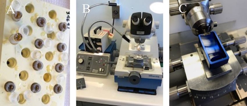
minutes each change. The samples were into en bloc stain (Figure 1(A)) (0.5%
uranyl acetate in veronal acetate buffer) for 1 hour and dehydrated in a series of
ascending acetone concentration [70%, 95%, 100% (X2)] for at least 10 minutes
each. Furthermore, the samples were placed in a 50/50 mixture of acetone and
100% epoxy resin for at least 1 hour on a rotator. The 50/50 mixture was re-
placed with 100% epoxy resin for 30 minutes at room temperature on a rotator.
The second change of 100% epoxy was made and incubated for another 30 mi-
nutes at room temperature on a rotator. The samples were placed into molds
with a label and baked for 24 hours for embedding. Sections of grid were cut
Figure 1. Thin-section process. Samples in bloc (A), ultra-microtome ((B) and (C)), Sec-
tions are cut under water (C).
DOI: 10.4236/aim.2023.131003 34 Advances in MicrobiologyU. B. Iloghalu et al.
with a diamond knife on an ultramicrotome (Figure 1(B) and Figure 1(C)) to
produce thin slices of samples that were semitransparent to electrons. The sec-
tions were about 70 nm thick and stained with uranyl acetate. Uranyl acetate is a
heavy metal that gives quality contrast to the image as electrons are scattered for
good eye contrast. The grids were air-dried and carefully loaded into the electron
microscope (EM) (Phillips CM12) for view while the room was kept dark.
Northern blotting: We employed the northern blotting technique to measure
the amount and size of RNA in both treated and untreated samples [24]. We
employed a previously described method [24] with modifications. Briefly, sam-
ples were run on a denaturing gel to separate RNA according to their sizes (28 S
& 18 S). The agarose gel (Invitrogen, Lot no 00607575) was run at 75 V for 55
mins and then moved to a positively charged Biodyne B Pre-Cut modified nylon
membrane (0.45 µM, Thermo Scientific Rockford, IL) for 2 hours using Nor-
thernMax-Gly kit (Thermofisher, AM1946).
The labeled probe corresponding to the gene of interest was generated with
RT-qPCR as well with traditional PCR products. We used Fotodyne (Incorpo-
rated Foto/Analyst luminary FX with darkroom controls) to determine probe ef-
ficiency. We hybridized the generated probe to the nylon membrane with trans-
ferred RNA in the nylon membrane. Unhybridized probes were removed by
washing in several changes of buffer. The solid nylon membrane with probe pre-
cisely bound to RNA of interest subjected to analysis (data not shown).
The reverse transcriptase of polymerase chain reaction of ORF 1, ORF 2,
and ORF 3 Genes: ORF (1, 2, 3) levels of gene ID 4246735-7 (A-4) were deter-
mined using quantitative reverse-transcriptase real-time polymerase chain reac-
tion (RT-qPCR). ORF (Open Reading Frame) is a norovirus gene which is of
three parts: 1, 2, and 3. Each of them encodes different proteins. ORF1 has the
specific function of encoding polypeptide with regions of close relationship to
cysteine proteinase, helicase, and RNA-dependent RNA polymerase (RdRp)-
encoding regions. ORF2 encodes viral capsid protein (VP1) while ORF3 encodes
small structural protein (VP2) linked with VP1 strength.
Briefly, total RNA was isolated from control and experimental cells using the
isolation RNeasy Mini Kit (50) (Qiagen Inc. catalog number 74104), according
to the manufacturer’s instruction. The total RNA 2 µg was then reverse tran-
scribed using SuperScript-III enzyme (Invitrogen, New York, USA), a 2 pmol of
gene specific primer. A non-viral tag sequence attached at the 5’ end of the
strand specific viral sequence was part of the reverse transcription primer (RT-
primer) (A-8). The RT-qPCR targeted the ORFs (1, 2, and 3) genes using
gene-specific primers (2 pmol) from Integrated DNA Technologies (IDT) (Ref.
no. 187223698). The resulting cDNAs were subjected to RT-qPCR using the
MESA Blue qPCR Master Mix Plus for Syber Assay (Eurogentech, Seraing, Bel-
gium). We used a previously described method for the RT-qPCR [25]. Briefly,
2× Mesa blue master mix and the respective primers were mixed with 2 μl of di-
luted cDNA (A-7). Each strand was at a final concentration of 125 nM before
enzyme activation by incubation at 95˚C for 10 min. The cycling parameters in
DOI: 10.4236/aim.2023.131003 35 Advances in MicrobiologyU. B. Iloghalu et al.
an Eppendorf realplex2 (Mastercycler ep gradient S) real time PCR were as fol-
lows: 50 cycles of 94˚C, 15 secs; 58˚C, 20 secs; 72˚C, 20 sec. Each sample and a
standard curve were run in triplicate to ensure reproducibility.
The ORF 1-3 primers obtained from IDT, Inc. (A-8). ORF 1, 2, 3 PCR primers
(TposGpos:
3’-CGGGAAGGCGACTGGAGTGCCCAAACATCTTTCCCTTGTTC-5),
qPCR-Tpos Forward: 3’-CGGGAAGGCGACTGGAGTGCC-5’, qPCR-RGneg
Reverse: 3’-TGGACAACGTGGTGAAGGAT-5’) and TnegGneg
3’-GGCCGTCATGGTGGCGCGAATAATGGACAACGTGGTGAAGGAT-5’,
and qPCR-FTneg GGCCGTCATGGTGGCGAATAA, qPCR-RGpos
3’-CAAACATCTTTCCCTTGTTC-5’ utilized were custom synthesized. The log10-
transcribed RNA genomic replicas were plotted against the threshold cycle (Ct)
value. The positive amplification control and negative amplification control (Esche-
richia coli) were incorporated in each RT-qPCR run.
Statistical Analysis
Data are shown as means ± standard error (SE). T-test and one-way ANOVA
were performed to compare the means. All statistical analyses were performed
using Graph-Pad Prism version 7. Differences of p < 0.05 were considered sig-
nificant.
3. Result
To evaluate the mechanisms of action of the virus, we examined virus-infected
RAW 264.7 cells by negative staining [18] and thin sectioning. Thin sectioning is
a diagnostic procedure, which enhances beam penetration in tissues and cells.
Cells were centrifuged and fixed with glutaraldehyde to hold them together. Our
result showed that virus-infected cells were left potentially void of all the cell
machinery (Figure 2(A1) and Figure 2(A2)), whereas uninfected cells depict
healthy normal and dividing cells (Figure 2(B1) and Figure 2(B2)). Organelles
like the mitochondria, endoplasmic reticulum, cytoplasmic membrane were evi-
dent in Figure 2(B1) and Figure 2(B2). Treated cells with extracts showed a no-
ticeable difference from the untreated virus-infected cells. The changes in the
treated depicted the gentle restoration of some of the virus eroded cell orga-
nelles. Typical is the presence of cytoplasmic membrane, dense cytoplasm, and
the nucleus (Figure 3). In Figure 4, we showed the presence and the devastative
impact of the murine norovirus in the cytoplasm. Note that this figure consists
of untreated virus-infected cells. Murine norovirus is an RNA non-enveloped
virus. Our RT-qPCR result shows levels of ORF (1, 2 & 3) gene expression in
both treated and untreated cells (Figure 5).
To show effects of the extracts Hibiscus sabdariffa (HS) calyces and Zanthox-
ylum armatum (ZA) seeds on expression of ORF genes during norovirus infec-
tion, active doses of the Hibiscus sabdariffa (HS) calyces and Zanthoxylum ar-
matum (ZA) seed extracts were added to murine norovirus, a surrogate of hu-
man norovirus. Subsequently, the cells were infected with norovirus for 2 hours.
mRNA expression levels for ORF family were investigated by utilizing RT-qPCR
DOI: 10.4236/aim.2023.131003 36 Advances in MicrobiologyU. B. Iloghalu et al.
Figure 2. Electron micrographs of untreated samples. MNV+ RAW264.7 ((A1) and
(A2)); RAW 264.7 cell line only ((B1) and (B2)).
Figure 3. EM micrographs of treated infected cells. Treated with Hibiscus sabdariffa ex-
tracts ((A1) - (A3)) and extracts of Zanthoxylum armatum ((B1) - (B3)).
DOI: 10.4236/aim.2023.131003 37 Advances in MicrobiologyU. B. Iloghalu et al.
Figure 4. Images of murine norovirus in the cytoplasm. Arrow shows murine norovirus.
Figure 5. qPCR representation of treated and untreated samples against MNV. P: Positive
control, N: Negative, SP1 & SN1: Treated with Hs (+ve & −ve), SP2: Treated with Za (+ve
& −ve).
using the MESA Blue qPCR Master Mix. Uninfected cells were used as a negative
control. The infected but not plant extract exposed cells were used as positive
control. Based on the ORF genes expression analysis results, the expression le-
vels were mainly up (Figure 5). The ORF affected by extracts were Hibiscus
sabdariffa (HS) calyces and Zanthoxylum armatum (ZA) seed extracts. Treat-
ment with Hibiscus sabdariffa (HS) calyces and Zanthoxylum armatum (ZA)
seed extracts during norovirus infection resulted in the upregulation or expres-
sion of ORF as indicated by the Ct values. The Ct values associated with the
treated cells (Sp1 and Sp2) were significantly (p < 0.05) lower the Ct associated
with the control cells (Sn1 and Sn2) (Figure 5). The number of expressed gene is
inversely proportional to Ct values. The Ct value is inversely proportional to the
amount of viral nucleic acid in specimens (i.e., lower Ct values indicate higher
amount of virus), so it can be used as a proxy of viral load.
DOI: 10.4236/aim.2023.131003 38 Advances in MicrobiologyU. B. Iloghalu et al.
4. Discussion
Plants naturally contain a wide range of secondary metabolites (phytochemicals)
which indeed protect them from microbial attack [26] [27]. Phytochemicals are
also known to treat an array of human ailments [28] such as cardiovascular dis-
eases [29], diabetes mellitus [30], gastrointestinal tract infection [31], obesity
[32], and periodontal diseases [33]. The mechanism through which the studied
plants destroy or affect the viral unit is not yet known. In this study, murine no-
rovirus acts as a model to human norovirus to evaluate the inhibitory effects of
crude extracts of Zanthoxylum armatum and Hibiscus sabdariffa.
The plants of interest in this study, Hibiscus sabdariffa and Zanthoxylum ar-
matum. H. sabdariffa, have been widely used as herbal medicine and drinks,
food, hot and cold beverages, and as a flavoring agent [34]. The extract has evi-
dently proven to possess antioxidant, neuro-protective [35], antidiabetic, an-
ti-hypertensive [36], anti-cholesterol effects [37]. Zanthoxylum armatum has
been shown to be anthelminthic, stomachic, and carminative [38]. Both the
fruits and seeds control dyspepsia and fever [39].
The objective of this study, therefore, is to determine the mechanism of action
of calyces of Hibiscus sabdariffa and seeds of Zanthoxylum armatum extracts on
murine norovirus. Transmission electron microscopy (TEM) was used to ob-
serve any structural changes to the extract-treated-cell-murine norovirus com-
plex directly. Transmission electron microscopy (TEM) has aided many virus
discoveries—evaluations of virus-host cell interactions as well as diagnosis of
various viral infections [40]. TEM has long been used to discover and describe
the structure of viruses [41]. The use of TEM has also revealed to have an insight
into different virus-cell interactions [42]. In our study, with the help of TEM, we
observed the extract-treated-cell-murine norovirus complex directly. We were
able to see restoration of cytoplasmic membrane, dense cytoplasm, and the nuc-
leus in the treated cells and the control showing grievously eroded by norovirus-
es (Figure 3) suggesting the extracts acted by arresting viral replication.
Negative staining [18] and thin sectioning were both used in our study. Nega-
tive staining is a quick technique used for viewing tiny particles in fluids, typi-
cally viruses [43] [44]. Untreated virus-infected-RAW 264.7 exhibited a higher
infection rate characterized by the dead cells. Also, the mitochondria cytoplasm,
endoplasmic reticulum, ribosome, and nuclei lost their structural integrity (Figure
2 upper). In contrast, uninfected cells were healthy (Figure 2 lower). Loss of
normal cell integrity (organelles) possibly suggests that the cells may have lost
normal physiological processes. Our result agrees with a study of Usutu virus on
human neural cells. The study shows that non-treated cells show higher viral in-
fection rate and more cell death [45].
There was evidence of clathrin-mediated endocytosis, a process through which
cells absorb proteins, viruses, hormones, and metabolites [46] [47]. Nuclear
proteins imported into the nucleus through the nuclear pore complexes (NPCs)
[48]. NPCs are large molecular machines that attach to the nuclear envelope
DOI: 10.4236/aim.2023.131003 39 Advances in MicrobiologyU. B. Iloghalu et al.
[49]. Stages of cell divisions were seen in this group, chromatins which package
RNA were beginning to stick together in the healthy cells. The chromatin aids
cell’s mitosis and avoids chromosome rupture [50] and hence sustains cell RNA
integrity and controls RNA replication and subsequently gene expression [51].
Moreover, the extract treated virus-infected cells showed evidence of change in
ultrastructure. The extract acted as a therapy to the infected cells. Our result
showed that the extract ordered the cell’s machinery in a way that virus could
not further replicate. There was evidence of dense cytoplasm, the distinct nuc-
lear membrane separating the nucleus from the cytoplasm. This simply implies
that these extracts could potentially be used to treat or control norovirus infec-
tion.
Studies have shown that plant extracts have gained a science-based foundation
in the treatment of bacterial diseases [52]. For example, the alcoholic extracts of
Hemidesmus indicus, Leucas aspera, and Tridax procumbens have shown to in-
volve blebbing and leakage of cellular contents and disruption of membrane po-
tential [14] Antifungal activity of Momordica charantia seed extracts is known to
cause a loss of integrity of cell wall, disruption of the cell membrane, and defor-
mation of cells with irregular budding [53]. A study of the antiviral effect of Bra-
zillian Cerrado against the avian metapneumovirus suggested that the extract
acted during the adsorption phase as 99% viral replication was inhibited [54].
Z. armatum has a wide medicinal use. Both the fruits and seeds are typically
used in controlling fever and dyspepsia, anthelmintic, stomachic, and carmina-
tive, asthma, bronchitis. It has been scientifically proven to be anti-oxidative, an-
ti-inflammatory, antimicrobial, anti-tumor, hepatic-protective, piscidal, insecti-
cidal and larvicidal activities [55].
H. sabdariffa is widely used as herbal medicine and drinks, food, hot and cold
beverages, as flavoring agent. H. sabdariffa is known to treat various cardiovas-
cular risk factors including hypertension, hypotension, hyperlipidemia in Jor-
dan, Greece, Brazil, and obesity [56] [57] [58]. The extract of H. sabdariffa has
evidently proven to possess antioxidant, hepatic- and nephroprotective, antidia-
betic, anti-hypertensive, anti-cholesterol effects. Anti-toxicity effect of this plant
has been proven to be culprit for these multifactorial jobs [59].
The Real-time quantitative polymerase chain reaction (RT-qPCR) is the stan-
dard method for gene expression analysis. It is a convenient and fast method
that gives the user the opportunity to monitor the PCR reaction in real time [60].
Gene expression is inversely proportional to Ct values (threshold cycle) [61].
The threshold is a point where threshold line intersects reaction curve and a lev-
el above background fluorescence usually located at the beginning of the expo-
nential phase. Our study has shown that the positive control had a Ct value of 28
whereas the negative control has a Ct value of 38. This suggests more expression
of the gene of interest. We found that higher gene expression of ORF was signif-
icantly associated cells treated were Hibiscus sabdariffa (HS) calyces and Zan-
thoxylum armatum (ZA) seed extracts. The expression of ORF genes could po-
tentially code for proteins thus providing evidence or support the theory of the
DOI: 10.4236/aim.2023.131003 40 Advances in MicrobiologyU. B. Iloghalu et al.
role of restoration of cells organelles in response to plants extracts from the ef-
fect of norovirus infection.
It has been shown that Ct values below 29 cycles indicate much availability of
targeted nucleic acid and Ct values above 38 cycles show very small amounts of
targeted nucleic acids [62]. Our treated samples showed very low Ct values of 10
and 8 (HS and ZA positive) and 15 and 12 (HS and ZA negative), respectively.
These values suggest that the amount of gene expression in ZA is insignificantly
higher than that those in HS. The genome of interest embodies three open read-
ing frames (ORFs), the gene. The ORF1 has the specific function of encoding
polypeptide with regions of close relationship to cysteine proteinase, helicase,
and RNA-dependent RNA polymerase (RdRp)-encoding regions. The second
ORF (ORF2) encodes viral capsid protein (VP1), and ORF3 encodes small
structural protein (VP2) linked with VP1 strength [63]. Treated HS and ZA have
shown approximately three-fold and four-fold changes respectively. The fold
change reflects the impact of the extracts on the ORF 1 and 2 genes. This further
strengthened the activities of macrophages to maintaining structural integrity of
the cell. Murine norovirus acts in different ways to the cells including eroding
the cytoplasmic contents and invariably affecting the intact nucleus. These noro-
virus influences invariably cause changes in gene expression of cells treated with
plant extracts [64]. In another study, doxorubicin involved in enzyme inhibition
and DNA damage subsequently cause changes in gene expression of treated cells
[65]. Viral replication depends on the metabolic pathways of the host cell. Many
viruses have also evolved to modify many host pathways [66]. Studies have
shown that the MNV genome shows a high degree of structural resemblance to
that of human norovirus, with the three essential ORFs encoded inside the hu-
man norovirus genome having a straight homolog in MNV (A-4) [67]. The murine
norovirus genes of interest are three overlapping open reading frames (ORFs). The
ORFs encode cysteine proteinase, helicase (ORF-1), viral capsid (ORF2), and
minor structural protein connected with VP1 firmness [68].
5. Conclusion
Current advances in molecular diagnostics have helped to establish norovirus as
the most common cause of outbreaks of acute gastroenteritis across all ages. Hi-
biscus sabdariffa (HS) calyces and Zanthoxylum armatum (ZA) exhibit antiviral
activities. Cells treated with HS and ZA extracts were associated with dense cy-
toplasm, the distinct nuclear membrane separating the nucleus from the cytop-
lasm from the extract-treated cells which impair replication. Replication was also
affected by the expression of several genes.
Acknowledgements
This research was financially supported in part by Agriculture and Food Re-
search Initiative grant No. 2011-6800-30395 from the USDA National Institute
of Food and Agriculture through the NoroCore project. The authors are grateful
DOI: 10.4236/aim.2023.131003 41 Advances in MicrobiologyU. B. Iloghalu et al.
to Hal Makeel and Dr. Ricardo Vancin from Duke Pathology Department for their
contributions. We also extend our sincere gratitude to Mr. John Teleha for his as-
sistance with the transfer from EndNote to RefWorks citation management.
Conflicts of Interest
The authors declare no conflicts of interest regarding the publication of this pa-
per.
References
[1] Niendorf, S., Jacobsen, S., Faber, M., Eis-Hübinger, A.M., Hofmann, J. and Bock,
C.T. (2016) Steep Rise in Norovirus Cases and Emergence of a New Recombinant
Strain GII.P16-GII.2, Germany, Winter 2016. Eurosurveillance, 22, 30447.
https://doi.org/10.2807/1560-7917.ES.2017.22.4.30447
[2] Cannon, J.L., Barclay, L., Collins, N.R., Wikswo, M.E., Castro, C.J., Magaña, L.C.
and Vinjé, J. (2017) Genetic and Epidemiologic Trends of Norovirus Outbreaks in
the United States from 2013 to 2016 Demonstrated Emergence of Novel GII.4 Re-
combinant Viruses. Journal of Clinical Microbiology, 55, 2208-2221.
https://doi.org/10.1128/JCM.00455-17
[3] Fu, J.-G., Shi, C., Xu, C., Lin, Q., Zhang, J., Yi, Q. and Xing, Z. (2017) Outbreaks of
Acute Gastroenteritis Associated with a Re-Emerging GII.P16-GII.2 Norovirus in
the Spring of 2017 in Jiangsu, China Public Library of Science. PLOS ONE, 12,
e0186090. https://doi.org/10.1371/journal.pone.0186090
[4] Iaconelli, M., Muscillo, M., Della Libera, S., Fratini, M., Meucci, L., De Ceglia, M.
and La Rosa, G. (2017) One-Year Surveillance of Human Enteric Viruses in Raw
and Treated Wastewaters, Downstream River Waters, and Drinking Waters. Food
and Environmental Virology, 9, 79-88. https://doi.org/10.1007/s12560-016-9263-3
[5] Iloghalu, U. and Khatiwada, P. (2015) Phytochemicals: Natural Remedies for Emerging
Viral Infection. Medicinal & Aromatic Plants, 4, Article 213.
[6] Moore, M.D., Goulter, R.M. and Jaykus, L. (2015) Human Norovirus as a Food-
borne Pathogen: Challenges and Developments. Annual Review of Food Science
and Technology, 6, 411-433. https://doi.org/10.1146/annurev-food-022814-015643
[7] Saxena, K., Blutt, S.E., Ettayebi, K., Zeng, X., Broughman, J.R., Crawford, S.E. and
Estes, M.K. (2016) Human Intestinal Enteroids: A New Model to Study Human Ro-
tavirus Infection, Host Restriction, and Pathophysiology. Journal of Virology, 90,
43-56. https://doi.org/10.1128/JVI.01930-15
[8] Rajagopalan, S. and Yoshikawa, T.T. (2016) Norovirus Infections in Long-Term
Care Facilities. Journal of the American Geriatrics Society (JAGS), 64, 1097-1103.
https://doi.org/10.1111/jgs.14085
[9] Lucht, J.M. (2015) Public Acceptance of Plant Biotechnology and GM Crops.
Viruses, 7, 4254-4281. https://doi.org/10.3390/v7082819
[10] Scarpellini, E., Ianiro, G., Attili, F., Bassanelli, C., De Santis, A. and Gasbarrini, A.
(2015) The Human Gut Microbiota and Virome: Potential Therapeutic Implications.
Digestive and Liver Disease, 47, 1007-1012.
https://doi.org/10.1016/j.dld.2015.07.008
[11] Nisa, H., Kamili, A.N., Nawchoo, I.A., Shafi, S., Shameem, N. and Bandh, S.A.
(2015) Fungal Endophytes as Prolific Source of Phytochemicals and Other Bioactive
Natural Products: A Review. Microbial Pathogenesis, 82, 50-59.
https://doi.org/10.1016/j.micpath.2015.04.001
DOI: 10.4236/aim.2023.131003 42 Advances in MicrobiologyU. B. Iloghalu et al.
[12] Shakeri, A., Zirak, M.R. and Sahebkar, A. (2018) Ellagic Acid: A Logical Lead for
Drug Development? Current Pharmaceutical Design, 24, 106-122.
https://doi.org/10.2174/1381612823666171115094557
[13] Mwitari, P.G., Ayeka, P.A., Ondicho, J., Matu, E.N. and Bii, C.C. (2013) Antimi-
crobial Activity and Probable Mechanisms of Action of Medicinal Plants of Kenya:
Withania Somnifera, Warbugia Ugandensis, Prunus Africana and Plectrunthus
Barbatus. PLOS ONE, 8, e65619. https://doi.org/10.1371/journal.pone.0065619
[14] Saritha, K., Rajesh, A., Manjulatha, K., Setty, O.H. and Yenugu, S. (2015) Mechan-
ism of Antibacterial Action of the Alcoholic Extracts of Hemidesmus indicus (L.) R.
Br. ex Schult, Leucas aspera (Wild.), Plumbago zeylanica L., and Tridax procum-
bens (L.) R. Br. ex Schult. Frontiers in Microbiology, 6, Article 577.
https://doi.org/10.3389/fmicb.2015.00577
[15] Tran, T.T., Kim, M., Jang, Y., Lee, H., Nguyen, H., Nguyen, T., Park, H., Dang, Q.
and Kim, J.-C. (2017) Characterization and Mechanisms of Anti-Influenza Virus
Metabolites Isolated from the Vietnamese Medicinal Plant Polygonum chinense.
BMC Complementary and Alternative Medicine, 17, Article No. 162.
https://doi.org/10.1186/s12906-017-1675-6
[16] Raina, H., Soni, G., Jauhari, N., Sharma, N. and Bharadvaja, N. (2014) Phytochemi-
cal Importance of Medicinal Plants as Potential Sources of Anticancer Agents. Tur-
kish Journal of Botany, 38, 1027-1035. https://doi.org/10.3906/bot-1405-93
[17] Domaszewska-Szostek, A., Puzianowska-Kuźnicka, M. and Kuryłowicz, A. (2021)
Flavonoids in Skin Senescence Prevention and Treatment. International Journal of
Molecular Sciences, 22, Article 6814. https://doi.org/10.3390/ijms22136814
[18] Iloghalu, U., Holmes, B., Khatiwada, J. and Williams, L.L. (2019) Selected Plant Ex-
tracts Show Antiviral Effects against Murine Norovirus Surrogate. Advances in Mi-
crobiology, 9, 372-384. https://doi.org/10.4236/aim.2019.94022
[19] Su, X. and D’Souza, D. (2013) Naturally Occurring Flavonoids against Human No-
rovirus Surrogates. Food and Environmental Virology, 5, 97-102.
https://doi.org/10.1007/s12560-013-9106-4
[20] Laue, M. and Bannert, N. (2010) Detection Limit of Negative Staining Electron Mi-
croscopy for the Diagnosis of Bioterrorism-Related Micro-Organisms. Journal of
Applied Microbiology, 109, 1159-1168.
https://doi.org/10.1111/j.1365-2672.2010.04737.x
[21] Garcia, A., Pratap, P.R., Lüpfert, C., Cornelius, F., Jacquemin, D., Lev, B., Allen, T.
and Clarke, R.J. (2017) The Voltage-Sensitive Dye RH421 Detects a Na+, K+-ATPase
Conformational Change at the Membrane Surface. Bochimica et Biophysica Ac-
ta-Biomembranes, 1859, 813-823. https://doi.org/10.1016/j.bbamem.2017.01.022
[22] Carmona-Rivera, C. and Kaplan, M.J. (2016) Induction and Quantification of NE-
Tosis. Current Protocols in Immunology, 115, 14.41.1-14.41.14.
https://doi.org/10.1002/cpim.16
[23] Miller, S.E. (2010) Electron Microscopy of Viral Infections. In: Jerome, K.R., Ed.,
Lennette’s Laboratory Diagnosis of Viral Infections, CRC Press, Boca Raton, 173-196.
https://doi.org/10.3109/9781420084962.011
[24] He, S.L. and Green, R. (2013) Northern Blotting. In: Colowick, S.P. and Kaplan,
N.O., Eds., Methods in Enzymology, Elsevier Science & Technology, United States,
75-87. https://doi.org/10.1016/B978-0-12-420037-1.00003-8
[25] Vashist, S., Urena, L. and Goodfellow, I. (2012) Development of a Strand Specific
Real-Time RT-qPCR Assay for the Detection and Quantitation of Murine Norovi-
rus RNA. Journal of Virological Methods, 184, 69-76.
DOI: 10.4236/aim.2023.131003 43 Advances in MicrobiologyU. B. Iloghalu et al.
https://doi.org/10.1016/j.jviromet.2012.05.012
[26] Soliman, S., Mohammad, M.G., El-Keblawy, A.A., Omar, H., Abouleish, M., Mad-
kour, M. and Hosni, R.M. (2018) Mechanical and Phytochemical Protection Me-
chanisms of Calligonum Comosum in Arid Deserts Public Library of Science. PLOS
ONE, 13, e0192576. https://doi.org/10.1371/journal.pone.0192576
[27] Nunes, M.A., Rodrigues, F., Alves, R.C. and Oliveira, M.B.P.P. (2017) Herbal Prod-
ucts Containing Hibiscus sabdariffa L., Crataegus spp., and Panax spp.: Labeling
and Safety Concerns. Food Research International, 100, 529-540.
https://doi.org/10.1016/j.foodres.2017.07.031
[28] Ashafa, A.O.T. and Alayande, K.A. (2017) Evaluation of Cytotoxic Effects and An-
timicrobial Activities of Lecaniodiscus cupanioides (Planch.) Leaf Extract. Transac-
tions of the Royal Society of South Africa, 72, 33-38.
https://doi.org/10.1080/0035919X.2016.1214851
[29] Šmejkal, K., Malaník, M., Zhaparkulova, K., Sakipova, Z., Ibragimova, L., Ibadul-
laeva, G. and Žemlička, M. (2016) Kazakh Ziziphora Species as Sources of Bioactive
Substances. Molecules (Basel, Switzerland), 21, Article 826.
https://doi.org/10.3390/molecules21070826
[30] Raafat, K.M. and Samy, W. (2018) Phytochemical and Biological Evaluation of Ul-
trasound-Assisted Spray Dried Lonicera Etrusca for Potential Management of Di-
abetes. Records of Natural Products, 12, 367-379.
https://doi.org/10.25135/rnp.40.17.10.171
[31] Anand, S., Mandal, S., Patil, P. and Tomar, S.K. (2016) Pathogen-Induced Secretory
Diarrhea and Its Prevention. European Journal of Clinical Microbiology & Infec-
tious Diseases, 35, 1721-1739. https://doi.org/10.1007/s10096-016-2726-5
[32] Zhang, T., Wen, F., Wu, Y., Goh, G.S.H., Ge, Z., Tan, L.P. and Yang, Z. (2015)
Cross-Talk between TGF-Beta/SMAD and Integrin Signaling Pathways in Regulat-
ing Hypertrophy of Mesenchymal Stem Cell Chondrogenesis under Deferral Dy-
namic Compression. Biomaterials, 38, 72-85.
https://doi.org/10.1016/j.biomaterials.2014.10.010
[33] Varela-López, A., Bullón, P., Giampieri, F. and Quiles, J.L. (2015) Non-Nutrient,
Naturally Occurring Phenolic Compounds with Antioxidant Activity for the Pre-
vention and Treatment of Periodontal Diseases. Antioxidants, 4, 447-481.
https://doi.org/10.3390/antiox4030447
[34] Zheng, D., Zou, Y., Cobbina, S.J., Wang, W., Li, Q., Chen, Y. and Wu, X. (2016) Pu-
rification, Characterization and Immunoregulatory Activity of a Polysaccharide
Isolated from Hibiscus sabdariffa L. Journal of the Science of Food and Agriculture,
97, 1599-1606. https://doi.org/10.1002/jsfa.7908
[35] Rajab, N.F., Musa, S.M., Munawar, M.A., Mun, L.L., Yen, H.K., Ibrahim, F.W. and
Meng, C.K. (2016) Anti-Neuroinflammatory Effects of Hibiscus Sabdariffa Linn.
(Roselle) on Lipopolysaccharides-Induced Microglia and Neuroblastoma Cells. Ke-
san Anti-Neuroinflammatori Hibiscus sabdariffa Linn. (Roselle) Pada Aruhan Li-
popolisakarida Sel Mikroglia dan Neuroblastoma. Jurnal Sains Kesihatan Malaysia,
14, 111-117. https://doi.org/10.17576/jskm-2016-1402-13
[36] Riaz, G. and Chopra, R. (2018) A Review on Phytochemistry and Therapeutic Uses
of Hibiscus sabdariffa L. Biomedicine & Pharmacotherapy, 102, 575-586.
https://doi.org/10.1016/j.biopha.2018.03.023
[37] Hajifaraji, M., Matlabi, M., Ahmadzadeh-Sani, F., Mehrabi, Y., Rezaee, M.S., Haji-
mehdipour, H. and Roghani, K. (2018) Effects of Aqueous Extracts of Dried Calyx
of Sour Tea (Hibiscus sabdariffa L.) on Polygenic Dyslipidemia: A Randomized
DOI: 10.4236/aim.2023.131003 44 Advances in MicrobiologyU. B. Iloghalu et al.
Clinical Trial. Avicenna Journal of Phytomedicine, 8, 24-32.
[38] Purohit, S., Jugran, A.K., Bhatt, I.D., Palni, L.M.S., Bhatt, A. and Nandi, S.K. (2017)
In Vitro Approaches for Conservation and Reducing Juvenility of Zanthoxylum
armatum Dc: An Endangered Medicinal Plant of Himalayan Region. Trees: Struc-
ture & Function, 31, 1101-1108. https://doi.org/10.1007/s00468-016-1494-2
[39] Negi, J.S., Bisht, V.K., Bhandari, A.K., Bisht, R. and Negi, S.K. (2012) Major Con-
stituents, Antioxidant and Antibacterial Activities of Zanthoxylum armatum DC.
Essential Oil. Iranian Journal of Pharmacology & Therapeutics, 11, 68-72.
[40] Roingeard, P. (2008) Viral Detection by Electron Microscopy: Past, Present and
Future. Biology of the Cell, 100, 491-501. https://doi.org/10.1042/BC20070173
[41] Goldsmith, C.S. and Miller, S.E. (2009) Modern Uses of Electron Microscopy for
Detection of Viruses. Clinical Microbiology Reviews, 22, 552-563.
https://doi.org/10.1128/CMR.00027-09
[42] Madani, T.A., El-Tayb, M.E.A., et al. (2017) Electron Microscopy of Alkhumra
Hemorrhagic Fever Virus. Mary Ann Liebert, 17, 195-199.
https://doi.org/10.1089/vbz.2016.2064
[43] Monninger, M.K., Nguessan, C.A., Blancett, C.D., Kuehl, K.A., Rossi, C.A., Olsch-
ner, S.P., et al. (2016) Preparation of Viral Samples within Biocontainment for Ul-
trastructural Analysis: Utilization of an Innovative Processing Capsule for Negative
Staining. Journal of Virological Methods, 238, 70-76.
https://doi.org/10.1016/j.jviromet.2016.10.005
[44] Yu, Y., Tan, Q., Zhao, W., Zhang, X., Ma, J., Wu, Z., Zhu, Z. and Cui, Y. (2017)
Characterization of an Orf Virus Isolated from an Outbreak in Heilongjiang Prov-
ince, China. Archives of Virology, 162, 3134-3149.
https://doi.org/10.1007/s00705-017-3426-x
[45] Carrasco, L. (2018) Mechanisms of Viral Toxicity in Animal Cells. CRC Press, Boca
Raton. https://doi.org/10.1201/9781351074353
[46] Ailte, I., Lingelem, A.B.D., Kvalvaag, A.S., Kavaliauskiene, S., Brech, A., Koster, G.
and Sandvig, K. (2017) Exogenous Lysophospholipids with Large Head Groups
Perturb Clathrin-Mediated Endocytosis. Traffic, 18, 176-191.
https://doi.org/10.1111/tra.12468
[47] Prosser, D.C. and Wendland, B. (2016) DePFth Perception in Clathrin-Mediated
Endocytosis. Developmental Cell, 37, 387-388.
https://doi.org/10.1016/j.devcel.2016.05.017
[48] Ruba, A. and Yang, W. (2016) O-GlcNAc-ylation in the Nuclear Pore Complex.
Cellular and Molecular Bioengineering, 9, 227-233.
https://doi.org/10.1007/s12195-016-0440-0
[49] Lin, D.H., Stuwe, T., Schilbach, S., Rundlet, E.J., Perriches, T., Mobbs, G. and Hoelz,
A. (2016) Architecture of the Symmetric Core of the Nuclear Pore. Science (Ameri-
can Association for the Advancement of Science), 352, aaf1015.
https://doi.org/10.1126/science.aaf1015
[50] Lieberman, P.M. (2008) Chromatin Organization and Virus Gene Expression.
Journal of Cellular Physiology, 216, 295-302. https://doi.org/10.1002/jcp.21421
[51] Wang, X., Ma, Z., Kong, X. and Lv, Z. (2016) Effects of RNAs on Chromatin Acces-
sibility and Gene Expression Suggest RNA-Mediated Activation. The International
Journal of Biochemistry & Cell Biology, 79, 24-32.
https://doi.org/10.1016/j.biocel.2016.08.004
[52] Schmidt, H.B. and Görlich, D. (2016) Transport Selectivity of Nuclear Pores, Phase
DOI: 10.4236/aim.2023.131003 45 Advances in MicrobiologyU. B. Iloghalu et al.
Separation, and Membraneless Organelles. Trends in Biochemical Sciences, 41,
46-61. https://doi.org/10.1016/j.tibs.2015.11.001
[53] Wang, S.Z., Zheng, Y.L., Xiang, F., Li, S. and Yang, G.L. (2016) Antifungal Activity
of Momordica charantia Seed Extracts toward the Pathogenic Fungus Fusarium so-
lani L. Journal of Food and Drug Analysis, 24, 881-887.
https://doi.org/10.1016/j.jfda.2016.03.006
[54] Kohn, L.K., Foglio, M.A., Rodrigues, R.A., Sousa, I.D.O., Martini, M.C., Padilla,
M.A. and Arns, C.W. (2015) In-Vitro Antiviral Activities of Extracts Of plants of
the Brazilian Cerrado against the Avian Metapneumovirus (aMPV). Revista Brasileira
de Ciência Avícola, 17, 275-280. https://doi.org/10.1590/1516-635X1703275-280
[55] Latika, B., Aseesh, P. and Sushma, T. (2013) An Overview on Phytomedicinal Ap-
proaches of Zanthoxylum armatum DC. An Important Magical Medicinal Plant.
Journal of Medicinal Plants Research, 7, 366-370.
[56] Abu-Irmaileh, B.E. and Afifi, F.U. (2003) Herbal Medicine in Jordan with Special
Emphasis on Commonly Used Herbs. Journal of Ethnopharmacology, 89, 193-197.
https://doi.org/10.1016/S0378-8741(03)00283-6
[57] AbouZid, S.F. and Mohamed, A.A. (2011) Survey on Medicinal Plants and Spices
Used in Beni-Sueif, Upper Egypt. Journal of Ethnobiology and Ethnomedicine, 7,
Article No. 18. https://doi.org/10.1186/1746-4269-7-18
[58] Alzweiri, M., Al Sarhan, A., Mansi, K., Hudaib, M. and Aburjai, T. (2011) Ethno-
pharmacological Survey of Medicinal Herbs in Jordan, the Northern Badia Region.
Journal of Ethnopharmacology, 137, 27-35.
https://doi.org/10.1016/j.jep.2011.02.007
[59] Da-Costa-Rocha, I., Bonnlaender, B., Siever, H., Pischel, I. and Heinrich, M. (2014)
Hibiscus sabdafiffa L.—A Phytochemical and Pharmacological Review. Food Che-
mistry, 165, 424-443. https://doi.org/10.1016/j.foodchem.2014.05.002
[60] Wagner, E.M. and Jones, J. (2013) Monitoring Gene Expression: Quantitative
Real-Time RT-PCR. In: Freeman, L., Ed., Lipoproteins and Cardiovascular Disease.
Methods in Molecular Biology, Humana Press, Totowa, NJ, 19-45.
https://doi.org/10.1007/978-1-60327-369-5_2
[61] Nguyen, T.T.-T.N., Shynlova, O. and Lye, S.J. (2016) Matrix Metalloproteinase Ex-
pression in the Rat Myometrium during Pregnancy, Term Labor, and Postpartum.
Biology of Reproduction, 95, 1-14. https://doi.org/10.1095/biolreprod.115.138248
[62] Farrar, J.S. and Wittwer, C.T. (2017) High-Resolution Melting Curve Analysis for
Molecular Diagnostics. In: Patrinos, G.P., Ed., Molecular Diagnostics, Academic
Press, Cambridge, MA, 79-102.
https://doi.org/10.1016/B978-0-12-802971-8.00006-7
[63] Bull, R.A., Hansman, G.S., Clancy, L.E., Tanaka, M.M., Rawlinson, W.D. and
White, P.A. (2005) Norovirus Recombination in ORF1/ORF2 Overlap. Emerging
Infectious Diseases, 11, 1079-1085. https://doi.org/10.3201/eid1107.041273
[64] Fraisse, A., Coudray-Meunier, C., Martin-Latil, S., Hennechart-Collette, C., Delan-
noy, S., Fach, P. and Perelle, S. (2017) Digital RT-PCR Method for Hepatitis A Virus
and Norovirus Quantification in Soft Berries. International Journal of Food Micro-
biology, 243, 36-45. https://doi.org/10.1016/j.ijfoodmicro.2016.11.022
[65] Drozd, E., Krzysztoń-Russjan, J. and Gruber, B. (2016) Doxorubicin Treatment of
Cancer Cells Impairs Reverse Transcription and Affects the Interpretation of
RT-qPCR Results. Cancer Genomics & Proteomics, 13, 161-170.
[66] Sanchez, E.L. and Lagunoff, M. (2015) Viral Activation of Cellular Metabolism. Vi-
rology, 479-480, 609-618. https://doi.org/10.1016/j.virol.2015.02.038
DOI: 10.4236/aim.2023.131003 46 Advances in MicrobiologyU. B. Iloghalu et al.
[67] Herod, M.R., Salim, O., Skilton, R.J., Prince, C.A., Ward, V.K., Lambden, P.R. and
Clarke, I.N. (2014) Expression of the Murine Norovirus (MNV) ORF1 Polyprotein
is Sufficient to Induce Apoptosis in a Virus-Free Cell Model. PLOS ONE, 9, e90679.
https://doi.org/10.1371/journal.pone.0090679
[68] de Graaf, M., van Beek, J. and Koopmans, M.P.G. (2016) Human Norovirus Trans-
mission and Evolution in a Changing World. Nature Reviews Microbiology, 14,
421-433. https://doi.org/10.1038/nrmicro.2016.48
DOI: 10.4236/aim.2023.131003 47 Advances in MicrobiologyYou can also read