Effect of electrochemical oxidation and drug loading on the antibacterial properties and cell biocompatibility of titanium substrates - Nature
←
→
Page content transcription
If your browser does not render page correctly, please read the page content below
www.nature.com/scientificreports
OPEN Effect of electrochemical
oxidation and drug loading
on the antibacterial properties
and cell biocompatibility
of titanium substrates
Fateme Nowruzi1, Rana Imani1* & Shahab Faghihi2*
A combination of TiO2 nanotube array (TON) and controlled drug release system is employed to
provide enhanced surface properties of titanium implants. Electrochemical anodization process is used
to generate TON for introducing, vancomycin, an effective antibacterial drug against Staphylococcus
aureus. TON loaded vancomycin is then coated with a number of layers of 10% gelatin using spin
coating technique. The gelatin film is reinforced with graphene oxide (GO) nanoparticles to improve
the surface bioactivity. The surface of the samples is characterized by field emission electron
microscopy (FESEM), energy-dispersive X-ray spectroscopy (EDS), and contact angle measurement.
The results illustrate that the TON was constructed and vancomycin molecules are successfully loaded.
The drug release study shows that the amount of released vancomycin is controlled by the thickness
of gelatin layers. With an increase in gelatin film layers from 3 to 7, the release of vancomycin in the
burst release phase decreased from 58 to 31%, and sustained release extended from 10 to 17 days.
The addition of GO nanoparticles seems to reduce drug release in from 31 to 22% (burst release
phase) and prolonged drug release (from 17 to 19 days). MTT assay indicates that samples show no
cytotoxicity, and combination of GO nanoparticles with gelatin coating could highly promote MG63
cell proliferation. Soaking the samples in SBF solution after 3 and 7 days demonstrates that hydroxy
apatite crystals were deposited on the TON surface with GO-gelatin coating more than surface of
TON with gelatin. Moreover, based on the results of disc diffusion assay, both samples (loaded with
Vancomycin and coated with gelatin and gelatin-GO) with the inhibition zones equaled to 20 mm
show effective antibacterial properties against S. aureus. The evidence demonstrates that titania
nanotube loaded with vancomycin and coated with gelatin-GO has a great potential for general
applicability to the orthopedic implant field.
Titanium and its alloys are among the most widely used metallic materials in the field of orthopedic and dental
implants1–3. Desirable properties, including tensile strength and elastic modulus, high biocompatibility, and
low risk of allergy make them successful candidate for these a pplications4–6. However, Ti-based implants some
deficiencies including poor osteointegration, low osteogenesis, and bacterial infection in the implant site which
may lead to implants failure especially in prolonged use7–9. Generally, poor osteointegration of Ti-based implants
occurs because of bio-inert surface, production of excessive reactive oxygen species (ROS) at the interface, and
bacterial infection after s urgery10. As a result, two strategies are commonly considered to moderate these prob-
lems including increasing osteoblast cell proliferation and inhibition of bacterial infection11,12.
Long-term intravenous or oral antibiotics are the main traditional approaches to treat bacterial infection after
implant surgery13, which have severe drawbacks including harmful side effects, toxicity, uneven bio-distribution,
and less bioavailability14,15. Staphylococcus aureus is considered the main bacteria that causes implant infec-
tion after surgery16–18. The secretion of these bacteria on implant surfaces forms biofilms, which protect them
against the immune system and antibacterial agents. Consequently, treatment with antibiotics has become a
1
Department of Biomedical Engineering, Amirkabir University of Technology, Tehran 15875/4413, Iran. 2Stem
Cell and Regenerative Medicine Group, National Institute of Genetic Engineering and Biotechnology (NIGEB),
Tehran 14965/161, Iran. *email: r.imani@aut.ac.ir; sfaghihi@nigeb.ac.ir
Scientific Reports | (2022) 12:8595 | https://doi.org/10.1038/s41598-022-12332-z 1
Vol.:(0123456789)
www.nature.com/scientificreports/
vital issue19. Since bacteria and osteoblast cells are in competition for attachment, implants having antibacterial
surface characteristics can decrease bacterial attachment and colony formation so they can be used for treating
bone defects20,21. Advanced biomaterials equipped with sustained and localized release of antibacterial agents
could promote the healing/regeneration process of tissues which are more susceptible to bacterial infections
(e.g. bone, skin, cardiac tissue)22–25.
With the convergence of material science and biology, a combination of surface modification and drug
delivery systems can be employed to tackle the above mentioned complications. The electrochemical anodizing
is a prevalent process among surface modifications that constructs TiO2 nanotube arrays (TON) on titanium
substrates26–29. TON arrays have beneficial properties including hosting a vast range of drugs and higher biocom-
patibility due to their highly porous nanostructures. Moreover, their fabrication is low cost and simple29–33. The
main disadvantage of TON arrays as drug nano-reservoirs is uncontrollable drug release behavior that may cause
toxicity in the implant site. However, modified TON, such as TON coated with biopolymers, exhibits enhanced
drug release b ehavior34–36. TON coated by biopolymers can provide more controllability on drug release behavior
by selecting a biopolymer based on composition and degradation rate properties37. In addition, the polymer
coating can inherently have osteointegration and antibacterial properties38–40, and they can also carry a second
drug as a multidrug s ystem41–45.
In this study, in order to delay the drug release while enhancing the bioactivity of titanium substrates, a
multilayer nanocomposite hydrogel was used as a cover on the TON surface. Among the biocompatible hydro-
gels, gelatin with FDA approval serves as the most important biopolymer which frequently used in biomedical
applications, especially in tissue engineering and drug delivery r esearches46. According to literatures and our
previous experience, GO, as hydrophilic nanoparticle provides many advantages in combination with hydrogels
as a nanocomposite s tructure47,48. In dental and bone tissue engineering, GO nanoparticles enhance osseointe-
gration/osteoblast bioactivity via acceleration of apatite f ormation49. In addition, graphene and its derivatives
show great potential for osteogenic differentiation of stem c ells50 and have been suggested as a coating agent on
the orthopedic implants51,52. Here, we aimed to superpose gelatin and GO properties in order to modify TON
surface where the coated gelatin/GO multilayer structure improves osteogenic properties at the same time as
delaying drug release from nanotube reservoir.
For this purpose, TON arrays were formed by anodizing process on the surface of titanium substrates and
Vancomycin, an antibacterial drug against S. aureus was introduced into nanotube a rrays53,54. Graphene oxide-
gelatin nano-composite was used to coat TON arrays loaded with Vancomycin. The modified titanium samples
then were characterized by field emission scanning electron microscopy (FESEM), energy-dispersive X-ray
spectroscopy (EDX), wettability measurement, and degradability. In addition, the drug release behavior was
assessed on samples having different layers of coating. The effect of surface modification of titanium samples was
assessed on the growth of MG63 cells and formation of hydroxyapatite crystals by MTT assay and bioactivity
test. Finally, disc diffusion assay was employed to elucidate antibacterial activity of the samples.
Experimental
Materials. Commercially available pure titanium (CP-Ti-grade 2) used in this study were supplied by
McMaster Carr Company, Los Angeles, CA, USA. Graphene oxide was obtained from Central Laboratory of
Amirkabir University, Iran, Tehran. Vancomycin Hydrochloride was purchased from Daana pharmaceutical
company, Iran, Tehran. Dulbecco’s modified eagle medium (DMEM) and trypsin were purchased from Gibco
BRL (France). Fetal bovine serum (FBS), MTT, DMSO, PBS, and penicillin/streptomycin (PS) were purchased
from Sigma-Aldrich. Simulated body fluid (SBF) was provided by APATECH, Iran, Yazd. Other chemicals were
supplied by Merck, Germany.
Preparation of TiO2 nanotube arrays (TON). Highly-ordered titania nanotube arrays were fabri-
cated on titanium discs of 10 mm diameter using electrochemical anodization method. The samples were first
grounded with SiC abrasive paper (grit size: P800, P1000, P1200, and P1500) and sequentially cleaned in ethanol
70%, acetone, and deionized water using an ultrasonic bath for 25 min. The samples were dried at ambient tem-
perature before anodization. For anodization process, stainless steel and titanium discs having smooth surfaces
were used as cathode and anode electrodes, respectively. The cell was connected to DC power supply (PEQ lab,
EV843, made in Belgium and ethylene glycol, 38 wt% ammonium fluoride, and 2 vol% deionized water was used
as electrolyte solution. Processing parameters including temperature, voltage, and time have significant effects
on the structure of anodized titanium. The anodization process was carried out at voltages of 45, 60, 70, and 95,
for 1.5 and 3 h (Table 1), at a controlled temperature (4 °C). All anodized samples were ultrasonically cleaned
in ethanol and acetone for a minute and rinsed with deionized water. Finally, samples were dried at room tem-
perature.
Vancomycin loaded into TON arrays on Ti surface. The Vancomycin was introduced into titania
nanotubes according to a method of pipetting and drying. 25 mg/ml solution of Vancomycin in phosphate buffer
saline (PBS) was prepared and 10 µL of drug solution was pipetted onto nanotubes. The samples were dried at air
temperature and cleaned with soft tissue. Finally, to remove the drug residues, the samples were rinsed with the
PBS solution. To enhance the drug loading efficiency and achieve the required concentration, the drug loading
process was repeated four times.
Coating of gelatin‑based film on TON arrays. Gelatin-based films with different thicknesses were
coated on titania nanotube substrates using spin-coating method. A 10 wt% gelatin aqueous solution was pre-
pared and spin-coated on the substrates at a speed of 3800 RPM for 60 s. The polymer film thickness would affect
Scientific Reports | (2022) 12:8595 | https://doi.org/10.1038/s41598-022-12332-z 2
Vol:.(1234567890)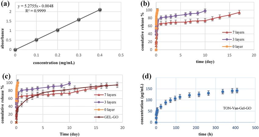
www.nature.com/scientificreports/

                                  Voltage (V)    Time (h)
                                  45             1.5
                                  45             3
                                  60             1.5
                                  60             3
                                  70             1.5
                                  70             3
                                  90             3

                                 Table 1.  Time and voltage used in anodizing process.

                                  Samples                                                                              Abbreviations
                                  TiO2 nanotube arrays (Titania nanotube)                                              TON
                                  TiO2 nanotubes loaded with Vancomycin                                                TON-Van
                                  TiO2 nanotubes loaded with Vancomycin and coated with Gelatin 10% (3 layers)         TON-Van-Gel C3
                                  TiO2 nanotubes loaded with Vancomycin and coated with Gelatin 10% (7 layers)         TON-Van-Gel C7
                                  TiO2 nanotubes loaded with Vancomycin and coated with gelatin10%-GO0.5% (7 layers)   TON-Van-Gel-GO

                                 Table 2.  Samples and related abbreviations.

                                 sustained release behavior of the drug, therefore, samples with varied coating layers were prepared to obtain
                                 desired drug release profile. To modify gelatin film, GO was added as an agent. For GO/gelatin composite syn-
                                 thesis, GO nano-powders were first dispersed in deionized water using an ultrasonic homogenizer for 30 min.
                                 The gelatin10%-GO0.5% solution was then prepared and spin-coated on the samples. All the samples were
                                 subsequently glutaraldehyde vapor treated with glutaraldehyde 25%, at 25 °C. To remove excess glutaraldehyde,
                                 samples were then immersed in a glycine aqueous solution (7.5 mg/mL) for 5 min and dried at room tempera-
                                 ture. Finally, for sterilization of the samples Ultraviolet radiation was used for 20 min. Table 2 lists the samples
                                 with their abbreviations.

                                 Surface characterization. Field emission scanning electron microscopy (FESEM) was utilized to observe
                                 the morphology of anodized samples as well as gelatin-based coated ones. After the samples were coated with
                                 an ultrathin gold layer via sputter coater (Emitech, K450X), images were captured at an accelerating voltage of
                                 25 kV in high vacuum mode. The titania nanotube dimensions were analyzed using the Digimizer software. The
                                 non-destructive ellipsometry (SENpro, SENtech, Germany) test and SEM analysis were performed to measure
                                 the thickness of polymer coating. Energy-dispersive X-ray spectroscopy (EDS) was used to determine the types
                                 of elements in the samples (TON and TON-Van), especially to verify the presence of Vancomycin.

                                 Wettability measurement. The sessile water drop technique was used to measure the static contact angle
                                 and wettability of the samples. Deionized water droplet (0.5 µL) was firstly deposited on the top surface of
                                 samples through micro-syringe. Images were then captured and the static contact angle was measured using
                                 CAG-20, Jikan Co. The experiment measured the average contact angle at five different points in each sample at
                                 ambient temperature.

                                 Degradability evaluation. Degradability of coating has a direct effect on drug release behavior of the sam-
                                 ples. The degradation of biopolymers is related to their composition, structure, charge, and surface properties. In
                                 general, weight loss measurement is the most common method to investigate hydrogel degradation. However, in
                                 this study, weight loss measurement was not applied because there was a significant difference between coating
                                 weight and titanium weight. Therefore, degradability was evaluated by comparing the SEM images of coating
                                 before and after drug release in PBS solution. Samples were placed in clean and sterile glass bottles containing
                                 PBS. Then they were sealed and placed in an incubator (GFL, Germany) at 37 °C, at a speed of 60 RPM. The
                                 degradation was measured after 10 and 17 days. Eventually, freeze-drying was performed to remove the water
                                 that was absorbed by the hydrogel coatings.

                                 Vancomycin release. In-vitro release of Vancomycin from bare TON, Gelatin coated, and gelatin-GO
                                 coated samples loaded with the drug were investigated by immersion into 10 mL of PBS (pH = 7.4) at 37 °C. Dur-
                                 ing the first 6 h, 300 µL of the medium was taken out at short intervals to monitor the burst release of the drug.
                                 For delayed release of the drug, 300 µL PBS solution was collected every 24 h until the whole drug was released
                                 into the medium. The calibration curve was plotted based on the Vancomycin in PBS, the release of the drug
                                 was measured by UV-spectroscopy (Lambda 900, PerkinElmer, USA) at 280 nm. The release was considered

Scientific Reports |   (2022) 12:8595 |                  https://doi.org/10.1038/s41598-022-12332-z                                                3

                                                                                                                                              Vol.:(0123456789)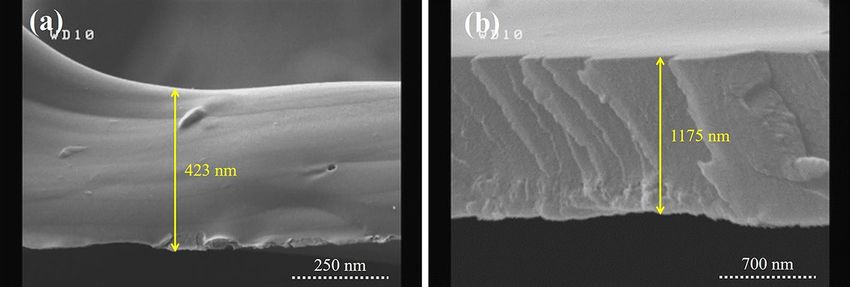
www.nature.com/scientificreports/
complete when there was no change in the absorbance at 280 nm. The data were analyzed using mathematical
kinetic modeling.
Cell culture. The osteosarcoma cell line Human Osteoblast-Like cells (HOS) MG-63, from the National
Cell Bank of Iran (NCBI; Pasteur Institute), was used for cell culture experiments. The cells were cultured in a
culture medium (DMEM supplemented with 10% FBS, 1% penicillin/streptomycin) at 37 °C in the humidified
incubator with 5% CO2. After the culture reached approximately 90% confluence, MG-63 cells were removed
from culture flasks by trypsinization and centrifuging (1500 RPM, 5 min). They were then suspended in the
fresh medium.
Cell viability. Samples including TON, TON-Van-Gel C7, TON-Van-Gel-GO and culture plate as a control
were sterilized by UV and washed with PBS and culture medium before they were placed in a 24-well plate. Then,
200 µL of culture medium containing 80,000 cells were seeded onto the surface of the samples. After incubation
for 3 h, 1 mL of culture medium was added to each well. Subsequently, after 1, 3, and 7 days of incubation, to
remove the unattached cells, the culture medium was removed and samples were rinsed with sterile PBS twice
and incubated with fresh culture medium supplemented with 1 mg/mL MTT solution for 4 h to allow formazan
formation. After removing the medium, 50 µL of dimethyl sulfoxide (DMSO) solution was added to each well
and incubated for 10 min to dissolve the formazan crystals. The medium was transferred to a 96-well plate to
measure the absorbance of the resulting purple solution using a micro-plate reader at 580 nm. The cell viability
directly depends on the amount of formazan crystals, calculated by dividing the average optical density of sam-
ples by the average optical density of control.
Bioactivity. Samples (TON-Van-Gel and TON-Van-Gel-Go) were evaluated by soaking in 5 mL of SBF
solution similar to human blood plasma. The samples were kept at 37 °C for 3 and 7 days. The medium was
replaced every 2 days to avoid insufficient presence of ions (Ca and P). The samples were removed from the
SBF solution and dried using freeze-drying. The structure and morphology of the SBF-immersed samples were
characterized by FESEM.
Antibacterial assay. A zone of inhibition test, also called a Kirby-Bauer Test, was utilized to investigate the
antibacterial characteristics of the samples. Gram-positive S. aureus (ATCC 25923), one of the most infectious
bacteria in bone tissue engineering was employed. Plates containing Mueller–Hinton agar medium (Merck, Ger-
many) were spread with S. aureus at a concentration of 1.5 × 10⁸ CFU/mL. Then, the samples (TON-Van-Gel-GO
and TON-Van-Gel C7) were placed gently into the agar nutrient. Ciprofloxacin antibiotic discs were placed on
the plates as a positive control. Finally, the plates were incubated for 24 h at 37 °C in order for bacteria to grow
in agar media. The size of the inhibition zone that appeared around the discs indicates the antibacterial activity
of each sample.
Statistical analysis. Statistical analysis was carried out using SPSS software (v 17.0; IBM New York, NY,
USA) when statistical differences were detected, a t-student comparison test was performed. Data are reported
as mean ± SD at a significance level of p < 0.05.
Results and discussion
Fabrication and characterization of titania nanotube arrays. Recently, more attention has been
focused on the electrochemical anodization to construct self-order titania nanotube structures. Strong adher-
ent nanotube structures with open tops and closed ends are the most favorable drug nano-carrier systems.
The parameters such as voltage and time of the anodization process have the main impact on the morphology
of titanium dioxide. Here, the impact of anodizing parameters on the morphology of titania, which has great
importance for the consequent drug loading was investigated. The anodizing process with different voltages (45,
60, 70, 90 V) and various times (1.5, 3 h) was carried out. The experiments were performed in the electrolyte
containing ammonium fluoride at the controlled temperature (4 °C). The first reaction of anodizing oxidation
was the electrolysis of water (reaction 1). A compact layer of titanium dioxide was then formed on the titanium
substrates (reaction 2) which was dissolved by fluoride ions, subsequently. As the results, the pits were formed on
the titanium surfaces (reaction 3). At the suitable voltage and time, pits were converted into nanotube structures.
At the lowest applied voltage (45 V), the pits with infinitesimally small diameters were formed on the Ti surfaces,
however, the nanotube morphology was not apparent (Fig. 1a). When the voltage was increased to 60 V, the
titania nanotubes were formed, yet they were not highly ordered (Fig. 1b). At the voltage of 70 V, the nanotubes
showed a more distinguishable morphology with increased diameter (Fig. 1c).
2H2 O ↔ O2 + 4H+ + 4e (1)
Ti + O2 ↔ TiO2 (2)
TiO2 + 6F− + 4H+ ↔ [TiF6 ]2− + 2H2 O (3)
With increasing time of anodizing from 1.5 to 3 h, no significant changes were observed in the nanotube
structure at the low voltage (45 V) (Fig. 1d). In the higher voltage though, extending the anodizing time made
Scientific Reports | (2022) 12:8595 | https://doi.org/10.1038/s41598-022-12332-z 4
Vol:.(1234567890)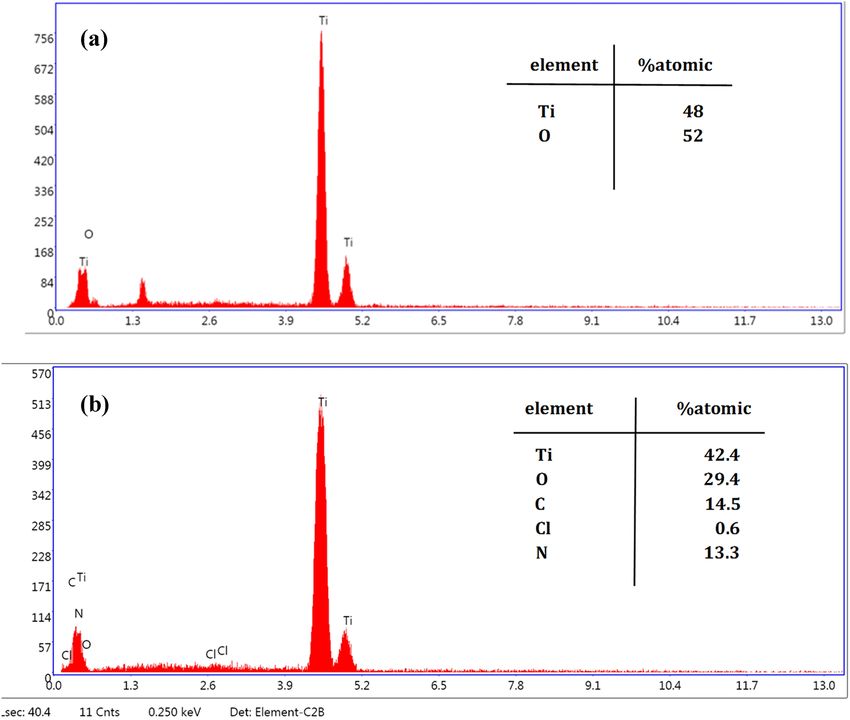
www.nature.com/scientificreports/

                                 Figure 1.  SEM images of samples after anodizing process with different voltages and times (a) 45 V for 1.5 h,
                                 (b) 60 V for 1.5 h, (c) 70 V for 1.5 h, (d) 45 V for 3 h, (e) 60 V for 3 h, (f) 70 V for 3 h, (g) 90 V for 3, (h) opened
                                 top and (i) closed bottom of TON array constructed by anodizing process with voltage of 70 V and 3 h.

                                 the nanotubes provided a more intrinsic structures (Fig. 1e,f). Increasing the voltage to 90 V was gradually
                                 destroyed the top portion of the nanotubes (Fig. 1g).
                                    A more desirable morphology of nanotubes was obtained at the anodizing voltage of 70 for 3 h, where the
                                 SEM images revealed that they possess a diameter of about 94 ± 4 nm and length of about 3 µm, aligned vertically
                                 and highly-ordered with opened top and closed bottom (Fig. 1h,i).

                                 EDS analysis of TON and TON‑Van. EDS was utilized to prove that Vancomycin was loaded into tita-
                                 nia nanotube arrays. Chemical compositions of samples (TON and TON-Van) were detected, as illustrated by
                                 Fig. 2a,b. Ti and O elements could be seen in both samples, confirming TiO2 formation on the surface of tita-
                                 nium samples. The presence of Cl, N, and C in the EDS spectrum of TON-Van (Fig. 2a) indicates that Vancomy-
                                 cin (C66H75Cl2N9O24) has been successfully loaded into the nanotube arrays.

                                 Current transient curve and anodizing process evaluation. Current transient curves demonstrate
                                 the behavior of current versus time during the anodizing process. This curve can help to explain the formation
                                 of titania structure during the process. Figure 3 displays the current transient curve at the voltage of 70, for 3 h,
                                 at the temperature of 4 °C. The application of initial voltage showed a sharp rise up to a which is attributed to the
                                 electrolysis of water and the beginning of the formation of the compact oxide layer. The observed gas evolution
                                 in the anode cell seems to be related to the transfer of electric charge and indicates the electrical conductivity of
                                 the titanium surfaces. Subsequently, the current declines sharply due to the formation of a dense and compact
                                 oxide layer with a low electrical conductivity. As the transfer of electric charge decreased, ion transportation
                                 in the electrolyte increased. At the next step the compact layer was dissolved by fluoride ions and pores in the
                                 dense layer were nucleated which causes a slight increase in the current, followed by a dropdown. However,
                                 the small increase in current was not observed here due to the fast ion transportation (especially fluoride ions)
                                 and rapid dissolution of the oxide layer. The other reason could be the limitation on the time resolution of our
                                 measurement device to record the rapid changes in the current. The decrease in current would gradually turn to
                                 a plateau. There was a competition between oxidation and dissolution of the oxide layer which controls the ano-
                                 dizing process. Nanotubes were formed on the compact oxide layer as the anodization proceeds. As can be seen
                                 in Fig. 3, current increases to higher values with the increase of voltage. This is due to the increase of electron
                                 exchange during the anodization process. As the voltage of the anodization process increases, the oxidation rate
                                 improves and the exchange of electrons and current density enhances.

                                 Water contact angle measurement. Surface hydrophilicity or wettability of implants has a significant
                                 impact on cell behavior. Moreover, the water-soluble drug loading efficiency also improves with increasing wet-

Scientific Reports |   (2022) 12:8595 |                https://doi.org/10.1038/s41598-022-12332-z                                                       5

                                                                                                                                                  Vol.:(0123456789)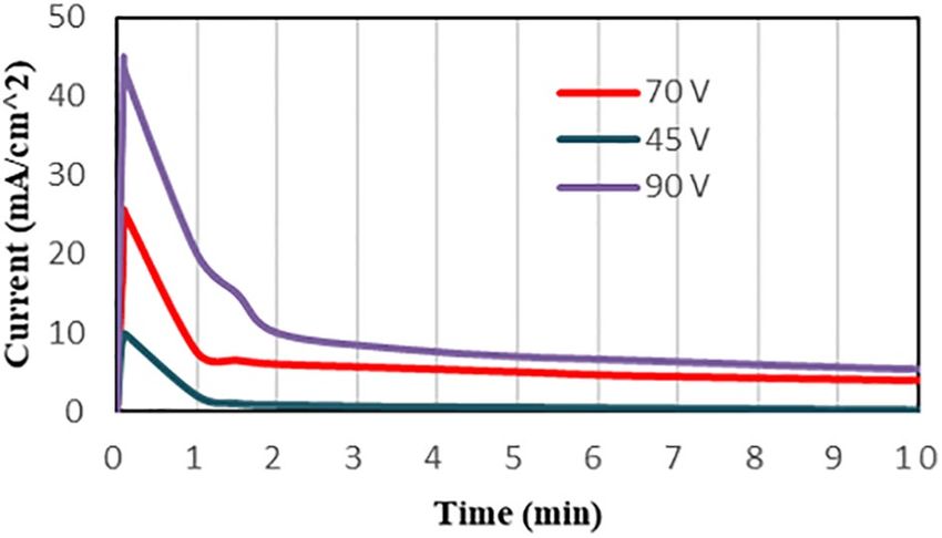
www.nature.com/scientificreports/

                                           Figure 2.  EDS spectrum of (a) TON, (b) TON-Van.

                                           Figure 3.  Transient curve at voltages of 45, 70, and 90 V for 3 h (4 °C).

                                           tability. Therefore, contact angle measurement, the primary way to investigate the degree of affinity between
                                           water and the surfaces, becomes vital to characterize the surfaces of drug delivery implants. Here, the wettability
                                           of samples (TON, TON-Gel C7, and TON-Gel-GO) was studied and depicted in Table 3. The TON was hydro-
                                           philic due to water penetration into the nanotubes. The contact angle of TON-Van-Gel C7 and TON-Gel-Go
                                           were 71.9° and 70.2°, respectively, which are in a similar hydrophilic range (smaller than 90°) (Fig. 4a,b). The
                                           similar contact angle indicates that even by modifying gelatin with graphene oxide as a hydrophilic agent, the
                                           contact angle was unchanged. This could be partly due to graphene oxide nanoparticles which fill and block the
                                           micro-pores among the gelatin molecules. The micro-pores structure provides the channels for penetration and
                                           absorption of water molecules. This could also be related to the strong crosslink between the chains of polymer
                                           which prevents the water absorption.

          Scientific Reports |   (2022) 12:8595 |               https://doi.org/10.1038/s41598-022-12332-z                                                  6

Vol:.(1234567890)
www.nature.com/scientificreports/
Samples Contact angle (degree)
TON < 5°
TON-Van-Gel C7 71.9 ± 1.34
TON-Van-Gel-GO 70.2 ± 0.28
Table 3. Water contact angle measurements of samples.
Figure 4. Water contact angle measurements of (a) TON-Gel, (b) TON-Gel-GO.
Figure 5. SEM images of TON-Van-Gel C7 soaked in PBS and freeze dried (a) before drug release (b) after
10 days, and (c) after 17 days.
In‑vitro degradation of gelatin‑coating. The degradation of coating is an important factor for drug
release behavior, therefore, the in-vitro degradation of the TON-Van-Gel C7 was evaluated. FESEM was utilized
to observe the changes in the microscopic morphology of gelatin coating during the degradation process in PBS.
Figure 5a reveals the dense structure of gelatin coating with some cracks before degradation. These cracks are
probably due to the process of freeze-drying. Figure 5b,c show no holes and collapse in the surface coatings after
10 and 17 days which implies the dense structure of the coating possibly due to the strong crosslink between the
chains of polymer.
Vancomycin release. The effect of gelatin thickness on the release of Vancomycin was investigated from
TON-Van, TON-Van-Gel C3, and TON-Van-Gel C7. The thickness of 3 and 7 layers of gelatin was 445 nm and
1038.06 nm, obtained by ellipsometry. As it can be seen in Fig. 6a,b, the ellipsometry results were confirmed
by the SEM cross sectional images. Figure 7a shows the Vancomycin calibration curve (in PBS, at 280 nm) that
used to obtain Vancomycin concentrations. The total amount of Vancomycin loaded into titania nanotubes was
measured 490 µg/cm2 using a UV–Vis spectrophotometer. Figure 7b presents a comparative drug release profile
of Vancomycin from TON-Van, TON-Van-Gel C3, and C7. As anticipated, the release behavior of Vancomycin
from all the samples displayed biphasic behavior, including a burst release and sustained release. Drug molecules
in the upper part of the nanotubes were released immediately in the media at the initial stage (burst release
phase), inhibiting the bacterial invasion and improve the antibacterial efficacy in early hours of implantation.
Drug release profile of the samples are provided in Table 4. In the burst release phase, the Vancomycin released
from TON-Van was approximately 83% in the first hour whereas the released drug from TON-Van-Gel C3
and C7 were 58% and 31%, respectively. In the sustained release phase, the drug was released from TON-Van,
TON-Van-Gel C3, and C7 in 1, 10, and 17 days, respectively. The slower release of Vancomycin from TON-Van
in the second stage demonstrates that the nanotube structure could act as a nano-reservoir for the drug. In addi-
tion, the results show that the gelatin-coating reduced the amount of released drug in the burst release phase.
With increasing the number of layers of spin-coated gelatin, the thickness would increase which resulted in the
significant decrease in the rate of drug release. This is probably because the polymer chains could restrict the
Scientific Reports | (2022) 12:8595 | https://doi.org/10.1038/s41598-022-12332-z 7
Vol.:(0123456789)www.nature.com/scientificreports/
Figure 6. Cross sectional SEM images of Gelatin-coating (a) 3 layers and (b) 7 layers.
Figure 7. (a) Calibration curve of serial dilutions of Vancomycin in PBS, (b) Vancomycin release profile from
different layers of gelatin coating, (c) Vancomycin release profile from gelatin-GO coating and (d) concentration
of released drug from TON-Gel-GO.
% Drug release
Sample 1h 1 day 7 days Drug totally released (day)
TON-Van 83.47 100 100 1
TON-van-Gel C3 58.28 80.56 90.98 10 ± 1
TON-van-Gel C7 31.13 66.28 71.92 17 ± 1
TON-van-Gel-GO 22.34 45.9 79.17 19 ± 1
Table 4. Vancomycin release profile from different samples.
movement of drug molecules and limits their release. The limitation in the rate of drug release would extend the
time which provides more control on drug release profile that enhances the antibacterial property of the samples.
The effect of graphene oxide on the release profile of the Vancomycin was investigated by TON-Van-Gel-
GO. As it can be seen in Fig. 7c, a biphasic behavior for GO-gelatin sample was detect. The percentage of drug
release was decreased from 31 to 22% in the burst release stage by adding the GO nanoparticles (Table 4). The
negative charge of graphene oxide and carboxyl groups could electrostatically interact with chains of gelatin.
Hence, the physical links between gelatin chains create difficulty for Vancomycin molecules to get released into
PBS which results in prolonged release period. In fact, the GO-gelatin coating could prolong Vancomycin release
up to 19 days. In comparison, the GO-gelatin coated samples could provide longer and sustained drug release
required for successful anti-infection bone therapy as compared to TON-Van and TON-Van-Gel C7 samples.
Scientific Reports | (2022) 12:8595 | https://doi.org/10.1038/s41598-022-12332-z 8
Vol:.(1234567890)www.nature.com/scientificreports/
Zero order Qt = Q0 + k0 t
First order ln Qt = Q0 − k1 t
Higuchi Qt = kH t 0.5
Hixson-Crowell Q0
1/3 1/3
− Qt = Khc t
Table 5. The mathematical function equation used to fit the release data.
Kinetic model Samples First order Zero order Hixson-Crowell Higuchi
R2 TON-Van-Gel C7 0.8346 0.7097 0.8239 0.8053
R2 TON-Van-Gel-GO 0.9703 0.8115 0.9281 0.9561
R2 TON-Van 0.9647 0.7333 0.911 0.8711
Table 6. R2 values of Vancomycin-delivery kinetics.
Figure 8. Cell viability of MG-63 cells cultured on TON-Van, TON-Van-Gel, TON-Van-Gel-GO at different
time point using MTT assay. (*p < 0.05, **p < 0.01, ***p < 0.001, n = 3).
In order to have an effective local drug delivery system, the concentration of antibacterial drug release in the
media should be blow the toxic level and above the minimum inhibitory concentration (MIC). Here, Vancomycin
did not show toxic behavior at 800 µg/mL for MG-63 Cells. Moreover, Vancomycin has been reported to have a
MIC of 0.5–10 µg/mL for S. aureus55–59. As shown in Fig. 7d, the Vancomycin concentration for TON-Van-Gel-
GO in the media was within the therapeutic window. This could mean that the drug delivery system should be
able to kill the bacteria and avoid the formation of biofilm without interference with the cellular process.
Mathematical modeling was used to investigate the mechanism of drug release and analyze the release kinet-
ics. Model-dependent methods based on different mathematical functions were employed to select the best fit
for Vancomycin release profile with a higher value. Table 5 summarizes the equation of mathematical functions
used to fit the release experiments data including zero-order, first-order, Higuchi, and Hixson-Crowell. The
obtained R2 values for the samples (TON-Van, TON-Van-Gel C7, and TON-Van-Gel-GO) are listed in Table 6.
The comparison between the R2 values revealed that the Vancomycin release was perfectly fitted with the first-
order model in all the samples. This is an indication for water-soluble drug release from a porous matrix or a
matrix with diffusion-controlled release system.
Cell viability. The cell viability and proliferation on the surface of the samples were evaluated by MTT assay.
Figure 8 shows MG-63 cell proliferation after 1, 3, and 7 days of culture. As it can be seen, no statistically signifi-
cant difference between TON without coating and control sample was observed. Moreover, the number of viable
cells on the surface of TON-Van was significantly lower than the control after the day 1. The gelatin-based coat-
ing showed to remarkably improve cell viability and the addition of GO nanoparticles had a beneficial impact
on cell viability of the samples. It is known gelatin could provide cells with the biomimetic bone environment
since it is the major protein of the extracellular matrix. Gelatin has the arginine–glycine–aspartic acid (RGD)
sequence which is known for establishing cell-substrate interactions. Moreover, the dispersion of GO nanoparti-
cles into gelatin increases the chance to create more cellular niches for osteoblast cells due to the enlargement of
the surface area and the increase of surface roughness. It is known that the surface oxygen-containing functional
groups trigger the adsorption of the surrounding serum proteins on the surface. This could be the reason that
GO-enriched surfaces would absorb the exogenous proteins that results in eliciting efficient interactions with the
Scientific Reports | (2022) 12:8595 | https://doi.org/10.1038/s41598-022-12332-z 9
Vol.:(0123456789)www.nature.com/scientificreports/
Figure 9. SEM images of TON coated by gelatin with and without GO soaking in SBF solution for different
times (a) TON-Gel for 3 days (b) TON-Gel-GO for 3 days, (c) TON-Gel for 7 days, and (d) TON-Gel-GO for
7 days.
cells51. It is shown that the presence of GO nanoparticles could engage in gene expression profile and upregulate
mRNA expression levels of all osteogenic markers60,61.
In addition, GO nanosheets could interfere in apatite nucleation and hydroxyl apatite (HA) formation. It
is shown by Wang et al. this could be originated from ion interactions. The –OH and carboxyl groups from
GO could attract C a2+ and HPO4 2- of microenviroment solution resulting in the accelerated hydroxy apatite
formation50.
The TON-Van-Gel-GO sample showed the best cell substrate interactions with MG-63 cells. Here, Vancomy-
cin at 800 µg/mL showed about 80% cell viability at day 1. Therefore, the results show no cytotoxicity whether
by the samples or the dosage of the drug used according to ISO-10993-5.
Bioactivity. Previous studies have demonstrated that both pure titanium and TON surfaces are bio-inert62–64.
FESEM was utilized to determine the formation of bone-like apatite on the samples (Gelatin and Gelatin-GO)
soaked in SBF solution for 3 and 7 days. As shown in Fig. 9a, a few apatite nucleates were formed on the sur-
face of gelatin coated sample after 3 days whereas GO-gelatin coated sample was covered by flake-like apatite
(Fig. 9b). The negative charge of GO with deprotonation of –COOH and –OH groups could attract Ca2+ ions
and enhance the apatite nucleation50. After 7 days of immersion in SBF, the amount of apatite crystals on both
samples were increased (Fig. 9c,d). However, the mineralization on the surface of GO reinforced sample was
higher than the sample without GO, suggesting that the sample coated by gelatin and reinforced with GO has the
highest bioactivity among the samples. The amino groups in gelatin can attract calcium ions followed by PO3− 4
which could results in accelerated biomineralization process. GO also promotes the flake-like hydroxyapatite
deposition through electrostatic interaction. It is fair to assume that TON-Van-Gel-GO sample, could provide a
more stable connection between the surface of samples and bone tissues.
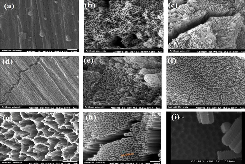
The antibacterial assay. The antibacterial characteristic of the samples was investigated by disc diffusion
assay after 24 h of incubation against gram-positive S. aureus bacteria, the most common pathogenic bacteria
in bone infections. Figure 10, shows the inhibition zones of the samples including TON-Van-Gel and TON-
Van-Gel-GO. The inhibition zones diameters were also measured and depicted in Table 7 which confirm our
observation. The inhibition zones of both samples were about 20 mm which indicates both samples provide
Vancomycin delivery models that can inhibit S. aureus infection. It is apparent that Vancomycin could provide
an effective antibacterial property against gram-positive bacteria such as S. aureus with minimal cytotoxicity.
This data was in agreement with Liu et al.65 finding which confirmed that Vancomycin sustained delivery from
titanium surface mediated by nanoparticles provided the similar inhibition zone and showed efficient antibacte-
rial effect against S. aureus. The proper pattern of drug release is essential for the effective antibacterial ability of
the implant`s surface. As discussed in 3.6, in this study, the concentration of released vancomycin from coating
reached the concentration above the MIC in the first hour when the possibility of infection is higher. Also, based
on Zhang et al.66 study which developed an electrospun Vancomycin-loaded coating on titanium substrate, the
obtained profile of release and the concentration of the released Vancomycin during 19 days may support treat-
ing implant-associated infection in-vivo.
Scientific Reports | (2022) 12:8595 | https://doi.org/10.1038/s41598-022-12332-z 10
Vol:.(1234567890)www.nature.com/scientificreports/
Figure 10. Disc diffusion assay to evaluate the antibacterial property of (a) TON-Van-Gel C7, and (b) TON-
Van-Gel-GO.
Samples Control TON-Van-Gel C7 TON-Van-Gel-GO
Zone of inhibition (mm) 25 ± 1.1 20 ± 1.6 20 ± 1.3
Table 7. The values ± SD for inhibition zone obtained from disc diffusion assay against S. aureus.
Conclusion
In the present study, a layer of TiO2 nanotubes on pure titanium samples was fabricated through electrochemi-
cal anodization in order to carry the vancomycin, as an antibacterial agent. A gelatin-based coating which was
reinforced with graphene oxide was spin-coated on the surface of the samples to control the release profile while
improve the biological activity of the samples. The abundant formation of apatite crystals on the Gelatin-GO
substrate implies that TON-Van-Gel-GO has the superior bioactivity and possible improved osteointegration.
Since the TON-Van-Gel-GO also showed effective antibacterial properties against gram-positive S. aureus, it is
believed that it could present a promising potential for bone tissue engineering applications.
Data availability
The datasets used and/or analyzed during the current study available from the corresponding author on reason-
able request.
Received: 14 February 2022; Accepted: 10 May 2022
References
1. de Freitas Quadros, F. et al. Preparation, structural and microstructural characterization of Ti-25Ta-10Zr alloy for biomedical
applications. J. Market. Res. 8(5), 4108–4114 (2019).
2. Chowdhury, P. R. Surface Modification of Titanium for Orthopedic and Drug Delivery Applications (Northern Illinois University,
2020).
3. Van den Borre, C. E. et al. How surface coatings on titanium implants affect keratinized tissue: A systematic review. J. Biomed.
Mater. Res. Part B: Appl. Biomater. (2022).
4. Wu, J. et al. Growth factors enhanced angiogenesis and osteogenesis on polydopamine coated titanium surface for bone regenera-
tion. Mater. Des. 196, 109162 (2020).
5. Nicholson, W. J. Titanium alloys for dental implants: A review. Prosthesis 2(2), 100–116 (2020).
6. Khodaei, M. et al. Surface treatment of titanium dental implant with H 2 O 2 solution. Int. J. Miner. Metall. Mater. 27(9), 1281–1286
(2020).
7. Yu, Y. et al. Enzyme responsive titanium substrates with antibacterial property and osteo/angio-genic differentiation potentials.
Colloids Surf., B 185, 110592 (2020).
8. Zhang, L.-C., Chen, L.-Y. & Wang, L. Surface modification of titanium and titanium alloys: Technologies, developments, and future
interests. Adv. Eng. Mater. 22(5), 1901258 (2020).
9. Li, X. et al. Surface treatments on titanium implants via nanostructured ceria for antibacterial and anti-inflammatory capabilities.
Acta Biomater. 94, 627–643 (2019).
10. Kim, K. T. et al. General review of titanium toxicity. Int. J. Implant Dentis. 5(1), 10 (2019).
11. Wang, M. & Tang, T. Surface treatment strategies to combat implant-related infection from the beginning. J. Orthopaedic Transl.
17, 42–54 (2019).
12. Ahmadiyan, S. et al. Antibacterial activity and biocompatibility of Ag-coated Ti implants: Importance of surface modification
parameters. Trans. IMF 1–10 (2022).
13. Hong, L. et al. Rapid biofilm elimination on bone implants using near-infrared-activated inorganic semiconductor heterostructures.
Adv. Healthcare Mater. 8(19), 1900835 (2019).
14. Niu, X. et al. Fabrication and antibacterial properties of cefuroxime-loaded TiO 2 nanotubes. Appl. Microbiol. Biotechnol. 104(7),
2947–2955 (2020).
15. Ma, X. et al. Titanium implants and local drug delivery systems become mutual promoters in orthopedic clinics. Nanomaterials
12(1), 47 (2022).
Scientific Reports | (2022) 12:8595 | https://doi.org/10.1038/s41598-022-12332-z 11
Vol.:(0123456789)www.nature.com/scientificreports/
16. Li, Y. et al. Near-infrared light triggered phototherapy and immunotherapy for elimination of methicillin-resistant staphylococcus
aureus biofilm infection on bone implant. ACS Nano 14(7), 8157–8170 (2020).
17. Caplin, J. D. & García, A. J. Implantable antimicrobial biomaterials for local drug delivery in bone infection models. Acta Biomater.
93, 2–11 (2019).
18. Kates, S. L., Hurni, S. & Chen, M. S. Development and challenges in setting up an international bone infection registry. Arch.
Orthopaedic Trauma Surg. 140, 1–9 (2019).
19. Krok, E. et al. Modification of titanium implants using biofunctional nanodiamonds for enhanced antimicrobial properties.
Nanotechnology 31(20), 205603 (2020).
20. Tao, B. et al. Surface modification of titanium implants by ZIF-8@ Levo/LBL coating for inhibition of bacterial-associated infection
and enhancement of in vivo osseointegration. Chem. Eng. J. 390, 124621 (2020).
21. Thukkaram, M. et al. Fabrication of microporous coatings on titanium implants with improved mechanical, antibacterial, and
cell-interactive properties. ACS Appl. Mater. Interfaces. 12(27), 30155–30169 (2020).
22. Tao, B. et al. Fabrication of gelatin-based and Zn2+-incorporated composite hydrogel for accelerated infected wound healing.
Mater. Today Bio. 13, 100216 (2022).
23. Tao, B. et al. Fabrication of copper ions-substituted hydroxyapatite/polydopamine nanocomposites with high antibacterial and
angiogenesis effects for promoting infected wound healing. J. Ind. Eng. Chem. 104, 345–355 (2021).
24. Li, K. et al. Gallium (Ga)–strontium (Sr) layered double hydroxide composite coating on titanium substrates for enhanced osteo-
genic and antibacterial abilities. J. Biomed. Mater. Res., Part A 110(2), 273–286 (2022).
25. Tao, B. et al. Osteoimmunomodulation mediating improved osteointegration by OGP-loaded cobalt-metal organic framework on
titanium implants with antibacterial property. Chem. Eng. J. 423, 130176 (2021).
26. Li, J. et al. Hydrodynamic control of titania nanotube formation on Ti-6Al-4V alloys enhances osteogenic differentiation of human
mesenchymal stromal cells. Mater. Sci. Eng., C 109, 110562 (2020).
27. Brammer, K. S. et al. Biomaterials and biotechnology schemes utilizing TiO2 nanotube arrays. Biomater. Sci. Eng. 193–210 (2011).
28. Lin, Q. et al. Nano-hydroxyapatite crystal formation based on calcified TiO2 nanotube arrays. Appl. Surf. Sci. 478, 237–246 (2019).
29. Zhang, G. et al. A multifunctional antibacterial coating on bone implants for osteosarcoma therapy and enhanced osteointegration.
Chem. Eng. J. 428, 131155 (2022).
30. İzmir, M. & Ercan, B. Anodization of titanium alloys for orthopedic applications. Front. Chem. Sci. Eng. 13(1), 28–45 (2019).
31. Mohan, L., Anandan, C. & Rajendran, N. Electrochemical behaviour and bioactivity of self-organized TiO2 nanotube arrays on
Ti-6Al-4V in Hanks’ solution for biomedical applications. Electrochim. Acta 155, 411–420 (2015).
32. Saharudin, K. A. et al. Surface modification and bioactivity of anodic Ti6Al4V alloy. J. Nanosci. Nanotechnol. 13(3), 1696–1705
(2013).
33. Indira, K., Mudali, U. K. & Rajendran, N. In-vitro biocompatibility and corrosion resistance of strontium incorporated TiO2
nanotube arrays for orthopaedic applications. J. Biomater. Appl. 29(1), 113–129 (2014).
34. Shen, K. et al. The sustained release of dexamethasone from TiO2 nanotubes reinforced by chitosan to enhance osteoblast function
and anti-inflammation activity. Mater. Sci. Eng., C 116, 111241 (2020).
35. Zhang, F., Xie, C. & Xiao, X. pH-responsive release of TiO2 nanotube arrays/mesoporous silica composite based on tannic acid-Fe
(III) complex coating. Micro & Nano Letters 15(12), 797–801 (2020).
36. Fathi, M., Akbari, B. & Taheriazam, A. Antibiotics drug release controlling and osteoblast adhesion from Titania nanotubes arrays
using silk fibroin coating. Mater. Sci. Eng., C 103, 109743 (2019).
37. Liu, Y. et al. pH-responsive TiO2 nanotube drug delivery system based on iron coordination. J. Nanomater. (2019).
38. Li, Z. et al. Growing vertical aligned mesoporous silica thin film on nanoporous substrate for enhanced degradation, drug delivery
and bioactivity. Bioact. Mater. 6(5), 1452–1463 (2021).
39. Ahmadabadi, H. Y., Yu, K. & Kizhakkedathu, J. N. Surface modification approaches for prevention of implant associated infections.
Colloids Surf. B Biointerfaces 193, 111116 (2020).
40. You, K. et al. Versatile polymer-based strategies for antibacterial drug delivery systems and antibacterial coatings. J. Mater. Chem.
B (2022).
41. Kunrath, M. F. et al. Antibacterial potential associated with drug-delivery built TiO 2 nanotubes in biomedical implants. AMB
Express 9(1), 51 (2019).
42. Gunputh, U. F. & Le, H. A review of in-situ grown nanocomposite coatings for titanium alloy implants. J. Compos. Sci. 4(2), 41
(2020).
43. Sasireka, A., Rajendran, R. & Raj, V. In vitro corrosion resistance and cytocompatibility of minerals substituted apatite/biopolymers
duplex coatings on anodized Ti for orthopedic implant applications. Arab. J. Chem. 13(8), 6312–6326 (2020).
44. Jariya, S. I., Ravichandran, K. & Narayanan, T. S. Development of novel multi-functional composite coatings on titanium: Evalu-
ation of structural characteristics, bioactivity and corrosion behaviour. J. Alloys Comp. 855, 157290 (2021).
45. Wei, L. et al. Dual-drug delivery system based on hydrogel/micelle composites. Biomaterials 30(13), 2606–2613 (2009).
46. Jaipan, P., Nguyen, A. & Narayan, R. J. Gelatin-based hydrogels for biomedical applications. Mrs Commun. 7(3), 416–426 (2017).
47. Khorshidi, S. et al. Electrospun fibroin/graphene oxide nanocomposite mats: An optimization for potential wound dressing
applications. Fibers Polym. 21(3), 480–488 (2020).
48. Ghasemi, A. et al. Studying the potential application of electrospun polyethylene terephthalate/graphene oxide nanofibers as
electroconductive cardiac patch. Macromol. Mater. Eng. 304(8), 1900187 (2019).
49. Shang, L. et al. Graphene and graphene oxide for tissue engineering and regeneration, in Theranostic Bionanomaterials. 165–185
(Elsevier, 2019).
50. Wang, C. et al. Enhanced osseointegration of titanium alloy implants with laser microgrooved surfaces and graphene oxide coating.
ACS Appl. Mater. Interfaces. 11(43), 39470–39483 (2019).
51. Shin, Y. C. et al. Enhanced osseointegration of dental implants with reduced graphene oxide coating (2022).
52. Pooshidani, Y., Ghofrani, R. & Shabani, I. Nanostructured self-healing polymers and composites, in Fundamentals of Nanoparticles.
401-423 (Elsevier, 2018).
53. Cong, Y., Yang, S. & Rao, X. Vancomycin resistant Staphylococcus aureus infections: A review of case updating and clinical features.
J. Adv. Res. 21, 169–176 (2020).
54. Zirak, N. et al. Fabrication, drug delivery kinetics and cell viability assay of PLGA-coated vancomycin-loaded silicate porous
microspheres. Ceram. Int. 48(1), 48–54 (2022).
55. Swanson, T., Cheng, X. & Friedrich, C. Development of chitosan–vancomycin antimicrobial coatings on titanium implants. J.
Biomed. Mater. Res., Part A 97(2), 167–176 (2011).
56. Noel, S. P. et al. Chitosan sponges to locally deliver amikacin and vancomycin: A pilot in vitro evaluation. Clin. Orthopaedics Relat.
Res. 468(8), 2074–2080 (2010).
57. Joosten, U. et al. Effectiveness of hydroxyapatite-vancomycin bone cement in the treatment of Staphylococcus aureus induced
chronic osteomyelitis. Biomaterials 26(25), 5251–5258 (2005).
58. Loc-Carrillo, C. et al. Local intramedullary delivery of vancomycin can prevent the development of long bone Staphylococcus
aureus infection. PLoS ONE 11(7), e0160187 (2016).
59. Zhang, H. et al. Improved antibacterial activity and biocompatibility on vancomycin-loaded TiO2 nanotubes: In vivo and in vitro
studies. Int. J. Nanomed. 8, 4379 (2013).
Scientific Reports | (2022) 12:8595 | https://doi.org/10.1038/s41598-022-12332-z 12
Vol:.(1234567890)www.nature.com/scientificreports/
60. Nayak, T. R. et al. Graphene for controlled and accelerated osteogenic differentiation of human mesenchymal stem cells. ACS Nano
5(6), 4670–4678 (2011).
61. Kang, M. S. et al. Reduced graphene oxide coating enhances osteogenic differentiation of human mesenchymal stem cells on Ti
surfaces. Biomater. Res. 25(1), 1–9 (2021).
62. Shi, Y. et al. Electrophoretic deposition of graphene oxide reinforced chitosan–hydroxyapatite nanocomposite coatings on Ti
substrate. J. Mater. Sci. - Mater. Med. 27(3), 48 (2016).
63. Jariya, S. I., Ravichandran, K. & Narayanan, T. S. Development of novel multi-functional composite coatings on titanium: Evalu-
ation of structural characteristics, bioactivity and corrosion behaviour. J. Alloy. Compd. 855, 157290 (2021).
64. Camargo, W. A. et al. Effect of surface alkali-based treatment of titanium implants on ability to promote in vitro mineralization
and in vivo bone formation. Acta Biomater. 57, 511–523 (2017).
65. Liu, Z. et al. Construction of poly (vinyl alcohol)/poly (lactide-glycolide acid)/vancomycin nanoparticles on titanium for enhancing
the surface self-antibacterial activity and cytocompatibility. Colloids Surf., B 151, 165–177 (2017).
66. Zhang, L. et al. Electrospun vancomycin-loaded coating on titanium implants for the prevention of implant-associated infections.
Int. J. Nanomed. 9, 3027 (2014).
Acknowledgements
The authors thank the technical guidance of Dr. Shahsanam Abbasi during this work.
Author contributions
F.N. designed, performed analyzed the experiments and wrote the first draft of the paper. S.F. and R.I. coordi-
nated the study. S.F. also wrote the final draft of the paper. All authors reviewed the results and approved the
final version of the manuscript.
Competing interests
The authors declare no competing interests.
Additional information
Correspondence and requests for materials should be addressed to R.I. or S.F.
Reprints and permissions information is available at www.nature.com/reprints.
Publisher’s note Springer Nature remains neutral with regard to jurisdictional claims in published maps and
institutional affiliations.
Open Access This article is licensed under a Creative Commons Attribution 4.0 International
License, which permits use, sharing, adaptation, distribution and reproduction in any medium or
format, as long as you give appropriate credit to the original author(s) and the source, provide a link to the
Creative Commons licence, and indicate if changes were made. The images or other third party material in this
article are included in the article’s Creative Commons licence, unless indicated otherwise in a credit line to the
material. If material is not included in the article’s Creative Commons licence and your intended use is not
permitted by statutory regulation or exceeds the permitted use, you will need to obtain permission directly from
the copyright holder. To view a copy of this licence, visit http://creativecommons.org/licenses/by/4.0/.
© The Author(s) 2022
Scientific Reports | (2022) 12:8595 | https://doi.org/10.1038/s41598-022-12332-z 13
Vol.:(0123456789)You can also read