Dysregulated APOBEC3G causes DNA damage and promotes genomic instability in multiple myeloma - Nature
←
→
Page content transcription
If your browser does not render page correctly, please read the page content below
Blood Cancer Journal                                                                                                                            www.nature.com/bcj

ARTICLE                 OPEN

Dysregulated APOBEC3G causes DNA damage and promotes
genomic instability in multiple myeloma
Srikanth Talluri1,2, Mehmet K. Samur 1, Leutz Buon1, Subodh Kumar1,2, Lakshmi B. Potluri1,2, Jialan Shi1,2, Rao H. Prabhala1,2,3,
                                                    ✉
Masood A. Shammas 1,2 and Nikhil C. Munshi 1,2,3

This is a U.S. government work and not under copyright protection in the U.S.; foreign copyright protection may apply 2021

   Multiple myeloma (MM) is a heterogeneous disease characterized by significant genomic instability. Recently, a causal role for the
   AID/APOBEC deaminases in inducing somatic mutations in myeloma has been reported. We have identified APOBEC/AID as a
   prominent mutational signature at diagnosis with further increase at relapse in MM. In this study, we identified upregulation of
   several members of APOBEC3 family (A3A, A3B, A3C, and A3G) with A3G, as one of the most expressed APOBECs. We investigated
   the role of APOBEC3G in MM and observed that A3G expression and APOBEC deaminase activity is elevated in myeloma cell lines
   and patient samples. Loss-of and gain-of function studies demonstrated that APOBEC3G significantly contributes to increase in DNA
   damage (abasic sites and DNA breaks) in MM cells. Evaluation of the impact on genome stability, using SNP arrays and whole
   genome sequencing, indicated that elevated APOBEC3G contributes to ongoing acquisition of both the copy number and
   mutational changes in MM cells over time. Elevated APOBEC3G also contributed to increased homologous recombination activity, a
   mechanism that can utilize increased DNA breaks to mediate genomic rearrangements in cancer cells. These data identify
   APOBEC3G as a novel gene impacting genomic evolution and underlying mechanisms in MM.

   Blood Cancer Journal (2021)11:166 ; https://doi.org/10.1038/s41408-021-00554-9

INTRODUCTION                                                                           unclear. Functionally, they can deaminate cytosine/deoxycytosine
Multiple myeloma (MM) is a plasma cell malignancy associated                           to uracil/deoxyuridine. Deamination of cytidine residues by
with a marked genomic instability. Whole exome and whole-                              APOBECs primarily occurs in single-stranded DNA (ssDNA) that is
genome sequencing analyses indicate a complex mutational                               exposed during replication, transcription, or during DNA damage
spectrum [1–7]. The clonal heterogeneity plays a critical role in                      repair. Dysregulated activity of APOBECs has been shown to cause
development of resistance to treatment and relapse [8, 9]. The                         C>T transitions or C>G, C>A transversions in the DNA. In somatic
high DNA damage and dysregulated repair are among important                            cells, such mutations can facilitate oncogenesis.
factors contributing to genomic instability. Previous data has                            Recently, a role for APOBECs in inducing mutations in cancer
established the role of dysregulated homologous recombination                          has been proposed [9]. A number of cancers including breast,
(HR) activity in the ongoing genomic rearrangements and                                lung, bladder, cervix and head and neck have been shown to
instability in MM [1]. Recently, a role for the AID/APOBEC family                      frequently display mutational signatures attributed to the
of cytidine deaminases in generation of somatic mutations in                           deoxycytidine deamination activity of one or more APOBEC
cancer has been proposed, and APOBEC signature mutations have                          enzymes [9, 10]. A3B has been shown to be the source of
been identified in a variety of human cancers [2]. Data from our                        mutations in breast cancer and is upregulated in several
and other laboratories have also demonstrated that APOBEC                              different cancers and can predict treatment respose [11–18].
mutational signature is prevalent in MM cell genome [3–6].                             Interestingly, APOBECs seems to be more active later in tumor
Furthermore, the APOBEC mutational signature correlates with                           evolution, as seen by enrichment of these mutational signatures
sub-clonal diversity in myeloma [4, 7].                                                in sub-clonal cancer gene mutations in breast, head/neck
   The APOBEC family of proteins is comprised of AID (activity                         squamous, bladder, and lung carcinoma [9]. The preferred
induced deaminase) and 10 related APOBEC enzymes (A1, A2,                              target regions for APOBEC-induced mutations seem to be ssDNA
A3A, A3B, A3C, A3D, A3F, A3G, A3H, and A4) [8, 9]. AID has been                        intermediates that are produced during DNA replication, and
well studied for its role in somatic hyper mutation and class switch                   repair and transcription [9, 19]. We and others have identified
recombination of immunoglobulin genes. On the other hand,                              the prevalence of APOBEC signature associated mutations in
APOBECs (apolipoprotein B mRNA editing enzyme and catalytic                            myeloma which increases with disease progression [2, 20].
polypeptide-like) have been shown to have roles in mRNA editing                        APOBEC mutational signatures are found to be associated with
and in anti-viral immunity. However, a detailed understanding of                       high mutational load and MAF/MAFB translocations that
the specific biological roles of each of these proteins is still                        correlate with poor prognosis in myeloma [6].

1
 Dana Farber Cancer Institute, Boston, MA 02115, USA. 2Veterans Administration Boston Healthcare System, West Roxbury, MA 02132, USA. 3Harvard Medical School, Boston, MA
02215, USA. ✉email: Nikhil_Munshi@DFCI.Harvard.edu

Received: 13 March 2021 Revised: 14 August 2021 Accepted: 1 September 2021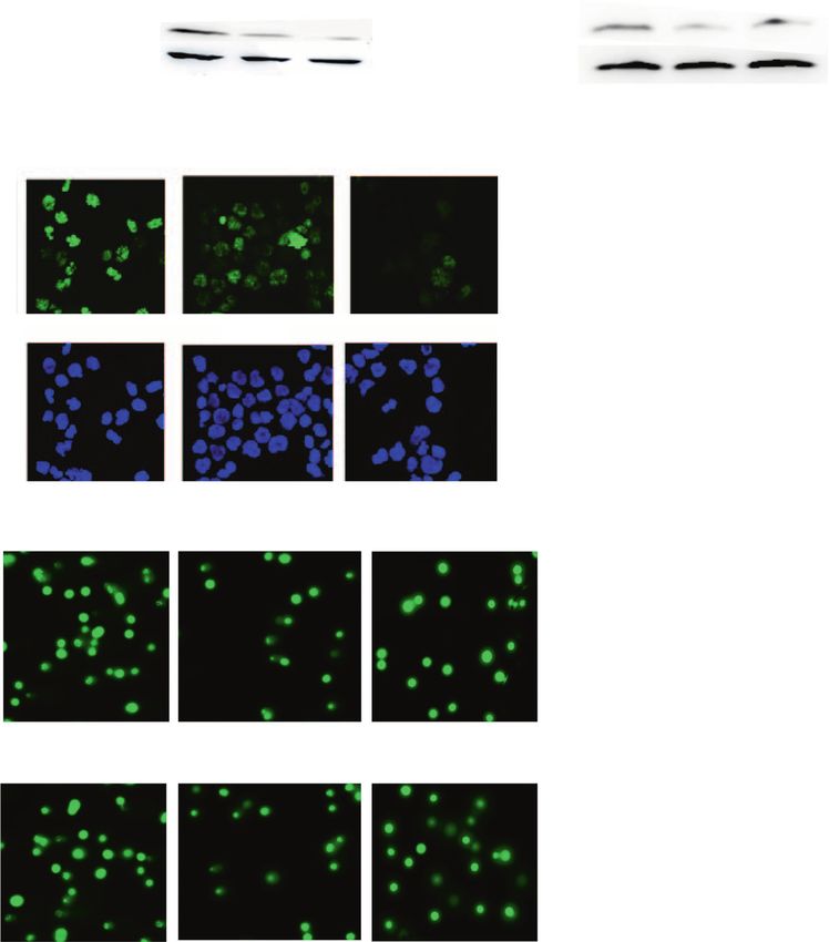
S. Talluri et al.
                  2
1234567890();,:

                      Fig. 1 APOBEC expression and activity is dysregulated in Multiple Myeloma. A Expression of AID/APOBEC family of genes was evaluated in
                      CD138 + plasma cells from newly diagnosed multiple myeloma (NDMM) patients (n = 409), using RNA sequencing; B Deaminase activity
                      evaluated in the lysates of CD138 + plasma cells from normal donors (NPC) (n = 5), newly diagnosed multiple myeloma patients (n = 5), and
                      myeloma cell lines (n = 12); C Expression of APOBEC3G in CD138 + plasma cells from normal donors (NPC) (n = 5), newly diagnosed multiple
                      myeloma patients (n = 5), and myeloma cell lines (n = 12), evaluated by qRT-PCR. Asterisk indicates a p value < 0.05.

                         In this study, we demonstrate that multiple APOBECs are highly        The APOBEC family of proteins deaminate cytosines to uracil in
                      expressed in MM. The impact of APOBEC3G (A3G), one of the most           single-stranded DNA substrates. Using a modified fluorescent
                      expressed APOBECs in MM, is further investigated in detail to            oligonucleotide-based assay [21], we measured the deaminase
                      report its influence on various parameters of genome stability in         activity in cell lysates from normal plasma cells from healthy
                      MM. These data identify A3G as a novel gene involved in increased        donors (NPC), plasma cells from MM patients (MMPC), and
                      DNA damage and dysregulation of DNA repair and genome                    myeloma cell lines (MMCL). The deaminase activity was signifi-
                      stability.                                                               cantly elevated in patient MM cells and MM cell lines, compared to
                                                                                               NPC, suggesting significantly elevated APOBEC activity is in
                                                                                               myeloma (Fig. 1B). Since APOBEC3G was identified as one of the
                      RESULTS                                                                  most expressed APOBECs, we confirmed its expression in MM
                      ABOBEC3G is dysregulated in multiple myeloma                             patient samples and cell lines using quantitative real-time
                      We evaluated the expression of APOBEC family of proteins in              PCR (Fig. 1C).
                      myeloma patient samples and cell lines. RNA sequencing of
                      CD138 + plasma cells from patients with newly diagnosed MM               Knockdown of APOBEC3G reduces DNA abasic sites and
                      (n = 409) showed that APOBECs 3A, 3B, 3C, and 3G are highly              breaks in myeloma cells
                      expressed with 3C and 3G being the most expressed in                     To investigate the impact of elevated APOBEC3G expression on
                      MM (Fig. 1A). Consistently, the evaluation of expression data in         genomic integrity, we investigated the cellular impact of shRNA-
                      MM cell lines also showed that APOBECs 3B, 3C, and 3G as three           mediated knockdown of A3G in MM.1S and H929 myeloma cell
                      most expressed APOBEC genes in MM (Supplementary Fig. 1).                lines (Fig. 2A). Knockdown of A3G with shRNA1 and shRNA2

                                                                                                                    Blood Cancer Journal (2021)11:166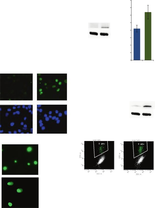
S. Talluri et al.
                                                                                                                                     3

resulted in reduction of deaminase activity by 50 ± 6% and 59 ±   contributes to the overall deaminase activity in these cells
8% (p < 0.001) in MM.1 S cells and by 38±5% and 65±5% (p <        (Supplementary Fig. 2). Since APOBEC-mediated deamination of
0.007) in H929 cells respectively. We also observed a strong      cytosine to uracil can lead to generation of abasic sites, we
correlation between A3G expression and deaminase activity in      investigated the impact of A3G knockdown in MM cells on
both cell lines (R2 > 0.8), suggesting that A3G significantly      generation of abasic sites. A3G-knockdown resulted in 36 and 33%

Blood Cancer Journal (2021)11:166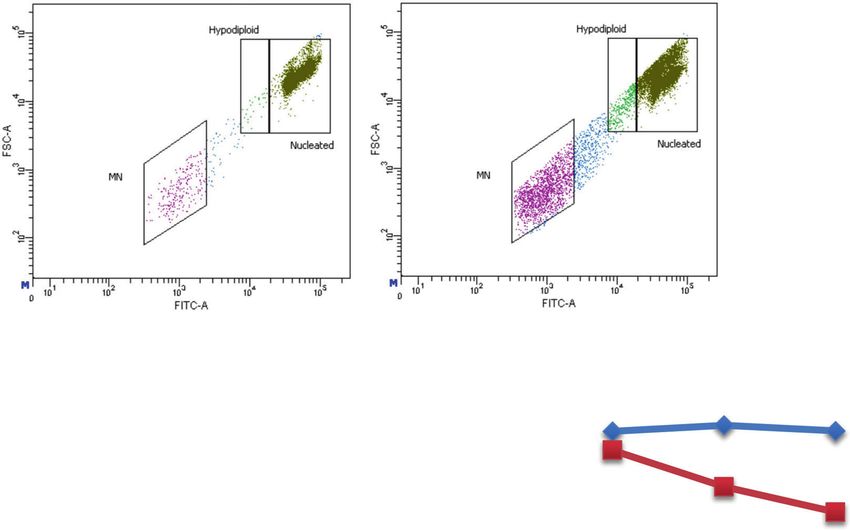
S. Talluri et al.
4
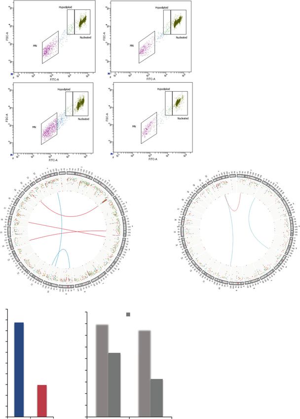
Fig. 2 APOBEC3G knockdown reduces DNA breaks and HR activity in myeloma cells. MM.1S and H929 cells were transduced with lentivirus
particles carrying control shRNA (Sh-C) or two different shRNAs against APOBEC3G (Sh-A3G-1 and 2) and selected in puromycin. A I. Cells were
evaluated for A3G expression (by qRT-PCR) and deaminase activity (as described in Methods section); error bars represent SDs of triplicate
assays. II. Cells were evaluated for A3G expression by Western blotting. B Genomic DNA was isolated from control and A3G knockdown cells
and abasic sites were quantified using OxiSelect™ Oxidative DNA Damage Quantitation Kit (AP sites) from Cell Biolabs. C I. Western blot
analysis of γH2AX in control and A3G knockdown cells; relative expression values of γH2AX (normalized to GAPDH) are shown above
corresponding bands. II. Immunofluorescent staining of γH2AX foci in control and A3G knockdown cells. The number of cells with >5 foci/
nucleus were quantified (n > 100 nuclei) and plotted in the graph. DAPI staining was used to detect nuclei (D) DNA breaks in control and A3G
knockdown cells evaluated by Comet assay using Cell Biolabs OxiSelectTM Comet Assay kit. The number of nuclei with comets were quantified
(n > 100 nuclei) and plotted in the graph. E Homologous recombination (HR) activity was evaluated in the lysates from control and A3G
knockdown MM.1S and H929 cells as described in Methods; and error bars represent SDs of multiple experiments.
reduction in the number of genomic abasic sites in MM.1S and DNA samples evaluated by whole genome sequencing
H929 cells, respectively (p < 0.05; Fig. 2B). Since repair of abasic (in Fig. 3B), were also investigated using SNP 6.0 array
sites involves cleavage of DNA by base excision repair endonu- (Affymetrix). The genome of “Day 0” cells was used as baseline
clease, we investigated the impact of elevated A3G on DNA breaks to identify new copy-number events in control and A3G-
in MM cells by evaluating the expression of γH2AX (a DNA break knockdown cells at 3 weeks. As shown in Supplementary Fig.
marker) by both immunofluorescence staining and Western 3A, the copy number events acquired throughout chromosomes
blotting. Knockdown of A3G resulted in reduced γH2AX expres- were reduced in A3G-knockdown, relative to control cells.
sion (Fig. 2C, panel I). Consistently, the knockdown of A3G with Assessment of total copy number change events detected
two different shRNAs reduced the number of γH2AX foci in MM.1S throughout genome (Supplementary Fig. 3B) and on individual
and H929 cells by 45−65 and 40−53%, respectively (p < 0.05; chromosomes (Supplementary Fig. 3C) indicate that the
Fig. 2C, panel II). In order to further confirm these results, we also acquisition of new genomic changes in A3G knockdown cells,
performed a single-cell gel electrophoresis (comet assay) to detect relative to control cells, was reduced by 60%. These data
DNA breaks. A3G knockdown significantly reduced the DNA demonstrate that A3G knockdown reduces the acquisition of
damage in MM cells as determined by comet assay [22]. Relative new genomic changes in MM cells.
to control cells, A3G knockdown in MM.1S and H929 cells with two
different shRNAs reduced the comets by 54−76 and 79−83%, Evaluation of impact on acquisition of mutations in a plasmid
respectively (p ≤ 0.05) (Fig. 2D). These data demonstrate that substrate. APOBEC proteins induce deamination of Cytosines in
elevated A3G contributes to increased DNA breaks in MM cells. ssDNA converting them to Uracil which usually results in C>T or
We have previously demonstrated that elevated/dysregulated C>G mutations or much less frequently in C>A mutations. We
homologous recombination (HR) activity is involved in ongoing transduced both control and A3G knockdown cells with a EGFP
genomic rearrangements and instability in MM [1]. Since HR can expressing plasmid. EGFP-positive cells were purified and cultured
be induced by DNA breaks, we investigated the impact of A3G for additional 72 hr. Genomic DNA was isolated and EGFP
knockdown on HR activity and observed reduced HR activity by 78 sequences amplified using specific primers. The PCR products
and 48% in A3G knockdown MM1S and H929 cells, respectively were purified and cloned into a plasmid vector to transfect into
(p = T, C>G and C>A) in a
Knockdown of APOBEC3G reduces genomic instability in plasmid substrate by 68 and 51%, respectively (p ≤ 0.05).
myeloma cells
We also evaluated the impact of A3G knockdown on genome Overexpression of APOBEC3G induces DNA double stand
stability using various approaches including evaluation of micro- breaks, HR activity, and genomic instability in myeloma cells
nuclei (a marker of genomic instability), mutational changes in a To further confirm the role of APOBEC3G in genomic instability, we
plasmid substrate and copy number and genomic changes over used ectopically overexpressed V5 tagged A3G in U266 MM cells
time, using whole-genome sequencing. that have relatively low endogenous A3G expression. As predicted,
these A3G-overexpressing cells has significantly upregulated
Impact on micronuclei. Ongoing genomic rearrangements and deaminase activity (Fig. 4A, panel I). The overexpression lead to
instability is associated with generation of DNA/chromosomal significant increase in the number of abasic sites by 49% (p < 0.05) in
fragments and formation of micronuclei, which are used as marker A3G-overexpressing cells relative to control cells (Fig. 4B). To
of genomic instability [23, 24]. Knockdown of A3G protein resulted monitor the impact of A3G overexpression on DNA breaks, the cells
in 52 ± 14% and 58 ± 9% reduction in the number of micronuclei were evaluated for DNA breaks by γH2AX foci using immunofluor-
in MM.1S and H929 cells, respectively (p =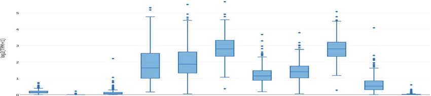
S. Talluri et al.
                                                                                                                                           5

HR activity, we used both the MM and U2OS (osteosarcoma) cell        the fluorescence which is detected by flow cytometry. As seen in
lines. U2OS osteosarcoma cells, stably integrated with HR reporter   Fig. 4E (panels I–II), A3G overexpression resulted in 1.7-fold
substrate (DRGFP), were transduced with control plasmid or           increase (p ≤ 0.05) in HR activity in U2OS cells. Consistently, the
expressing A3G. Transduced cells were then infected with             evaluation of HR activity in MM (U266) cell lysates showed a 2.5-
adenovirus expressing I-SceI endonuclease to initiate HR. In this    fold increase (p ≤ 0.05) in A3G-overexpressing, relative to control
assay system, the HR generates a functional GFP gene resulting in    cells (Fig. 4E, panel III).

Blood Cancer Journal (2021)11:166
S. Talluri et al.
6
Fig. 3 APOBEC3G knockdown reduces genomic instability in myeloma cells. A Control (sh-C) and APOBEC3G knockdown (sh-A3G) MM.1S
and H929 cells were evaluated for the presence of micronuclei, using In Vitro MicroFlow Kit (Litron Labs). Images of micronuclei (I) and bar
graphs (II) showing percentage of micronuclei are presented. B MM.1S cells were transduced with control shRNA (sh-C) or that targeting A3G
(sh-A3G) and following puromycin selection, cultured for 3 weeks. DNA from these and day 0 (baseline control) cells was extracted and the
acquisition of new genomic changes in cultured relative to day 0 cells monitored, using whole genome sequencing (30x). Circos plots
showing translocations and other genomic changes (I) and bar graphs showing total number of SNVs and indels (II) are presented. C Control
and A3G knockdown MM cells were transfected with a plasmid expressing EGFP. EGFP-positive cells were purified, cultured for additional 72 h,
genomic DNA isolated, and EGFP sequences amplified using specific primers. The PCR products were cloned into a plasmid vector to transfect
into competent E. coli cells and plated on LB agar. Plasmid DNA from 10 colonies/sample were isolated and sequenced to analyze for
mutations. Bar graphs show APOBEC-like mutations.
We also observed significant increase (3.8-fold; p ≤ 0.05) in Here we report that several members of APOBEC3 family (A3A,
micronuclei in A3G-overexpressing U266 cells relative to control A3B, A3C, and A3G) are highly expressed in MM. Consistently, the
cells (Fig. 5A). To further investigate the effect of A3G over- APOBEC deaminase activity was also elevated in MM cells and cell
expression on the acquisition of mutations, we stably transduced a lines, relative to normal plasma cells. In specific, A3G, is one of the
EGFP expressing plasmid in control and A3G-overexpressing U266 most expressed APOBEC genes in MM. In normal cells, A3G is
cells. EGFP positive cells were purified using cell sorter and predominantly found in the cytoplasm where it acts as an innate
cultured for additional 72 h. Genomic DNA was isolated, and EGFP immune barrier against viral infections, targeting viral DNA [33].
sequences were amplified and sequenced as in Fig. 3C. Over- APOBEC3G has been extensively studied in the context of its anti-
expression of A3G increased the number of APOBEC-like (C>T, viral functions inhibiting the infectivity of HIV, hepatitis B, human T
C>G, and C>A) mutations by 3.9-fold (p ≤ 0.05) in the EGFP cell leukemia virus type 1, and human papillomavirus [34–38].
plasmid substrate (Fig. 5B). We also observed a 2.2-fold increase in APOBEC enzymes exert their anti-viral activity by targeted
overall mutations in the EGFP plasmid in A3G-overexpressing cells deamination of cytidine residues in the ssDNA produced during
compared to control cells. However, the number of overall reverse transcription of viral genomic RNA. However, it has been
mutations is not significantly different than the APOBEC-like shown that DNA damage could trigger the translocation of A3G to
mutations, suggesting that A3G-overexpression predominantly the nucleus and A3G has been shown to inhibit retro-transposition
caused APOBEC-like (C>T, C>G, and C>A) mutations in the plasmid of endogenous retroviruses. A3G is constitutively expressed in
substrate. These data demonstrate that A3G overexpression is immune cells and is further induced by interferon (IFN) [39]. With
associated with increased mutational instability. We also investi- regards to cancer, A3G has been shown to promote liver
gated if this increased mutation rate could lead to the loss of EGFP metastasis in an orthotopic mouse model of colorectal cancer
signal. In fact, as seen in (Fig. 5B, panel II), the A3G overexpression [40], sensitize mesenchymal gliomas to radiation-induced cell
resulted in the loss of EGFP + cells over time, suggesting that death [41], and enhance lymphoma cell radio resistance by
overexpression of APOBECs can cause deleterious mutations promoting cytidine deaminase-dependent DNA repair [42, 43].
leading to loss of function. Importantly, the role of APOBEC has been imputed in the
Overall, using various approaches, we demonstrate that A3G development of number of malignancies, especially MM. Early
knockdown significantly inhibits whereas its overexpression studies evaluating progression of SMM to MM, identified APOBEC
increases abasic sites, DNA breaks, HR activity, and genomic activity as the key late event shaping the progressor phenotype to
instability in MM cells. MM [4]. Similarly, early Exome sequencing identified APOBEC
driven mutational signature as one of the 2 key events in MM [3].
A study evaluating mutational profile in newly diagnosed MM
using deep whole genome sequencing identified APOBEC
DISCUSSION mutational events shaping the landscape of later stages of MM
Genome instability is one of the prominent features observed [44]. This study utilized clonality of the mutations and signatures
early at MGUS stage and persists and may even be enhanced on driving those mutations to recreate the evolution of the disease
progression to MM [25–27]. Genomic instability underlies the and described that especially in the high-risk group, the APOBEC
development of clonal heterogeneity in MM which is associated signature played a predominant role in the middle phase of
with disease progression including development of drug resis- progression. This study also identified differential utilization of
tance and relapse. The factors contributing to genomic instability various mutational processes in various subgroups and reported
in cancer may include increased DNA damage [28], dysregulated that the APOBEC-related mutational process was significantly
DNA repair [1, 29], replication stress [30], oxidative stress [31], and higher in t(14;16) MM. Interestingly, t(14;16) MM has the highest
mitotic segregation errors [32]. The molecular mechanisms and mutational burden which is associated with poor survival
sequence of events driving genomic instability and clonal outcome. This adverse role of APOBEC-mediated mutational
evolution in cancer are not fully understood, and understanding signature was also confirmed by an independent large study
these mechanisms has important implications in devising transla- reporting association with poor prognosis primary and secondary
tional strategies to treat and/or prevent cancer. Previously we translocations [6].
have demonstrated that homologous recombination (HR) is Suppression of A3G in MM cells significantly reduced, whereas
dysregulated in MM and contributes to genomic instability and its overexpression increased the number of abasic sites. Since
drug resistance [1]. Investigating the mechanisms underlying APOBEC-mediated deamination of cytosine to uracil can lead to
dysregulated HR, we reported base excision repair related generation of abasic site, this result is consistent with the
apurinic/apyrimidinic (APEX) nucleases to contribute to regulation functional nature of APOBEC proteins. Abasic sites are repaired
of recombinase RAD51 and HR in MM [29]. Moreover, the whole by base excision repair related APEX nucleases, which cleave the
genome sequencing data has also identified APOBEC contributing DNA 5′ to the abasic site [45]. Our previous studies have
mutations associated with progression to MM with further demonstrated that APEX nucleases are overexpressed and
increase at relapse. Since APOBEC-mediated deamination of contribute to increased DNA breaks (unpublished data) and HR
cytosine to uracil can lead to generation of abasic site, the activity in MM cells [29]. Results here with A3G are consistent and
substrate of APEX activity, we investigated the role of specific in line with the previous observation and indirectly provides a
APOBEC in genomic instability. relationship to a proximal pathway impacting genomic integrity in
Blood Cancer Journal (2021)11:166S. Talluri et al.
7
MM. The observation that suppression of A3G significantly different HR assay systems in 2 different cell types. One possible
reduced, whereas its overexpression increased DNA breaks in interpretation of these results is that increased DNA breaks by
MM cells suggest that A3G-induced abasic sites could at least elevated A3G expression increase the need for HR-mediated
partly be attributed to the observed upregulation of APEX repair. DNA breaks are known to induce HR, which has been
nucleases in MM cells [29]. The results here also show a link shown to be dysregulated and involved in the acquisition of
between elevated A3G and increased HR activity using two ongoing genomic rearrangements in MM [1]. Alternatively, A3G
Blood Cancer Journal (2021)11:166S. Talluri et al.
8
Fig. 4 Overexpression of APOBEC3G induces DNA breaks and HR activity in myeloma cells. U266 cells were transduced with lentivirus
particles carrying control plasmid (OE-C) or APOBEC3G-ORF with V5 tag (OE-A3G) and selected in puromycin. A I. Cells were evaluated for A3G
expression by qRT-PCR and in vitro deaminase activity as described in methods; II. Western blot showing A3G expression, evaluated using
anti-V5 tag antibody; B Genomic DNA from these cells was isolated and abasic sites quantified, using OxiSelect™ Oxidative DNA Damage
Quantitation Kit (Cell Biolabs). C I–II. Images showing immunofluorescence staining of γH2AX foci and DAPI staining to detect nuclei (I), and
the bar graphs showing the number of cells with >5 foci/nucleus (II) in control and A3G overexpression cells; III. Western blot analysis of
γH2AX in control and A3G overexpression cells; D DNA breaks in control and A3G overexpression cells evaluated by Comet assay, using Cell
Biolabs OxiSelectTM Comet Assay kit. Images of Comets (I) and bar graphs showing the number of nuclei with comets in >100 nuclei examined
(II) are presented; E Homologous recombination (HR) activity was evaluated in control and A3G-overexpressing U2OS cells, using DRGFP assay
(I–II), and in the lysates of control and A3G-overexpressing U266 cells using an in vitro assay described in Methods section.
Fig. 5 APOBEC3G overexpression induces genomic instability in myeloma cells. Control and A3G-overexpressing U266 cells (described in
Fig. 4) were evaluated for genomic instability. A Following selection, the cells were evaluated for the presence of micronuclei, using In Vitro
MicroFlow Kit (Litron Labs). Images of micronuclei (I) and bar graphs showing percentage of micronuclei (II) are presented. B Control and A3G-
overexpressing cells described above were transfected with a plasmid expressing EGFP. EGFP-positive cells were purified, cultured for
additional 72 h, genomic DNA isolated, and EGFP sequences amplified using specific primers. The PCR products were cloned into a plasmid
vector to transfect into competent E. coli cells and plated on LB agar. Plasmid DNA from 10 colonies/sample were isolated and sequenced to
analyze for mutations. I. Bar graphs show APOBEC-like mutations as well as total (or overall) mutations; II. Percent GFP + cells were analyzed by
flow cytometry at different time points and plotted in the graph.
could also play a direct role in homologous recombination. Due to the low sample size, we were not able to detect specific
Evaluation in lymphoma cells has shown that A3G promotes repair mutational signatures in this investigation. However, we do
of ionizing radiation-induced double strand breaks mainly demonstrate the impact of A3G modulation in a plasmid substrate
through non-homologous end joining [42]. However, a role of showing that the knockdown of A3G reduced, whereas its
A3G in HR has not been reported previously. To our knowledge, overexpression increased, the incidence of overall as well as
this study is the first report establishing a possible link between APOBEC-like (C>T, C>G, and C>A) mutations (Figs. 3B and 5B).
A3G and HR in myeloma cells. Although a direct role of A3G in HR These data demonstrate that in addition to known C>T, C>G, and
cannot be ruled out, it needs to be investigated further. C>A mutations, A3G could probably also contribute to other
Blood Cancer Journal (2021)11:166S. Talluri et al.
9
Detection of DNA breaks
DNA breaks were estimated by evaluating the levels of γ-H2AX (DNA
break-associated protein) and Comet, a gel-based assay for the detection
of DNA breaks. Levels of γ-H2AX protein were measured by immuno-
fluorescence staining and/or by quantification of γ-H2AX by Western
blotting. Comet assay was performed using OxiSelect™ Comet Assay Kit
(Cell Biolabs Inc., San Diego, CA) following manufacture’s protocol.
DNA cytidine deaminase activity assay
Cytidine deaminase activity was measured in the myeloma cell lysates
using a fluorescent oligonucleotide-based assay as described before [21].
Briefly, cell lysates were incubated at 37 °C for 2 h in the presence of a
Fig. 6 Proposed model for the role of APOBEC3G in genomic single-stranded (ss) DNA oligonucleotide 5′(6-FAM) AAATTCTAATAGA-
instability in multiple myeloma. Elevated A3G-induced deaminase TAATGTGA (TAMRA)3′. In the absence of deaminase activity, the
activity leads to generation of abasic sites. Processing of increased fluorescence is quenched by TAMRA. In the presence of cytidine
abasic sites then leads to increase in DNA breaks. Previous data from deaminase activity in the lysates, cytidine is converted to uridine. This is
our laboratory demonstrate that APEX1, the nuclease which is subsequently excised by the addition of Uracil DNA glycosylase (UDG)
involved in the repair of abasic sites by cutting the DNA 5′ to abasic resulting in the creation of abasic sites. The abasic sites are cleaved by
site, is elevated and contributes to dysregulation of RAD51 and incubation at 95 °C resulting in the increased fluorescence signal, which is
homologous recombination [29], a mechanism of genomic instabil- measured using a fluorescence plate reader.
ity in MM [1]. Increased abasic sites by A3G and the replication stress
caused by abasic sites can also contribute to genomic instability by HR activity assays
increasing the likelihood of translesion synthesis [50]. Homologous recombination (HR) activity was assessed either in cell lysates,
using in vitro HR conditions [46] as described previously [29] or in intact
cells, using a fluorescence-based HR substrate (Addgene, Cambridge, MA)
[47] as described by us previously [48].
mutation types in myeloma cells. Taken together, these data
demonstrate that elevated A3G contributes to dysregulation of HR
and genome stability. Micronucleus assay
To evaluate the impact on genomic instability, control and A3G knock-
These data, in light of our previous observations, suggest a
down cells were analyzed for micronuclei, using a flow cytometry-based
significant role of A3G in genomic instability in MM cells (Fig. 6). Micronucleus Assay (MicroFlow kit, Litron Laboratories, New York, USA) as
We propose that the elevated A3G-induces deaminase activity described by us previously [29].
leading to increased generation of abasic sites with subsequent
increase in DNA breaks and HR activity. Moreover, a direct role of
A3G in dysregulation of HR in multiple myeloma and its additional Evaluation of impact on mutations in a plasmid substrate
Control and A3G-knockdown or A3G-overexpression cells were transduced
roles in genomic instability cannot be ruled out and requires with lentivirus particles carrying EGFP expression vector. GFP positive cells
further investigation. These data also identify A3G as a novel were purified, using flow cell sorter and cultured for additional 72 h.
target to inhibit/reduce DNA damage, HR activity, and genomic Genomic DNA was isolated using PureLink Genomic DNA Mini Kit from
instability in MM cells. Thermofisher. EGFP sequences were PCR amplified, using specific primers.
Purified PCR products were cloned into plasmid vector, transfected into
competent E. coli cells and grown on LB agar plates overnight. Plasmid
MATERIALS AND METHODS DNA was isolated from 10 individual colonies per sample and sent for
Cell lines and patient samples sequencing. The number of APOBEC-like (C>T, C>G, C>A) conversions were
Multiple Myeloma (MM) cell lines were purchased from the American Type quantified and the average number of mutations were plotted in
Tissue Culture Collection (Rockville MD). Cell lines were cultured in the graph.
RPMI1640 medium containing 10% fetal bovine serum and antibiotic and
maintained in logarithmic growth. Samples of bone marrow aspirates from Evaluation of impact on genomic instability using SNP/WGS
myeloma patients and normal individuals were obtained following Control and transgenically-modulated cells were cultured for three weeks.
informed consent under the protocol approved by Institutional Review Live cells were separated, using Ficoll density gradient centrifugation and
Board of Dana Farber Cancer Institute. MM cells were isolated by Magnet genomic DNA from these and “day 0 cells” (representing baseline genome)
Assisted Cell Sorting (MACS, Miltenyi Biotech), according to the was isolated, using PureLink Genomic DNA Mini Kit, and analyzed using
manufacturer’s protocol. single nucleotide polymorphism (SNP6.0) arrays (Affymetrix) or whole-
genome sequencing (WGS). For both the SNP and WGS experiments, the
Western blotting, immunocytochemical detection of proteins, genome of “day 0” cells (harvested and stored in the beginning of each
experiment) was used as baseline to identify the changes in control and
and antibodies used
transgenically-modulated MM cells that were cultured for three weeks. SNP
For Western blotting, lysates boiled in sample buffer, were fractionated on
and WGS data were analyzed as reported previously [1, 4].
gradient SDS-polyacrylamide gel and subsequently electroblotted onto
nitrocellulose paper. The blots were incubated with indicated primary
antibodies, washed and incubated in either anti-rabbit or anti-mouse, Statistics and reproducibility
horseradish peroxidase conjugates. After washing, specific proteins were RNA Sequencing data were analyzed as described previously [49].
detected using an enhanced chemiluminescence, according to the Experiments were conducted in triplicate and bar graphs with error bars
instructions provided in the manual (Amersham Life Sciences Inc., indicating SD values presented; two-tailed p values were derived by
Arlington Heights, IL). For immunocytochemical detection of γ-H2AX, Student’s t test. To confirm the genomic impact of APOBEC3G, both the
cytospins of normal plasma cells and MM cells were fixed in methanol/ gain and loss of function, a total of three MM cell lines and the evaluation
acetone (1:1) for 10 min at –20 °C. Fixed cells were rinsed, rehydrated in of different parameters of genome maintenance, using multiple
PBS, and incubated with mouse monoclonal antibody to γ-H2AX (Ser139), approaches, were used. The overexpression of APOBEC3G in one MM cell
clone JBW301 (Catalog #05–636; Millipore). Other antibodies used were line and its knockdown in two MM cell lines were evaluated for impact on
anti-GAPDH (14C10) HRP conjugated antibody (Catalog #3683; Cell deaminase activity, abasic sites, spontaneous DNA breaks (using Western
Signaling Technology), anti-V5 tag antibody SV5-Pk1 (Catalog #ab27671; blotting, immunofluorescence and Comet assay), homologous recombina-
Abcam), anti-rabbit IgG, HRP-linked antibody (Catalog #7074; Cell Signaling tion activity and genomic instability (using micronucleus assay, and
Technology) and anti-mouse IgG, and HRP-linked antibody (Catalog #7076; evaluation of the acquisition of genomic changes over time using single
Cell Signaling Technology). nucleotide polymorphism arrays and whole-genome sequencing).
Blood Cancer Journal (2021)11:166S. Talluri et al.
10
REFERENCES 29. Kumar S, Talluri S, Pal J, Yuan X, Lu R, Nanjappa P, et al. Role of apurinic/
1. Shammas MA, Shmookler Reis RJ, Koley H, Batchu RB, Li C, Munshi NC. Dys- apyrimidinic nucleases in the regulation of homologous recombination in mye-
functional homologous recombination mediates genomic instability and pro- loma: mechanisms and translational significance. Blood Cancer J. 2018;8:92.
gression in myeloma. Blood. 2009;113:2290–7. 30. Gaillard H, García-Muse T, Aguilera A. Replication stress and cancer. Nat Rev
2. Alexandrov LB, Nik-Zainal S, Wedge DC, Aparicio SA, Behjati S, Biankin AV, et al. Cancer. 2015;15:276–89.
Signatures of mutational processes in human cancer. Nature. 2013;500:415–21. 31. Klaunig JE, Kamendulis LM, Hocevar BA. Oxidative stress and oxidative damage in
3. Bolli N, Avet-Loiseau H, Wedge DC, Van Loo P, Alexandrov LB, Martincorena I, carcinogenesis. Toxicol Pathol. 2010;38:96–109.
et al. Heterogeneity of genomic evolution and mutational profiles in multiple 32. Levine MS, Holland AJ. The impact of mitotic errors on cell proliferation and
myeloma. Nat Commun. 2014;5:2997. tumorigenesis. Genes Dev. 2018;32:620–38.
4. Bolli N, Maura F, Minvielle S, Gloznik D, Szalat R, Fullam A, et al. Genomic patterns 33. Mangeat B, Turelli P, Caron G, Friedli M, Perrin L, Trono D. Broad antiretroviral
of progression in smoldering multiple myeloma. Nat Commun. 2018;9:3363. defence by human APOBEC3G through lethal editing of nascent reverse tran-
5. Hoang PH, Cornish AJ, Dobbins SE, Kaiser M, Houlston RS. Mutational processes scripts. Nature 2003;424:99–103.
contributing to the development of multiple myeloma. Blood Cancer J. 2019;9:60. 34. Ziegler SJ, Liu C, Landau M, Buzovetsky O, Desimmie BA, Zhao Q, et al. Insights
6. Walker BA, Wardell CP, Murison A, Boyle EM, Begum DB, Dahir NM, et al. APOBEC into DNA substrate selection by APOBEC3G from structural, biochemical, and
family mutational signatures are associated with poor prognosis translocations in functional studies. PLoS ONE. 2018;13:e0195048.
multiple myeloma. Nat Commun. 2015;6:6997. 35. Bonvin M, Greeve J. Hepatitis B: modern concepts in pathogenesis–APOBEC3
7. Maura F, Bolli N, Angelopoulos N, Dawson KJ, Leongamornlert D, Martincorena I, cytidine deaminases as effectors in innate immunity against the hepatitis B virus.
et al. Genomic landscape and chronological reconstruction of driver events in Curr Opin Infect Dis. 2008;21:298–303.
multiple myeloma. Nat Commun. 2019;10:3835. 36. Sasada A, Takaori-Kondo A, Shirakawa K, Kobayashi M, Abudu A, Hishizawa M, et al.
8. Conticello SG. The AID/APOBEC family of nucleic acid mutators. Genome Biol. APOBEC3G targets human T-cell leukemia virus type 1. Retrovirology. 2005;2:32.
2008;9:229. 37. Vartanian JP, Guétard D, Henry M, Wain-Hobson S. Evidence for editing of human
9. Swanton C, McGranahan N, Starrett GJ, Harris RS. APOBEC enzymes: mutagenic papillomavirus DNA by APOBEC3 in benign and precancerous lesions. Science.
fuel for cancer evolution and heterogeneity. Cancer Discov. 2015;5:704–12. 2008;320:230–3.
10. Roberts SA, Lawrence MS, Klimczak LJ, Grimm SA, Fargo D, Stojanov P, et al. An 38. Wiegand HL, Cullen BR. Inhibition of alpha retrovirus replication by a range of
APOBEC cytidine deaminase mutagenesis pattern is widespread in human can- human APOBEC3 proteins. J Virol. 2007;81:13694–9.
cers. Nat Genet. 2013;45:970–6. 39. Koning FA, Newman EN, Kim EY, Kunstman KJ, Wolinsky SM, Malim MH. Defining
11. Zou J, Wang C, Ma X, Wang E, Peng G. APOBEC3B, a molecular driver of muta- APOBEC3 expression patterns in human tissues and hematopoietic cell subsets. J
genesis in human cancers. Cell Biosci. 2017;7:29. Virol. 2009;83:9474–85.
12. Burns MB, Lackey L, Carpenter MA, Rathore A, Land AM, Leonard B, et al. APO- 40. Ding Q, Chang CJ, Xie X, Xia W, Yang JY, Wang SC, et al. APOBEC3G promotes
BEC3B is an enzymatic source of mutation in breast cancer. Nature. liver metastasis in an orthotopic mouse model of colorectal cancer and predicts
2013;494:366–70. human hepatic metastasis. J Clin Investig. 2011;121:4526–36.
13. Kosumi K, Baba Y, Ishimoto T, Harada K, Nakamura K, Ohuchi M, et al. APOBEC3B 41. Wang Y, Wu S, Zheng S, Wang S, Wali A, Ezhilarasan R, et al. APOBEC3G acts as a
is an enzymatic source of molecular alterations in esophageal squamous cell therapeutic target in mesenchymal gliomas by sensitizing cells to radiation-
carcinoma. Med Oncol. 2016;33:26. induced cell death. Oncotarget. 2017;8:54285–96.
14. Yan S, He F, Gao B, Wu H, Li M, Huang L, et al. Increased APOBEC3B predicts 42. Nowarski R, Wilner OI, Cheshin O, Shahar OD, Kenig E, Baraz L, et al. APOBEC3G
worse outcomes in lung cancer: a comprehensive retrospective study. J Cancer. enhances lymphoma cell radioresistance by promoting cytidine deaminase-
2016;7:618–25. dependent DNA repair. Blood. 2012;120:366–75.
15. Tsuboi M, Yamane A, Horiguchi J, Yokobori T, Kawabata-Iwakawa R, Yoshiyama S, 43. Prabhu P, Shandilya SM, Britan-Rosich E, Nagler A, Schiffer CA, Kotler M. Inhibition
et al. APOBEC3B high expression status is associated with aggressive phenotype of APOBEC3G activity impedes double-stranded DNA repair. FEBS J.
in Japanese breast cancers. Breast Cancer. 2016;23:780–8. 2016;283:112–29.
16. Sieuwerts AM, Willis S, Burns MB, Look MP, Meijer-Van Gelder ME, Schlicker A, 44. Samur MK, Aktas Samur A, Fulciniti M, Szalat R, Han T, Shammas M, et al.
et al. Elevated APOBEC3B correlates with poor outcomes for estrogen-receptor- Genome-wide somatic alterations in multiple myeloma reveal a superior out-
positive breast cancers. Hormones Cancer. 2014;5:405–13. come group. J Clin Oncol. 2020;38:3107–18.
17. Leonard B, Hart SN, Burns MB, Carpenter MA, Temiz NA, Rathore A, et al. APO- 45. Mosbaugh DW, Linn S. Further characterization of human fibroblast apurinic/
BEC3B upregulation and genomic mutation patterns in serous ovarian carcinoma. apyrimidinic DNA endonucleases. The definition of two mechanistic classes of
Cancer Res. 2013;73:7222–31. enzyme. J Biol Chem. 1980;255:11743–52.
18. Yamazaki H, Shirakawa K, Matsumoto T, Hirabayashi S, Murakawa Y, Kobayashi M, 46. Liu J, Sneeden J, Heyer WD. In vitro assays for DNA pairing and recombination-
et al. Endogenous APOBEC3B overexpression constitutively generates dna sub- associated DNA synthesis. Methods Mol Biol. 2011;745:363–83.
stitutions and deletions in myeloma cells. Sci Rep. 2019;9:7122. 47. Pierce AJ, Johnson RD, Thompson LH, Jasin M. XRCC3 promotes homology-
19. Hoopes JI, Cortez LM, Mertz TM, Malc EP, Mieczkowski PA, Roberts SA. APOBEC3A directed repair of DNA damage in mammalian cells. Genes Dev. 1999;13:2633–8.
and APOBEC3B preferentially deaminate the lagging strand template during DNA 48. Lu R, Pal J, Buon L, Nanjappa P, Shi J, Fulciniti M, et al. Targeting homologous
replication. Cell Rep. 2016;14:1273–82. recombination and telomerase in Barrett’s adenocarcinoma: Impact on
20. Morgan GJ, Walker BA, Davies FE. The genetic architecture of multiple myeloma. telomere maintenance, genomic instability, and tumor growth. Oncogene.
Nat Rev Cancer. 2012;12:335–48. 2014;33:1495–505.
21. Stenglein MD, Burns MB, Li M, Lengyel J, Harris RS. APOBEC3 proteins mediate 49. Samur MK, Minvielle S, Gulla A, Fulciniti M, Cleynen A, Aktas Samur A, et al. Long
the clearance of foreign DNA from human cells. Nat Struct Mol Biol. intergenic non-coding RNAs have an independent impact on survival in multiple
2010;17:222–9. myeloma. Leukemia. 2018. https://doi.org/10.1038/s41375-018-0116-y.
22. Gyori BM, Venkatachalam G, Thiagarajan PS, Hsu D, Clement MV. OpenComet: an 50. Gelot C, Magdalou I, Lopez BS. Replication stress in mammalian cells and its
automated tool for comet assay image analysis. Redox Biol. 2014;2:457–65. consequences for mitosis. Genes. 2015;6:267–98.
23. Balmus G, Karp NA, Ng BL, Jackson SP, Adams DJ, McIntyre RE. A high-throughput
in vivo micronucleus assay for genome instability screening in mice. Nat Protoc.
2015;10:205–15.
24. Terradas M, Martín M, Genescà A. Impaired nuclear functions in micronuclei
ACKNOWLEDGEMENTS
This work was supported by Department of Veterans Affairs Merit Review Award
results in genome instability and chromothripsis. Arch Toxicol. 2016;90:2657–67.
I01BX001584-01 (NCM), NIH grants P01-155258 and P50 CA100707 (NCM and MAS),
25. van Nieuwenhuijzen N, Spaan I, Raymakers R, Peperzak V. From MGUS to multiple
and Paula and Rodger Riney grant (NCM).
myeloma, a paradigm for clonal evolution of premalignant cells. Cancer Res.
2018;78:2449–56.
26. Dutta AK, Fink JL, Grady JP, Morgan GJ, Mullighan CG, To LB, et al. Subclonal
evolution in disease progression from MGUS/SMM to multiple myeloma is AUTHOR CONTRIBUTIONS
characterised by clonal stability. Leukemia. 2019;33:457–68. NCM envisioned the study, analyzed and interpreted data, and prepared manuscript;
27. Aktas Samur A, Minvielle S, Shammas M, Fulciniti M, Magrangeas F, Richardson MAS assisted in the evaluation of impact on genomic instability, data interpretation
PG, et al. Deciphering the chronology of copy number alterations in Multiple and critical review, and preparation of manuscript; LB and MKS conducted
Myeloma. Blood Cancer J. 2019;9:39. bioinformatic and statistical analyses; ST contributed to major experiments and
28. Tubbs A, Nussenzweig A. Endogenous DNA damage as a source of genomic manuscript preparation; SK, LBP, JS, and RHP contributed to specific experiments and
instability in cancer. Cell. 2017;168:644–56. data analyses.
Blood Cancer Journal (2021)11:166S. Talluri et al.
11
COMPETING INTERESTS Open Access This article is licensed under a Creative Commons
The authors declare no competing interests. Attribution 4.0 International License, which permits use, sharing,
adaptation, distribution and reproduction in any medium or format, as long as you give
appropriate credit to the original author(s) and the source, provide a link to the Creative
ADDITIONAL INFORMATION Commons license, and indicate if changes were made. The images or other third party
Supplementary information The online version contains supplementary material material in this article are included in the article’s Creative Commons license, unless
available at https://doi.org/10.1038/s41408-021-00554-9. indicated otherwise in a credit line to the material. If material is not included in the
article’s Creative Commons license and your intended use is not permitted by statutory
Correspondence and requests for materials should be addressed to Nikhil C. Munshi. regulation or exceeds the permitted use, you will need to obtain permission directly
from the copyright holder. To view a copy of this license, visit http://creativecommons.
Reprints and permission information is available at http://www.nature.com/ org/licenses/by/4.0/.
reprints
This is a U.S. government work and not under copyright protection in the U.S.; foreign
Publisher’s note Springer Nature remains neutral with regard to jurisdictional claims
copyright protection may apply 2021
in published maps and institutional affiliations.
Blood Cancer Journal (2021)11:166You can also read