Endoplasmic Reticulum Stress Induced Proliferation Remains Intact in Aging Mouse b-Cells
←
→
Page content transcription
If your browser does not render page correctly, please read the page content below
ORIGINAL RESEARCH
                                                                                                                                          published: 31 August 2021
                                                                                                                                    doi: 10.3389/fendo.2021.734079

                                           Endoplasmic Reticulum Stress
                                           Induced Proliferation Remains
                                           Intact in Aging Mouse b-Cells
                                           Jarin T. Snyder 1, Christine Darko 2, Rohit B. Sharma 2* and Laura C. Alonso 2*
                                           1 Graduate School of Biomedical Sciences, UMass Medical School, Worcester, MA, United States, 2 Division of

                                           Endocrinology, Diabetes and Metabolism and the Weill Center for Metabolic Health, Weill Cornell Medicine,
                                           New York, NY, United States

                                           Aging is associated with loss of proliferation of the insulin-secreting b-cell, a possible
                                           contributing factor to the increased prevalence of type 2 diabetes in the elderly. Our group
                                           previously discovered that moderate endoplasmic reticulum (ER) stress occurring during
                                           glucose exposure increases the adaptive b-cell proliferation response. Specifically, the
                                           ATF6a arm of the tripartite Unfolded Protein Response (UPR) promotes b-cell replication
                                           in glucose excess conditions. We hypothesized that b-cells from older mice have reduced
                                           proliferation due to aberrant UPR signaling or an impaired proliferative response to ER
                           Edited by:      stress or ATF6a activation. To investigate, young and old mouse islet cells were exposed
                      Wenbiao Chen,        to high glucose with low-dose thapsigargin or activation of overexpressed ATF6a, and b-
  Vanderbilt University, United States
                                           cell proliferation was quantified by BrdU incorporation. UPR pathway activation was
                       Reviewed by:
                  Scott Soleimanpour,      compared by qPCR of target genes and semi-quantitative Xbp1 splicing assay.
 University of Michigan, United States     Intriguingly, although old b-cells had reduced proliferation in high glucose compared to
                          Barak Blum,
    University of Wisconsin-Madison,
                                           young b-cells, UPR activation and induction of proliferation in response to low-dose
                         United States     thapsigargin or ATF6a activation in high glucose were largely similar between young and
                *Correspondence:           old. These results suggest that loss of UPR-led adaptive proliferation does not explain the
                   Laura C. Alonso         reduced cell cycle entry in old b-cells, and raise the exciting possibility that future therapies
           lca4001@med.cornell.edu
                   Rohit B. Sharma         that engage adaptive UPR could increase b-cell number through proliferation even in
           ros4015@med.cornell.edu         older individuals.

                  Specialty section:       Keywords: pancreatic beta cells, proliferation, endoplasm Reticulum Stress, unfolded protein response, ATF6
        This article was submitted to      (activating transcription factor 6), thapsigargin, aging/aging beta cells
    Diabetes: Molecular Mechanisms,
              a section of the journal
           Frontiers in Endocrinology
                                           INTRODUCTION
           Received: 30 June 2021
         Accepted: 05 August 2021          Type 2 diabetes is an age-associated chronic disease, with a peak incidence of 25% in the US
         Published: 31 August 2021
                                           population aged ≥65 (1). Lifestyle interventions targeting people with prediabetes delay onset and
                            Citation:      reduce incidence of disease (2), but most people pass through the prediabetes phase without being
Snyder JT, Darko C, Sharma RB and
                                           diagnosed, and multiple barriers limit implementation of successful lifestyle changes. Treatments
     Alonso LC (2021) Endoplasmic
                                           targeting aging as a risk factor for diabetes are lacking. As an epidemic of obesity coincides with
           Reticulum Stress Induced
          Proliferation Remains Intact
             in Aging Mouse b-Cells.       Abbreviations: ER, endoplasmic reticulum; UPR, unfolded protein response; PERK, Protein kinase R-like ER kinase; IRE1,
       Front. Endocrinol. 12:734079.       Inositol-requiring enzyme 1; ATF4, Activating transcription factor 4; ATF6, Activating transcription factor 6; CDK, Cyclin
   doi: 10.3389/fendo.2021.734079          dependent kinase.

Frontiers in Endocrinology | www.frontiersin.org                                  1                                       August 2021 | Volume 12 | Article 734079
Snyder et al.                                                                                                    Stress-Induced Proliferation in Aging b-Cells

population aging in the coming decades, the incidence of                        and Use Committees. “Young” (10-14 weeks) and “old” (10-25
diabetes is expected to increase around the world (3).                          months) C57BL/6J mice of both sexes were obtained from litters
Complications of diabetes including cardiovascular disease,                     bred in-house or purchased from the Jackson Laboratory. Old
renal disease, blindness and neuropathy are more frequent and                   and young groups included a similar balance of male and female
severe in the elderly (4), underscoring the need for better                     mice. Mice were housed in a 12-hour light/dark cycle with
prevention and treatment of diabetes in this growing population.                unrestricted access to chow and water.
    Aging is associated with increased insulin resistance (5).
Progression to diabetes is determined by the ability of the                     Islet Isolation, Dispersion, and Culture
pancreatic b-cell to increase insulin production to adapt to                    Islets were isolated using collagenase P and density gradient
increased insulin demand. In non-diabetic obese individuals,                    centrifugation, handpicked, cultured, and dispersed in 0.05%
insulin resistance is increased but b-cell mass and insulin                     trypsin as previously described (19). Since the islet mass of old
secretion are also increased to maintain glucose control (6),                   mice is larger compared to that of young mice, for each replicate
suggesting that healthy b-cells may proliferate to adapt to                     islets from 2 young mice were combined to provide sufficient
increased insulin demand. Autopsy studies show that b-cell mass                 material to match with each old mouse. Dispersed cells were
is lost in late stage diabetes (7), highlighting b-cell regeneration as a       plated in 500 µL islet complete media (ICM; RPMI containing
therapeutic goal for both type 1 and type 2 diabetes patients.                  10% FBS (Atlanta Biologics), penicillin/streptomycin, and 5 mM
    Reports suggest that adaptive proliferation is reduced or                   glucose) on uncoated glass coverslips for immunostaining [50
entirely lost with aging in mouse and human b-cells (8–10),                     islet equivalents (IEQ)] or directly onto Nunc-treated
posing a barrier to devising novel therapies to prevent and treat               polystyrene 24-well plates (ThermoFisher) for RNA extraction
diabetes through b-cell regeneration in this population. However,               (100 IEQ). 16-24 hours later the media was replaced with ICM
the mechanisms that cause b-cells to cease proliferating with age               containing 15mM glucose simultaneously with the addition of
remain controversial. Some evidence suggests that aged b-cells are              experimental treatments. Thapsigargin (Sigma-Aldrich) was
mostly unresponsive to signals that promote proliferation in                    added to a final concentration of 20 nM to induce moderate
young b-cells, indicative of a permanent cell-cycle arrest typical              UPR (Figure 3). Ad-ATF6:DHFR (20) was added at a
of senescence or terminal differentiation (10), while other studies             multiplicity of infection of 10, and cells were treated with 10
report that proliferation can still be induced in old b-cells through           µM trimethoprim (TMP) to activate ATF6a or DMSO control at
stimuli such as exposure to the youthful microenvironment (11–                  the time virus was added. All samples exposed to experimental
13) or partial b-cell ablation (14). To increase relevance to the               treatments were matched with control-treated samples from the
real-world population, studies on mechanisms that promote                       same pool of dispersed cells. Dispersed islet cells were then
proliferation in young b-cells need to be investigated in old b-                cultured for 72 hours. To allow quantification of S-phase entry,
cells to test for age-dependence.                                               coverslip-containing wells were treated with 10 µg/mL
    The UPR supports ER protein folding capacity critical for b-                Bromodeoxyuridine (BrdU; Sigma-Aldrich) for the last 24 hours.
cells to produce and secrete insulin (15). UPR initiators ATF6,
PERK, and IRE1 are conserved ER transmembrane proteins that                     Immunostaining, Microscopy,
communicate ER protein folding stress to other organelles                       and Cell Counting
[Reviewed in (16)]. We previous discovered that acute                           Following fixation for 10 minutes in 4% paraformaldehyde and
knockdown of ATF6a reduces, and overexpression increases,                       unmasking for 25 minutes in 1N HCl, immunostaining for
high glucose-induced adaptive proliferation in b-cells from                     insulin and BrdU were executed as previously described (19)
mouse and human islets from a wide donor age range (17).                        using primary antibodies from Abcam (BrdU: #ab6326, insulin:
However, whether UPR activation and UPR-dependent                               #ab7842), DyLight secondary antibodies from Jackson
proliferation differed between young and old islets were not                    ImmunoResearch Laboratories and DAPI (Sigma-Aldrich).
examined. Since loss of proteostasis is commonly considered a                   Fluorescence images were acquired using a Nikon microscope.
hallmark of aging (18), impaired UPR-responsive proliferation is                Images were blinded and the number of Insulin + and
a plausible explanation for the age-associated deficit in adaptive               BrdU+Insulin+ cells were manually counted. 3149 ± 657 b-cells
b-cell proliferation. Here we test the hypothesis that impaired                 were counted for the glucose dose range experiment, 1214 ± 597
UPR signaling is responsible for the reduction of glucose-                      b-cells were counted for the thapsigargin experiment, and 1314 ±
responsive proliferation observed in aged mouse b-cells.                        379 b-cells were counted for the ATF6 experiment. Data are
Intriguingly, contrary to the hypothesis, we find that UPR-                      reported as the percent of Insulin+ cells that were BrdU+Insulin+.
induced proliferation remains intact in old mouse b-cells.
                                                                                Cell Lysis, RNA Extraction, cDNA
                                                                                Synthesis, and qPCR
MATERIALS AND METHODS                                                           Cells were washed 3x in cold PBS before lysis. Cells were lysed and kept
                                                                                at -80°C until RNA was extracted using the RNA/Protein Purification
Mice                                                                            Plus Kit (NorgenBiotek) per the manufacturer’s instructions. 300-600
All mouse procedures were approved by the UMass Medical                         ng of total RNA was used to synthesize cDNA using the SuperScript IV
School and Weill Cornell Medicine Institutional Animal Care                     VILO Master Mix (ThermoFisher) per the manufacturer’s

Frontiers in Endocrinology | www.frontiersin.org                            2                                    August 2021 | Volume 12 | Article 734079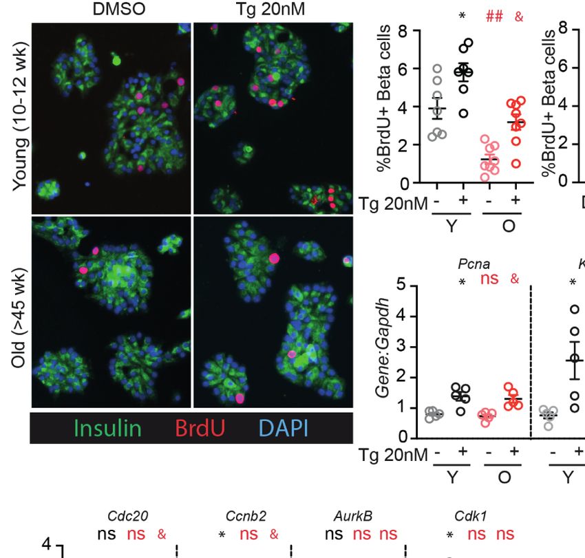
Snyder et al.                                                                                                               Stress-Induced Proliferation in Aging b-Cells

instructions. qPCR was performed using SYBR Green and the listed                       weeks) and old (>45 weeks) mouse islets were trypsinized and
primers (Supplementary Table 1). Relative changes in gene                              cultured in a range of glucose concentrations (5-25 mM).
expression were quantified using the DDCt method.                                       Proliferation was measured by BrdU incorporation, which is a
                                                                                       reliable marker for b-cell S-phase entry in young and old islet
Semi-Quantitative Gel-Based Assay for                                                  cells in our hands (19). Old b-cells had significantly reduced
Xbp1 Splicing                                                                          BrdU incorporation compared to young b-cells at high glucose
The assay was performed according to a previously published                            (15-20 mM); a trend towards reduced proliferation was observed
technique (21). Products were separated by electrophoresis through                     at all glucose concentrations (Figures 1A, B; n=4). Abundance
a 3% agarose gel, and bands were quantified using Image J.                              of mRNA of two proliferation-associated markers were
                                                                                       inconsistent; at 15 mM glucose, Ki67 but not Pcna was
Statistical Analysis                                                                   reduced in old dispersed islets (Figure 1C). Proliferation
Data were analyzed using GraphPad Prism 8. For all                                     markers reported to be upregulated in cycling b-cells [Cdc20,
experiments, the interaction between age and the response to                           Ccnb2, AurkB and Cdk1 (22)] trended towards lower expression
treatment was investigated using the Repeated Measures Two-                            in old dispersed islet cultures but the differences did not reach
Way ANOVA followed by Sidak’s multiple comparisons testing,                            statistical significance (Figure 1D). Note that bulk RNA
or Student’s t-test. The a-level was set to 0.05. Data are presented                   assessment has limited sensitivity to detect transcriptional
as individual data points or mean with standard error; the                             differences in the small proportion of dividing b-cells in dispersed
number of replicates is included in each figure.                                        islet cultures, reinforcing the utility of immunofluorescence
                                                                                       measurements that quantify proliferation on a per-cell basis.

RESULTS                                                                                ER Stress Response Pathways Are Intact
                                                                                       in b-Cells From Older Mice
Aging Restricts Glucose-Induced                                                        Since UPR activation is required for glucose-dependent b-cell
Proliferation in Mouse b-Cells                                                         proliferation (17), and a loss of proteostasis is a hallmark of aging
To determine if age-associated loss of b-cell proliferation was                        (18), we hypothesized that aging islets may have aberrant UPR
dependent on the degree of glucose stimulus, young (10-12                              signaling which could explain impaired glucose-responsive

                                     A

                                    B                                       C                      D

 FIGURE 1 | b-cells from older mice have a reduced proliferative response to high glucose compared to younger mice. Dispersed young (Y:10-12 weeks) or old
 (O:>45 weeks) mouse islet cells were cultured in each glucose concentration for 72 hours. BrdU was added for the final 24 hours of culture to label b-cells in S
 phase. (A) Representative immunofluorescence images of young and old dispersed mouse islet cells in each glucose concentration stained for insulin (green), BrdU
 (red) and DAPI (blue). Proliferating Ins+ b-cells are marked by BrdU+ nuclei. (B) Brdu+Ins+ cells were manually counted and represented as a % of Ins+ cells at each
 glucose concentration (n=4). Statistical analysis was performed by two-way repeated measures ANOVA and post-hoc Sidak’s multiple comparisons test.
 (C, D) qPCR of islet cells cultured in 15mM glucose quantified the relative abundance of proliferation-associated transcripts Pcna and Ki67 (C) or Cdc20, Ccnb2,
 AurkB and Cdk1 (D) in comparison to reference gene Gapdh. Statistical analysis was performed by t-test. *P < 0.05. ns, non significant.

Frontiers in Endocrinology | www.frontiersin.org                                   3                                        August 2021 | Volume 12 | Article 734079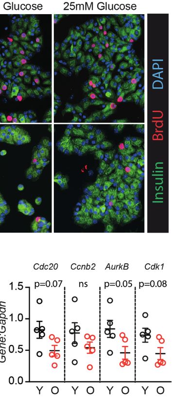
Snyder et al.                                                                                                              Stress-Induced Proliferation in Aging b-Cells

proliferation. To investigate, young and old dispersed mouse islet                    (Figures 2E–J). In 15 mM glucose, old islet cells contained
cells were cultured in high glucose media (15mM) and treated                          slightly increased levels of unspliced Xbp1 (Figure 2C), ATF6
with low dose thapsigargin (20 nM) or DMSO for 72 hours,                              targets HerpUD1 and Sel1L (Figure 2G), as well as Atf4
followed by RNA extraction (Figure 2A). Thapsigargin triggers                         transcripts (Figure 2I), suggesting a slightly more active UPR
the UPR by blocking activity of the sarco/endoplasmic reticulum                       in old, dispersed mouse islets cultured in high glucose. However,
Ca2+ ATPase (SERCA), preventing calcium uptake into the ER                            upon addition of low dose thapsigargin, young and old islets
necessary for proper protein folding (23). Xbp1 splicing was                          responded similarly in activating Xbp1 splicing and expression of
measured as a readout for IRE1 activity (Figures 2B–D) using an                       XBP1, ATF6 and ATF4 targets (Figures 2B, C, E, G, I). Aging
RT-PCR gel-based method (21), and canonical IRE1, ATF6, and                           did not alter the fold change in transcript abundance after
PERK/ATF4 pathway targets were quantified by qPCR                                      thapsigargin treatment for any UPR marker (Figures 2D, F,

                                  A                                                        B

                                  C                                                            D

                                  E                                                            F

                                  G                                                                H

                                  I                                                                J

 FIGURE 2 | Adaptive-dose ER stressor thapsigargin induces UPR pathway activation in old islet cells similarly to young islet cells. (A) Timeline of thapsigargin
 experiments. After allowing 24 hours to recover from trypsinization in normal glucose (5mM), dispersed islets from young and old mice were cultured in 15mM
 glucose in the presence of low-dose thapsigargin (Tg; 20nM) or vehicle (DMSO) for 72 hours, then harvested for RNA isolation. (B) RT-PCR gel-based assay for
 Xbp1 splicing, comparing young and old mouse islet cells cultured in high glucose with low-dose Tg or DMSO (n=5). (C, D) was used to quantify the relative
 abundance of unspliced and spliced Xbp1 (uXbp1 and sXbp1) compared to Actin (C); fold change is shown in (D). (E, F) qPCR assays for XBP1 targets (E); fold
 changes are shown in (F). (G, H) qPCR assays for Atf6 targets (G); fold changes are shown in (H). (I, J) qPCR assays for Atf4 pathway targets (I); fold changes are
 shown in (J). (C, E, G, I) Statistical analyses were performed by two-way repeated measures ANOVA and post-hoc Sidak’s multiple comparisons test. *Y-DMSO vs
 Y-Tg20. #Y-DMSO vs O-DMSO. &O-DMSO vs O-Tg20. ns, non significant. (D, F, H, J) Statistical analyses performed by t-test. One symbol, p < 0.05; two symbols,
 p < 0.01, three symbols, p < 0.001. ns, non significant.

Frontiers in Endocrinology | www.frontiersin.org                                  4                                        August 2021 | Volume 12 | Article 734079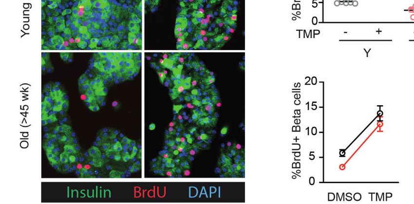
Snyder et al.                                                                                                                Stress-Induced Proliferation in Aging b-Cells

H, J). These results suggest that loss of UPR pathway activation                       thapsigargin did not differ between young and old for any gene, if
does not explain the age-associated reduction in glucose induced                       anything trending higher in the older samples (Figures 3E–H).
b-cell proliferation.                                                                  Thus, consistent with the similar UPR pathway activation between
                                                                                       young and old islet cells, aged mouse b-cells do not have impaired
Older b-Cells Retain the Proliferative                                                 UPR-induced proliferation in this ex vivo culture system.
Response to Low-Dose Thapsigargin
Although aged b-cells have impaired glucose-responsive                                 ATF6a Activation Robustly Induces
proliferation [Figure 1 and (24)], it is unknown whether aged                          Proliferation in Older b-Cells
b-cells can be induced to proliferate by low dose ER-stress as                         We previously identified ATF6a as a UPR component that is
previously illustrated for young b-cells (17). As expected, old                        required for glucose-induced b-cell proliferation, and when
b-cells had reduced BrdU incorporation compared to young b-                            overexpressed further increases proliferation in the context of
cells when cultured in 15mM glucose under DMSO control                                 15mM glucose (17). To test whether aging b-cells have impaired
conditions (Figures 3A–C). However, low-dose thapsigargin                              responsiveness to ATF6a, we applied stress-independent
significantly increased BrdU incorporation in both young and old                        activation of ATF6a using the ATF6a-dihydrofolate reductase
b-cells. Although fewer old b-cells stimulated with thapsigargin had                   (DHFR) fusion system previously developed by Shoulders et al.
BrdU incorporation compared to young b-cells under the same                            (20). Binding of the DHFR inhibitor trimethoprim (TMP) to the
conditions (Figure 3B), the magnitude of the pro-proliferative effect                  DHFR-domain stabilizes its folding structure; in the absence of
of thapsigargin was similar between young and old islet samples                        TMP the protein remains unfolded and is rapidly degraded (20).
(Figures 3C, D). Proliferation-associated mRNA transcripts were                        Young and old mouse b-cells were transduced with Ad-ATF6a-
increased in thapsigargin treated young and old dispersed islet                        DHFR and exposed to 10uM TMP or DMSO for 72 hours
cultures, and the fold change of expression upon stimulation with                      (Figure 4A). Activation of ATF6a-target genes was similar in

                                A                                           B                   C                     D

                                                                            E                                         F

                                G                                                                      H

 FIGURE 3 | Old b-cells increase proliferation in response to adaptive-dose ER stress similarly to young b-cells. Young (Y:10-14 weeks) and old (O: >45 weeks)
 dispersed mouse islet cells were cultured in 15mM glucose with or without low-dose thapsigargin as described in Figure 2. (A) Representative images of young and
 old dispersed mouse islet cells stained for insulin (green), BrdU (red) and DAPI. (B–D) Brdu+Ins+ cells were represented as a % of Ins+ cells (n=7-8) and graphed as
 individual data points (B) or in a grouped analysis (C); fold change (FC) is shown in (D). (E, F) qPCR results for proliferation marker transcripts Pcna and Ki67 (E);
 fold changes are shown in (F). (G, H) qPCR results for proliferation-associated transcripts Cdc20, Ccnb2, AurkB and Cdk1 (G); fold changes are shown in (H).
 (B, E, G) Statistical analyses were performed by two-way repeated measures ANOVA and post-hoc Sidak’s multiple comparisons test. *Y-DMSO vs Y-Tg20 #Y-
 DMSO vs O-DMSO &O-DMSO vs O-Tg20. ns, non significant. (D, F, H) Statistical analyses performed by t-test. One symbol, p < 0.05; two symbols, p < 0.01.
 ns, non significant.

Frontiers in Endocrinology | www.frontiersin.org                                   5                                         August 2021 | Volume 12 | Article 734079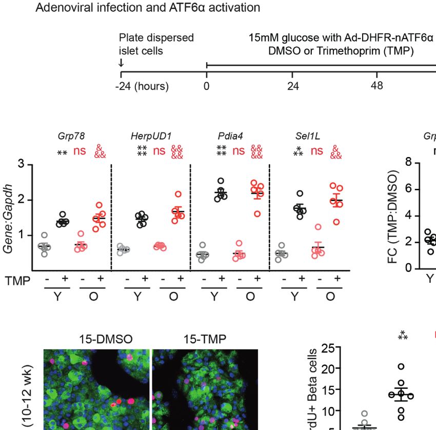
Snyder et al.                                                                                                               Stress-Induced Proliferation in Aging b-Cells

young and old dispersed islet cells (Figures 4B, C). Strikingly,                           dose thapsigargin. Remarkably, despite a lower proportion entering
stabilization of ATF6a with TMP caused a dramatic and                                      the cell cycle at baseline in 15mM glucose, old mouse b-cells also
significant increase in the fraction of b-cells incorporating                               retained a robust proliferation response to mild ER stress and to
BrdU (Figures 4D–G). The increased proliferation frequency                                 ATF6 activation. These results suggest the exciting possibility that if
of young and old b-cells upon activation of ATF6a were                                     UPR pathways can be harnessed therapeutically to increase b-cell
remarkably similar; the proliferation fold change was actually                             mass, the treatment might retain efficacy in older subjects.
higher in the old condition (Figures 4F, G). These data suggest                                Age-associated loss of b-cell proliferation may be due to
that ATF6a-driven b-cell proliferation is not lost in aging b-cells.                       quiescence, terminal differentiation, or senescence (9).
                                                                                           Although these states all feature cell cycle arrest, they are
                                                                                           physiologically distinct. Quiescence is a normal physiological
DISCUSSION                                                                                 state in which cells are non-proliferative but are poised to enter
                                                                                           the cell cycle when induced by pro-mitogenic signals. Terminal
This study tested the hypothesis that reduced proliferation in high                        differentiation is also a normal developmental program in which
glucose in aging b-cells is due to impaired activation of UPR                              an irreversible block of cell division occurs as cells acquire
pathways or UPR-dependent proliferation. Contrary to the                                   mature functional properties. In contrast, senescence is a
hypothesis, the results suggest that islet cells from older mice do                        pathological response to potentially oncogenic stimuli such as
not have impaired activation of UPR pathways in response to low                            DNA damage (25). Senescent cells impact tissue function both by

                                     A

                                     B                                                                C

                                     D                                              E

                                                                                       F                      G

 FIGURE 4 | Old b-cells increase proliferation in response to ATF6a activation similarly to young b-cells. (A) Timeline of experiments showing Ad-DHFR-ATF6a
 transduction and stabilization (activation) of the DHFR-ATF6a fusion protein with trimethoprim (TMP). After 24 hours recovery, young and old dispersed mouse islet
 cells were transduced with Ad-DHFR-ATF6a (MOI=10) with simultaneous treatment of 10 uM trimethoprim (TMP) or vehicle (DMSO) and addition of 15mM glucose.
 Cultures were harvested for RNA isolation or immunofluorescence staining at 72 hours. (B, C) qPCR assays showed similar activation of Atf6a targets in young and
 old islet cells (n=5) (B); fold changes are shown in (C). (D) Representative images of young and old Ad-DHFR-ATF6a transduced islet cells stained for insulin (green),
 BrdU (red) and DAPI (blue). (E, F) %Brdu+ were remarkably similar between young and old Ad-DHFR-ATF6a-expressing beta cells upon activation with TMP (n=7)
 expressed as individual data points (E), grouped analysis (F) and fold change (G). (B, E) Statistical analyses were performed by two-way repeated measures ANOVA
 and post-hoc Sidak’s multiple comparisons test. *Y-DMSO vs Y-Tg20 #Y-DMSO vs O-DMSO &O-DMSO vs O-Tg20. (G) Statistical analyses were performed by
 t-test. One symbol, p < 0.05; two symbols, p < 0.01, three symbols, p < 0.001, four symbols, p < 0.0001. ns, non significant.

Frontiers in Endocrinology | www.frontiersin.org                                   6                                        August 2021 | Volume 12 | Article 734079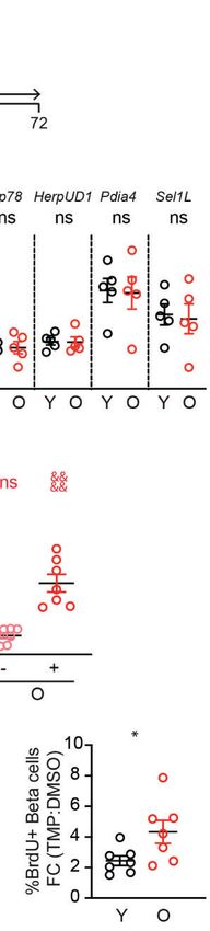
Snyder et al.                                                                                              Stress-Induced Proliferation in Aging b-Cells

cell-intrinsic restriction of regenerative capacity as well as               proliferation, nor whether ATF6a activation increases b-cell
through paracrine effects known as the “Senescence Associated                proliferation in vivo in any mice, young or old.
Secretory Phenotype” (SASP) (26, 27).                                            This study did not assess human b-cells for loss of
    All three of these processes occur in b-cells and could                  UPR signaling or UPR-induced proliferation. We previously
potentially be modified by UPR activation, either through                     reported that low dose ER stress and ATF6a overexpression
increasing likelihood that proliferation-competent b-cells enter             activated b-cell proliferation in dispersed human islet cells, but
the cell cycle, or by increasing the replication-competent pool. The         did not compare the proliferation between young and old donors
b-cell quiescence period is lengthened by older age and lower                (17). In that study, islet donors ranged in age from 15-65 years;
ambient glucose (28). Whether UPR activation modulates the b-                b-cells from the three oldest donors, ages 61, 63 and 65,
cell quiescence period remains unknown, but the current                      all increased BrdU incorporation in response to low dose
observations are consistent with the possibility that high glucose           thapsigargin and/or tunicamycin [see (17) Supplemental
and UPR activation additively or synergistically shorten the                 Figures 13C, G, K]. Intriguingly, a single-cell RNA sequencing
quiescence period. Age-related loss of proliferation has been                study reported that b-cells isolated from aged non-diabetic
associated with increased insulin secretion (29–31). The short               cynomolgus monkeys had aberrantly increased UPR expression
time frame of the current experiments argues against a mechanism             (36). It is unknown whether UPR triggers b-cell proliferation in
involving prevention of terminal differentiation, but UPR                    primate b-cells.
activation could potentially recall b-cells from a modified                       With respect to specific UPR pathways, this study only
terminally differentiated state. Finally, senescent b-cells are              tested the impact of ATF6a activation on b-cell proliferation.
known to accumulate in the islet with age and in diabetes, and               The IRE1/XBP1 pathway also impacts b-cell proliferation,
high fat diet or free fatty-acids induce senescence in the b-cells via       although both activation and inhibition reduced proliferation,
p16INK4a (27, 32–34). Derepression of the p16INK4a locus by                  suggesting this pathway may be less therapeutically useful
polycomb repressive complex 1 (PRC1) component BMI1 is                       as a tool to increase b-cell mass (17). Inhibition of the PERK
another important mechanism limiting b-cell proliferation in                 pathway did not impact mouse b-cell proliferation (17).
aging (35). Although loss of p16INK4a was not sufficient to                       Our study has weaknesses that may be addressed in future
restore the proliferative response to glucose in mouse islets (24),          studies. We did not assess the effect of aging on the induction of
the impact of UPR activation on b-cell senescence, SASP or BMI1              proliferation and cell cycle genes across a glucose range, focusing
remain unknown.                                                              instead on the effect of adding UPR in the context of 15mM
    Intriguingly, we observed slightly increased UPR activation in           glucose. We also did not test whether mouse sex impacts the
old mouse b-cells at baseline in high glucose, despite lower                 proliferation response to thapsigargin or ATF6a activation in
proliferation in high glucose compared to young b-cells. This                young or old b-cells.
effect was more pronounced when the qPCR results were                            In summary, the current study suggests the promising
normalized to Actin than when normalized to Gapdh (data not                  observation that older mouse b-cells retain proliferative
shown), which was possibly due to a systematic reduction in Actin            responsiveness to UPR, and to ATF6a activation specifically.
abundance in older islet cell cultures. Islet cell cultures are              However, many important questions remain. It is unclear
prepared following islet isolation and trypsinization, both of               whether UPR-induced b-cell proliferation can be sustained
which can cause ER stress; it is unknown whether aging alters                over a longer duration, or how the functional state of these
the ER stress response to these procedures. The conundrum of                 newly created daughter b-cells compares to the parent cells.
why older b-cells had reduced proliferation in “basal” high                  Whether this process can be harnessed to increase b-cell mass
glucose conditions despite slightly activated UPR, whereas                   in vivo in mice, or even humans, also remains untested. Future
proliferation was robustly activated in the same cultures when               studies will be necessary to determine if modulation of ATF6a
further stress was applied, might be explained by time frame or              has therapeutic utility for the treatment diabetes.
intensity of stress. The low-level stress response may have been
active for multiple days, bypassing a window for inducing
proliferation, or it may have been of insufficient intensity or
breadth to trigger cell cycle entry.                                         DATA AVAILABILITY STATEMENT
    How these results relate to b-cell proliferation in the
                                                                             The original contributions presented in the study are included in
aging in vivo environment remains unknown. However, the
                                                                             the article/Supplementary Material. Further inquiries can be
diminished proliferation frequency in high glucose consistently
                                                                             directed to the corresponding authors.
observed in the current studies suggests that at least some
portion of the in vivo aging lower-proliferation phenotype
was replicated in these ex vivo cultures. Some effects of aging
on b-cells are not cell-intrinsic, but rather are the result of              ETHICS STATEMENT
interaction with the aged extracellular environment. For
example, transplantation of old islets to young hosts restored               The animal study was reviewed and approved by Institutional
the proliferation of aged mouse b-cells (11, 12). It is not known            Care and Use Committees of the UMass Medical School and the
whether the aged microenvironment impacts UPR-induced                        Weill Cornell Medical College.

Frontiers in Endocrinology | www.frontiersin.org                         7                                 August 2021 | Volume 12 | Article 734079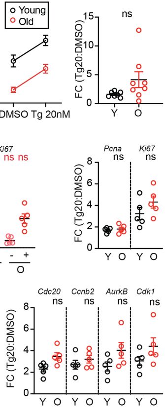
Snyder et al.                                                                                                                Stress-Induced Proliferation in Aging b-Cells

AUTHOR CONTRIBUTIONS                                                                    ACKNOWLEDGMENTS
The study was conceptualized by JS, RS, and LA. Experiments                             We are grateful to the Beta Cell Biology Groups at Weill Cornell
were performed and analysed by JS, CD, and RS. The first draft                           Medicine and at the UMass Medical School for helpful
of the manuscript was prepared by JS (text) and RS (figures).                            discussion. Some aged mice were donated by the laboratories
All authors contributed to the article and approved the                                 of Dr. John Keaney and Dr. Susan Swain. We gratefully thank Dr.
submitted version.                                                                      Luke Wiseman for providing the DHFR-ATF6a construct. Parts
                                                                                        of this manuscript were adapted from a master’s thesis submitted
                                                                                        by JS to the UMass Medical School.

FUNDING
                                                                                        SUPPLEMENTARY MATERIAL
This work was supported by NIH/NIDDK: R01DK114686
(LCA), R01DK113300 (LCA), R01DK124906 (LCA), NIH/                                       The Supplementary Material for this article can be found online
NIGMS: R25GM113686 (JTS) and George F. and Sybil H.                                     at: https://www.frontiersin.org/articles/10.3389/fendo.2021.
Fuller Foundation (LCA).                                                                734079/full#supplementary-material

                                                                                        14. Stolovich-Rain M, Hija A, Grimsby J, Glaser B, Dor Y. Pancreatic Beta Cells in
REFERENCES                                                                                  Very Old Mice Retain Capacity for Compensatory Proliferation. J Biol Chem
 1. Fishman EI, Stokes A, Preston SH. The Dynamics of Diabetes Among                        (2012) 287:27407–14. doi: 10.1074/jbc.M112.350736
    Birth Cohorts in the U.S. Diabetes Care (2014) 37:1052–9. doi: 10.2337/             15. Wang M, Kaufman RJ. Protein Misfolding in the Endoplasmic Reticulum as a
    dc13-1982                                                                               Conduit to Human Disease. Nature (2016) 529:326–35. doi: 10.1038/
 2. Diabetes Prevention Program Research Group. 10-Year Follow-Up of                        nature17041
    Diabetes Incidence and Weight Loss in the Diabetes Prevention Program               16. Sharma RB, Snyder JT, Alonso LC. Atf6a Impacts Cell Number by
    Outcomes Study. Lancet (2009) 374:1677–86. doi: 10.1016/S0140-6736(09)                  Influencing Survival, Death and Proliferation. Mol Metab (2019) 27:S69–80.
    61457-4                                                                                 doi: 10.1016/j.molmet.2019.06.005
 3. Charvat H, Goto A, Goto M, Inoue M, Heianza Y, Arase Y, et al. Impact of            17. Sharma RB, O’Donnell AC, Stamateris RE, Ha B, McCloskey KM, Reynolds
    Population Aging on Trends in Diabetes Prevalence: A Meta-Regression                    PR, et al. Insulin Demand Regulates b Cell Number via the Unfolded Protein
    Analysis of 160,000 Japanese Adults. J Diabetes Invest (2015) 6:533–42.                 Response. J Clin Invest (2015) 125:3831–46. doi: 10.1172/JCI79264
    doi: 10.1111/jdi.12333                                                              18. Ló pez-Otı́n C, Blasco MA, Partridge L, Serrano M, Kroemer G. The
 4. Huang ES, Laiteerapong N, Liu JY, John PM, Moffet HH, Karter AJ. Rates of               Hallmarks of Aging. Cell (2013) 153:1194. doi: 10.1016/j.cell.2013.05.039
    Complications and Mortality in Older Patients With Diabetes Mellitus: The           19. Sharma RB, Darko C, Zheng X, Gablaski B, Alonso LC. DNA Damage Does
    Diabetes and Aging Study. JAMA Internal Med (2014) 174:251–8.                           Not Cause Brdu Labeling of Mouse or Human b-Cells. Diabetes (2019) 68
    doi: 10.1001/jamainternmed.2013.12956                                                   (5):975–87. doi: 10.2337/db18-0761
 5. Liu J, Wu YY, Huang XM, Yang M, Zha BB, Wang F, et al. Ageing and Type 2            20. Shoulders MD, Ryno LM, Genereux JC, Moresco JJ, Tu PG, Wu C, et al.
    Diabetes in an Elderly Chinese Population: The Role of Insulin Resistance and           Stress-Independent Activation of XBP1s and/or ATF6 Reveals Three
    Beta Cell Dysfunction. Eur Rev Med Pharmacol Sci (2014) 18:1790–7.                      Functionally Diverse ER Proteostasis Environments. Cell Rep (2013)
 6. Linnemann AK, Baan M, Davis DB. Pancreatic b-Cell Proliferation in                      3:1279–92. doi: 10.1016/j.celrep.2013.03.024
    Obesity. Adv Nutr (2014) 5:278–88. doi: 10.3945/an.113.005488                       21. Prachasilchai W, Sonoda H, Yokota-Ikeda N, Oshikawa S, Aikawa C, Uchida
 7. Chen C, Cohrs CM, Stertmann J, Bozsak R, Speier S. Human Beta Cell Mass                 K, et al. A Protective Role of Unfolded Protein Response in Mouse Ischemic
    and Function in Diabetes: Recent Advances in Knowledge and Technologies                 Acute Kidney Injury. Eur J Pharmacol (2008) 592:138–45. doi: 10.1016/
    to Understand Disease Pathogenesis. Mol Metab (2017) 6:943–57.                          j.ejphar.2008.06.108
    doi: 10.1016/j.molmet.2017.06.019                                                   22. Klochendler A, Caspi I, Corem N, Moran M, Friedlich O, Elgavish S, et al. The
 8. Perl SY, Kushner JA, Buchholz BA, Meeker AK, Stein GM, Hsieh M, et al.                  Genetic Program of Pancreatic b-Cell Replication In Vivo. Diabetes (2016)
    Significant Human b-Cell Turnover is Limited to the First Three Decades of               65:2081–93. doi: 10.2337/db16-0003
    Life as Determined by In Vivo Thymidine Analog Incorporation and                    23. Oslowski CM, Urano F. Measuring ER Stress and the Unfolded Protein
    Radiocarbon Dating. J Clin Endocrinol Metab (2010) 95(10):E234–9.                       Response Using Mammalian Tissue Culture System. In: Methods in
    doi: 10.1210/jc.2010-0932                                                               Enzymology. (2011) 490:71–92. doi: 10.1016/B978-0-12-385114-7.00004-0
 9. Kushner JA. The Role of Aging Upon B Cell Turnover. J Clin Invest (2013)            24. Moreno-Asso A, Castaño C, Grilli A, Novials A, Servitja JM. Glucose
    123:990–5. doi: 10.1172/JCI64095.990                                                    Regulation of a Cell Cycle Gene Module Is Selectively Lost in Mouse
10. Tschen SI, Dhawan S, Gurlo T, Bhushan A. Age-Dependent Decline in b-Cell                Pancreatic Islets During Ageing. Diabetologia (2013) 56:1761–72.
    Proliferation Restricts the Capacity of b-Cell Regeneration in Mice. Diabetes           doi: 10.1007/s00125-013-2930-0
    (2009) 58:1312–20. doi: 10.2337/db08-1651                                           25. Campisi J, D’Adda Di Fagagna F. Cellular Senescence: When Bad Things
11. Chen X, Zhang X, Chen F, Larson CS, Wang LJ, Kaufman DB. Comparative                    Happen to Good Cells. Nat Rev Mol Cell Biol (2007) 8:729–40. doi: 10.1038/
    Study of Regenerative Potential of b Cells From Young and Aged Donor Mice               nrm2233
    Using a Novel Islet Transplantation Model. Transplantation (2009) 88:496–           26. Coppé JP, Patil CK, Rodier F, Sun Y, Muñoz DP, Goldstein J, et al.
    503. doi: 10.1097/TP.0b013e3181b0d2ee                                                   Senescence-Associated Secretory Phenotypes Reveal Cell-Nonautonomous
12. Almaça J, Molina J, Drigo RA, Abdulreda MH, Jeon WB, Berggren PO, et al.               Functions of Oncogenic RAS and the P53 Tumor Suppressor. PloS Biol
    Young Capillary Vessels Rejuvenate Aged Pancreatic Islets. Proc Natl Acad Sci           (2008) 6(12):2853-68. doi: 10.1371/journal.pbio.0060301
    USA (2014) 111:17612–7. doi: 10.1073/pnas.1414053111                                27. Aguayo-Mazzucato C, Andle J, Lee TB, Midha A, Talemal L, Chipashvili V,
13. Salpeter SJ, Khalaileh A, Weinberg-Corem N, Ziv O, Glaser B, Dor Y. Systemic            et al. Acceleration of b Cell Aging Determines Diabetes and Senolysis
    Regulation of the Age-Related Decline of Pancreatic b-Cell Replication.                 Improves Disease Outcomes. Cell Metab (2019) 30:129–42.e4. doi: 10.1016/
    Diabetes (2013) 62:2843–8. doi: 10.2337/db13-0160                                       j.cmet.2019.05.006

Frontiers in Endocrinology | www.frontiersin.org                                    8                                        August 2021 | Volume 12 | Article 734079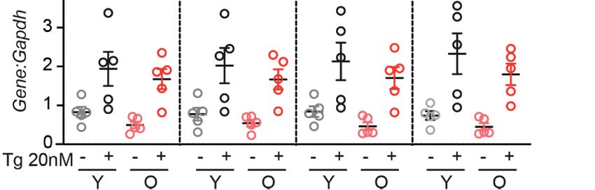
Snyder et al.                                                                                                                  Stress-Induced Proliferation in Aging b-Cells

28. Salpeter SJ, Klein AM, Huangfu D, Grimsby J, Dor Y. Glucose and Aging              35. Dhawan S, Tschen S-I, Bhushan A. Bmi-1 Regulates the Ink4a/Arf Locus to
    Control the Quiescence Period That Follows Pancreatic Beta Cell Replication.           Control Pancreatic b-Cell Proliferation. Genes Dev (2009) 23:906–11.
    Development (2010) 137:3205–13. doi: 10.1242/dev.054304                                doi: 10.1101/gad.1742609
29. Helman A, Klochendler A, Azazmeh N, Gabai Y, Horwitz E, Anzi S, et al. p16         36. Li J, Zheng Y, Yan P, Song M, Wang S, Sun L, et al. A Single-Cell
    Ink4a-Induced Senescence of Pancreatic Beta Cells Enhances Insulin                     Transcriptomic Atlas of Primate Pancreatic Islet Aging. Natl Sci Rev (2021)
    Secretion. Nat Med (2016) 22:412–20. doi: 10.1038/nm.4054                              8(2):nwaa.127. doi: 10.1093/nsr/nwaa127
30. Avrahami D, Li C, Zhang J, Schug J, Avrahami R, Rao S, et al. Aging-
    Dependent Demethylation of Regulatory Elements Correlates With                     Conflict of Interest: The authors declare that the research was conducted in the
    Chromatin State and Improved b Cell Function. Cell Metab (2015) 22:619–            absence of any commercial or financial relationships that could be construed as a
    32. doi: 10.1016/j.cmet.2015.07.025                                                potential conflict of interest.
31. Puri S, Roy N, Russ HA, Leonhardt L, French EK, Roy R, et al. Replication
    Confers b Cell Immaturity. Nat Commun (2018) 9(1):485. doi: 10.1038/               Publisher’s Note: All claims expressed in this article are solely those of the authors
    s41467-018-02939-0                                                                 and do not necessarily represent those of their affiliated organizations, or those of
32. Sone H, Kagawa Y. Pancreatic Beta Cell Senescence Contributes to the               the publisher, the editors and the reviewers. Any product that may be evaluated in
    Pathogenesis of Type 2 Diabetes in High-Fat Diet-Induced Diabetic Mice.            this article, or claim that may be made by its manufacturer, is not guaranteed or
    Diabetologia (2005) 48:58–67. doi: 10.1007/s00125-004-1605-2                       endorsed by the publisher.
33. Krishnamurthy J, Ramsey MR, Ligon KL, Torrice C, Koh A, Bonner-Weir S,
    et al. p16INK4a Induces an Age-Dependent Decline in Islet Regenerative             Copyright © 2021 Snyder, Darko, Sharma and Alonso. This is an open-access article
    Potential. Nature (2006) 443(7110):453–7. doi: 10.1038/nature05092                 distributed under the terms of the Creative Commons Attribution License (CC BY).
34. Pascoe J, Hollern D, Stamateris R, Abbasi M, Romano LC, Zou B, et al. Free         The use, distribution or reproduction in other forums is permitted, provided the
    Fatty Acids Block Glucose-Induced b-Cell Proliferation in Mice by Inducing         original author(s) and the copyright owner(s) are credited and that the original
    Cell Cycle Inhibitors P16 and P18. Diabetes (2012) 61:632–41. doi: 10.2337/        publication in this journal is cited, in accordance with accepted academic practice. No
    db11-0991                                                                          use, distribution or reproduction is permitted which does not comply with these terms.

Frontiers in Endocrinology | www.frontiersin.org                                   9                                           August 2021 | Volume 12 | Article 734079
You can also read