DYNAMICS OF BINDING ABILITY PREDICTION BETWEEN SPIKE PROTEIN AND HUMAN ACE2 REVEALS THE ADAPTIVE STRATEGY OF SARS COV 2 IN HUMANS
←
→
Page content transcription
If your browser does not render page correctly, please read the page content below
www.nature.com/scientificreports
OPEN Dynamics of binding ability
prediction between spike
protein and human ACE2
reveals the adaptive strategy
of SARS‑CoV‑2 in humans
Xia Xue1,3,10, Jianxiang Shi1,5,10, Hongen Xu1,10, Yaping Qin1,2, Zengguang Yang1,
Shuaisheng Feng1, Danhua Liu1,2, Liguo Jian2, Linlin Hua2, Yaohe Wang3,4, Qi Zhang6,
Xueyong Huang7, Xiaoju Zhang8, Xinxin Li9, Chunguang Chen9, Jiancheng Guo1,2,5*,
Wenxue Tang1,2,5* & Jianbo Liu2*
SARS-CoV-2 (severe acute respiratory syndrome coronavirus 2) is a novel coronavirus causing the
COVID-19 pandemic in 2020. High adaptive plasticity on the spike protein of SASR-CoV-2 enables
it to transmit across different host species. In the present study, we collected 2092 high-quality
genome sequences of SARS-CoV-2 from 160 regions in over 50 countries and reconstructed their
phylogeny. We also analyzed the polymorphic interaction between spike protein and human ACE2
(hACE2). Phylogenetic analysis of SARS-CoV-2 suggests that SARS-CoV-2 is probably originated from
a recombination event on the spike protein between a bat coronavirus and a pangolin coronavirus
that endows it humans infectivity. Compared with other regions in the S gene of SARS-CoV-2, the
direct-binding sites of the receptor-binding domain (RBD) is more conserved. We focused on 3,860
amino acid mutations in spike protein RBD (T333-C525) of SARS-CoV-2 and simulated their differential
stability and binding affinity to hACE2 (S19-D615). The results indicate no preference for SARS-CoV-2
infectivity on people of different ethnic groups. The variants in the spike protein of SARS-CoV-2
may also be a good indicator demonstrating the transmission route of SARS-CoV-2 from its natural
reservoir to human hosts.
Coronavirus is commonly found in nature but infects only mammals and birds1–3. Among the characterized
46 species, seven of them are human-susceptible4,5. Aside from SARS-CoV and MERS-CoV that cause deadly
pneumonia in humans by crossing the species b arrier3,6,7, SARS-CoV-2 is now causing a global pandemic of
respiratory disease (COVID-19) since the first confirmed cases in Wuhan city of China8. SARS-CoV-2 has been
identified as a novel β-coronavirus in the family of Coronaviridae2. Up to date, COVID-19 results in more than
80 million people infected and almost two million deaths worldwide. Compared with SARS-CoV and MERS-
CoV, SARS-CoV-2 spreads more rapidly due to its higher human i nfectivity8–10. Revealing the origin of SARS-
CoV-2 and its strategy for adapting to human hosts is therefore valuable to control the COVID-19 pandemic
and develop effective therapeutics and vaccines.
1
Academy of Medical Sciences, Precision Medicine Center of The Second Affiliated Hospital of Zhengzhou
University, Zhengzhou University, Zhengzhou, China. 2The Second Affiliated Hospital of Zhengzhou University,
Zhengzhou, China. 3National Centre for International Research in Cell and Gene Therapy, Academy of Medical
Science, Zhengzhou University, Zhengzhou, China. 4Center for Biomarkers and Biotherapeutics, Barts Cancer
Institute, Queen Mary University of London, London, UK. 5BGI College and Henan Institute of Medical and
Pharmaceutical Sciences, Zhengzhou University, Zhengzhou, Henan, China. 6State Key Laboratory of Esophageal
Cancer Prevention and Treatment, School of Pharmaceutical Sciences, Zhengzhou University, Zhengzhou,
China. 7Henan Province Center for Disease Control and Prevention, Zhengzhou, China. 8Henan Provincial
People’s Hospital, Zhengzhou, China. 9Henan Hospital of Infectious Diseases, Zhengzhou, China. 10These
authors contributed equally: Xia Xue, Jianxiang Shi and Hongen Xu. *email: gjc@zzu.edu.cn; twx@zzu.edu.cn;
jbliuzz@163.com
Scientific Reports | (2021) 11:3187 | https://doi.org/10.1038/s41598-021-82938-2 1
Vol.:(0123456789)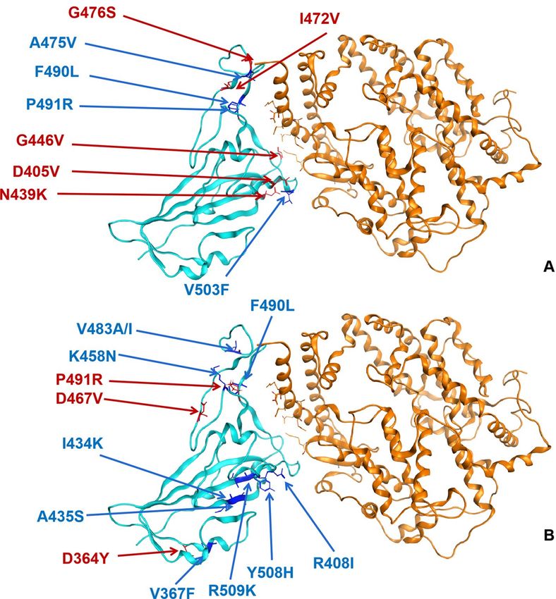
www.nature.com/scientificreports/
The genome structure of SARS-CoV-2 is similar to other beta-coronaviruses, which is composed of 5′-rep-
licase ORF1ab-S-envelope(E)-membrane(M)-N-3′ containing numbers of open reading frames (ORFs). The
phylogenetic relationship of various c oronavirus11 shows around 70% genome sequence similarity between
SARS-CoV-2 and SARS-CoV11. SARS-CoV-2 is more closely related to bat coronavirus RaTG13 particularly in
the spike (S) gene1. It is noted that although bat coronavirus RaTG13 is highly similar to SARS-CoV-2 especially
between genes, they differed significantly in some crucial genomic s tructures12. One of the most notable features
is a polybasic (furin) cleavage site insertion (PRRA residue) at the junction between two subunits (S1, S2) of S
protein1,2,12,13, which is uniquely present in SARS-CoV-2. Although some studies show bats could be the reservoir
host for many coronaviruses including SARS-CoV, the natural host of SARS-CoV-2 remains u ncertain4,10,14.
Given the global spread of COVID-19, it draws much attention to reveal the origins of the pandemic event.
A detailed evolutionary analysis of SARS-CoV-2 may explain its infectivity and transmissibility in different
animal hosts and provide evidence indicating whether this virus evolved naturally or created by manipulated
engineering.
Spike glycoprotein on the surface is critical for SARS-CoV-2 entry into the target cells by forming protrud-
ing homotrimers to recognize the host cell receptor and subsequently cause membrane fusion15. Spike protein
includes two linked subunits (S1, S2), while the receptor-binding domain (RBD) on the S1 subunit mediates the
binding to the peptidase domain (PD) of angiotensin-converting enzyme 2 (ACE2) in host cells. S2 subunit is
responsible for subsequent membrane fusion during viral i nfection14,16. In SARS-CoV, spike protein RBD is the
most diverse region in the whole genome, among which six amino acids (Y442, L472, N479, D480, T487, and
Y4911) have been confirmed to play a pivotal role for ACE2 receptor binding and further its transmission across
species boundary16. Similar to SARS-CoV, RBD variants acquired in different hosts have also been observed
in SARS-CoV-216. Thus, identifying the spike protein RBD variants affecting its binding to the ACE2 receptor
would be necessaryfor understanding the transmission mechanism of SARS-CoV-2 in human cells. Furthermore,
recent studies have shown that the capacity of hACE2 binding to spike protein of SARS-CoV-2 determines the
transmissibility of this coronavirus across species and in different p opulations14,17.
The present study intends to (1) reconstruct the phylogeny of SARS-CoV-2 strains from different populations
at the genomic level, (2) identify the variants on spike protein of SARS-CoV-2 based on the S gene sequences
and predict their differential stability and the binding affinities to hACE2, (3) explore the potential whether the
variants in hACE2 from the different ethnic groups may affect the infectivity of SARS-CoV-2. To this end, we
analyzed the variants of ACE2 in a large cohort including 1000 Chinese people and other ethnic groups to identify
the polymorphisms that may influence the binding of hACE2 and spike protein of SARS-CoV-2. Furthermore,
we predicted the affinities of spike protein RBD that binds to hACE2 variants to reveal whether those changes
would influence individuals’ susceptibility to SARS-CoV-2. Herein, our study partially explains the origin of
SARS-CoV-2 based on genomic and multiple key gene sequences, which allows us to elucidate the population
risk profiles and also help advance therapeutics such as a rationally designed soluble ACE2 receptor for the
management of COVID-19.
Materials and methods
Data preparation. Two thousand and ninety-two genome sequences of SARS-CoV-2 from 160 regions
including 50 countries were downloaded from GenBank (https://www.ncbi.nlm.nih.gov/genbank/) and GISAID
(https://www.gisaid.org/) databases, metadata of all the SARS-CoV-2 was shown in Supplementary Table S1.
Ten genomic sequences of SARS-CoV and MERS-CoV (five for each), ten genome sequences of pangolin coro-
navirus and three bat coronaviruses were collected from GISAID as well. The genome-sequencing of a local
donor has been completed in a laboratory from the local CDC, and the relevant data is stored in the local CDC
laboratory database for further analysis (unreleased). The whole-genome sequences were aligned with Clustal
Omega (V1.2.3)18 under the default setting of this program. Then we extracted the sequences of S, E, M, N genes
from the genome sequences of all beta coronaviruses in our analysis. SARS-CoV-2 Wuhan-01 (NC_045512) was
selected as the reference for gene sequences extraction, after alignment, a custom Python script (Supplementary
Script 1) was used to extract sequences. In order to analyze the S gene and S, E, M, N genes, we aligned them
individually with Clustal Omega (V1.2.3)18 and filtered them by sequences (Supplementary Script 1) contain-
ing more than 15 continuous unknown bases (N). Three hundred and twenty-seven non-repetitive sequences
of S gene and 469 of S–E–M–N sequences of SARS-CoV-2 were extracted from the filtered genome sequences,
respectively.
Phylogenetic analysis. After sequence alignment, Modelfinder in Iqtree (v1.6.12)19 was used to deter-
mine the most suitable model for each sequence dataset. We chose GTR + G model for genome sequences and
JC + I + G for gene tree construction. The maximum likelihood tree was reconstructed using MEGA v5.220 and
Iqtree (v1.6.12)19. We performed with 1000 Maximum number of iterations and applied approximate Bayes
test on our phylogenetic analysis, bootstrap values under replicating resample 1000 times. Figtree v1.421 was
performed on editing and screening the evolutionary tree topology. The calculation of genetic distance (whole
genome and individual genes) was carried out by Kimura two factor correction method for nucleic acid level
calculation. To avoid the prediction error caused by the selection of outgroups with a far evolutionary relation-
ship, the sequence of MERS-CoV and SARS-CoV were included as outgroups to predict the genetic relationship.
Variants of the spike protein and hACE2 prediction. In silico mutagenesis of SARS-CoV-2 spike
protein receptor-binding domain bound to the ACE2 receptor complex (PDB ID: 6M0J) was used to predict
the variable influence on binding affinity and protein stability. The proposed residue sites were substituted to
19 other amino acids and an ensemble of the conformations (the number of conformations was limit to 25)
Scientific Reports | (2021) 11:3187 | https://doi.org/10.1038/s41598-021-82938-2 2
Vol:.(1234567890)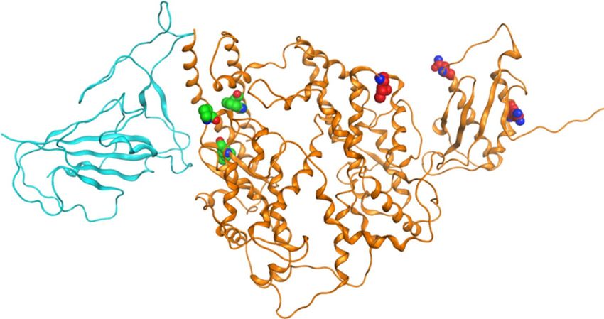
www.nature.com/scientificreports/
was generated for each mutant by low-mode MD (Molecular Dynamics), the parameters we used including
iteration limit 50, RMSD limit 0.25, energy window 10, conformation limit 25, fix residues farther than 4.5,
0 tether sidechains and one tether backbone. MM/GBVI was applied to calculate the binding affinity of each
conformation and ACE2 molecules. The force field used for calculation was OPLS-AA, and the implicit solvent
was the reaction field (R-Field) model. All calculations were performed in MOE (2019.01) (Molecular Operat-
ing Environment) software22,23. The structure of ACE2 receptor (PDB ID: 6M17) was used to perform in silico
mutagenesis. The genomic variants in the human ACE2 gene for different populations were downloaded from
the gnomAD database (https://gnomad.broadinstitute.org/). The proposed residue sites were substituted to the
amino acids that have the reported point mutations according to gnomAD. The parameters used to predict the
polymorphism of hACE2 binding to the spike protein of SARS-CoV-2 are the same as the previous parameters
for predicting the variants of the spike protein binding to hACE2. The statistical analysis for affinity and stability
of the complex was calculated in the MOE, the cut off of the dAffinity refers to being strong affinity was set as
definite three while definite one for high stability24,25.
Results
Phylogeny of SARS‑CoV‑2. The Maximum Likelihood (ML) phylogenic trees were constructed based on
2,112 genome sequences of beta coronaviruses derived from different hosts, with SARS-CoV and MERS-CoV
selected as the out-groups (Fig. 1a). After alignment, we merged identical sequences into one clade with labels
kept as one. The genomic tree showed all SARS-CoV-2 is closely related to the beta coronavirus isolated from the
horseshoe bat (RaTG13 and RmYN02), and pangolin coronaviruses were the ancestor that give rise to bat coro-
naviruses and SARS-CoV-2 in humans. According to sequence alignment, SARS-CoV-2 is not related closely
with SARS-CoV at the nucleic acid level despite the fact that they have over 72% sequence similarity. Moreover,
SARS-CoV-2 strains from different regions were hard to solved completely based on genome sequences with
multiple polytomies presented on the genome tree, particularly in the later time of this pandemic (Supplemen-
tary Fig. S1). We focused on the strains that are closer to the original direction and collected earlier in China.
Since the branches on the basal of the tree have higher bootstrap values and divergent more than others (Fig. 1b),
it may indicate that SARS-CoV-2 is slowly adapting to the human hosts’ environment in the course of COVID-
19. The complete phylogenomic tree is shown in Supplementary Fig. S1.
We extracted and aligned the S gene sequences of coronavirus from pangolin, bat (Rhinolophus spp) corona-
virus RaTG13 and SARS-CoV-2 (Fig. 2). We found an insertion that caused frameshift in S genes of pangolin
coronavirus, which demonstrated less similarity to the S gene in SARS-CoV-2 compared with that of RaTG13.
However, the insertions exhibited in pangolin indicated potential recombination in the spike protein of coro-
navirus could occur during its cross-species transmission. According to S gene sequences in different strains,
we found the similarity between SARS-CoV-2 and bat coronavirus RaTG13 is 98%, significantly higher than
the similarity with pangolins (85%). S gene from different hosts showed higher divergence on the phylogenetic
relationship (Fig. 3) compared with the genomic phylogenetic tree. Nevertheless, the phylogeny reconstructed
based on genome, S gene, and multiple structural genes (S, E, M, N) all indicate the SARS-CoV-2 is more closely
related to bat coronavirus than the coronaviruses isolated from pangolin.
The phylogenetic trees were also reconstructed based on the sequences of S, E, M, N genes (Supplementary
Fig. S1), which were extracted from the genome sequences and SARS-CoV-2 Wuhan-01 was used as the refer-
ence genome. We believe those structural protein-coding genes are fundamental to viral infection26. Unlike
the genomic phylogeny, structural protein-coding genes showed more divergence on two polar areas than the
strains in the middle part of the tree. Similar to the genomic tree, multiple gene sequences of SARS-CoV-2 are
divergent from outgroups and insertion or deletion were detected according to S gene sequences comparison.
Polymorphism prediction of spike protein in SARS‑CoV‑2. S gene has been studied as the key for
SARS-CoV-2 binding to host cells’ receptors. This gene showed less conservation compared with the rest of the
genome sequence of SARS-CoV-2. Some studies suggested that the high divergence in spike protein RBD and
specifically the direct binding sites to the ACE2 receptor might play an important role in SARS-CoV-2′s adapta-
tion to different animal hosts, and different human p opulations16,27. Combined with the phylogeny based on the
S gene, we also simulated all possible missense mutations in the RBD of the S gene and evaluated their binding
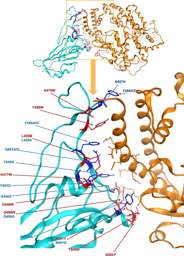
capacity with the ACE2 receptor of humans. Twenty-three sequence variants were predicted to show differential
affinity and stability of the spike protein (Fig. 4), among which nine variants exhibited increased affinity and sta-
bility while 14 with decreased affinity and stability (Table 1) compared to the wild-type complex. All simulated
outcomes of 3860 variants in spike protein are shown in Supplementary Table S2.
Figure 4 and Table 1 showed the missense mutations in spike protein RBD that have a strong affinity (Cut-
off = 3) and stability (Cutoff = 1) with hACE2 receptors. Interestingly, residues L455, Q498, and N501 would have
two potential mutations that leading to conflicting affinity changes, including increased affinity if leucine 455
changed to methionine, glutaurine 498 changed to tryptophan and asparagine 501 changed to tyrosine. At the
same time, reduced affinity is predicted when leucine 455 changed to alanine, glutamine 498 changed to alanine,
or asparagine 501 changed to glycine. The variants on the same residue causing opposite affinity fluctuation
suggest the mutations on those amino acids may lead to distinct adaptation directions for SARS-CoV-2 to gain
fitness in different hosts. Furthermore, Phe456, Gln493, and Phe486 carried two mutations that both result in
affinity reduction. The stability of spike protein with different mutations was summarized in Table 1. In summary,
we only found five mutants that could increase the stability of spike protein including G446W, G496A, Q498W,
N501Y and G502Y, which was not consistent with the affinity.
We then investigated the mutations of spike protein reported by SARS-CoV-2 database of China National
Center for Bioinformation (https: //bigd.big.ac.cn/ncov) compared with our predictions. A total of 1,150 variants
Scientific Reports | (2021) 11:3187 | https://doi.org/10.1038/s41598-021-82938-2 3
Vol.:(0123456789)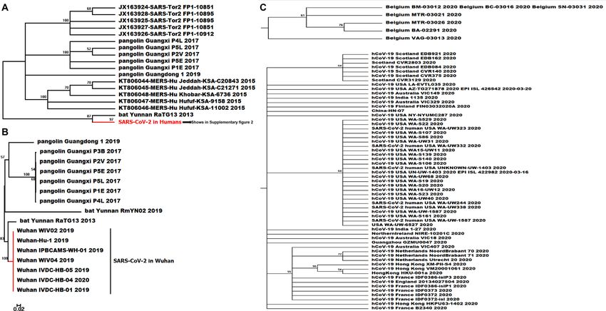
www.nature.com/scientificreports/

                                            Figure 1.  The ML phylogeny tree of different strains SARS-CoV-2 from the various region all around the world
                                            based on whole-genome sequences (Partial, complete phylogenetic tree is showen in Supplementary Fig. S1), the
                                            bootstrap values were mapped on the branch as long as the colors annotated for all clades. (A) The phylogenetic
                                            relationship reconstructed with genome sequences of SARS-CoV, MERS-CoV and pangolin/bat coronaviruses,
                                            SARS-CoV-2 strains from different regions showed in one consensus branch on the bottom of the sub-tree with
                                            bootstrap value 100, SARS-CoV-2 human on the bottom includes branches of SARS-CoV-2 from 164 regions
                                            globally (Supplementary Fig. S1); (B) The phylogenetic relationship of SARS-CoV-2 collected from Wuhan city
                                            of China in the early time of COVID-19 pandemic and beta coronaviruses derived from bat and pangolin.

                                            in spike protein were collected from the database and 643 missense variants (Before May 27th, 2020) were
                                            selected and analyzed their affinity and stability binding to hACE2 (Supplementary Table S3). Seventy-six mis-
                                            sense variants in region T333-C525 of spike protein and nine variants showed changes in the affinity of it to ACE2
                                            (Fig. 5A), and 13 variants cause structural stability change in spike protein RBD (Fig. 5B).

                                            Polymorphisms in ACE2 affect the binding ability to spike protein of SARS‑CoV‑2.                       We calcu-
                                            lated the population frequency of 388 missense variants in ACE2 collected from both gnomAD (Supplementary
                                            Table S4) and the local population (Table 2). Analysis of their stability and affinity to spike protein of SARS-
                                            COV-2 were performed in this study, and the results showed no obvious trend of changes on affinity and stability
                                            found in all variants compared with the wild type hACE2 based on our cutoff of defining strong/weak affinity
                                            and stability.

          Scientific Reports |   (2021) 11:3187 |                https://doi.org/10.1038/s41598-021-82938-2                                                 4

Vol:.(1234567890)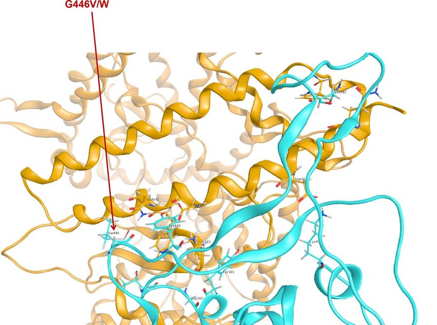
www.nature.com/scientificreports/

                                  Figure 2.  The sequence and amino acid alignment of the S gene in SARS-CoV-2 and coronaviruses found in
                                  bat and pangolin. The genomic structure of SARS-CoV-2 is shown in upper, the nucleic sequences alignment is
                                  shown in the middle while the amino acid is shown at the bottom of the figure.

                                  Figure 3.  Phylogenetic tree based on S gene (Partial, the complete tree was found in Supplementary
                                  Fig. S2), the bootstrap values were mapped on the branch as long as the colors annotated for all clades (A)
                                  The phylogenetic relationship of SARS-CoV, MERS-CoV and coronaviruses in pangolin and bat, SARS-
                                  CoV-2 in human on the bottom includes branches of SARS-CoV-2 from 164 regions based on S sequences
                                  (Supplementary Fig. S2); (B) The phylogenetic relationship based on S gene sequences of SARS-CoV-2 collected
                                  from Wuhan city of China in the early time of COVID-19 pandemic and beta coronaviruses derived from
                                  bat and pangolin; (C) Clades of SARS-CoV-2 from the regions show more divergent in S gene along with the
                                  COVID-19 pandemic.

                                  Discussion and conclusions
                                  Reconstructing the phylogeny of SARS-CoV-2 is of great value to understand its cross-species transmission
                                  pathway and to provide a reference for long-term infection prevention of zoonotic coronavirus. As a novel coro-
                                  navirus, the morphological information of SARS-CoV-2 is limited for phylogenic analysis, while the genomic
                                  data provides a timely and accurate resource to identify its divergence during this pandemic of COVID-191,4. The

Scientific Reports |   (2021) 11:3187 |               https://doi.org/10.1038/s41598-021-82938-2                                                5

                                                                                                                                           Vol.:(0123456789)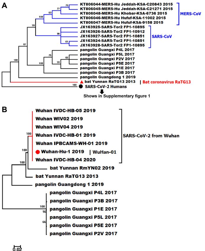
www.nature.com/scientificreports/
Figure 4. Identified variants in spike protein RBD mapped to the structure of spike protein in SARS-CoV-2 in
complex with ACE2 in humans. Blue = spike protein, Orange = ACE2 direct binding to spike protein. Twenty-
three point mutation causing affinity significant change on direct binding of spike protein of SARS-CoV-2, dark
blue presents decreasing affinity while red shows increasing.
genomes of SARS-CoV-2 has been reported about ~ 80% similar to SARS-CoV11 and ~ 66% similar to MERS-
CoV with low query coverage as 34%, but due to the structural and biological differences among them, they are
strikingly different coronaviruses s pecies9,28. The phylogeny of SARS-CoV-2 from over 50 countries in the present
study shows both the genomic and S sequences of SARS-CoV-2 are more closely related to SARS-CoV compared
with MERS-CoV. Despite the high genomic sequence similarity between SARS-CoV and SARS-CoV-2, the stabil-
ity and viability of SARS-CoV-2 are obviously higher than SARS-CoV in both aerosol environments and objects
surfaces29. This discrepancy indicates SARS-CoV-2 might adapt to human transmission better while SARS-CoV
failed to do s o6,9,28,30. Consistent with other s tudies1,2,11,31, we found all SARS-CoV-2 in our analysis were sister
taxon with coronavirus from horseshoe bat. Bat coronavirus RaTG13 is highly suspected as the natural reservoir
host of c oronavirus17,31 and the similarity between SARS-CoV-2 and SARS-bat is as high as 96.12%. Meanwhile,
Scientific Reports | (2021) 11:3187 | https://doi.org/10.1038/s41598-021-82938-2 6
Vol:.(1234567890)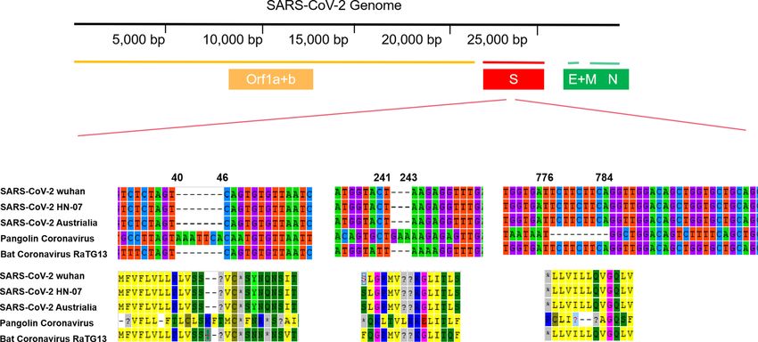
www.nature.com/scientificreports/
Mutants Affinity (kcal/mol) dAffinity (kcal/mol) Stability (kcal/mol) dStability (kcal/mol)
K417W − 90.17 − 3.68 − 3096.01 1.08
G446W − 98.31 − 7.56 − 3100.18 − 0.42
Y449A − 88.71 5.75 − 3098.95 2.41
Y453Q − 85.63 3.36 − 3100.47 1.40
L455M − 92.15 − 6.35 − 3102.42 1.05
F456A − 80.25 4.66 − 3100.11 3.05
F456G − 80.11 4.80 − 3099.85 3.31
A475W − 101.02 − 7.18 − 3101.49 0.28
F486A − 84.93 6.49 − 3099.87 1.99
F486G − 84.72 6.70 − 3099.50 2.37
N487H − 86.01 5.65 − 3098.24 0.79
Y489W − 96.85 − 5.74 − 3101.93 0.35
Q493A − 90.20 3.63 − 3099.00 1.21
Q493G − 89.33 4.50 − 3098.68 1.52
G496A − 85.87 4.21 − 3102.19 − 0.18
Q498A − 83.68 3.42 − 3100.48 1.45
Q498W − 92.51 − 5.41 − 3102.17 − 0.25
T500W − 96.40 − 5.19 − 3101.31 0.27
N501G − 85.81 3.33 − 3098.31 1.94
N501Y − 92.70 − 3.55 − 3100.42 − 0.17
G502Y − 95.82 − 6.55 − 3102.78 − 0.84
Y505A − 82.39 5.33 − 3101.12 1.98
Table 1. The affinity and stability of different variants of spike protein RBD bound to hACE2.
the coronavirus isolated from pangolins were detected 91.02% identical with SARS-CoV-2 at the nucleotide
level, which makes pangolins a potential intermediate host between the natural reservoir and human beings.
It is still uncertain that a common ancestral CoV that gave rise to SARS-CoV-2, bat coronavirus RaTG13 and
pangolin coronavirus, but we found bat coronavirus RaTG13 is more closely related to SARS-CoV-2 in humans
based on phylogenetic analysis of S gene. Furthermore, based on both genetic and genomic phylogeny, SARS-
CoV-2 is highly likely to come from a bat coronavirus which has also been studied in some other a nalyses1,31. We
found multiple insertions and deletions in S gene of SARS-pangolin compared with SARS-CoV-2 and SARS-bat,
which suggests the pangolin might not be the only direct or intermediate host from cross-species transmission.
Unique furin (polybasic) cleavage site insertion (PRRA) was found at the in-between region of S1 and S2 subunits
of spike protein of SARS-CoV-2, and this structure has uncharacterized potentials to enhance the infectivity of
SARS-CoV-2. Furthermore, this insertion has not been identified in SARS-pangolin and SARS-bat, which also
indicates the essential intermediate host of SARS-CoV-2 remains u nidentified14,15,27.
To most SARS-coronavirus, spike protein is the critical initiator for viral infection by recognizing the recep-
tor of host c ells5,17. S gene shows more divergence in sequences and protein structures relative to other regions
of coronaviruse14. Along with gradual adaptation to the cellular environment of humans, mutations occur in
spike protein, which might change the binding affinity and stability of the spike protein-hACE2 complex. An
enhancement of such binding affinity and stability would increase the transmissibility of SARS-CoV-2 among
humans and cause more severe disease16. For instance, the transmissibility and pathogenicity of SARS-CoV
decreased comparing strains isolated in 2003–2004 with the ones from 2002 to 200332,33. Similarly, MERS-CoV
might have been controlled literally by itself along with spreading in human beings9. We predicted the affinity and
stability of spike protein RBD (T333-C525) of SARS-CoV-2 by using MOE2019 (Molecular Operating Environ-
ment) to hACE2 (S19-D615), point mutations on spike protein were simulated with amino acid scan and 3860
mutations were set up for analysis (Supplementary Table S1). We selected 23 variants of spike protein RBD that
causing significantly higher or lower stability and binding affinity to hACE2 with the cutoff set up as three for
determining significance (Table 1). Nine mutations were found to significantly enhance the binding between
spike protein and hACE2, which gives us an important direction in monitoring the arisal of mutations influenc-
ing the transmissibility of SARS-CoV-2 in the long run. Amino acid change on G446 (Fig. 6) was also reported
through the pandemic from CDC (Fig. 6, Supplementary Table S2), and this sample was documented as collected
in early March that is closer to the original direction of the phylogenetic tree based on genomic data (Fig. 1).
According to 1,150 variants in the spike protein of SASR-CoV-2 that including 634 missense mutations
reported by CDC, we found 76 missense variants located in the region between T333-C525 and 5 variants
enhanced its affinity binding to hACE2 (Table 3) and 3 of them increased the stability of spike-hACE2 complex
(Table 4). Furthermore, we collected the genome sequences of SARS-CoV-2 harboring those variants in spike
protein and found the variants V483A occured in 26 strains from the USA, V367F occured in 12 strains from
Hong Kong, Australia, and other European countries, and a variant G446V with predicted highly enhanced affin-
ity was found in a strain from Australia (Supplementary Table S2). We mapped those strains onto our genomic
Scientific Reports | (2021) 11:3187 | https://doi.org/10.1038/s41598-021-82938-2 7
Vol.:(0123456789)www.nature.com/scientificreports/
Figure 5. Reported variants in spike protein RBD mapped to the structure of spike protein in SARS-CoV-2 in
complex with ACE2 in humans. Blue = spike protein, Orange = ACE2, dark blue presents decreasing affinity and
stability while red presents increasing ones. (A) Affinity; (B) Stability.
Mutants Affinity (kcal/mol) dAffinity (kcal/mol) Stability (kcal/mol) dStability (kcal/mol)
I468V − 65.09 6.88E−12 − 3107.75 0.85
N638S − 65.09 − 4.76E−12 − 5930.30 0.33
R708Q − 64.99 0 − 2906.26 2.17
Table 2. Analysis of affinity and stability of polymorphism in ACE2 from local population binding to spike
protein of SARS-COV-2.
phylogenetic tree and found they dispersed distributed in the position close to the bat coronaviruses and pangolin
coronaviruses. As COVID-19 spreads globally, few reports analyze the origin of SARS-CoV-2 according to the
divergent pattern in spike protein. We found multiple variants in spike protein from different countries and
clustered in the ancestral direction on phylogeny which might suggest the plasticity of spike protein is essential
to the host-adaption of SARS-CoV-2 infection in humans and other hosts. However, due to the limited detec-
tion capability and restricted availability of samples from infected animals, the variants available in the national
database are incompleted, more data is needed for identifying the patient zero.
Given the SARS-CoV-2 strains have variants on spike protein relative to a reference sequence of SARS-
CoV-2 (NC_045512.2) were found more divergence on the phylogenetic position that closer to coronaviruses
isolated from bat and pangolin, it might indicate an evolutionary pattern occurred in S gene enabling SARS-
CoV-2 adaptation to human hosts. However, since it is difficult to find the exact time the patient zero who got
infected by SARS-CoV-2, the date of the sample collection could mislead the results. We believe further clinical
information from all the countries would be needed and deeper cooperation and collaboration could be help-
ful. We shared all the analysis of over 3000 variants in spike protein to help the world tracking the mutations of
Scientific Reports | (2021) 11:3187 | https://doi.org/10.1038/s41598-021-82938-2 8
Vol:.(1234567890)www.nature.com/scientificreports/
Figure 6. Residue G446 variants in documents from CDC (G446V) and in our prediction (G446W).
Mutants Affinity (kcal/mol) dAffinity (kcal/mol) Stability (kcal/mol) dStability (kcal/mol)
D405V − 88.38 − 0.35 − 791.79 0.24
N439K − 88.46 − 0.46 − 796.46 0.57
I472V − 87.92 − 0.80 − 799.30 0.99
G446V − 91.90 − 1.15 − 3099.43 0.33
G476S − 91.72 − 1.14 − 3100.24 0.63
A475V − 90.61 3.24 − 3101.96 − 0.19
F490L − 90.59 2.05 − 3100.21 1.28
P491R − 88.25 2.31 − 803.62 − 0.59
V503F − 91.94 1.54 − 3100.99 0.23
Table 3. The affinity of the variants of SARS-CoV-2 spike RBD bound to human ACE2, red marks the
enhanced ones while black represents the decreased ones.
Mutants Affinity (kcal/mol) dAffinity (kcal/mol) Stability (kcal/mol) dStability (kcal/mol)
D364Y − 88.26 − 0.00 − 792.45 − 0.87
D467V − 88.26 − 0.00 − 793.14 − 0.68
P491R − 88.25 2.31 − 803.62 − 0.59
V367F − 88.26 6.85E−12 − 799.28 1.06
R408I − 88.26 − 0.00 − 792.67 1.44
I434K − 88.26 1.27E−07 − 800.49 1.84
A435S − 88.26 − 4.22E−05 − 799.69 1.04
K458N − 88.186 0.06 − 793.07 1.29
V483A − 88.606 − 0.012 − 797.29 1.01
V483I − 88.726 − 0.13 − 797.21 1.09
F490L − 90.596 2.05 − 3100.21 1.28
R509K − 88.26 5.45E−05 − 797.38 2.04
Y508H − 88.18 0.05 − 798.96 2.41
Table 4. The stability of the variants of SARS-CoV-2 spike RBD bound to human ACE2, red marks the
enhanced ones while black represents the decreased ones.
SARS-CoV-2 and select the potential druggable targets or neutral inhibitors to prevent further life loss brought
by the pandemic of COVID-19.
Host-virus interaction over time makes a natural selection on both virus and host c ells34. It is believed that
the variants in hACE2 receptor play a role in SARS-CoV-2 infection. Cao and his colleagues (2020)35 found the
polymorphisms of hACE2 did not bring differences in resisting SARS-CoV-2 infection, but the data they used is
limited by sample size. A study published on bioRxiv showed hACE2 with the variants K26R, S16P, T27A, K31R,
Scientific Reports | (2021) 11:3187 | https://doi.org/10.1038/s41598-021-82938-2 9
Vol.:(0123456789)www.nature.com/scientificreports/
Figure 7. Spike protein polymorphous points from local population and Italy37, the Italian ones are marked as
green while local ones marked as red.
H34R, E35K, E37K, D38V, N51S, N64K, K68E, F72V, T921, Q102P, G326E, G352V, D355N, H378R, Q388L,
and D509Y were able to increase the susceptibility of the individuals who carry these variations, while K31R,
E35K, E37K, D38V, N33I, H34R, Q388L, and Y83H decreased binding capacity between hACE2 and spike pro-
tein of SARS-CoV-2. Herein, we identified all the variants in hACE2 in GnomAD and local dataset, with their
population frequency in different ethnic groups calculated. Same with Eric’s results36, no significant differences
in hACE2 variant frequency were found from gnomAD while three variants (I468V, N638S, and R708Q) with
high allele frequency hACE2 were identified in the local population, and their allele frequencies were very low
in other populations (Fig. 7). Researchers found three unique variants in hACE2 from the Italian population
that might be corresponding to the high fatality of COVID-19 in Italy, which are P389H, W69C, and L 351V37.
We compared the unique variants from the local and Italian populations by mapping them on the hACE
protein structure (Fig. 7). The variants found in Italian were closer to the binding region of spike protein than
Chinese variants; however, in silico simulation indicated that none of them change the affinity and stability
of spike protein of SARS-CoV-2 and hACE2 complex significantly (Table 2). We did not find any variants in
hACE2 that would increase or decrease the affinity and stability of spike protein binding to hACE2, and which
may indicate SARS-CoV-2 enables indiscriminately infect to all humans ethnic groups. Due to various diets and
health conditions, the efficiency and virulence of SARS-CoV-2 might be different, and the mortality rate is also
more likely to be linked to the medical and health conditions of patients.
Up to date, there is no effective therapeutics approved universally for both treatment and prevention of
COVID-19. To develop an efficient inhibitor or vaccine to prevent SARS-CoV-2 leading to COVID-19, it is still
urgent to understand the mechanism of SARS-CoV-2 adaptation and transmission in different hosts, particularly
in humans. The variants in the spike protein of SARS-CoV-2 and hACE2 would provide a database for tracking
the adaptive mutation of SARS-CoV-2 and potential recombination events across different species. Our ongoing
study shows ORF1ab of SARS-CoV-2 and MERS-CoV may undergo recombination and result in more severe
disease (unpublished). The analysis shared in this study would provide useful genetic information to prevent
the recurrence of this epidemic, and protect human beings from zoonotic coronavirus infection in the future.
Received: 1 July 2020; Accepted: 27 January 2021
References
1. Andersen, K. G., Rambaut, A., Lipkin, W. I., Holmes, E. C. & Garry, R. F. The proximal origin of SARS-CoV-2. Nat. Med. 26,
450–452. https://doi.org/10.1038/s41591-020-0820-9 (2020).
2. Singhal, T. A review of coronavirus disease-2019 (COVID-19). Indian J. Pediatrics 87, 281–286. https://doi.org/10.1007/s1209
8-020-03263-6 (2020).
3. Shi, Z. & Hu, Z. A review of studies on animal reservoirs of the SARS coronavirus. Virus Res. 133, 74–87. https: //doi.org/10.1016/j.
virusres.2007.03.012 (2008).
4. Cui, J. et al. Evolutionary relationships between bat coronaviruses and their hosts. Emerg. Infect. Dis. 13, 1526–1532. https://doi.
org/10.3201/eid1310.070448 (2007).
5. van der Hoek, L. et al. Identification of a new human coronavirus. Nat. Med. 10, 368–373. https://doi.org/10.1038/nm1024 (2004).
6. Di Mascio, D. et al. Outcome of Coronavirus spectrum infections (SARS, MERS, COVID 1 -19) during pregnancy: a systematic
review and meta-analysis. Am J Obstet Gynecol MFM, 100107, https://doi.org/10.1016/j.ajogmf.2020.100107 (2020).
7. Ji, J. S. Origins of MERS-CoV, and lessons for 2019-nCoV. Lancet Planetary Health 4, e93. https://doi.org/10.1016/s2542
-5196(20)30032-2 (2020).
Scientific Reports | (2021) 11:3187 | https://doi.org/10.1038/s41598-021-82938-2 10
Vol:.(1234567890)www.nature.com/scientificreports/
8. Zhang, J. J. et al. Clinical characteristics of 140 patients infected with SARS-CoV-2 in Wuhan, China. Allergy https://doi.
org/10.1111/all.14238(2020).
9. Jiang, X., Rayner, S. & Luo, M. H. Does SARS-CoV-2 has a longer incubation period than SARS and MERS?. J. Med. Virol. 92,
476–478. https://doi.org/10.1002/jmv.25708 (2020).
10. Lee, P. I. & Hsueh, P. R. Emerging threats from zoonotic coronaviruses-from SARS and MERS to 2019-nCoV. J. Microbiol. Immunol.
Infect. https://doi.org/10.1016/j.jmii.2020.02.001 (2020).
11. Zhang, Y. Z. & Holmes, E. C. A genomic perspective on the origin and emergence of SARS-CoV-2. Cell 181, 223–227. https://doi.
org/10.1016/j.cell.2020.03.035 (2020).
12. Wu, A. et al. Genome composition and divergence of the novel coronavirus (2019-nCoV) originating in China. Cell Host Microbe
27, 325–328. https://doi.org/10.1016/j.chom.2020.02.001 (2020).
13. Hoffmann, M. et al. SARS-CoV-2 cell entry depends on ACE2 and TMPRSS2 and is blocked by a clinically proven protease inhibi-
tor. Cell 181, 271–280 e278, https://doi.org/10.1016/j.cell.2020.02.052 (2020).
14. Lan, J. et al. Structure of the SARS-CoV-2 spike receptor-binding domain bound to the ACE2 receptor. Nature https://doi.
org/10.1038/s41586-020-2180-5 (2020).
15. Walls, A. C. et al. Structure, Function, and Antigenicity of the SARS-CoV-2 Spike Glycoprotein. Cell 181, 281–292 e286, https://
doi.org/10.1016/j.cell.2020.02.058 (2020).
16. Ortega, J. T., Serrano, M. L., Pujol, F. H. & Rangel, H. R. Role of changes in SARS-CoV-2 spike protein in the interaction with the
human ACE2 receptor: an in silico analysis. EXCLI J 19, 410–417. https://doi.org/10.17179/excli2020-1167 (2020).
17. Shang, J. et al. Structural basis of receptor recognition by SARS-CoV-2. Nature 581, 221–224. https://doi.org/10.1038/s41586-020-
2179-y (2020).
18. Sievers, F. et al. Fast, scalable generation of high-quality protein multiple sequence alignments using Clustal Omega. Mol. Syst.
Biol. 7, 539–539. https://doi.org/10.1038/msb.2011.75 (2011).
19. Minh, B. Q. et al. IQ-TREE 2: new models and efficient methods for phylogenetic inference in the genomic Era. Mol. Biol. Evol.
37, 1530–1534. https://doi.org/10.1093/molbev/msaa015 (2020).
20. Kumar, S., Stecher, G., Li, M., Knyaz, C. & Tamura, K. MEGA X: molecular evolutionary genetics analysis across computing
platforms. Mol. Biol. Evol. 35, 1547–1549. https://doi.org/10.1093/molbev/msy096 (2018).
21. Rambaut, A. FigTree, version 1.4.3. Computer program distributed by the author, website: http://www.tree.bio.ed.ac.uk/software/
figtree/ [accessed January 4, 2011] (2009).
22. Inc, C. C. G. Molecular operating environment (MOE). (2016).
23. Chemical Computing Group ULC, 1010 Sherbrooke St. West, Suite #910, Montreal, QC, Canada, H3A 2R7, 2019. (2019).
24. Du, J. et al. The design of high affinity human PD-1 mutants by using molecular dynamics simulations (MD). Cell Commun. Signal
16, 25. https://doi.org/10.1186/s12964-018-0239-9 (2018).
25. Dehouck, Y. et al. Fast and accurate predictions of protein stability changes upon mutations using statistical potentials and neural
networks: PoPMuSiC-2.0. Bioinformatics 25, 2537–2543, https://doi.org/10.1093/bioinformatics/btp445 (2009).
26. Srinivasan, S. et al. Structural genomics of SARS-CoV-2 indicates evolutionary conserved functional regions of viral proteins.
Viruses 12, https://doi.org/10.3390/v12040360 (2020).
27. Zhang, H., Penninger, J. M., Li, Y., Zhong, N. & Slutsky, A. S. Angiotensin-converting enzyme 2 (ACE2) as a SARS-CoV-2 receptor:
molecular mechanisms and potential therapeutic target. Intensive Care Med. 46, 586–590. https://doi.org/10.1007/s00134-020-
05985-9 (2020).
28. Mahase, E. Coronavirus covid-19 has killed more people than SARS and MERS combined, despite lower case fatality rate. BMJ
368, m641. https://doi.org/10.1136/bmj.m641 (2020).
29. van Doremalen, N. et al. Aerosol and Surface Stability of SARS-CoV-2 as Compared with SARS-CoV-1. N. Engl. J. Med. 382,
1564–1567. https://doi.org/10.1056/NEJMc2004973 (2020).
30. Prompetchara, E., Ketloy, C. & Palaga, T. Immune responses in COVID-19 and potential vaccines: lessons learned from SARS and
MERS epidemic. Asian Pac. J. Allergy Immunol. 38, 1–9. https://doi.org/10.12932/AP-200220-0772 (2020).
31. Xiao, K. et al. Isolation of SARS-CoV-2-related coronavirus from Malayan pangolins. Nature https://doi.org/10.1038/s41586-020-
2313-x (2020).
32. Molecular evolution of the SARS coronavirus during the course of the SARS epidemic in China. Science 303, 1666, https://doi.
org/10.1126/science.1092002 (2004).
33. Li, W. et al. Bats are natural reservoirs of SARS-like coronaviruses. Science 310, 676. https://doi.org/10.1126/science.1118391
(2005).
34. Malik, R. & Johnston, D. Dendritic GIRK channels gate the integration window, plateau potentials, and induction of synaptic
plasticity in dorsal but not ventral CA1 neurons. J. Neurosci. 37, 3940. https://doi.org/10.1523/JNEUROSCI.2784-16.2017 (2017).
35. Cao, Y. et al. Comparative genetic analysis of the novel coronavirus (2019-nCoV/SARS-CoV-2) receptor ACE2 in different popula-
tions. Cell Discov. 6, 11. https://doi.org/10.1038/s41421-020-0147-1 (2020).
36. Stawiski, E. W. et al. Human ACE2 receptor polymorphisms predict SARS-CoV-2 susceptibility. bioRxiv, 2020.2004.2007.024752,
https://doi.org/10.1101/2020.04.07.024752 (2020).
37. Renieri, A. et al. ACE2 variants underlie interindividual variability and susceptibility to COVID-19 in Italian population. medRxiv,
2020.2004.2003.20047977, https://doi.org/10.1101/2020.04.03.20047977 (2020).
Acknowledgements
We thank the supports provided by the Academy of Medical sciences Zhengzhou University and the data analy-
sis was supported by the Supercomputing Center in Zhengzhou University (Zhengzhou). The study is funded
by the Collaborative Innovation Project of Zhengzhou (Zhengzhou University) (Grant No. 18XTZX12004) to
WT, Henan health and Planning Commission (Grant No. SBGJ2018041) to JG, the Department of Science and
Technology of Henan Province (Grants No. 201100312100) to JL, the Key Scientific and Technological Research
Projects in Henan Province of China (Grant No. 182102310094) to QZ, Project of Basic Research Fund of Henan
Institute of Medical and Pharmacological Sciences to JS (Grant No.: 2019BP0202). Any opinions, findings,
conclusions, or recommendations expressed here are those of the author(s) and do not necessarily reflect the
views of the funding agencies.
Authors contributions
X.X., J.X.S., H.E.X. designed and conducted the current study. Y.P.Q., Z.G.Y., S.S.F., D.H.L. provided bio-infor-
matics supports and performed the main analysis. W.X.T., J.C.G., Y.H.W., J.B.L. supported the methodological
and pathological information. X.X., J.X.S., H.E.X., Y.P.Q. wrote the manuscript. All authors revised and approved
the final manuscript.
Scientific Reports | (2021) 11:3187 | https://doi.org/10.1038/s41598-021-82938-2 11
Vol.:(0123456789)www.nature.com/scientificreports/
Additional information
Supplementary Information The online version contains supplementary material available at https://doi.
org/10.1038/s41598-021-82938-2.
Correspondence and requests for materials should be addressed to J.G., W.T. or J.L.
Reprints and permissions information is available at www.nature.com/reprints.
Publisher’s note Springer Nature remains neutral with regard to jurisdictional claims in published maps and
institutional affiliations.
Open Access This article is licensed under a Creative Commons Attribution 4.0 International
License, which permits use, sharing, adaptation, distribution and reproduction in any medium or
format, as long as you give appropriate credit to the original author(s) and the source, provide a link to the
Creative Commons licence, and indicate if changes were made. The images or other third party material in this
article are included in the article’s Creative Commons licence, unless indicated otherwise in a credit line to the
material. If material is not included in the article’s Creative Commons licence and your intended use is not
permitted by statutory regulation or exceeds the permitted use, you will need to obtain permission directly from
the copyright holder. To view a copy of this licence, visit http://creativecommons.org/licenses/by/4.0/.
© The Author(s) 2021
Scientific Reports | (2021) 11:3187 | https://doi.org/10.1038/s41598-021-82938-2 12
Vol:.(1234567890)You can also read