Downstaging and Resection of Initially Unresectable Hepatocellular Carcinoma with Tyrosine Kinase Inhibitor and Anti-PD-1 Antibody Combinations
←
→
Page content transcription
If your browser does not render page correctly, please read the page content below
Original Paper

                                                          Liver Cancer 2021;10:320–329                                                         Received: September 3, 2020
                                                                                                                                               Accepted: January 7, 2021
                                                          DOI: 10.1159/000514313                                                               Published online: March 30, 2021

Downstaging and Resection of Initially
Unresectable Hepatocellular Carcinoma with
Tyrosine Kinase Inhibitor and Anti-PD-1 Antibody
Combinations
Xiao-Dong Zhu a Cheng Huang a Ying-Hao Shen a Yuan Ji b Ning-Ling Ge c Xu-Dong Qu d
Lingli Chen b Wen-Kai Shi e Mei-Ling Li a Jin-Jin Zhu a Chang-Jun Tan a Zhao-You Tang a
Jian Zhou a Jia Fan a Hui-Chuan Sun a
aDepartment
              of Liver Surgery and Transplantation, Liver Cancer Institute and Zhongshan Hospital, Fudan
University, Shanghai, China; bDepartment of Pathology, Zhongshan Hospital, Fudan University, Shanghai, China;
cDepartment of Hepatic Oncology, Liver Cancer Institute and Zhongshan Hospital, Fudan University, Shanghai,

China; dDepartment of Interventional Radiology, Zhongshan Hospital, Fudan University, Shanghai, China;
eDepartment of Hepatobiliary and Pancreatic Surgery, The First Affiliated Hospital of Zhengzhou University,

Zhengzhou, China

Keywords                                                                                    3.2 months (range: 2.4–8.3 months) after the initiation of com-
Hepatocellular carcinoma · Conversion therapy ·                                             bination therapy. At baseline, these 10 patients had a median
Anti-angiogenic therapy · Anti-PD-1 antibody                                                largest tumor diameter of 9.3 cm, 7 had Barcelona Clinic Liver
                                                                                            Cancer stage C (vascular invasion) disease, 2 had stage B, and 1
                                                                                            had stage A. Before surgery, 6 patients were evaluated as a par-
Abstract                                                                                    tial response, 3 stable disease, and 1 partial response in the in-
Background: Combined therapy with tyrosine kinase inhibi-                                   trahepatic lesion but a new metastatic lesion in the right adre-
tors (TKIs) and anti-PD-1 antibodies has shown high tumor re-                               nal gland. Six patients (60%) achieved a pathological complete
sponse rates for patients with unresectable hepatocellular car-                             response. One patient died from immune-related adverse ef-
cinoma (HCC). However, using this treatment strategy to con-                                fects 2.4 months after hepatectomy. After a median follow-up
vert initially unresectable HCC to resectable HCC was not                                   of 11.2 months (range: 7.8–15.9 months) for other 9 patients, 8
reported. Methods: Consecutive patients with unresectable                                   survived without disease recurrence, and 1 experienced tumor
HCC who received first-line therapy with combined TKI/anti-                                 recurrence. Conclusions: Combination of TKI/anti-PD-1 anti-
PD-1 antibodies were analyzed. Tumor response and resect-                                   bodies is a feasible conversion therapy for patients with unre-
ability were evaluated via imaging every 2 months (±2 weeks)                                sectable HCC to become resectable. This study represents the
using RECIST v1.1. Resectability criteria were (1) R0 resection                             largest patient cohort on downstaging role of combinational
could be achieved with sufficient remnant liver volume and                                  systemic therapy on TKI and PD-1 antibody for HCC.
function; (2) intrahepatic lesions were evaluated as partial re-                                                                                © 2021 The Author(s)
sponses or stable disease for at least 2 months; (3) no severe or                                                                               Published by S. Karger AG, Basel

persistent adverse effects occurred; and (4) hepatectomy was
not contraindicated. Results: Sixty-three consecutive patients                              Xiao-Dong Zhu, Cheng Huang, and Ying-Hao Shen contributed
were enrolled. Of them, 10 (15.9%) underwent R0 resection in                                equally to this work.

karger@karger.com      © 2021 The Author(s)                                                 Hui-Chuan Sun
www.karger.com/lic     Published by S. Karger AG, Basel                                     Department of Liver Surgery and Transplantation
                       This article is licensed under the Creative Commons Attribution-
                                                                                            Liver Cancer Institute and Zhongshan Hospital, Fudan University
                       NonCommercial-NoDerivatives 4.0 International License (CC BY-        No. 180, Fenglin Road, Shanghai 200032 (China)
                       NC-ND) (http://www.karger.com/Services/OpenAccessLicense).           sun.huichuan @ zs-hospital.sh.cn
                       Usage and distribution for commercial purposes as well as any dis-
                       tribution of modified material requires written permission.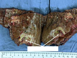
Background                                                       Here, we report 10 patients with initially advanced or
                                                                 unresectable HCC who received anti-angiogenic tyrosine
    Primary liver cancer is the second leading cancer-related    kinase inhibitor (TKI) and anti-PD-1 antibody combina-
cause of death in China [1]. Although the incidence and          tion therapy, followed by R0 resection. To our knowledge,
mortality of liver cancer are decreasing in China [2, 3], they   this represents the largest reported cohort of patients with
are increasing in the USA and Europe [4]. Over 90% of pri-       unresectable HCC to undergo R0 resection following sys-
mary liver cancers are hepatocellular carcinomas (HCCs),         temic therapy.
and life expectancy following a diagnosis of HCC is lower
than many other cancers [4]. One contributing factor to this
poor prognosis is that the majority of patients with HCC are        Patients and Methods
diagnosed at an advanced stage at which they have already
missed the opportunity for curative resection [5]. There-            Patients
                                                                     Consecutive patients with unresectable or advanced HCC re-
fore, systemic therapy is the standard of care for most pa-      ceived first-line systemic therapy with combined TKI/anti-PD-1 an-
tients with HCC. In recent years, a number of novel anti-        tibodies were retrospectively analyzed. HCC was diagnosed based
cancer agents have shown effectiveness for the treatment of      on standard imaging examinations, with or without elevated serum
advanced or unresectable HCC in terms of tumor response          tumor markers, alpha-fetoprotein, and/or protein induced by vita-
and patient survival. However, overall survival (OS) re-         min K absence-II, based on local and international guidelines [18,
                                                                 19]. Tumors were considered unresectable either because they were
mains poor for patients with HCC receiving systemic ther-        already advanced-stage HCC, intermediate stage, or because of in-
apy [6].                                                         sufficient remnant liver volume after liver resection (
63 patients treated as first-line
systemic therapy
51 patients did not meet 12 patients meet
the criteria of R0 resection the criteria of R0 resection
10 underwent surgery 2 did not underwent surgery
1 unresolved adverse effects
1 refused surgery
Fig. 1. Patient flowchart.
remnant liver volume is below 30% in non-cirrhotic patients or 40% Statistical Analyses
in cirrhotic patients, or tumor stage is Barcelona Clinic Liver Cancer Statistical analyses were performed using PASW Statistics v.18.0
(BCLC) stage B and up-to-seven criteria out [27], or stage C, or tu- for Windows (IBM Corp., Armonk, NY, USA). OS was defined as the
mor recurrence is diagnosed within 12 months after initial resection. interval between the date that combination therapy was initiated and
Except in the first patient, liver biopsies were performed to assess the the date of the patient’s death. Recurrence-free survival was defined
severity of liver necroinflammation to exclude latent hepatotoxicity as the interval between the date of surgery and the date of diagnosis
induced by combination therapy. Patients were classified as having of tumor recurrence or patient death from any cause. Kaplan-Meier
resectable HCC if (1) R0 resection could be achieved with sufficient analysis was used to determine the survival rates at each time points.
remnant liver volume and function, (2) intrahepatic lesions were
evaluated as partial response (PR) or stable disease (SD) for at least 2
months, (3) no severe or persistent adverse effects occurred from
systemic therapy, and (4) no contraindications for hepatectomy ex- Results
isted.
Post-hepatectomy liver failure was diagnosed and graded based Patients and Treatment
on an increased international normalized ratio of prothrombin time From September 2018 to December 2019, 63 consecu-
and hyperbilirubinemia on or after postoperative day 5 as proposed
by the International Study Group of Liver Surgery [28]. Postopera- tive patients who received TKI plus anti-PD-1 antibody
tive complications were classified using the Clavien-Dindo classifica- treatment as first-line systemic therapy were analyzed. Of
tion [29]. A standard 7-point baseline sampling protocol were ap- them, 10 patients (15.9%) underwent R0 resection. Other 2
plied for resected tumor specimen [30]. If no viable tumor cells were patients were evaluated as resectable after achieving a PR of
found, the whole tumor specimen were completely sampled. Patho- the original tumor (e.g., met the first and second criterion
logical complete response (pCR) was defined as no residual viable
tumor cells on hematoxylin and eosin staining on slide sections from of resection as mentioned above) but did not undergo liver
completely resected primary tumors, tumor thrombosis, and meta- resection. One experienced unresolved adverse effect from
static lesions. anti-PD-1 antibody treatment (grade II myocarditis), and 1
refused surgery (Fig. 1).
Postoperative Management The demographics and baseline characteristics of all 63
Combination therapy was resumed 4–6 weeks post-surgery, after
postoperative complications were resolved. Follow-up imaging ex- patients who received combination therapy are summa-
aminations (contrast-enhanced MRI/CT or abdominal ultrasound) rized in Table 1. No patients with extrahepatic spread
were performed every 2–3 months or when tumor recurrence was (n = 20) underwent surgery after treatment, whereas 10
suspected based on elevated serum tumor biomarkers. Post-recur- out of 43 patients without extrahepatic spread underwent
rence treatments were administered according to local guidelines surgery (p = 0.023). All 10 patients who underwent sur-
[18].
gery were men, with a median age of 52 years (range: 44–
322 Liver Cancer 2021;10:320–329 Zhu/Huang/Shen/Ji/Ge/Qu/Chen/Shi/Li/
DOI: 10.1159/000514313 Zhu/Tan/Tang/Zhou/Fan/SunTable 1. Baseline patient demographics and disease characteristics
Characteristics Patients who Patients who did not p values
underwent surgery undergo surgery
(n = 10) (n = 53)*
Sex (male/female), n 10/0 46/7 0.352
Median age, years (range) 52 (44–72) 57 (25–74) 0.547
ECOG performance status (0/1/2), n 5/5/0 12/33/8 0.155
Etiology of HCC (HBV/HCV/non-viral), n 10/0/0 51/0/2 1.000
BCLC stage (A/B/C), n 1/2/7 1/13/39 0.466
China liver cancer stage (Ib/IIa/IIb/IIIa/IIIb), n 1/1/1/7/0 1/1/12/19/20 0.087
Macrovascular invasion (yes/no), n 7/3 27/26 0.319
Portal vein tumor thrombus (Vp0/Vp1-2/Vp3/Vp4), n 5/0/2/3 28/1/10/14 1.000
Hepatic vein tumor thrombus (Vv0-1/ Vv2/Vv3), n 8/1/1 46/1/6 0.538
Extrahepatic disease (yes/no), n 0/10 20/33 0.023
Child-Pugh class (A/B), n 10/0 50/3 1.000
Baseline AFP, median (range), ng/mL 1,122 (17.5–60,500) 860 (1–60,500) 0.807
Baseline AFP ≥400 ng/mL, n (%) 7 (70.0) 32 (60.4) 0.729
Baseline PIVKA-II, median (range), mAU/mL 3,927 (111–75,000) 16,245 (43–75,000) 0.194
Baseline PIVKA-II ≥1,000 mAU/mL, n (%) 8 (80.0) 35 (66.0) 0.481
ECOG, Eastern Cooperative Oncology Group; HCC, hepatocellular carcinoma; HBV, hepatitis B; HCV,
hepatitis C; BCLC, Barcelona Clinic Liver Cancer; AFP, alpha-fetoprotein; PIVKA-II, protein induced by vitamin
K absence-II. * Including 2 patients who met the criteria for R0 resection but not did not undergo surgery.
Table 2. Characteristics of surgical and postoperative features
Patient Intrahepatic Number of BCLC CNLC Vascular TKI Anti-PD-1 Tumor Tumor
No tumor size, intrahepatic stage stage invasion used antibody response, by response, by
cm tumors used RECIST v1.1 mRECIST
1 10.2 1 C IIIa Vp4 Len Niv PR CR
2 9.0 >3 C IIIa Vp4 Len Pem SD SD
3 11.6 >3 B IIb – Len Pem PR PR
4 9.0 >3 C IIIa Vp4 Len Sin PR CR
5 4.2 2 B IIa – Len Pem SD SD
6 17.6 >3 C IIIa Vp3 Apa Sin PR PR
7 9.6 1 C IIIa Vv2 Len Pem SD SD
8 6.8 1 C IIIa Vv3 Apa Cam PR PR
9 15.4 1 C IIIa Vp3 Apa Sin PD PD
10 7.3 1 A Ib – Len Pem PR CR
BCLC, Barcelona Clinic Liver Cancer; CNLC, Chinese Liver Cancer Stage; TKIs, tyrosine kinase inhibitors; Len, lenvatinib; Apa,
apatinib; Niv, nivolumab; Pem, pembrolizumab; Sin, sintilimab; Cam, camrelizumab; CR, complete response; PR, partial response; SD,
stable disease; PD, progressive disease.
72 years), 7 had BCLC Stage C disease with macrovascular disease with insufficient future remnant liver volume if
invasion, including Vp3 in 2 patients, Vp4 in 3 patients resected. The median diameter of the largest liver nodule
according to Liver Cancer Study Group of Japan (LCSGJ) was 9.3 cm (range: 4.2–17.6 cm), and 5 patients had mul-
classification [31], invasion of the hepatic vein in 1 patient tiple lesions in the liver (up-to-seven criteria out) (Ta-
(Vv2 by LCSGJ classification [31]), and invasion of the ble 2).
inferior vena cava/right atrium (Vv3) in 1 patient. Two The TKIs and the anti-PD-1 antibodies of the 10 patients
patients had BCLC stage B HCC, and 1 had BCLC stage A received are listed in Table 2. Tumor responses evaluated
Initial Experience of Conversion Therapy Liver Cancer 2021;10:320–329 323
for Unresectable HCC DOI: 10.1159/0005143139 weeks
Pretreatment MR
(before surgery)
Portal vein thrombosis
100 µm
a Resected specimen H&E staining of resected specimen
MHV
Right pedicle
RHV
Pretreatment MR Pretreatment CT 9 weeks
(before surgery)
RPV
Tumor
b Resected specimen H&E staining of resected specimen
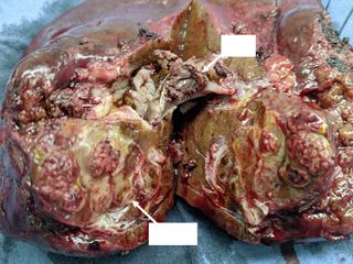
Fig. 2. Two representative cases. a Patient 4 was diagnosed with ficient (the ratio of left external liver lobe volume to standard liver
multiple HCCs involving the right liver lobe and invading the volume was 18.7%). The patient received PVE and lenvatinib 8 mg/
main branch of the portal vein (BCLC stage C). The patient re- day and pembrolizumab 200 mg every 3 weeks. Nine weeks after
ceived lenvatinib 8 mg/day and sintilimab 200 mg every 3 weeks treatment initiation, the ratio of the residual to standard liver vol-
for 9 weeks. Following systemic therapy, the primary tumors ume increased to 31.5%, and curative liver resection was per-
showed no arterial enhancement on contrast-enhanced MRI, and formed. H&E staining of the surgically resected specimen showed
the portal vein tumor thrombosis regressed. Curative liver resec- a pCR. HCC, hepatocellular carcinoma; H&E, hematoxylin and
tion was performed. H&E staining of the surgically resected speci- eosin; MR, magnetic resonance; CT, computed tomography;
men showed a pCR. b Patient 10 was diagnosed with a solitary BCLC, Barcelona Clinic Liver Cancer; PVE, portal vein embolism;
HCC lesion involving the right hepatic vein, middle hepatic vein, pCR, pathological complete response; MHV, middle hepatic vein;
and right pedicle (BCLC stage A). However, the tumor was unre- RHV, righ hepatic vein; RPV, right portal vein.
sectable because the ratio of the remnant liver volume was insuf-
324 Liver Cancer 2021;10:320–329 Zhu/Huang/Shen/Ji/Ge/Qu/Chen/Shi/Li/
DOI: 10.1159/000514313 Zhu/Tan/Tang/Zhou/Fan/SunPretreatment
Before
surgery
Patient 1 Patient 2 Patient 3 Patient 5
Pretreatment
Before
surgery
Patient 6 Patient 7 Patient 8 Patient 9
Fig. 3. Pretreatment MR or CT scan showed that the patients (ex- tients 3 and 5) (only 1 nodule was shown on this image for patient
cept patients 4 and 10) had a unresectable HCC before systemic 5). Before surgery, obvious tumor regression was observed in all
treatment and imaging scan before surgery. The major reason of the cases, except sustained stable disease were seen for patient 2,
unresectability were tumor invasion into major portal vein (pa- and partial response in the intrahepatic lesion but a new metastat-
tients 1 and 2), into the first branch of the portal vein (patients 6 ic lesion in the right adrenal gland (green arrow) for patient 9.
and 9), into middle hepatic vein (patient 7), or into right atrium HCC, hepatocellular carcinoma; MR, magnetic resonance; CT,
(patient 8; the intrahepatic nodule was not shown on this image) computed tomography.
(red arrows); multiple intrahepatic lesions (yellow arrows) (pa-
via pre-hepatectomy imaging were PR (n = 6), SD (n = 3), tinib plus pembrolizumab) because the baseline remnant
and progressive disease (n = 1; the intrahepatic tumor was liver volume was 18.7%. Nine weeks after the initiation of
evaluated as a PR; however, a new metastatic lesion was combination therapy, the remnant liver volume was in-
found in the right adrenal gland during systemic therapy). creased to 31.5%. The patient then received right trisectio-
Figure 2 shows 2 representative cases. Patient 4 (Fig. 2a) nectomy. pCR was also diagnosed by pathological study.
was diagnosed with BCLC stage C HCC, as tumor invaded MR/CT scans before systemic treatment and before surgery
into main branch of the portal vein. After the treatment for other 8 patients are shown in Figure 3.
with lenvatinib plus sintilimab for 9 weeks, significant ne-
crosis was found in the portal vein thrombosis by MRI Surgery and Perioperative Findings
study. No viable tumor cell can be found in the resected The median interval between initiation of systemic ther-
liver lesion and tumor thrombus by pathological study. Pa- apy and surgery was 3.2 months (range: 2.4–8.3 months).
tient 10 (Fig. 2b) received a PVE to enlarge the future liver For patients with BCLC stage C disease, only those with
volume before initiation of combination therapy (lenva- vascular invasion (7 out of 34 patients) could be converted
Initial Experience of Conversion Therapy Liver Cancer 2021;10:320–329 325
for Unresectable HCC DOI: 10.1159/000514313Table 3. Patient characteristics before and after surgery
Patient Days from TKI withdrawal Anti-PD-1 antibody Major PHLF* Postoperative Postoperative pCR
No. systemic therapy days before withdrawal days resection complication† hospital stay,
to surgery surgery before surgery days
1 73 7 19 Y y V 68 y
2 128 7 64 Y n 0 11 n
3 185 9 24 Y n IIIa 14 y
4 73 8 39 Y y 0 22 y
5 127 7 43 N y 0 13 n
6 95 8 37 Y y I 26 y
7 99 7 34 N n 0 11 n
8 79 7 36 N n IIIa 14 y
9 251 8 183‡ N y 0 14 n
10 73 7 31 Y n IIIa 18 y
PHLF, post-hepatectomy liver failure; TKIs, tyrosine kinase inhibitors; pCR, pathological complete response. * Classified according
to the International Study Group of Liver Surgery. † Clavien-Dindo criteria. ‡ Patient 9 had a G3 immune-related dermatitis and stopped
sintilimab therapy.
to resectable disease, whereas none of those with extrahe- fore surgery. Retrospective analysis of the surgical specimen
patic metastasis (n = 20) could be converted (Table 1). All revealed an active inflammation of the liver parenchyma.
the patients underwent surgery were classified as Child- Thereafter, all the other patients underwent liver biopsies
Pugh A before surgery. As shown in Table 3, 7 patients un- before surgery to exclude active inflammation in the liver
derwent a major liver resection (≥3 segments), with a mean parenchyma. We did not encounter any prominent liver
intraoperative blood loss of 950 ± 746 mL. The median inflammation in the following liver biopsy, and no im-
postoperative hospital stay was 14 days (range: 11–68 days). mune-related adverse events were observed in the subse-
The prevalence of post-hepatectomy liver failure was 50.0%, quent 9 patients following hepatectomy.
with grade A failure experienced by 4 patients and grade C Six patients (60%) achieved a pCR in all surgical speci-
by 1 patient. Five patients experienced postoperative com- mens. For patient 7, a pCR was found in the resected tumor
plications, including 1 patient with grade I (bile leakage), 3 thrombosis in the middle hepatic vein, but viable tumor
with grade IIIa (subphrenic collection requiring additional cells were found in the intrahepatic lesion, indicating a
puncture), and 1 with grade V, who died from immune- downstaging from BCLC stage C to stage A.
related adverse effects (patient 1).
Patient 1 died 2.4 months post-surgery after experienc- Follow-Up
ing immune-related adverse effects that began from post- The cutoff date for the present analysis was November
operative day 7 in the liver, skin, lung, and pancreas, dem- 3, 2020. One patient died from multisystemic immune-re-
onstrated via liver and skin biopsies. Preoperative labora- lated adverse events without tumor recurrence (patient 1).
tory examination did not show any sign of liver After a median follow-up of 11.2 months (range: 7.8–15.9
inflammation. His liver function was classified as Child- months), the other 9 patients remained alive. Tumor recur-
Pugh A6 without ascites or hepatic encephalopathy, serum rence was detected in 1 patient, who received locoregional
total bilirubin was 13.8 μmol/L, serum albumin was 33 g/L, therapy plus combination therapy with lenvatinib and anti-
international normalized ratio was 1.14, serum alanine PD-1 antibodies and remained tumor-free at the last fol-
transaminase was 9 U/L, aspartate aminotransferase was 19 low-up; the other 8 patients are tumor-free. All surviving
U/L, γ-glutamyl transferase was 71 U/L, serum HBV-DNA patients received combination therapy and regular surveil-
was undetectable, the shockwave elastography of liver pa- lance. The 12-month survival rate of the 10 patients after
renchyma was 21.0 kPa, and the ratio of future liver volume the initiation of combination therapy was 90.0% (standard
to standard liver volume was 44.6%. The last dosage of anti- error, 9.5%), and 12-month recurrence-free survival rate
PD-1 antibody (nivolumab) was given 19 days before sur- after surgery was 80.0% (standard error, 12.6%).
gery and last dose of TKI (lenvatinib) was given 7 days be-
326 Liver Cancer 2021;10:320–329 Zhu/Huang/Shen/Ji/Ge/Qu/Chen/Shi/Li/
DOI: 10.1159/000514313 Zhu/Tan/Tang/Zhou/Fan/SunDiscussion The present study also demonstrated that major hepa-
tectomy after the combination treatment is safe. The inci-
The present study showed that conversion therapy al- dence of postoperative complications was similar to the
lowed successful R0 resection in 15.9% (10/63) of the pa- previous reports [40, 41]. One patient died following hepa-
tients with initially unresectable HCC. The results suggest tectomy, which was attributed to severe immune-related
that combination therapy with an anti-angiogenic TKI and adverse effects (liver tissue necrosis) began on postopera-
anti-PD-1 antibodies is a feasible conversion therapy for tive day 7, as demonstrated via liver biopsy. Subsequently,
patients with unresectable HCC to achieve a successful re- the patient experienced immune-related adverse effects on
section with potential long-term recurrence-free survival. the skin as indicated by skin biopsies, lungs, pancreas and
Macrovascular invasion is not indicated for resection as pituitary gland, and the onset of type 1 diabetes. The patient
recommended by BCLC guideline, while some Asian guide- eventually died from multisystem failure on postoperative
lines or consensus recommends surgical resection to pa- day 72. Following this case, pre-hepatectomy liver biopsies
tients with portal vein invasion [18, 32, 33], and R0 resec- became mandatory to exclude active inflammation in the
tion has been achieved in highly selected patients meeting liver parenchyma in all the other patients. Further studies
these criteria. However, the median recurrence-free surviv- are warranted to fully examine the value of liver biopsies
al for these patients following upfront surgery is around 6 before surgery, particularly following downstaging with
months, and the median OS is approximately 1 year [34]. immune therapy.
One study found that in patients with HCC who received Only a small proportion of patients with unresectable
adjuvant apatinib therapy following surgery, the median HCC are able to achieve downstaging and undergo resec-
recurrence-free survival was 7.6 months [35]. So, the out- tion following sorafenib [42] because the ORR associated
comes of surgery with or without postoperative adjuvant with sorafenib treatment istion in this study. The efficacy and safety of the combination proval Number: B2020-177R). All patients provided written in-
therapy in patients with other etiological factors are un- formed consent before receiving combined TKI/anti-PD-1 antibody
treatment and before surgery.
known. Third, the combination of TKIs and anti-PD-1 an-
tibodies used in this study were not unified. All anti-PD-1
antibodies were off-label therapies for HCC and cannot be
Conflict of Interest Statement
reimbursed in China; therefore, patients’ choice will be an
important consideration (mostly the cost and updated in- H.C.S. has received speaker fees from Hengrui, Bayer, Eisai, and
formation from clinical trials). So far, no evidence showed MSD. X.D.Z. has received speaker fees from Eisai and MSD. Dr. Jia
the effects of these anti-PD-1 antibodies were different. Fan is an Editorial Board Member of Liver Cancer.
In summary, we reported the outcomes of 10 patients
with initially unresectable HCC who received successful
conversion therapy with combined TKI/anti-PD-1 antibod- Funding Sources
ies. The findings show that this conversion therapy strategy
This work was supported by the Leading Investigator Program of
is feasible for HCC, and subsequent liver resection is effec- the Shanghai municipal government (17XD1401100), the National
tive and safe given careful preparation and patient assess- Key Basic Research Program (973 Program; 2015CB554005) from
ment. Although the outcomes for patients who underwent the Ministry of Science and Technology of China, and the National
resection seems better than for those who did not undergo Natural Science Foundation of China (81672326 and 81871928, and
81871929).
resection, the exact role of liver resection in patients who
achieve downstaging from systemic therapy requires further
investigation in a prospective controlled study.
Author Contributions
Conception and design: H.-C. Sun, J. Fan, J. Zhou, and Z.-Y.
Acknowledgement Tang; administrative support: H.-C. Sun, and J. Zhou; provision of
study materials or patients: H.-C. Sun, X.-D. Zhu, C. Huang, Y.-H.
We thank the patients and their families. Shen, Y. Ji, N.-L. Ge, X.-D. Qu, L.-L. Chen, W.-K. Shi, and C.-J. Tan;
collection and assembly of data: X.-D. Zhu, C. Huang, Y.-H. Shen,
L.-L. Chen, M.-L. Li, and J.-J. Zhu; data analysis and interpretation:
Statement of Ethics H.-C. Sun and X.-D. Zhu; manuscript writing: H.-C. Sun, X.-D. Zhu,
C. Huang, and Y.-H. Shen; final approval of manuscript: all authors.
The study protocol was complied with the ethical guidelines of
the World Medical Association Declaration of Helsinki and was ap-
proved by Zhongshan Hospital Research Ethics Committee (Ap-
References
1 Zhou M, Wang H, Zeng X, Yin P, Zhu J, Chen diagnosis to death: the BRIDGE Study. Liver unresectable hepatitis B virus-related he-
W, et al. Mortality, morbidity, and risk factors Int. 2015;35(9):2155–66. patocellular carcinoma: a single center
in China and its provinces, 1990–2017: a sys- 6 Zhu X-D, Sun H-C. Emerging agents and reg- study of 45 patients. Ann Surg. 2020;
tematic analysis for the Global Burden of Dis- imens for hepatocellular carcinoma. J Hema- 271(3): 534–41.
ease Study 2017. Lancet. 2019; 394(10204): tol Oncol. 2019;12(1):110. 11 Tustumi F, Ernani L, Coelho FF, Bernardo
1145–58. 7 Tang ZY, Uy YQ, Zhou XD, Ma ZC, Lu JZ, Lin WM, Junior SS, Kruger JAP, et al. Preopera-
2 Zhou M, Wang H, Zhu J, Chen W, Wang L, ZY, et al. Cytoreduction and sequential resec- tive strategies to improve resectability for he-
Liu S, et al. Cause-specific mortality for 240 tion for surgically verified unresectable hepato- patocellular carcinoma: a systematic review
causes in China during 1990–2013: a system- cellular carcinoma: evaluation with analysis of and meta-analysis. HPB. 2018; 20(12): 1109–
atic subnational analysis for the Global Bur- 72 patients. World J Surg. 1995;19(6):784–9. 18.
den of Disease Study 2013. Lancet. 2016; 8 Lau WY, Lai EC. Salvage surgery following 12 Bertacco A, Vitale A, Mescoli C, Cillo U.
387(10015):251–72. downstaging of unresectable hepatocellular Sorafenib treatment has the potential to
3 Chen W, Zheng R, Baade PD, Zhang S, Zeng carcinoma: a strategy to increase resectability. downstage advanced hepatocellular carcino-
H, Bray F, et al. Cancer statistics in China, Ann Surg Oncol. 2007;14(12):3301–9. ma before liver resection. Per Med. 2020;
2015. CA Cancer J Clin. 2016;66(2):115–32. 9 Lau WY. Cure is possible with salvage surgery 17(2):83–7.
4 Siegel RL, Miller KD, Jemal A. Cancer statis- following downstaging of hepatocellular carci- 13 Irtan S, Chopin-Laly X, Ronot M, Faivre S,
tics, 2016. CA Cancer J Clin. 2019;66(1):7–30. noma. Chin-Ger J Clin Oncol. 2004;3(3):130–1. Paradis V, Belghiti J. Complete regression of
5 Park JW, Chen M, Colombo M, Roberts LR, 10 Wang Z, Peng Y, Hu J, Wang X, Sun H, Sun locally advanced hepatocellular carcinoma
Schwartz M, Chen PJ, et al. Global patterns of J, et al. Associating liver partition and por- induced by sorafenib allowing curative resec-
hepatocellular carcinoma management from tal vein ligation for staged hepatectomy for tion. Liver Int. 2011;31(5):740–3.
328 Liver Cancer 2021;10:320–329 Zhu/Huang/Shen/Ji/Ge/Qu/Chen/Shi/Li/
DOI: 10.1159/000514313 Zhu/Tan/Tang/Zhou/Fan/Sun14 Curtit E, Thiery-Vuillemin A, Nguyen T, 25 Eisenhauer EA, Therasse P, Bogaerts J, 35 Sun H-C, Zhu X-D, Zhou J, Gao Q, Shi Y-H, Heyd B, Pivot X, Di Martino V, et al. Com- Schwartz LH, Sargent D, Ford R, et al. New Ding Z-B, et al. Effect of postoperative apa- plete histologic response induced by sorafenib response evaluation criteria in solid tumours: tinib treatment after resection of hepatocel- in advanced hepatocellular carcinoma: a case revised RECIST guideline (version 1.1). Eur J lular carcinoma with portal vein invasion: a report. J Clin Oncol. 2011;29(12):e330–2. Cancer. 2009;45(2):228–47. phase II study. J Clin Oncol. 2020;38(4_Suppl 15 Zhu XD, Tang ZY, Sun HC. Targeting angio- 26 Lencioni R, Llovet JM. Modified RECIST l):514. genesis for liver cancer: past, present, and fu- (mRECIST) assessment for hepatocellular 36 Finn RS, Qin S, Ikeda M, Galle PR, Ducreux ture. Genes Dis. 2020;7(3):328–35. carcinoma. Semin Liver Dis. 2010; 30(1): 52– M, Kim TY, et al. Atezolizumab plus bevaci- 16 Kudo M, Ikeda M, Motomura K, Okusaka T, 60. zumab in unresectable hepatocellular carci- Kato N, Dutcus CE, et al. A phase Ib study of 27 Kudo M, Arizumi T, Ueshima K, Sakurai T, noma. N Engl J Med. 2020;382(20):1894–905. lenvatinib (LEN) plus nivolumab (NIV) in Kitano M, Nishida N. Subclassification of 37 Yau T, Kang Y-K, Kim T-Y, El-Khoueiry AB, patients (pts) with unresectable hepatocellu- BCLC B stage hepatocellular carcinoma and Santoro A, Sangro B, et al. Nivolumab lar carcinoma (uHCC): study 117. J Clin On- treatment strategies: proposal of modified (NIVO) + ipilimumab (IPI) combination col. 2020;38(Suppl 4):513. Bolondi’s subclassification (Kinki Criteria). therapy in patients (pts) with advanced hepa- 17 Finn RS, Ikeda M, Zhu AX, Sung MW, Baron Dig Dis. 2015;33(6):751–8. tocellular carcinoma (aHCC): results from AD, Kudo M, et al. Phase Ib study of lenva- 28 Rahbari NN, Garden OJ, Padbury R, Brooke- CheckMate 040. J Clin Oncol. 2019; 37(15_ tinib plus pembrolizumab in patients with Smith M, Crawford M, Adam R, et al. Pos- Suppl l):4012. unresectable hepatocellular carcinoma. J Clin thepatectomy liver failure: a definition and 38 Kaseb AO, Duda DG, Tran Cao HS, Abugabal Oncol. 2020;38(26):2960–70. grading by the International Study Group of YI, Vence LM, Rashid A, et al. Randomized, 18 Zhou J, Sun HC, Wang Z, Cong WM, Wang Liver Surgery (ISGLS). Surgery. 2011; 149(5): open-label, perioperative phase II study eval- JH, Zeng MS, et al. Guidelines for diagnosis 713–24. uating nivolumab alone or nivolumab plus and treatment of primary liver cancer in Chi- 29 Dindo D, Demartines N, Clavien PA. Classi- ipilimumab in patients with resectable HCC. na (2017 Edition). Liver Cancer. 2018; 7(3): fication of surgical complications: a new pro- J Clin Oncol. 2020;38(4_Suppl l):486. 235–60. posal with evaluation in a cohort of 6336 pa- 39 Dhir M, Sasson AR. Surgical management of 19 Bruix J, Sherman M. Management of hepato- tients and results of a survey. Ann Surg. 2004; liver metastases from colorectal cancer. J On- cellular carcinoma: an update. Hepatology. 240(2):205–13. col Pract. 2016;12(1):33–9. 2011;53(3):1020–2. 30 Cong WM, Bu H, Chen J, Dong H, Zhu YY, 40 Chen ZL, Zhang CW, Liang L, Wu H, Zhang 20 Ikeda M, Sung MW, Kudo M, Kobayashi M, Feng LH, et al. Practice guidelines for the WG, Zeng YY, et al. Major hepatectomy in Baron AD, Finn RS, et al. A phase 1b trial of pathological diagnosis of primary liver can- elderly patients with large hepatocellular car- lenvatinib (LEN) plus pembrolizumab (PEM) cer: 2015 update. World J Gastroenterol. cinoma: a multicenter retrospective observa- in patients (pts) with unresectable hepatocel- 2016;22(42):9279–87. tional study. Cancer Manag Res. 2020; 12: lular carcinoma (uHCC). J Clin Oncol. 2018; 31 Kudo M, Izumi N, Kokudo N, Matsui O, 5607–18. 36(15_Suppl l):4076. Sakamoto M, Nakashima O, et al. Manage- 41 Desai J, Deva S, Lee JS, Lin CC, Yen CJ, Chao 21 Xu J, Zhang Y, Jia R, Yue C, Chang L, Liu R, ment of hepatocellular carcinoma in Japan: Y, et al. Phase IA/IB study of single-agent et al. Anti-PD-1 antibody SHR-1210 com- consensus-based clinical practice guidelines tislelizumab, an investigational anti-PD-1 an- bined with apatinib for advanced hepatocel- proposed by the Japan Society of Hepatology tibody, in solid tumors. J Immunother Can- lular carcinoma, gastric, or esophagogastric (JSH) 2010 updated version. Dig Dis. 2011; cer. 2020;8(1):e000453. junction cancer: an open-label, dose escala- 29(3):339–64. 42 Liu K, McCaughan GW. How to select the ap- tion and expansion study. Clin Cancer Res. 32 Cheng S, Chen M, Cai J, Sun J, Guo R, Bi X, et propriate “neoadjuvant therapy” for hepato- 2019;25(2):515–23. al. Chinese expert consensus on multidisci- cellular carcinoma. Expert Opin Pharmaco- 22 Li Q, Qin S, Gu S, Chen X, Lin L, Wang Z, et plinary diagnosis and treatment of hepatocel- ther. 2018;19(11):1167–70. al. Apatinib as second-line therapy in Chinese lular carcinoma with portal vein tumor 43 Llovet JM, Ricci S, Mazzaferro V, Hilgard P, patients with advanced hepatocellular carci- thrombus (2018 Edition). Liver Cancer. 2020; Gane E, Blanc JF, et al. Sorafenib in advanced noma: a randomized, placebo-controlled, 9(1):28–40. hepatocellular carcinoma. N Engl J Med. double-blind, phase III study. J Clin Oncol. 33 Kudo M, Matsui O, Izumi N, Iijima H, Kadoya 2008;359(4):378–90. 2020;38(15_Suppl l):4507. M, Imai Y, et al. JSH consensus-based clinical 44 Cheng AL, Kang YK, Chen Z, Tsao CJ, Qin S, 23 Qin S, Ren Z, Meng Z, Chen Z, Chai X, Xiong practice guidelines for the management of he- Kim JS, et al. Efficacy and safety of sorafenib J, et al. Camrelizumab in patients with previ- patocellular carcinoma: 2014 update by the in patients in the Asia-Pacific region with ad- ously treated advanced hepatocellular carci- liver cancer study group of Japan. Liver Can- vanced hepatocellular carcinoma: a phase III noma: a multicentre, open-label, parallel- cer. 2014;3(3–4):458–68. randomised, double-blind, placebo-con- group, randomised, phase 2 trial. Lancet On- 34 Wei X, Jiang Y, Zhang X, Feng S, Zhou B, Ye trolled trial. Lancet Oncol. 2009;10(1):25–34. col. 2020;21(4):571–80. X, et al. Neoadjuvant three-dimensional con- 45 Kudo M, Finn RS, Qin S, Han KH, Ikeda K, 24 Zhang W, Bi X, Sun Y, Yu Y, Zhou J-G, Zeng formal radiotherapy for resectable hepatocel- Piscaglia F, et al. Lenvatinib versus sorafenib H, et al. Preliminary results of sintilimab plus lular carcinoma with portal vein tumor in first-line treatment of patients with unre- different dose of IBI305 (anti-VEGF mono- thrombus: a randomized, open-label, multi- sectable hepatocellular carcinoma: a ran- clonal antibody) in patients with advanced center Controlled Study. J Clin Oncol. 2019; domised phase 3 non-inferiority trial. Lancet. hepatocellular carcinoma: a phase Ib study. J 37(24):2141–51. 2018;391(10126):1163–73. Clin Oncol. 2020;38(15_Suppl l):3079. Initial Experience of Conversion Therapy Liver Cancer 2021;10:320–329 329 for Unresectable HCC DOI: 10.1159/000514313
You can also read