Downregulation of GJB2 and SLC26A4 Genes Induced by Noise Exposure is Associated with Cochlear Damage
←
→
Page content transcription
If your browser does not render page correctly, please read the page content below
Downregulation of GJB2 and SLC26A4 Genes
Induced by Noise Exposure is Associated with
Cochlear Damage
Amir Abbasi Garmaroudi
Tehran University of Medical Sciences
Monireh Khadem
Tehran University of Medical Sciences
Maryam Mirzaei Hotkani
Tehran University of Medical Sciences
Sajjad Mozaffari
Tehran University of Medical Sciences
MohammadReza Monazzam Esmaeilpoor
Tehran University of Medical Sciences
Elham Kazemirad ( kazemirad@tums.ac.ir )
Tehran University of Medical Sciences https://orcid.org/0000-0001-8634-5584
Research Article
Keywords: GJB2 & SLC26A4 genes, Real-time RT-PCR, Hearing impairment, Cochlear tissue, White &
Purple noise
Posted Date: June 29th, 2021
DOI: https://doi.org/10.21203/rs.3.rs-633325/v1
License: This work is licensed under a Creative Commons Attribution 4.0 International License.
Read Full License
Page 1/16Abstract
Noise can change the pattern of gene expression inducing sensorineural hearing impairment. There is no
investigation on effects of noise frequency on the expression of GJB2 and SLC26A4 genes involved in
congenital hearing impairment in cochlear tissue. This study investigated impacts of white and purple
noise on gene expression and pathologic changes of cochlear tissue. In this study, 32 adult male Westar
rats were selected and divided into experimental groups WN (animals exposed to white noise with a
frequency range of 100-20000 Hz), PN (animals exposed to purple noise with a frequency range of 4-20
kHz) and control groups. All experimental groups were exposed to a sound pressure level of 118-120 dB
for 8 hours per day. Cochlear tissue sampling was performed for tissue pathology studies, also RNA was
extracted at 1 hour & 1 week after cessation of noise exposure. The results showed that Both white and
purple noises caused permanent damage to the cortical, estrosilica systems of hair cells and ganglion of
the hearing nerve. GJB2 and SLC26A4 were downregulated in both groups exposed with white and purple
noise. However, differences are notably more significant in 1 weak post-exposure than 1 hour. Our
Findings suggest GJB2 and SLC26A4 can be considered as biomarkers of response to noise frequency
which is associated with the pathological response of cochlear tissue, leading to sensorineural hearing
impairment. It would be suggested the demand for a more conventional approach to assessment of
noise-induced hearing loss and subsequently the practice of hearing protection programs.
Introduction
Noise-induced hearing loss (NIHL) is the most common form of acquired hearing impairment in
developed countries and therefore it is considered a public health issue [1]. A wide range of workers is
exposed to noise pollution in the workplace, which has been shown to have detrimental effects, such as
hearing impairment [2]. Limited studies have been conducted on the effects of high-frequency noises on
the expression of genes involved in hearing impairment. One type of noise with a high-frequency range is
purple noise and the other which had a range of audible frequencies is white noise. White or octave band
noises centered on low frequency and purple noise centered on high frequency [3]. Hearing impairment
could be induced by these noises or after exposure to high-intensity noises [4]. studies were demonstrated
that exposure to sound pressure level (90–120 dB) and frequency range of 1–7 kHz could induce
upregulation of heat shock protein genes [5], Protein 45β (GADD45β), CDK-interacting protein 1 (p21Cip1)
[6], TACII, MHCII, [7] HDAC protein and Connexin 26 [8] in the atrium of inner ear suggesting them as
biomarkers of audible frequencies noise response. Moreover, exposure to sound pressure level of 120 dB
and frequency range of 7 kHz resulted in upregulation of C1s and Cfi proteins of the Corte system [9].
Stimulation with ultrasonic noises (16 kHz) and SPL = 130 dB significantly increased the expression of
CRH (Corticotropin-Releasing Hormone) genes and its CRH-R1 receptor in PVN, also the physical stress
caused by these noises had severely affected microglial cells [10]. Moreover, acute exposure to high-
frequency noise (10–20 kHz) and SPL = 110–120 dB resulted in upregulation of cochlear tissue different
proteins such as β-ACTIN, PRESTIN, and β-SPECTRIN [11]. Indeed, abnormal noise at different
frequencies and times exposure can induce mutation ad changes in the gene expression pattern in
Page 2/16ganglion and cochlear tissue. In this manner, noise can induce morphological and physiological changes
leading to hair-cell death and disorders in different sections of the human auditory system [6, 12–13].
Inner and particularly outer hair cells (IHC and OHC, respectively) especially those located in the basal
turn of the cochlea in the auditory system, are very sensitive to noise damage. Thus, the disruption of
stereocilia leads to a severe alteration in HC structural integrity that has been correlated to permanent
threshold shifts (PTS) in many species [14–19].
It was documented that several genes are involved in the physiological response of the cochlea to noise-
induced damage. These genes are associated with various biological processes such as oxidative stress
and transcriptional signaling [6, 20]. The gap junction beta-2 (GJB2) gene-encoded one of the most
important proteins involved in the regulation of potassium ions in the cochlea, which is located within the
spiral cells of the limbus and the spiral ligament fibroblasts [21–22]. The solute carrier 26a4 (SLC26A4)
gene consists of the DFNB4 locus, a recessive chromosomal locus, that causes sensorineural hearing
impairment in both non-syndromic and syndromic forms (pendred syndrome, PDS) [23]. Studies have
shown that pendred syndrome is alike an anion converter whose mutation leads to hearing impairment.
Pendred syndrome is most often diagnosed with nerve deafness associated with goiter. Sensorineural
hearing impairment due to pendred syndrome is progressive in some cases and causes varying degrees
of deafness [23]. Since no research has been performed directly on the effects of white and purple noises
exposure on the expression of SLC26A4 and GJB2 genes involved in congenital hearing impairment, in
the present study we investigate their expression pattern after exposure with white and purple noise at a
different time in the rat. Given that cochlear tissue is the most important sensorineural receptor of sound
when exposed to noises, the pathologic changes in this tissue were also investigated.
Materials And Methods
Study group selection
In this experimental study, 32 adult male Westar rats were selected and divided into experimental groups
(N = 12) WN (animals exposed to white noise with a frequency range of 100-20000 Hz), (N = 12) PN
(animals exposed to purple noise with a frequency range of 4–20 kHz) and the control group (N = 8)
(animals exposed to the same environmental conditions but without noise exposure). For each
experimental group, 4 rats were selected as the control group (Table 1). Both experimental groups were
exposed to a sound pressure level of 118–120 dB for 8 hours per day. 6 rats from the WN exposure group
were exposed to white noise for 3 days (WN-3D) and the other 6 rats were exposed to white noise for 6
days (WN-6D). Also, 6 rats from the PN exposure group were exposed to purple noise for 3 days (PN-3D)
and the other 6 rats were exposed to purple noise for 6 days (PN-6D). Samples of exposure groups were
anesthetized and after biopsy of rat's cochlea, afterward gene expression analysis was performed on
them at two-time period 1 hour and 1 week after cessation of noise exposure designated as 1h and 1w
for each group (Table 1). Accordingly, to determine the mean normal expression level of GJB2 and
SLC26A4 genes, control group samples were used under the same environmental conditions but without
exposure to the noise. For cochlear tissue pathology studies in experimental groups, 8 rats from the
Page 3/16exposure groups, 1 hour & 1 week after cessation of noise exposure (2 rats from the WN-3D group, 2 rats
from the WN-6D group, 2 rats from the PN-3D group, and 2 rats from the PN-6D group) were considered.
Also, for studies of cochlear tissue pathology in a state that was not under any stress or acoustic
stimulation, 1 rat was selected from the control group. In this study, the sample size and type of
exposures in the tested time-periods, according to the nature of this study, were obtained experimentally
and by surveying the results of similar studies in this field of research [5, 8, 24]. The study was done
under the principles of the Ethics Research Committee of the School of Public Health, Tehran University
of Medical Sciences (Code: IR.TUMS.SPH.REC.1398.220).
Table 1
The summary of groups exposed with white and purple noise in different time
Groups Sample size Noise Time of Time of RNA extraction after
(N) exposure exposure exposure
(hour per day)
WN-3D- 3 White noise* 8 hour/ 3D 1 hour
1h
WN-3D- 3 White noise 8 hour/ 3D 1 weak
1w
WN-6D- 3 White noise 8 hour/ 6D 1 hour
1h
WN-6D- 3 White noise 8 hour/ 6D 1 weak
1w
PN-3D- 3 Purple 8 hour/ 3D 1 hour
1h noise**
PN-3D- 3 Purple noise 8 hour/ 3D 1 weak
1w
PN-6D- 3 Purple noise 8 hour/ 6D 1 hour
1h
PN-6D- 3 Purple noise 8 hour/ 6D 1 weak
1w
*Frequency range: 100-20000 HZ, SPL (sound pressure level) = 118–120 dB
** Frequency range: 4–20 kHz, SPL (sound pressure level) = 118–120 dB
Laboratory conditions
Rats used in this study with a specific weight and age (7–8 weeks, 190-200g) were placed in a high-
performance four-cell audio chamber. One week before the start of exposure, they adapted to the
conditions of the experimental environment (12 hours of darkness and 12 hours of light, temperature of
25 ± 2 ° C and humidity of 45–52%) and then were exposed to noises at a specific time from 10 am to 6
pm [5, 8] and during the period the necessary tests, water, and food were sufficiently provided to the rats.
Page 4/16Exposure to white and purple noise took place inside a four-cell high-performance audio chamber
measuring 60×52×40 cm3. The sound transmission loss from inside the chamber to the outside was 50
decibels. The walls of this chamber are composed of layers of iron sheet, cellulose, air layer, cellulose,
iron sheet, and cellulose, respectively. Two holes on the walls of the chamber were used for ventilation of
the chamber and to supply enough oxygen for animal samples inside this chamber, a blowing peripheral
pump with a flow of 70 liters per minute was used.
Noise exposure
The noise pollution was generated using computer software (ADOBE AUDITION with version 13.0.7) and
distributed inside the chamber by an amplifier (Crown D75A, Elkhart IN) and 2 speakers (RadioShack
Corp, Ft Worth TX). Frequency and sound pressure level monitoring were used with SPECTRUM
ANALYZER software (version 5.0.2.). Also, to check the uniformity of the sound pressure level in different
parts of the chamber, the sound pressure level in different parts was measured by an analytical sound
meter (CELL491 catalog model).
Histology tests
First, the rats were anesthetized by exposure to carbon dioxide in a cubic chamber, then the skull bone
was dissected, and the brain of the specimens was removed. The cochlear tissue, which includes the
inner ear and is located inside the pterygium, was removed from the tympanic and temporal sections of
the auditory system by surgical instruments. The cochlear tissue inside the microtube for extract RNA, as
well as histological tests, were performed (tissue fixation, alcohol dehydration, clearing with xylene,
impregnation with Paraffin, molding, cutting from the samples with a microtome in thickness of 5 to 10
µm and painting of hematoxylin-eosin). In the end, morphological-pathological examination of cochlear
tissue was performed using light microscope images at a 200-micron scale [12, 25].
RNA extraction
Total RNA was extracted from tissue using Tripure reagent (Roch, Mannheim, Germany) according to the
manufacturer’s instruction. Briefly, 50 mg of tissue was homogenized and lysed using Tripure reagent.
The lysate was mixed with chloroform and vortexed vigorously and then centrifuged at 12,000 g for 15
min at 4˚C. Afterward, the upper colorless phase was precipitated by adding isopropanol and washed
with 75% ethanol. Finally, RNA was dried at room temperature and dissolved in RNase-free water. The
quantity and quality of RNA were evaluated by nanodrop (ND-1000, Thermo Scientific Fisher, Waltham,
Massachusetts, USA) and gel electrophoresis, respectively. RNA was treated with DNase to avoid
genomic contamination, (Qiagen, Hilden, Germany) according to the manufacturer's instructions.
cDNA synthesis
Complementary DNA (cDNA) was synthesized using 1 µg RNA, 2.5 µmol/µl oligo-dT (Roche, Mannheim,
Germany) and 60 µmol/µl random hexamers (Roche, Mannheim, Germany) incubated 10 min at 65°C
followed by addition of 10 mM of dNTP mix (Deoxy Nucleoside Triphosphate mix) (Roche, Mannheim,
Germany) 40U/µl RNase inhibitor (Roche, Mannheim, Germany), 4µL of 5X reverse transcriptase (RT)
Page 5/16buffer (Roche, Mannheim, Germany) and 20 U/µL reverse transcriptase enzyme (Roche, Mannheim,
Germany) and then incubated at 25°C for 10 min followed by incubation at 50°C for 60 min. The integrity
of cDNA was checked with GAPDH (Glyceraldehyde 3-Phosphate Dehydrogenase) primers (Table 1) as a
housekeeping gene. The PCR condition as follows: an initial denaturing step of 94°C for 5 min and 35
repetitions of denaturation at 94°C for 30 s, annealing at 49°C for 30s, and extension at 72°C for 45s with
a final extension of 72°C for 5 min.
Real-time RT-PCR analysis
To investigate the differences in the RNA expression level of target genes among exposure and control
group Real-time reverse transcriptase-PCR (RT-PCR) was used. Specific primers were designed by Primer 3
software version 0.4.0 (http://www.ncbi.nlm..nih.gov/tools/primer-blast) (Table 2). Two independent RNA
preparations were used for each real-time RT PCR experiment. The experiments were performed in 20 µl
reactions containing 1 µl cDNA target, 100 nM forward and reverse primers, and 1x SYBR® Premix Ex
TaqTM II (Takara, Tokyo, Japan). RT-PCR was carried out in duplicate for target genes and GAPDH as a
housekeeping gene using a Real-Time System (Applied Biosystems [ABI], Foster City, CA, USA). The PCR
condition was as follows: activation at 95°C for 3 min, amplification at 95°C for 10 s, 60°C for 32 s for 40
cycles followed by a melting curve, temperature increments of 0.2°C every 30 s to determine amplification
of the expected product [26].
Table 2
Primer sequences of target genes used in real-time RT-PCR
Oligo Name seq 5 − 3 TM
GJB2 Forward 5´-TCACTGTCCTCTTCATCTTCCG-3´ 59.51
Reverse 5´-CTTCCGTTTCTTTTCGTGTCTC-3´ 57.88
SLC26A4 Forward 5´-CATCATGCCTGGCTGGTTCT-3´ 60.40
Reverse 5´-TGGACACCAACATTCCGTCA-3´ 59.53
GAPDH Forward 5´-AAGTTCAACGGCACAGTCAAGG-3´ 61.58
Reverse 5´-CATACTCAGCACCAGCATCACC-3´ 61.32
Analysis of gene expression
The expression level of target genes is reported as the gene fold in the noise-exposure samples
normalized to the internal control gene (GAPDH) and relative to the control group using the 2−∆∆CT
method as previously described [27].
Statistical method
Statistical calculations were performed by prism Graph pad software with version 9.0.0.121. Relative
gene expression value data are mean ± standard deviation and were normalized to the internal control.
The normal distribution hypothesis for quantitative variables was evaluated by the Shapiro-Wilk test and
Page 6/16P-value < 0.05 was considered statistically significant. Differences among the exposure & control groups
were evaluated by Tukey's Multiple Comparison Test, followed by post hoc pairwise comparisons to
determine which differences account for the result of the more inclusive test.
Results
Chamber sound pressure level measurement reports
The measurement results showed that the arrangement of sound systems in the chamber was such that
the sound pressure level was constant in all parts of the chamber and all samples were in contact with
white and purple noise and with increasing distance from the floor surface of the chamber, the sound
pressure level has also increased. Also, the SPECTRUM ANALYZER software results showed that the
frequency range and produced sounds pressure level in the chamber were quite like the frequency range
and target sounds pressure level for the present study.
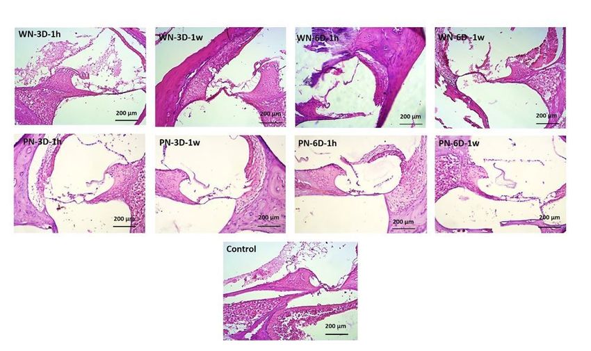
Results of pathological studies
Microscopic examination of tissues and their imaging showed that 1 week after cessation of noise
exposure, neurons in the samples of groups exposed to white noise (WN-3D and WN-6D) were damaged
more seriously than 1 hour after exposure termination (Fig. 1). Also, the nerve cells of the samples
exposed to purple noise (PN-3D and PN-6D) were damaged more significantly 1 week after cessation of
noise exposure compared to 1 hour after challenge. In the pathologic section, the auditory nerve ganglion
and Reisner membrane showed higher structural damage in samples exposed to purple noise (PN-3D and
PN-6D) compared to white noise (WN-3D and WN-6D). Besides, 1 week after termination of noise
exposure more seriously damaged was detected in samples exposed to purple noise (PN-3D and PN-6D)
in comparison with white noise (Fig. 1).
Expression pattern of GJB2 gene
The results showed that 1 week after cessation of noise exposure, the transcript level of GJB2 was
significantly decreased in exposure groups WN-3D, WN-6D, PN-3D, and PN-6D by 0.34-, 0.02-, 0.51- and
0.12-fold, p-value < 0.05. Also, 1 hour after cessation of noise exposure, the transcript level of GJB2 was
significantly decreased in exposure groups WN-3D and WN-6D by 0.51- and 0.12-fold, p-value < 0.05, while
it was increased in PN-3D (1.26-fold, p-value > 0.05) as compared to control. 1 hour after cessation of
noise exposure, the PN-6D exposure group was slightly downregulated which was not significantly (0.72-
fold, p-value > 0.05). Overall, 1 week after cessation of noise exposure, the average mRNA levels of GJB2
were significantly lower when compared to 1 hour after cessation of noise exposure in all of the exposure
groups (Fig. 2).
Expression pattern of SLC26A4 gene
The results showed that 1 week after termination of noise exposure, the transcript level of SLC26A4 was
significantly decreased in exposure groups WN-3D, WN-6D, PN-3D, and PN-6D by 0.31-, 0.009-, 0.43- and
Page 7/160.1-fold, p-value < 0.05. Also, 1 hour after cessation of noise exposure, the transcript level of SLC26A4 was
significantly decreased in exposure groups WN-3D, WN-6D, and PN-6D by 0.42-, 0.09- and 0.28-fold, p-
value < 0.05, while it was slightly increased in PN-3D (1.21-fold, p-value > 0.05) as compared to control.
Additionally, 1 week after cessation of noise exposure, the average expressions of SLC26A4 exhibited
more downregulation as compared to 1 hour after cessation of noise exposure in all of the exposure
groups (Fig. 3).
Discussion
The cochlear tissue and hair cells of the Corte system are the most important sensorineural receptors for
sound in the human auditory system. Noise exposure could trigger point mutation and alteration in gene
expression which resulted in many sensorineural hearing defects [13]. Therefore, it hypothesizes that the
cochlear tissue sample can be used as an ideal tissue sample to study the changes of target genes and
understanding the mechanisms that induced syndromic/non-syndromic sensorineural hearing
impairment. Accordingly, in this study, the effects of two noise types, which had different frequency limits
were investigated on the expression of GJB2 and SLC26A4 genes in rate cochlear tissue. One type of
noise had a high-frequency range (purple noise) and the other had a range of audible frequencies (white
noise). Most studies in the field of gene expression and its relationship with noise have examined only
the effects of noises with a certain frequency on the expression of genes [9–10, 24, 28], while in this
study, sound with a different frequency range in the human hearing range (20-20000 Hz) were analyzed.
The results of this study showed that GJB2 and SLC26A4 genes exhibited more downregulation after 6
days exposure of to both white and purple noises in comparison with 3 days exposure, suggested that
increasing the duration of time-exposure could induce more alteration in gene expression. Additionally,
when compare two-time points of RNA extraction after the termination of noise exposure, it was revealed
that the average expression of GJB2 and SLC26A4 genes exhibited more remarkable downregulation 1
week after the termination of noise exposure as compared to 1 hour in all the experiment groups.
Inconsistent with the results of gene expression, in pathologic examination of cochlear tissues, it was
demonstrated that neurons were damaged more seriously 1 week after cessation of white and purple
noise exposure compared to 1 hour after experiments. These results propose that more stable differences
in gene expression, as well as more pathologic damage, could be seen in the long-term period after
termination of noise exposure.
There is a lack of enough knowledge regarding the expression of GJB2 and SLC26A4 genes after white
and purple noise exposure. However, recently several studies have evaluated the expression of some
cochlear tissue and hair cells proteins, such as β-ACTIN, PRESTIN, and β-SPECTRIN after contact with
sound pressure level between 110–120 dB, and the results demonstrated that gene expression was
significantly increased in exposure groups [5, 9, 11, 24]. Furthermore, it was documented that after
exposure of rats with white noise (4kHz) for 2 days the expression of connexin 26 (CX26) protein in the
dorsal wall of the cochlear tissue increased significantly and the amount of protein in ligament fibrocytes
was increased [28].
Page 8/16In this context, a similar study showed that the expression of heat shock proteins (HSP 27, 70) increased
after 1 hour of noise exposure (100 dB), and the level of the hearing threshold was raised following 4
hours of exposure with the noises, suggesting that changes in HSP gene expression could lead to rapid
physiological changes at the level of the auditory threshold [29]. Inconsistent with the former studies,
Gratton et al. [6] stated that a sound pressure level of 105 dB (for 1 hour per day) induces overexpression
of HSP70, HSP40, Protein 45β (GADD45β), and CDK-interacting protein 1 (p21Cip1) genes in cochlear
tissue of mouse. However, the discrepancy between the results of these studies and the present study
may be due to the difference in time exposure, type of noise, and false-positive and negative results of the
real-time reverse transcriptase-PCR technique. Also, 1 hour after cessation of exposure, the expression of
both GJB2 and SLC26A4 genes in one group exposed to purple noise (PN-3D) up-regulated and the other
groups exposed to white and purple noise down-regulated significantly. These results are inconsistent
with a study by Alagramam et. al., [2] in which two groups of mice were exposed to noise with sound
pressure levels of 110 and 116 dB for 1 hour per day. The results showed that exposure to 110 dB
reduced the expression of 221 genes and increased the expression of 55 genes in the cochlea tissue,
while after exposure to 116 dB, the expression of 61 Genes was decreased and 243 genes enhanced.
Additionally, the expression of Fos Proto-Oncogene in the cochlea increased significantly after exposure
to 116 dB, while the expression of calcium-related genes decreased significantly after exposure to 110 dB
suggesting mitogen-activated protein kinase (MAPK) signaling was the major pathway in both levels of
noise exposure.
It was demonstrated that acute and chronic noise exposure could change gene expression differently. In
agreement with this notion, a study by Eraslan et al. [30] revealed that after acute noise exposure the
expression of both CRH-R1 and CRH-R2 (corticotropin-releasing hormone receptor) genes were
downregulated, while after chronic noise-exposure the expression of CRH-R1 and CRH-R2 genes increased
and decreased, respectively. These results are inconsistent with the results of the effects of purple noise
on the expression of GJB2 and SLC26A4 genes in the present study in which the gene expression is lower
after 6-day exposure compared to 3-day exposure. The results of this study are also in harmony with the
findings of a similar study [7] that have shown a significant relationship between changes in gene
expression pattern and the incidence of sensorineural hearing impairment. Furthermore, it was
documented that GJB2, SOD2, and CAT genes may be involved interactively or independently in the
development of NIHL [31]. Moreover, exposure of mice to 110 dB for 8 hours a day for 5 consecutive days
decreased the expression of connexin 26 (CX26) in cochlear tissue and lead to impairment of hair cells in
the cortical system and trigger the risk of NIHL [8].
Another important result in this study is that exposure to both white and purple noises has adverse
effects such as axonal changes of nerve neurons, destruction of cochlear tissue and hair cells on the
samples of the auditory system. These injuries have intensified with the increasing of time-exposure, also
purple noise due to the high energy has more destructive effects than white noise. Likewise, it was
reported that exposure to loud noises can lead to increased production of nitric acid in the spiral ganglion
neuron of pregnant women leading to alteration in the gene-expression pattern of ganglion cell and
thereby cell death, and development of congenital hearing impairment [32–33]. It is well documented that
Page 9/16after damage and destruction of the auditory system, restoration, and reconstruction of nerve cells such
as hair cells in mammals is not possible and subsequently can cause cell death in the Corte system, the
development of sensorineural hearing impairment, and permanent hearing threshold (PTS) changes [13].
In terms of pathology, cochlear injury induced by noise exposure is a multifactorial degradation process
that occurs due to inflammation and direct mechanical responses contributing to apoptosis and necrosis
[20]. In this line, changes in the neuron cell body's golico kanghogahse after noise exposure have been
reported in ear tissue and other tissues such as the heart [34]. Evidence has also shown that changes in
the expression level of proteins related to the auditory system noises cause permanent damage to the
auditory nerve ganglion and cochlear tissue neuron tissue to lead to non-syndromic hearing loss or
sensorineural syndrome [8, 13, 25]. Given that both white and purple noises have a significant effect on
the expression of genes involved in sensorineural hearing impairment (GJB2 and SLC26A4), therefore,
research on this issue should be concerned. The discovery of genes that are affected by noises and
cause sensorineural hearing impairment can be useful in pathophysiological studies and the
development of an appropriate solution for prevention [35].
Conclusion
Our findings suggest that GJB2 and SLC26A4 genes can be considered as biomarkers of response to
noise. Also, the pathological results show sensorineural hearing impairment and structural and cellular
changes in cochlear tissue following noise exposure. To prevent the effect of noise on genetic changes in
the auditory system, appropriate training, and advice should be suggested at the community level, also
using engineering or therapeutic control methods could decrease the risk of injury to the hearing system.
Declarations
Funding
This research was supported by Tehran University of Medical Sciences & Health Services, Grant Numbers:
98-3-99-45886.
Conflict of interest /Competing interests
The authors declare that they have no competing of interest.
Availability of data and materials
Data will be available on special request to corresponding author.
Authors’ contributions
AAG, MRME and EK collected and analyzed data. AAG and EK wrote the manuscript. MK, MMH and SM
reviewed and revised the manuscript. EK and MRME supervised the entire processes. All authors read and
approved the final manuscript.
Page 10/16Ethical approval and consent to participate
The study was approved by the Ethics Research Committee of the School of Public Health, Tehran
University of Medical Sciences (Code: IR.TUMS.SPH.REC.1398.220).
Consent for publication
All authors read and approved the final manuscript.
References
1. Sliwinska-Kowalska, M., and Davis, A (2012) Noise-induced hearing loss. Noise and Health. 14(61):
274–280. doi: 10.4103/1463-1741.104893
2. Alagramam KN, Stepanyan R, Jamesdaniel S, Chen DH-C, Davis RR (2014) Noise exposure immediately
activates cochlear mitogen-activated protein kinase signaling. Noise & health. 16(73):400. doi:
10.4103/1463-1741.144418
3. Park, S. N., Back, S. A., Park, K. H., Seo, J. H., Noh, H. I., Akil, O., et al (2013) Comparison of functional
and morphologic characteristics of mice models of noise-induced hearing loss. Auris Nasus Larynx.
40(1): 11–17. doi: 10.1016/j.anl. 2011.11.008
4. Kirchner, D. B., Evenson, E., Dobie, R. A., Rabinowitz, P., Crawford, J., Kopke, R., et al (2012) Occupational
noise-induced hearing loss: ACOEM task force on occupational hearing loss. J. Occup. Environ. Med.
54(1):106–108. doi: 10. 1097/JOM.0b013e318242677d
5. Ninomiya H, Ohgami N, Oshino R, Kato M, Ohgami K, Li X, et al (2018) Increased expression level of
Hsp70 in the inner ears of mice by exposure to low frequency noise. Hearing research. 36(3):49-54. doi:
10.1016/j.heares.2018.02.006
6. Gratton MA, Eleftheriadou A, Garcia J, Verduzco E, Martin GK, Lonsbury–Martin BL, et al (2011) Noise-
induced changes in gene expression in the cochleae of mice differing in their susceptibility to noise
damage. Hearing research. 277(1-2):211-26. doi: 10.1016/j.heares.2010.12.014
7. Yang W, Vethanayagam RR, Dong Y, Cai Q, Hu BH (2015) Activation of the antigen presentation
function of mononuclear phagocyte populations associated with the basilar membrane of the cochlea
after acoustic overstimulation. Neuroscience. 30(3):1-15. doi: 10.1016/j.neuroscience.2015.05.081
8. Zhou X-X, Chen S, Xie L, Ji Y-Z, Wu X, Wang W-W, et al (2016) Reduced Connexin26 in the mature
cochlea increases susceptibility to noise-induced hearing loss in mice. International journal of molecular
sciences. 17(3):301-308. doi: 10.3390/ijms17030301
9. Patel M, Hu Z, Bard J, Jamison J, Cai Q, Hu BH (2013) Transcriptome characterization by RNA-Seq
reveals the involvement of the complement components in noise-traumatized rat cochleae. Neuroscience.
Page 11/1624(8):1-16. doi: 10.1016/j.neuroscience.2013.05.038
10. Du F, Yin L, Shi M, Cheng H, Xu X, Liu Z, et al (2010) Involvement of microglial cells in infrasonic
noise-induced stress via upregulated expression of corticotrophin releasing hormone type 1 receptor.
Neuroscience. 167(3):909-19. doi: 10.1016/j.neuroscience.2010.02.060
11. Chen G-D (2006) Prestin gene expression in the rat cochlea following intense noise exposure. Hearing
research. 222(1-2):54-61. doi: 10.1016/j.heares.2006.08.011
12. Manikandan S, Srikumar R, Parthasarathy NJ, Devi RS (2005) Protective effect of acorus calamus L
INN on free radical scavengers and lipid peroxidation in discrete regions of brain against noise stress
exposed rat. Biological and pharmaceutical Bulletin. 28(12):2327-30. doi: 10.1248/bpb.28.2327
13. Han Y, Wang X, Chen J, Sha SH (2015) Noise‐induced cochlear F‐actin depolymerization is mediated
via ROCK 2/p‐ERM signaling. Journal of neurochemistry. 133(5):617-28. doi: 10.1111/jnc.13061
14. Chen, G. D., and Fechter, L. D (2003) The relationship between noise-induced hearing loss and hair cell
loss in rats. Hear. Res. 177(1-2): 81–90. doi: 10.1016/s03785955(02)00802-x
15. Hamernik, R. P., and Qiu, W (2000) Correlations among evoked potential thresholds, distortion product
optoacoustic emissions and hair cell loss following various noise exposures in the chinchilla. Hear. Res.
150(1-2): 245–257. doi: 10. 1016/s0378-5955(00)00204-5
16. Chen, Y. S., Liu, T. C., Cheng, C. H., Yeh, T. H., Lee, S. Y., and Hsu, C. J (2003) Changes of hair cell
stereocilia and threshold shift after acoustic trauma in guinea pigs: comparison between inner and outer
hair cells. ORL J. Otorhinolaryngol. Relat. Spec. 65(5): 266–274. doi: 10.1159/000075224
17. Hu, B.H., Henderson, D.N., T.M (2006) Extremely rapid induction of outer hair cell apoptosis in the
chinchilla cochlea following exposure to impulse noise. Hear. Res. 211(1-2):16–25. doi:
10.1016/j.heares.2005.08.006
18. Bohne, B. A., Harding, G. W., and Lee, S. C (2007) Death pathways in noise damaged outer hair cells.
Hear. Res. 223(1-2): 61–70. doi: 10.1016/j.heares.2006.10. 004
19. Harding, G.W., and Bohne, B. A (2009) Relation of focal hair-cell lesions to noise exposure parameters
from a4- or a 0.5- kHz octave band of noise. Hear. Res. 254(1-2): 54–63. doi:
10.1016/j.heares.2009.04.011
20. Yang S, Cai Q, Vethanayagam RR, Wang J, Yang W, Hu BH (2016) Immune defense is the primary
function associated with the differentially expressed genes in the cochlea following acoustic trauma.
Hearing research. 33(3):283-94. doi: 10.1016/j.heares.2015.10.010
Page 12/1621. Raphael Y, Wang Y, Lee MK (1994) Intercellular contacts between chick stereocilia after acoustic
overstimulation .Hearing research. 73(1):85-91. doi: 10.1016/0378-5955(94)90285-2
22. Martins FTA, Ramos PZ, Svidnicki MCCM, Castilho AM, Sartorato EL (2013) Optimization of
simultaneous screening of the main mutations involved in non-syndromic deafness using the TaqMan®
OpenArray™ Genotyping Platform. BMC medical genetics. 14(1):112. doi: 1471-2350/14/112
23. Nonose, Renata Watanabe and Lezirovitz, Karina and de Mello Auricchio, Maria Teresa Balester and
Batissoco, Ana Carla and Yamamoto, Guilherme Lopes and Mingroni-Netto, Regina C{\'e} Lia (2018)
Mutation analysis of SLC26A4 (Pendrin) gene in a Brazilian sample of hearing-impaired subjects. Journal
of BMC medical genetics. 19(1):1-10. Bio Med Central
24. Gong T-W, Fairfield DA, Fullarton L, Dolan DF, Altschuler RA, Kohrman DC, et al (2012) Induction of
heat shock proteins by hyperthermia and noise overstimulation in Hsf1−/− mice. Journal of the
Association for Research in Otolaryngology. 13(1):29-37. doi: 10.1007/s10162-011-0289-9
25. Kommareddi P, Nair T, Kakaraparthi BN, Galano MM, Miller D, Laczkovich I, et al (2015) Hair cell loss,
spiral ganglion degeneration, and progressive sensorineural hearing loss in mice with targeted deletion of
Slc44a2/Ctl2. Journal of the Association for Research in Otolaryngology. 16(6):695-712. doi:
10.1007/s10162-015-0547-3
26. Mohebali, M., Kazemirad, E., Hajjaran, H. et al (2019) Gene expression analysis of antimony
resistance in Leishmania tropica using quantitative real-time PCR focused on genes involved in
trypanothione metabolism and drug transport. Arch Dermatol Res. 311:9–17.
https://doi.org/10.1007/s00403-018-1872-2
27. Livak KJ, Schmittgen TD (2001) Analysis of relative gene expression data using real-time quantitative
PCR and the 2–ΔΔct Method. Methods. 25:402-408. doi: 10.1006/meth.2001
28. Hsu W-C, Wang J-D, Hsu C-J, Lee S-Y, Yeh T-H (2004) Expression of connexin 26 in the lateral wall of
the rat cochlea after acoustic trauma. Acta oto-laryngologica. 124(4):459-63. doi:
10.1080/00016480310000584
29. Sun W, Zhang L, Lu J, Yang G, Laundrie E, Salvi R (2008) Noise exposure–induced enhancement of
auditory cortex response and changes in gene expression. Neuroscience. 156(2):374-80. doi:
10.1016/j.neuroscience.2008.07.040
30. Eraslan E, Akyazi I, Ergül-Ekiz E, Matur E (2015) Noise stress changes mRNA expressions of
corticotropin-releasing hormone, its receptors in amygdala, and anxiety-related behaviors. Noise & health.
17(76):141. doi: 10.4103/1463-1741.155838
31. Wang SL, Yu LG, Liu RP, Zhu WZ, Gao WM, Xue LP, et al (2014) Gene-gene interaction of GJB2, SOD2,
and CAT on occupational noise-induced hearing loss in Chinese Han population. Europe PMC plus.
Page 13/1627(12):965-8. doi: 10.3967/bes2014.131
32. Sun D, Li X (2000) The effect of noise exposure on the expression of NOSmRNA in the cochlea. Lin
chuang er bi yan hou ke za zhi. Journal of clinical otorhinolaryngology. 14(8):373-4. PMID: 12563903
33. Pfenninger K, Maylie-Pfenninger M-F, Friedman L, Simkowitz P (1984) Lectin labeling of sprouting
neurons: III. Type-specific glycoconjugates on growth cones of different origin. Developmental biology.
106(1):97-108. doi: 10.1016/0012-1606(84)90066-6
34. Monsefi M, Talaei T (2005) Changes of heart glycoconjugates by noise stress in mouse as an
experimental model. Journal of Applied Animal Research. 27(2):121-4. doi:
10.1080/09712119.2005.9706554
35. Estivill X, Fortina P, Surrey S, Rabionet R, Melchionda S, D'Agruma L, et al (1998) Connexin-26
mutations in sporadic and inherited sensorineural deafness. Lancet. 351(8):394-98. doi: 10.1016/S0140-
6736(97)11124-2Get
Figures
Figure 1
Page 14/16Tissue image of different exposure group samples, 1 hour and 1 week after cessation of noise exposure.
WN-3D, group were exposed to white noise for 3 days; WN-6D, exposed to white noise for 6 days; PN-3D,
group were exposed to purple noise for 3 days; PN-6D; exposed to purple noise for 6 days. Pathologic
studies in experimental groups 1 hour (1h) & 1 week (1w) after cessation of noise exposure were
conducted
Figure 2
Effect of white & purple noise on Expression of GJB2 gene. Westar rats were exposed to white & purple
noise (SPL=118-120 dB) for 8hour, and Cochlear RNA was subjected to qRT-PCR for exposure group WN-
3D, WN-6D, PN-3D, and PN-6D conducted 1 hour (1h) and 1 weak (1w) after cessation of noise exposure.
Gene fold change is the ratio of values for noise-exposed rats relative to unexposed controls
Page 15/16Figure 3
Effect of white & purple noise on Expression of SLC26A4 gene. Westar rats were exposed to white &
purple noise (SPL=118-120 dB) for 8hour, and Cochlear RNA was subjected to qRT-PCR for exposure
group WN-3D, WN-6D, PN-3D, and PN-6D conducted 1 hour (1h) and 1 weak (1w) after cessation of noise
exposure. Gene fold change is the ratio of values for noise-exposed rats relative to unexposed controls
Page 16/16You can also read