DISPROPORTIONATE LEFT ATRIAL MYOPATHY IN HEART FAILURE WITH PRESERVED EJECTION FRACTION AMONG PARTICIPANTS OF THE PROMIS HFPEF STUDY - NATURE
←
→
Page content transcription
If your browser does not render page correctly, please read the page content below
www.nature.com/scientificreports
OPEN Disproportionate left atrial
myopathy in heart failure
with preserved ejection
fraction among participants
of the PROMIS‑HFpEF study
Ravi B. Patel1, Carolyn S. P. Lam2,3,4, Sara Svedlund5, Antti Saraste6, Camilla Hage7,
Ru‑San Tan2, Lauren Beussink‑Nelson1, Jasper Tromp2,3, Cynthia Sanchez1, Joyce Njoroge1,
Stanley A. Swat1, Ulrika Ljung Faxén7, Maria Lagerstrom Fermer8, Ashwin Venkateshvaran7,
Li‑Ming Gan8,9,10, Lars H. Lund7 & Sanjiv J. Shah1,11*
Impaired left atrial (LA) function in heart failure with preserved ejection fraction (HFpEF) is
associated with adverse outcomes. A subgroup of HFpEF may have LA myopathy out of proportion
to left ventricular (LV) dysfunction; therefore, we sought to characterize HFpEF patients with
disproportionate LA myopathy. In the prospective, multicenter, Prevalence of Microvascular
Dysfunction in HFpEF study, we defined disproportionate LA myopathy based on degree of LA
reservoir strain abnormality in relation to LV myopathy (LV global longitudinal strain [GLS]) by
calculating the residuals from a linear regression of LA reservoir strain and LV GLS. We evaluated
associations of disproportionate LA myopathy with hemodynamics and performed a plasma
proteomic analysis to identify proteins associated with disproportionate LA myopathy; proteins
were validated in an independent sample. Disproportionate LA myopathy correlated with better
LV diastolic function but was associated with lower stroke volume reserve after passive leg raise
independent of atrial fibrillation (AF). Additionally, disproportionate LA myopathy was associated
with higher pulmonary artery systolic pressure, higher pulmonary vascular resistance, and
lower coronary flow reserve. Of 248 proteins, we identified and validated 5 proteins (involved in
cardiomyocyte stretch, extracellular matrix remodeling, and inflammation) that were associated
with disproportionate LA myopathy independent of AF. In HFpEF, LA myopathy may exist out of
proportion to LV myopathy. Disproportionate LA myopathy is a distinct HFpEF subtype associated
with worse hemodynamics and a distinct proteomic signature, independent of AF.
While traditionally viewed as a syndrome of left ventricular (LV) diastolic dysfunction, heart failure (HF) with
preserved ejection fraction (HFpEF) may also be characterized by adverse changes to left atrial (LA) myocardial
structure and function, resulting in LA myopathy1,2. Speckle-tracking strain echocardiography, a sensitive meas-
ure of myocardial performance, can quantify severity of LA myopathy through assessment of abnormalities in
1
Division of Cardiology, Department of Medicine, Northwestern University Feinberg School of Medicine,
Chicago, IL, USA. 2National Heart Centre Singapore, Duke-National University of Singapore, Singapore,
Singapore. 3University Medical Centre, Groningen, The Netherlands. 4The George Institute for Global Health,
Sydney, Australia. 5Department of Clinical Physiology, Institute of Medicine, Sahlgrenska University Hospital,
University of Gothenburg, Gothenburg, Sweden. 6Heart Center, Turku University Hospital, University of Turku,
Turku, Finland. 7Department of Medicine, Cardiology Unit and Heart and Vascular Theme, Karolinska Institutet,
Stockholm, Sweden. 8Early Clinical Development, Research and Early Development, Cardiovascular, Renal and
Metabolism (CVRM), BioPhamaceuticals R&D, AstraZeneca, Gothenburg, Sweden. 9Department of Molecular
and Clinical Medicine, Institute of Medicine, Sahlgrenska Academy At the University of Gothenburg, Gothenburg,
Sweden. 10Department of Cardiology, Sahlgrenska University Hospital, Gothenburg, Sweden. 11Division of
Cardiology, Department of Medicine, Northwestern University Feinberg School of Medicine, 676 N. St Clair St,
Suite 600, Chicago, IL 60611, USA. *email: sanjiv.shah@northwestern.edu
Scientific Reports | (2021) 11:4885 | https://doi.org/10.1038/s41598-021-84133-9 1
Vol.:(0123456789)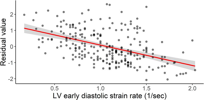
www.nature.com/scientificreports/
Figure 1. STROBE flow diagram for study inclusion.
intrinsic LA reservoir function3,4. Importantly, LA myopathy, as defined by reduced LA reservoir strain, appears
to be a stronger predictor of mortality among patients with established HFpEF than indices of ventricular
structure or f unction5. The spectrum and degree of LA myopathy in HFpEF are wide, as it may develop second-
ary to LV myopathy (secondary to high LV filling pressures), concurrent with LV myopathy as part of similar
pathophysiology affecting both LA and LV, or out of proportion to LV myopathy (due to intrinsic abnormalities
of the LA and/or atrial fibrillation [AF])6,7.
Despite the prognostic implications of LA dysfunction, the clinical, echocardiographic, and hemodynamic
profiles of HFpEF based on degree of LA myopathy in relation to LV myopathy have not been well defined.
Thus, our primary goal was to evaluate for the presence of “disproportionate LA myopathy” in HFpEF, and
comprehensively characterize this previously undefined phenotype. As such, we aimed to (1) phenotype a diverse
HFpEF cohort based on degree of LA myopathy as compared with LV myopathy; (2) evaluate the associations of
disproportionate LA myopathy with hemodynamics and coronary microvascular dysfunction; and (3) evaluate
the proteomic profile of disproportionate LA myopathy in participants enrolled in the multicenter, prospective
Prevalence of Microvascular Dysfunction in HFpEF (PROMIS-HFpEF) study. We hypothesized that LA myo-
pathy may exist out of proportion to LV myopathy and is associated with abnormal hemodynamics along with
distinct clinical and proteomic profiles, independent of AF.
Results
Clinical, laboratory, and echocardiographic variables associated with disproportionate LA
myopathy. This was a secondary analysis of the PROMIS-HFpEF cohort, which was an international study
designed to evaluate the prevalence and correlates of coronary microvascular dysfunction in chronic HFpEF.
We defined disproportionate LA myopathy based on degree of LA reservoir strain abnormality in relation to
LV myopathy (LV global longitudinal strain [GLS]) by calculating the residuals from a linear regression of LA
reservoir strain and LV GLS. Of 258 participants in the PROMIS cohort, 241 had adequate images for LA res-
ervoir strain and LV GLS at baseline and were thus included in our analyses (Fig. 1). There was a significant
but modest association of LV GLS with LA reservoir strain (Fig. 2). The clinical and laboratory characteristics
of the analytic cohort are shown in Table 1. Characteristics significantly associated with disproportionate LA
myopathy included older age, white race, AF, and chronic kidney disease. Higher levels of diastolic blood pres-
sure, N-terminal pro-B-type natriuretic peptide (NT-proBNP), and troponin T, and lower body mass index
(BMI) were also significantly associated with disproportionate LA myopathy. Findings were consistent upon
comparing participants by those with studentized residual values < 0 (indicative of LA myopathy) vs. those with
residuals > 0 (Supplemental Table S1).
There were several echocardiographic and hemodynamic correlates of disproportionate LA myopathy
(Table 2). Structural predictors of disproportionate LA myopathy included smaller LV end diastolic volume,
and larger LA and right atrial (RA) size. LV functional predictors of disproportionate LA myopathy included
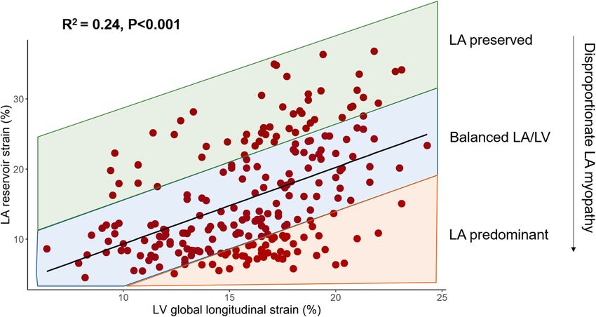
higher e’ tissue velocities and early diastolic strain rate (Fig. 3). Notably, LV systolic function indices were not
associated with disproportionate LA myopathy. Worse right ventricular (RV) systolic function as measured by
tricuspid annular plane systolic excursion (TAPSE) and RV free wall strain was associated with disproportionate
LA myopathy. Hemodynamic correlates of disproportionate LA myopathy included lower stroke volume/cardiac
index, higher pulmonary artery systolic pressure, and pulmonary vascular resistance. Finally, disproportionate
LA myopathy was associated with lower coronary flow reserve (CFR) (Supplemental Fig. S1).
Association of disproportionate LA myopathy with hemodynamics. Upon examination as a
continuous variable on linear regression analysis, disproportionate LA myopathy was significantly associated
with lower resting stroke volume (SV) after multivariable adjustment (β-coefficient per 1-SD lower studentized
residuals: − 6.8 [95% CI − 10.2, − 3.4] mL; P < 0.001; Table 3), including for AF. These findings were consistent
Scientific Reports | (2021) 11:4885 | https://doi.org/10.1038/s41598-021-84133-9 2
Vol:.(1234567890)
www.nature.com/scientificreports/
Figure 2. Association of LV global longitudinal strain with LA reservoir strain. Degree of LA myopathy (i.e.
disproportionate LA myopathy) was defined as studentized residual values derived from the linear regression
model of LV GLS and LA reservoir strain. Lower residual values thus correspond to disproportionately greater
LA myopathy compared with LV myopathy. GLS global longitudinal strain, LA left atrial, LV left ventricular.
on sensitivity analysis further adjusting for New York Heart Association (NYHA) class, 6-min walk distance, and
heart rate response to adenosine infusion (Supplemental Table S2).
There was a wide variation in change in SV after passive leg raise (i.e., intravascular volume challenge)
across the PROMIS cohort (range − 40.2 mL to + 39.6 mL; Supplemental Fig. S2). After multivariable adjust-
ment, disproportionate LA myopathy was associated with decrease in SV after intravascular volume challenge
(β-coefficient per 1-SD lower studentized residuals: − 2.2 [95% CI − 4.2, − 0.2] mL; P = 0.03; Table 3), independ-
ent of AF. These findings were consistent on sensitivity analysis (Supplemental Table S2).There was no interaction
by AF on the association of disproportionate LA myopathy with resting SV ( Pinteraction = 0.09) or change in SV
after passive leg raise maneuver (Pinteraction = 0.80).
Association of plasma proteins with disproportionate LA myopathy. Of the 248 candidate pro-
teins, there were 21 proteins significantly associated with disproportionate LA myopathy in PROMIS-HFpEF
cohort after controlling for multiple comparisons (Fig. 4, Supplemental Table S3). In the Northwestern HFpEF
validation cohort, 13 of the 21 proteins were also significantly associated with disproportionate LA myopathy
(Supplemental Table S3). Of the 13 proteins demonstrating associations on univariate analysis, 5 distinct pro-
teins remained significantly associated with disproportionate LA myopathy after adjustment for age, sex, and
AF in both PROMIS and Northwestern HFpEF cohorts (Table 4). These 5 proteins have previously demon-
strated pathway associations with cardiomyocyte stretch, collagen regulation, and inflammation (Supplemental
Figs. S5–S9).
Comparison of plasma proteins associated with disproportionate LA myopathy and atrial
fibrillation. Of the 248 candidate proteins, there were 11 proteins significantly associated with AF in
PROMIS-HFpEF cohort after controlling for multiple comparisons (Supplemental Fig. S3). Of these 11 pro-
teins, only 3 were validated in the Northwestern HFpEF cohort (Supplemental Table S4). Upon comparison of
validated proteins associated with disproportionate LA myopathy (13 proteins) and AF (3 proteins), 10 proteins
were uniquely associated with disproportionate LA myopathy and not AF (Supplemental Fig. S4). There were
no proteins that were only associated with AF and not disproportionate LA myopathy (Supplemental Table S4,
Fig. S4).
Discussion
In this prospective, multicenter study of an international and rigorously-phenotyped HFpEF cohort, we charac-
terized participants based on degree of LA myopathy as compared with LV myopathy. Despite favorable diastolic
function, disproportionate LA myopathy was associated with adverse RV function and elevated pulmonary
vascular resistance. Additionally, disproportionate LA myopathy was associated with diminished SV and SV
reserve, independent of AF. There was a significant correlation between coronary microvascular dysfunction as
measured by CFR and disproportionate LA myopathy. Finally, we identified and validated several plasma proteins
that were associated with disproportionate LA myopathy independent of clinical characteristics including AF. We
demonstrate that the proteome of disproportionate LA myopathy is distinct from that of AF. In aggregate, our
findings identify and characterize a distinct HFpEF phenotype of disproportionate LA myopathy that cannot be
Scientific Reports | (2021) 11:4885 | https://doi.org/10.1038/s41598-021-84133-9 3
Vol.:(0123456789)www.nature.com/scientificreports/
Characteristic PROMIS-HFpEF cohort (n = 241) β coefficient (SE) P value
Age, year 75 (70–81) 0.04 (0.01) < 0.001
Male sex, n (%) 107 (45) 0.19 (0.13) 0.14
White race, n (%) 209 (87) 0.84 (0.30) 0.006
NYHA class III/IV 61 (25.6) − 0.006 (0.15) 0.97
Comorbidities
Hypertension, n (%) 198 (83) − 0.09 (0.17) 0.59
Diabetes, n (%) 63 (26) 0.09 (0.15) 0.54
Hyperlipidemia, n (%) 128 (53) − 0.26 (0.13) 0.04
Atrial fibrillation, n (%) 99 (41.1) 1.20 (0.11) < 0.001
ASCVD, n (%) 100 (42) 3.8 × 1 0–3 (0.13) 0.98
Never Smokers 77 (32) − 0.13 (0.14) 0.38
Chronic kidney disease, n (%) 126 (52) 0.27 (0.13) 0.04
Pacemaker or ICD, n (%) 41 (17) − 0.05 (0.17) 0.77
Vital signs, physical characteristics, and laboratory data
Body mass index, kg/m2 28.0 (24.4–32.5) − 0.02 (0.01) 0.008
Heart rate, bpm 68 (60–78) 0.006 (0.005) 0.23
SBP, mmHg 139 (127–152) 0.002 (0.003) 0.57
DBP, mmHg 77 (68–85) 0.02 (0.01) 0.002
Waist circumference, cm 100 (89–113) − 0.002 (0.004) 0.61
NT-proBNP, pg/mL 960 (365–1770) 0.36 (0.05) < 0.001
Sodium, mEq/L 140 (138–142) 0.04 (0.02) 0.12
Potassium, mEq/L 4.2 (3.9–4.5) 0.26 (0.15) 0.09
Creatinine, mg/dL 1.06 (0.86–1.35) 0.31 (0.20) 0.12
Glomerular filtration rate, mL/min/1.73 m2 58.7 (45.6–69.7) − 0.29 (0.18) 0.12
Troponin T, ng/mL 13.0 (10.0–21.1) 0.17 (0.06) 0.007
Hemoglobin A1c, % 41 (38–49) 0.08 (0.31) 0.80
Urine albumin-to-creatinine ratio, mg/g 3.1 (1.3–9.7) 0.13 (0.07) 0.07
6-min walk distance, m 332 (210–412) − 0.15 (0.12) 0.21
Change in heart rate from baseline in response to adenosine, bpm 17 (9–28) − 0.002 (0.004) 0.62
Medications
Loop diuretic, n (%) 130 (54) 0.01 (0.13) 0.13
Thiazide diuretic, n (%) 26 (11) − 0.13 (0.21) 0.54
Mineralocorticoid receptor antagonist, n (%) 69 (29) − 0.06 (0.14) 0.67
ACE inhibitor, n (%) 71 (30) 0.07 (0.14) 0.62
ARB, n (%) 102 (42) 0.03 (0.13) 0.80
Table 1. Characteristics associated with disproportionate left atrial myopathy in PROMIS-HFpEF. Continuous
variables are reported as median (interquartile range). ACE angiotensin converting enzyme, ARB angiotensin
receptor blocker, ASCVD atherosclerotic vascular disease, DBP diastolic blood pressure, GFR glomerular
filtrate rate; ICD implantable cardioverter-defibrillator, NT-proBNP N-terminal pro-B-type natriuretic peptide,
NYHA New York Heart Association, SBP systolic blood pressure, UACRurinary albumin-to-creatinine ratio.
fully explained by the presence of comorbid AF. Ultimately, HFpEF patients with disproportionate LA myopathy
may ultimately benefit from targeted therapies to alleviate symptoms and improve outcomes.
Reduced LA reservoir strain has shown promise in identifying a high-risk HFpEF cohort. Indeed, LA res-
ervoir strain is associated with adverse clinical outcomes in H FpEF5,8,9. Impaired LA reservoir strain has been
more closely associated with poor prognosis in HFpEF than measures of LV function5. Furthermore, reduced LA
reservoir strain has recently been associated with increased pulmonary capillary wedge pressure with exercise
and lower peak oxygen consumption on cardiopulmonary exercise testing6,10–12. Despite this evidence, there has
been some concern that reduced LA reservoir strain may simply represent a marker of global LV dysfunction, as
there is a modest correlation between LA and LV strain in HFpEF13, and the association between LA reservoir
strain and adverse outcomes may be explained by severity of LV myopathy, as measured by reduced LV strain9,14.
Our study is the first, to our knowledge, to define HFpEF based on relative degree of LA myopathy com-
pared with LV myopathy to understand the degree to which disproportionate LA myopathy represents a unique
phenotype of HFpEF. Disproportionate LA myopathy appears to have a truly “LA-specific” echocardiographic
signature; disproportionate LA myopathy was associated with favorable diastolic function (higher e’ tissue veloc-
ity and early diastolic strain rate). Thus, our findings suggest LA myopathy may not simply be a consequence
of LV disease, but rather represents a unique pathophysiology among patients with HFpEF. Despite favorable
diastolic function, disproportionate LA myopathy correlated with a worse hemodynamic profile, highlighted by
Scientific Reports | (2021) 11:4885 | https://doi.org/10.1038/s41598-021-84133-9 4
Vol:.(1234567890)www.nature.com/scientificreports/
Echocardiographic variable PROMIS-HFpEF cohort (n = 241) β coefficient (SE) P value
Left heart structure/function
LV end-diastolic volume index, mL/m2 41.1 (33.8–50.1) − 0.55 (0.24) 0.02
LV end-systolic volume index, mL/m2 16.5 (12.5–21.4) − 0.24 (0.17) 0.15
LV mass index, g/m2 102.5 (83.5–124.5) 0.25 (0.22) 0.27
Relative wall thickness 0.45 (0.40–0.52) 0.17 (0.34) 0.62
LV ejection fraction, % 60 (55–64) 0.13 (0.43) 0.77
LA volume index, mL/m2 38.4 (30.7–44.8) 1.62 (0.17) < 0.001
e’ septal velocity, cm/s 6.8 (5.3–8.3) 0.09 (0.02) 0.001
e’ lateral velocity, cm/s 9.7 (7.8–12.1) 0.11 (0.02) < 0.001
Average E/e’ ratio 12.2 (9.3–15.9) − 0.03 (0.17) 0.87
Early diastolic strain rate, 1/s 1.10 (0.78–1.33) 1.26 (0.15) < 0.001
LV global longitudinal strain, % 16.5 (13.3–18.5) 1.4 × 10⁻⁴ (0.02) 0.99
LA reservoir strain, % 13.1 (9.2–22.2) − 0.11 (0.004) < 0.001
Right heart structure/function
RV end-diastolic area, cm2 18.5 (15.0–22.3) 0.02 (0.01) 0.06
Right atrial area, cm2 19.5 (15.9–24.6) 0.07 (0.01) < 0.001
TAPSE, cm 1.8 (1.6–2.1) − 0.73 (0.17) < 0.001
RV free wall strain, % 21.6 (17.8–25.4) − 0.04 (0.01) 0.001
Hemodynamics
PASP, mmHg 42 (35–51) 0.02 (0.005) < 0.001
Pulmonary vascular resistance, WU 2.2 (1.7–2.8) 1.17 (0.19) < 0.001
Stroke volume, mL 72 (54–85) − 1.05 (0.19) < 0.001
Cardiac index, L/min/m2 2.40 (2.00–2.86) − 0.36 (0.09) < 0.001
CFR 2.08 (1.78–2.50) − 0.42 (0.13) 0.002
Table 2. Echocardiographic variables associated with disproportionate left atrial myopathy. Continuous
variables are reported as median (interquartile range). CFR coronary flow reserve, LA left atrial, LV left
ventricular, PASP pulmonary artery systolic pressure, RV right ventricular, TAPSE tricuspid annular
plane systolic excursion.
Figure 3. Association of higher early diastolic strain rate with disproportionate LA myopathy. Shown is the
relationship between early diastolic strain rate and the residual values of the regression model between LV GLS
and LA reservoir strain (lower residual values indicate disproportionate LA myopathy). The red line represents
the linear regression model between the 2 variables. Higher early diastolic strain rate (i.e. better diastolic
function) was associated with disproportionate LA myopathy (β coefficient: − 1.26, SE: 0.15, P < 0.001).
Scientific Reports | (2021) 11:4885 | https://doi.org/10.1038/s41598-021-84133-9 5
Vol.:(0123456789)www.nature.com/scientificreports/
β-coefficient per 1-unit decrease in studentized residual (95% CI) P value
Resting SV
Model 1a − 5.2 (− 8.0, − 2.4) < 0.001
Model 2b − 6.8 (− 10.2, − 3.4) < 0.001
Change in SV after leg raisec
Model 1a − 1.5 (− 3.0, − 0.04) 0.04
Model 2b − 2.2 (− 4.2, − 0.2) 0.03
Table 3. Association of disproportionate left atrial myopathy with stroke volume and stroke volume reserve
(after passive leg raise maneuver). BMI body mass index, GLS global longitudinal strain, LA left atrial, LAV
left atrial volume, LV left ventricular, NT-proBNP N-terminal pro-B-type natriuretic peptide, SV stroke
volume. a Adjusted for enrollment site, age, sex, race, BMI, hypertension, diabetes, creatinine, and average E/e′.
b
Adjusted for Model 1 covariates plus LV GLS, LAV, NT-proBNP, and AF. c All models adjusted for resting SV.
Figure 4. Association of plasma proteins with disproportionate LA myopathy in PROMIS-HFpEF. The
volcano plot shows proteins positively and negatively associated with disproportionate LA myopathy. The y-axis
represents false-discovery rate adjusted P-values. There were 21 proteins associated with disproportionate LA
myopathy (red) after adjustment for multiple comparisons.
both higher pulmonary artery systolic pressure and pulmonary vascular resistance along with lower SV. Indeed,
disproportionate LA myopathy was independently associated with reduced SV, even after adjustment for sensitive
measures of LV function (LV GLS), LA dilation, biomarkers of HF severity (NT-proBNP), and AF, suggesting that
LA myopathy is a distinct entity and not simply a marker of either more advanced HFpEF or the presence of AF.
Reduced LA function carries important implications regarding the ability of HFpEF patients to withstand
intravascular volume challenges. Poor LA mechanical function in response to a passive leg raise maneuver has
demonstrated adequate predictive capability in diagnosing HFpEF from hypertensive controls15. In our study,
disproportionate LA myopathy was independently associated with inability to augment SV in response to a
Scientific Reports | (2021) 11:4885 | https://doi.org/10.1038/s41598-021-84133-9 6
Vol:.(1234567890)www.nature.com/scientificreports/
PROMIS-HFpEF (n = 241) Northwestern HFpEF cohort (n = 117)
Protein β-coefficient SE P value* β-coefficient SE P value*
NT-proBNPa 0.12 0.04 0.005 0.25 0.07 0.0006
BNPa 0.11 0.04 0.007 0.20 0.06 0.003
PRELP 0.52 0.18 0.006 1.00 0.40 0.01
TRAP − 0.29 0.13 0.03 − 0.37 0.18 0.04
VEGFD 0.24 0.07 0.002 0.52 0.20 0.009
DCN 0.25 0.12 0.04 0.56 0.25 0.03
Table 4. Plasma proteins associated with disproportionate LA myopathy on multivariable linear regression
in both PROMIS and Northwestern cohorts (validation). *False discovery rate-adjusted values. Adjusted
for age, sex, and AF. a NT-proBNP and BNP were categorized as 1 distinct protein given BNP is a product of
cleavage of NT-proBNP. BNP B-type natriuretic peptide, DCN decorin, NT-proBNP N-terminal pro-B-type
natriuretic peptide, PRELP proline-arginine-rich end leucine-rich repeat protein, TRAP tartrate-resistant acid
phosphatase, VEGFD vascular endothelial growth factor D.
passive leg raise maneuver. Thus, in HFpEF, preserved LA function appears to play a crucial role in the ability
to tolerate volume challenges through increased compliance to augment SV, which may be especially important
during exertion or at times of stress.
While improvement in LA function may result in reduced congestive symptoms in a tenuous subset of the
HFpEF cohort, identification of the specific HFpEF subgroup who may benefit from tailored, “LA-specific”
therapies has proven challenging. Our study supports a novel method to identify HFpEF patients with dispro-
portionate LA myopathy who may experience the most benefit from therapies to improve LA function. Patients
with disproportionate LA myopathy may be considered as an unique population for trials specifically targeting
the LA, such as the Reduce Elevated Left Atrial Pressure in Patients with Heart Failure II (REDUCE-LAP II;
NCT03088033) trial, a phase III, randomized, sham-control trial of an interatrial shunt device, which has been
shown to reduce pulmonary capillary wedge pressure during exercise in a mechanistic phase II t rial16.
The unique design of PROMIS-HFpEF allowed for insight into mechanisms responsible for LA myopathy. In
our study, there was a significant, modest correlation between disproportionate LA myopathy and CFR, suggest-
ing that LA function has a relationship with coronary microvascular dysfunction. It is possible the correlation
between disproportionate LA myopathy and CFR may be explained by lower coronary perfusion pressure in
the setting of increased LV filling pressures. Additionally, we identified and validated 5 distinct plasma proteins
that were associated with disproportionate LA myopathy independent of age, sex, and even AF. All validated
proteins are each linked to the LA specifically, and thus provide potential insight into the pathophysiology and
potential treatment targets for LA dysfunction. For example, as compared with the LV, BNP gene expression and
tissue BNP levels are markedly upregulated within the LA selectively in the setting of early LV d ysfunction17.
Proline-arginine-rich end leucine-rich repeat protein (PRELP) and decorin both belong to a broader family of
proteoglycans that regulates collagen formation, which has been implicated in the pathogenesis of AF and may
regulate growth factors involved in LA hypertrophy18. Vascular endothelial growth factor D is strongly linked to
AF development19. Finally, higher levels of tartrate-resistant acid phosphatase (TRAP) were associated with less
LA myopathy, which may be due to TRAP’s protective role as a negative regulator of the inflammatory response
and superoxide g eneration20.
In our study, AF was associated with disproportionate LA myopathy. It is possible that disproportionate
LA myopathy is a consequence of AF itself. However, we have previously demonstrated that LA myopathy, as
measured by LA reservoir strain, precedes and predicts AF development21. Furthermore, there was no effect
modification by AF status on the relationship between disproportionate LA myopathy and reduced SV in our
study, and the associations of disproportionate LA myopathy with poor hemodynamics were independent of
AF, suggesting that mechanical dysfunction of the LA uniquely contributes to worse hemodynamics. Further-
more, the 5 unique proteins we identified and validated were associated with disproportionate LA myopathy
independent of AF. Finally, we compared the proteomic signatures of disproportionate LA myopathy and AF in
both PROMIS-HFpEF and Northwestern cohorts and demonstrated that 10 proteins were uniquely associated
with disproportionate LA myopathy, but not AF. The proteomic differences suggest distinct pathophysiology
between LA mechanical dysfunction and LA electrical failure (i.e. AF). Taken together, our findings suggest that
disproportionate LA myopathy in HFpEF is driven by mechanisms independent of AF alone.
Our study has limitations. While the PROMIS-HFpEF cohort was relatively small, it represents a multina-
tional cohort of prospectively enrolled, well-phenotyped HFpEF. Echocardiographic hemodynamic measures are
subject to variability and require confirmation with invasive hemodynamics22,23. This investigation was designed
to test the hypothesis that there is a subgroup of HFpEF with disproportionate LA myopathy; further investiga-
tion is warranted to understand specific clinical thresholds of LA and LV strain that define disproportionate
LA myopathy in HFpEF. While the cross-sectional nature of our study prevented the evaluation of longitudinal
outcomes and the prognostic utility of disproportionate LA myopathy, LA mechanical dysfunction has been pre-
viously associated with adverse clinical outcomes5. Given the high rate of AF in those with disproportionate LA
myopathy, it is possible that AF is driving the adverse hemodynamic profile in this group, and the cross-sectional
nature of our study cannot evaluate the timing of onset of LA myopathy as compared with AF. However, the
Scientific Reports | (2021) 11:4885 | https://doi.org/10.1038/s41598-021-84133-9 7
Vol.:(0123456789)www.nature.com/scientificreports/
association of disproportionate LA myopathy with adverse hemodynamics was independent of AF, suggesting
that LA mechanical dysfunction contributes uniquely to poor hemodynamics.
Among an international cohort of HFpEF, we identified and characterized individuals with LA myopathy out
of proportion to LV myopathy. Despite favorable LV diastolic functional profiles, disproportionate LA myopathy
was associated with lower SV and reduced SV reserve, independent of AF. There is a unique proteomic profile that
is associated with abnormal LA mechanics in HFpEF independent of AF, highlighted by proteins of cardiomyo-
cyte stretch, collagen homeostasis, and inflammation. In aggregate, disproportionate LA myopathy represents a
unique subtype of HFpEF that cannot be fully explained by comorbid AF. HFpEF patients with disproportionate
LA myopathy may require distinct therapeutic targeting in order to improve symptoms and prognosis.
Methods
Study population. Details regarding the study design for PROMIS-HFpEF have been previously r eported24.
PROMIS-HFpEF enrolled participants with chronic HFpEF between December 2015 and January 2018 across
5 institutions: Karolinska University Hospital (Stockholm, Sweden); Sahlgrenska University Hospital (Gothen-
burg, Sweden); Turku University Hospital (Turku, Finland); Northwestern Memorial Hospital (Chicago, IL);
and National Heart Centre Singapore (Singapore). Detailed inclusion criteria are provided in the Supplemen-
tal Methods. For proteomic analyses, we also analyzed data from a validation cohort of HFpEF patients from
Northwestern University (n = 117). Detailed information regarding the validation cohort are provided in the
Supplemental Methods.
Echocardiography and coronary flow reserve. Participants underwent 2-dimensional echocardiog-
raphy, color, spectral, and tissue Doppler imaging, and speckle-tracking (Vivid 7/E9, GE Healthcare, General
Electric Corp., Waukesha, WI) as detailed previously24. All echocardiographic measurements were performed
by a central echocardiography core laboratory (Northwestern University, Chicago, IL). Chamber size, volume,
and function were analyzed as outlined by current societal guidelines22,25,26. LA and LV volumes were measured
by biplane method of discs method in the apical 4- and 2-chamber views. The primary hemodynamic outcome
of interest was SV, which was calculated using the LV outflow tract velocity time integral m ethod27.
Speckle-tracking echocardiography analysis was performed as previously d escribed24. Images were obtained
at a frame rate of 50–70 fps and an experienced research sonographer used a software package (GE EchoPAC,
GE Healthcare, Wakesha WI, USA) for strain analysis, which was verified by an investigator experienced in
echocardiography. Speckle-tracking was not performed in participants with poor image quality (more than 1
myocardial segment unable to be visualized, a missing view, or chamber foreshortening).
The LA endocardial border was traced manually in two views (apical 2- and 4-chamber) for creation of LA
longitudinal strain curves. Six segments of the LA were identified by strain software. Segments which did not
track appropriately were removed from the analysis and the average of the remaining segments was generated
from each view. LA reservoir strain was subsequently calculated by averaging the strain values from both apical
views. Given the ventricular cycle was the reference point, all LA strain values were positive. If participants were
in AF at the time of echocardiography, speckle-tracking was performed on 3 separate beats and subsequent strain
values were averaged. This method has been previously described and validated among those with AF5. LV GLS
was also calculated through the apical 4-, 3-, and 2-chamber views in similar method. In the case of LV GLS, 6
segments were identified and averaged in each view. RV free wall strain was determined by averaging the 3 RV
free wall segments that were identified in the apical 4-chamber RV-focused view. For all strain parameters, the
absolute values of strain for each chamber were reported for ease in interpretability (i.e. lower absolute strain
values indicate worse mechanical function).
As part of the PROMIS-HFpEF protocol, all participants underwent a standardized passive leg raise (intra-
vascular volume challenge) maneuver. In the supine position, a wedge-shaped pillow was placed under the legs
of participants, and they were instructed to keep their legs straight and resting comfortably on the wedge pillow
without tensing their leg muscles. Apical 4- and 2-chamber images were obtained during the passive leg raise
maneuver, and Doppler (pulsed wave at LV outflow tract and mitral leaflet tips) and tissue Doppler echocardi-
ography was performed.
The protocol for CFR measurement has been previously described24. Full details regarding CFR protocol can
be found in the Supplemental Methods.
Proteomic profiling. Plasma proteomic measurements were performed using the Olink Inflammation,
Cardiovascular II, and Cardiovascular III panels (Uppsala, Sweden). Full details regarding protein measurement
are detailed in the Supplemental Methods. In total, these panels consist of 266 distinct proteins. If protein levels
fell below limit of detection in ≥ 50% of the PROMIS-HFpEF cohort, they were excluded from further analysis
(n = 18), resulting 248 proteins for analysis.
Definition of disproportionate left atrial myopathy. We aimed to identify and characterize the
cohort of PROMIS-HFpEF by degree of LA myopathy compared with LV myopathy. We defined disproportion-
ate LA myopathy based on the association of LV GLS and LA reservoir strain using linear regression. We chose
these 2 indices because LA reservoir strain and LV GLS are 2 of the strongest echocardiographic predictors of
adverse cardiovascular outcomes in H FpEF5,28. Based on the regression model, continuous studentized residual
values were assigned to each participant. Studentized residuals greater than 0 indicate higher than expected LA
reservoir strain based on the regression model. Likewise, studentized residuals less than 0 indicate lower than
expected levels of LA reservoir strain based on the regression model (i.e., disproportionate LA myopathy).
Scientific Reports | (2021) 11:4885 | https://doi.org/10.1038/s41598-021-84133-9 8
Vol:.(1234567890)www.nature.com/scientificreports/
Statistical analysis. We used separate univariate linear regression models to identify clinical, laboratory,
and echocardiographic variables associated of disproportionate LA myopathy (i.e., lower studentized residu-
als). Candidate variables were log-transformed when appropriate to maintain homoscedasticity of residuals in
the regression models. We additionally evaluated clinical characteristics by dichotomizing studentized residuals
into < 0 vs. > 0 groups. We compared the 2 groups with χ2 tests for categorical variables and Student’s t tests or
Wilcoxon rank sum tests for continuous variables. Multivariable linear regression models evaluated the associa-
tion of disproportionate LA myopathy (i.e., continuous studentized residual values) with resting SV. Model 1
adjusted for the following variables: age, sex, race, study site, diabetes mellitus, hypertension, BMI, creatinine
and average E/e′. Model 2 adjusted for Model 1 covariates plus LV GLS, LA volume, AF, and NT-proBNP. There
was no evidence of multi-collinearity of covariates in the fully adjusted models. We assessed the association of
studentized residual values with change in SV after passive leg raise with similar linear regression models that
further adjusted for resting SV. In sensitivity analysis, we evaluated associations of disproportionate LA myopa-
thy with SV and change in SV after leg raise after further adjustment for NYHA class, 6-min walk distance, and
heart rate response to adenosine infusion in addition to Model 2 covariates. Additionally, interaction testing was
used to determine whether AF modified the relationship of disproportionate LA myopathy and SV at rest or
change in SV after passive leg raise.
Next we evaluated the associations of each of the 248 candidate plasma proteins with disproportionate LA
myopathy (i.e., lower residuals) in univariate linear regression analyses. To correct for multiple comparisons, the
false discovery rate controlling procedure was used to adjust for multiple h ypotheses29. We subsequently vali-
dated the associations of these plasma proteins in the Northwestern University validation cohort. Of the proteins
significantly associated with disproportionate LA myopathy on univariate analysis, we evaluated associations
after adjustment for age, sex, and AF in both PROMIS and Northwestern HFpEF cohorts. The protein–protein
interactions of top proteins associated with disproportionate LA myopathy in PROMIS and Northwestern HFpEF
cohorts were evaluated within the STRING database to further understand the molecular basis of LA myopathy30.
We performed a similar proteomic analysis to identify proteins associated with AF using logistic regression in
PROMIS-HFpEF and subsequently validated these associations in the Northwestern University HFpEF cohort.
Two-sided P-values were deemed significant at values < 0.05. All analyses were performed using R version 3.5.1
(R Foundation for Statistical Computing).
Study approval. This study was approved by the Northwestern University institutional review board and
at each of the participating institutions (Karolinska University Hospital; Sahlgrenska University Hospital; Turku
University Hospital; and National Heart Centre Singapore). All elements of the research were performed in
accordance with relevant guidelines and regulations and informed consent was obtained from all participants.
The PROMIS study complies with the Declaration of Helsinki.
Received: 15 October 2020; Accepted: 1 January 2021
References
1. Zile, M. R. et al. Prevalence and significance of alterations in cardiac structure and function in patients with heart failure and a
preserved ejection fraction. Circulation 124, 2491–2501 (2011).
2. Melenovsky, V. et al. Cardiovascular features of heart failure with preserved ejection fraction versus nonfailing hypertensive left
ventricular hypertrophy in the urban Baltimore community: The role of atrial remodeling/dysfunction. J. Am. Coll. Cardiol. 49,
198–207 (2007).
3. Saraiva, R. M. et al. Left atrial strain measured by two-dimensional speckle tracking represents a new tool to evaluate left atrial
function. J. Am. Soc. Echocardiogr. 23, 172–180 (2010).
4. Vianna-Pinton, R. et al. Two-dimensional speckle-tracking echocardiography of the left atrium: Feasibility and regional contrac-
tion and relaxation differences in normal subjects. J. Am. Soc. Echocardiogr. 22, 299–305 (2009).
5. Freed, B. H. et al. Prognostic utility and clinical significance of cardiac mechanics in heart failure with preserved ejection fraction:
Importance of left atrial strain. Circ. Cardiovasc. Imaging. https://doi.org/10.1161/CIRCIMAGING.115.003754 (2016).
6. Freed, B. H. & Shah, S. J. Stepping out of the left ventricle’s shadow: Time to focus on the left atrium in heart failure with preserved
ejection fraction. Circ. Cardiovasc. Imaging. https://doi.org/10.1161/CIRCIMAGING.117.006267 (2017).
7. Patel, R. B. & Shah, S. J. Therapeutic targeting of left atrial myopathy in atrial fibrillation and heart failure with preserved ejection
fraction. JAMA Cardiol. https://doi.org/10.1001/jamacardio.2020.0136 (2020).
8. Santos, A. B. et al. Impaired left atrial function in heart failure with preserved ejection fraction. Eur. J. Heart Fail. 16, 1096–1103
(2014).
9. Santos, A. B. et al. Prognostic relevance of left atrial dysfunction in heart failure with preserved ejection fraction. Circ. Heart Fail.
9, e002763. https://doi.org/10.1161/CIRCHEARTFAILURE.115.002763 (2016).
10. Telles, F. et al. Impaired left atrial strain predicts abnormal exercise haemodynamics in heart failure with preserved ejection frac-
tion. Eur. J. Heart Fail. 21, 494–505 (2019).
11. von Roeder, M. et al. Influence of left atrial function on exercise capacity and left ventricular function in patients with heart failure
and preserved ejection fraction. Circ. Cardiovasc. Imaging. https://doi.org/10.1161/CIRCIMAGING.116.005467 (2017).
12. Lundberg, A. et al. Left atrial strain improves estimation of filling pressures in heart failure: A simultaneous echocardiographic
and invasive haemodynamic study. Clin. Res. Cardiol. 108, 703–715 (2019).
13. Solomon, S. D. & LA Biering-Sorensen, T. Strain when ejection fraction is preserved: A new measure of diastolic function?. JACC
Cardiovasc. Imaging 10, 744–746 (2017).
14. Ersboll, M. et al. The prognostic value of left atrial peak reservoir strain in acute myocardial infarction is dependent on left ven-
tricular longitudinal function and left atrial size. Circ. Cardiovasc. Imaging 6, 26–33 (2013).
15. Obokata, M. et al. Incremental diagnostic value of la strain with leg lifts in heart failure with preserved ejection fraction. JACC
Cardiovasc. Imaging 6, 749–758 (2013).
Scientific Reports | (2021) 11:4885 | https://doi.org/10.1038/s41598-021-84133-9 9
Vol.:(0123456789)www.nature.com/scientificreports/
16. Feldman, T. et al. Transcatheter interatrial shunt device for the treatment of heart failure with preserved ejection fraction (REDUCE
LAP-HF I [Reduce elevated left atrial pressure in patients with heart failure]): A phase 2, randomized, sham-controlled trial.
Circulation 137, 364–375 (2018).
17. Luchner, A. et al. Differential atrial and ventricular expression of myocardial BNP during evolution of heart failure. Am. J. Physiol.
274, H1684-1689 (1998).
18. Barallobre-Barreiro, J. et al. Glycoproteomics reveals decorin peptides with anti-myostatin activity in human atrial fibrillation.
Circulation 134, 817–832 (2016).
19. Berntsson, J. et al. Increased vascular endothelial growth factor D is associated with atrial fibrillation and ischaemic stroke. Heart
105, 553–558 (2019).
20. Bune, A. J., Hayman, A. R., Evans, M. J. & Cox, T. M. Mice lacking tartrate-resistant acid phosphatase (Acp 5) have disordered mac-
rophage inflammatory responses and reduced clearance of the pathogen, Staphylococcus aureus. Immunology 102, 103–113 (2001).
21. Patel, R. B. et al. Characterization of cardiac mechanics and incident atrial fibrillation in participants of the cardiovascular health
study. JCI Insight. https://doi.org/10.1172/jci.insight.141656 (2020).
22. Rudski, L. G. et al. Guidelines for the echocardiographic assessment of the right heart in adults: A report from the American Society
of Echocardiography endorsed by the European Association of Echocardiography, a registered branch of the European Society of
Cardiology, and the Canadian Society of Echocardiography. J. Am. Soc. Echocardiogr. 23, 685–713 (2010).
23. Dubin, J., Wallerson, D. C., Cody, R. J. & Devereux, R. B. Comparative accuracy of Doppler echocardiographic methods for clinical
stroke volume determination. Am. Heart J. 120, 116–123 (1990).
24. Shah, S. J. et al. Prevalence and correlates of coronary microvascular dysfunction in heart failure with preserved ejection fraction:
PROMIS-HFpEF. Eur. Heart J. 39, 3439–3450 (2018).
25. Lang, R. M. et al. Recommendations for cardiac chamber quantification by echocardiography in adults: An update from the
American Society of Echocardiography and the European Association of Cardiovascular Imaging. J. Am. Soc. Echocardiogr. 28,
1–39 (2015).
26. Nagueh, S. F. et al. Recommendations for the evaluation of left ventricular diastolic function by echocardiography: An update from
the American Society of Echocardiography and the European Association of Cardiovascular Imaging. J. Am. Soc. Echocardiogr.
29, 277–314 (2016).
27. Kirkpatrick, J. N. & Lang, R. M. Heart failure: Hemodynamic assessment using echocardiography. Curr. Cardiol. Rep. 10, 240–246
(2008).
28. Shah, A. M. et al. Prognostic importance of impaired systolic function in heart failure with preserved ejection fraction and the
impact of spironolactone. Circulation 132, 402–414 (2015).
29. Benjamini, Y. & Hochberg, Y. Controlling the false discovery rate: A practical and powerful approach to multiple testing. J. R. Stat.
Soc. Ser. B (Methodol.) 57, 289–300 (1995).
30. Szklarczyk, D. et al. STRING v11: Protein-protein association networks with increased coverage, supporting functional discovery
in genome-wide experimental datasets. Nucleic Acids Res. 47, D607–D613 (2019).
Acknowledgements
We would like to acknowledge the following study staff for their work on the PROMIS-HFpEF study: Ann Lind-
ström, Ann Hultman-Cadring, Neal Pohlman, Juliet Ryan, Miriam Kåveryd Holmström, Tuija Vasankari, and
Sofie Andréen. We would like to acknowledge AstraZeneca for funding of the PROMIS-HFpEF study.
Author contributions
R.B.P.: study concept/design; analysis and interpretation of data; drafting the manuscript. C.S.P.L., S.S., A.S.,
C.H., R.-S.T., L.B.-N., J.T., C.S., J.N. S.S., U.F., M.F., L.-M.G. and L.L.: interpretation of data; critical manuscript
revisions for important intellectual content. A.V.: analysis, and interpretation of data; critical manuscript revi-
sions for important intellectual content. S.J.S.: Study concept/design; acquisition, analysis, and interpretation of
data; drafting the manuscript.
Funding
PROMIS-HFpEF was sponsored by AstraZeneca. Dr. Ravi Patel is supported by the National Institutes of Health’s
National Center for Advancing Translational Sciences (KL2TR001424). Dr. Sanjiv Shah is supported by the
U.S. National Institutes of Health grants (R01 HL107577, R01 HL127028, R01 HL140731, R01 HL149423) and
American Heart Association grants (#16SFRN28780016 and #15CVGPSD27260148).
Competing interests
Dr. Sanjiv Shah has received research grants from Actelion, AstraZeneca, Corvia, Novartis, and Pfizer; and has
received consulting fees from Abbott, Actelion, AstraZeneca, Amgen, Axon Therapies, Bayer, Boehringer-Ingel-
heim, Bristol-Myers Squibb, Cardiora, CVRx, Cytokinetics, Eisai, GSK, Ionis, Ironwood, Lilly, Merck, MyoKar-
dia, Novartis, Novo Nordisk, Pfizer, Sanofi, Shifamed, Tenax, and United Therapeutics. Dr. Carolyn S.P. Lam has
received research support from Boston Scientific, Bayer, Roche Diagnostics, AstraZeneca, Medtronic, and Vifor
Pharma; has served as consultant or on the Advisory Board/Steering Committee/Executive Committee for Boston
Scientific, Bayer, Roche Diagnostics, AstraZeneca, Medtronic, Vifor Pharma, Novartis, Amgen, Merck, Janssen
Research & Development LLC, Menarini, Boehringer Ingelheim, Novo Nordisk, Abbott Diagnostics, Corvia, Stealth
BioTherapeutics, JanaCare, Biofourmis, Darma, Applied Therapeutics, WebMD Global LLC, Radcliffe Group Ltd
and Corpus. Dr. Antti Saraste has received research grants from Academy of Finland and Finnish Foundation
for Cardiovascular Research during the conduct of the study; and consulting fees from GE healthcare, Novartis,
Abbot, Astra Zeneca. Dr. Camilla Hage has received consulting fees from Novartis and MSD. Dr. Maria Lagerstrom
Fermer and Dr. Li-Ming Gan are employees of AstraZeneca R&D. Dr. Lars H. Lund has received research grants
from Novartis, Boehringer-Ingelheim, Vifor Pharma, and AstraZeneca, and consulting fees from Novartis, Merck,
Boehringer-Ingelheim, Sanofi, Vifor Pharma, and AstraZeneca. The other authors declare no competing interests.
Additional information
Supplementary Information The online version contains supplementary material available at https://doi.
org/10.1038/s41598-021-84133-9.
Scientific Reports | (2021) 11:4885 | https://doi.org/10.1038/s41598-021-84133-9 10
Vol:.(1234567890)www.nature.com/scientificreports/
Correspondence and requests for materials should be addressed to S.J.S.
Reprints and permissions information is available at www.nature.com/reprints.
Publisher’s note Springer Nature remains neutral with regard to jurisdictional claims in published maps and
institutional affiliations.
Open Access This article is licensed under a Creative Commons Attribution 4.0 International
License, which permits use, sharing, adaptation, distribution and reproduction in any medium or
format, as long as you give appropriate credit to the original author(s) and the source, provide a link to the
Creative Commons licence, and indicate if changes were made. The images or other third party material in this
article are included in the article’s Creative Commons licence, unless indicated otherwise in a credit line to the
material. If material is not included in the article’s Creative Commons licence and your intended use is not
permitted by statutory regulation or exceeds the permitted use, you will need to obtain permission directly from
the copyright holder. To view a copy of this licence, visit http://creativecommons.org/licenses/by/4.0/.
© The Author(s) 2021
Scientific Reports | (2021) 11:4885 | https://doi.org/10.1038/s41598-021-84133-9 11
Vol.:(0123456789)You can also read