Did giraffe cardiovascular evolution solve the problem of heart failure with preserved ejection fraction?
←
→
Page content transcription
If your browser does not render page correctly, please read the page content below
248                                                                                         Evolution, Medicine, and Public Health [2021] pp. 248–255

 COMMENTARY                                                                                                                     doi:10.1093/emph/eoab016

              Did giraffe cardiovascular
              evolution solve the
              problem of heart failure

                                                                                                                                                                    Downloaded from https://academic.oup.com/emph/article/9/1/248/6296607 by guest on 01 November 2021
              with preserved ejection
              fraction?
              Barbara Natterson-Horowitz 1,2,3,*, Basil M. Baccouche2,4, Jennifer Mary Head5,
              Tejas Shivkumar6, Mads Frost Bertelsen 7, Christian Aalkjær 8,
              Morten H. Smerup 9, Olujimi A. Ajijola10, Joseph Hadaya10,11 and Tobias Wang12
              1
               Department of Medicine, Harvard Medical School, Boston, MA, USA; 2Department of Human Evolutionary Biology,
              Harvard University, Cambridge, MA, USA; 3Division of Cardiology, David Geffen School of Medicine at UCLA, Los
              Angeles, CA, USA; 4Department of Public Health and Primary Care, University of Cambridge, Cambridge, UK;
              5
               Zoobiquity Research Initiative at UCLA, Los Angeles, CA 90024, USA; 6Brentwood School, Los Angeles, CA, USA;
              7
               Copenhagen Zoo, Frederiksberg, Denmark; 8Department Biomedicine, Aarhus University, Aarhus, Denmark;
              9
               Department of Cardiothoracic Surgery, Copenhagen University Hospital, Rigshospitalet, Copenhagen, Denmark;
              10                                                                                                         11
                  UCLA Cardiac Arrhythmia Center, David Geffen School of Medicine at UCLA, Los Angeles, CA, USA;          Molecular,
                                                                                                12
              Cellular and Integrative Physiology Program, UCLA, Los Angeles, CA, USA and         Zoophysiology, Department of
              Biology, Aarhus University, Aarhus, Denmark
              *Corresponding author. Department of Human Evolutionary Biology, Harvard University, 11 Divinity Avenue, Peabody
              Museum, Room 47, Cambridge, MA 02138, USA. Tel: þ1-310-413-8131; E-mail: natterson-horowitz@fas.harvard.edu
              Received 29 October 2020; revised version accepted 4 June 2021

              ABSTRACT
              The evolved adaptations of other species can be a source of insight for novel biomedical innovation.
              Limitations of traditional animal models for the study of some pathologies are fueling efforts to find
              new approaches to biomedical investigation. One emerging approach recognizes the evolved adapta-
              tions in other species as possible solutions to human pathology. The giraffe heart, for example,
              appears resistant to pathology related to heart failure with preserved ejection fraction (HFpEF)—a
              leading form of hypertension-associated cardiovascular disease in humans. Here, we postulate that
              the physiological pressure-induced left ventricular thickening in giraffes does not result in the patho-
              logical cardiovascular changes observed in humans with hypertension. The mechanisms underlying
              this cardiovascular adaptation to high blood pressure in the giraffe may be a bioinspired roadmap for
              preventive and therapeutic strategies for human HFpEF.

              K E Y W O R D S : cardiovascular; left ventricular hypertrophy; heart failure with preserved ejection frac-
                      tion; resistance; comparative

              C The Author(s) 2021. Published by Oxford University Press on behalf of the Foundation for Evolution, Medicine, and Public Health. This is an Open
              V
              Access article distributed under the terms of the Creative Commons Attribution License (http://creativecommons.org/licenses/by/4.0/), which permits
              unrestricted reuse, distribution, and reproduction in any medium, provided the original work is properly cited.
Did giraffe cardiovascular evolution solve the problem of HFpEF?                                     Natterson-Horowitz et al. |   249

INTRODUCTION                                                         Evolved adaptations: finding solutions for a leading cause
                                                                     of heart failure
Bioinspired medicine
                                                                     Cardiovascular disease (CVD) is the leading cause of death in
Evolutionary medicine has emerged as a powerful lens through         the USA, killing one person every 37 s [16]. Heart failure (HF)—
which we can better understand the nature and origins of             the leading reason for hospitalization in patients over 65 years
human pathology. Evolutionary perspectives can also accelerate       of age in the USA—is a chronic progressive form of CVD that is
biomedical innovation. The emerging field of biomimicry can          characterized by the heart’s inability to pump sufficient blood to
serve as a source of novel approaches to human pathophysi-           meet the requirements of the body. HF syndromes are often
ology [1]. Bioinspired medicine (or biomimicry) recognizes the       grouped together based on whether the left ventricular (LV)
physiologic differences across species as a source of solutions      ejection fraction (EF) is preserved (HFpEF) or reduced (HFpEF)
to challenges encountered by the evolutionary ancestors of ex-       [17]. Heart failure with preserved ejection fraction (HFpEF)

                                                                                                                                          Downloaded from https://academic.oup.com/emph/article/9/1/248/6296607 by guest on 01 November 2021
tant individuals.                                                    accounts for half of all human HF diagnoses and has an esti-
   Biodiversity arises as organisms facing different challenges      mated 5-year survival of only 38% [18]. While advances in both
and opportunities evolve into phenotypes which are better            pharmacologic and device-based therapies over the past four
aligned with their environments. Thus, the physiologic adapta-       decades have significantly lowered mortality and morbidity in
tions of other species may be conceived of as solutions to these     patients with reduced LV ejection fraction [19], despite signifi-
challenges and optimized responses to opportunity. As such,          cant research investment, similar progress has yet to be made
contained within the biodiversity of physiologies of other spe-      in treating HFpEF.
cies on the planet may be countless solutions to physiologic
challenges to human health.
   Outside the field of medicine, biomimicry has been a source       HFpEF pathophysiology
of solutions for problems from biofouling [2] to architectural in-   The mammalian heart displays a high degree of plasticity in
stability [3]. Natural phenomena can serve as an inspiration for     order to adapt to changes in environmental conditions and
the creation of structures, products, services, and solutions.       increased workload. Cardiac remodeling—the ability of the
Among the earliest and most well-known commercial applica-           heart to adapt to increased workload demand by undergoing
tions of biomimicry was the invention of Velcro by Swiss engin-
                                                                     changes in size, shape, structure and function—is a vital adap-
eer George de Mestral, which was inspired by Mestral’s
                                                                     tive feature of the mammalian heart [20]. In humans, ventricular
observations that the burrs of the burdock plant stuck to his
                                                                     hypertrophy, a form of cardiac remodeling, may develop as a
clothes and dog’s fur [4]. Since the inception of Velcro in the
                                                                     physiologic (non-pathologic) response to exercise, develop-
early 1940s, there has been a steep increase in biomimicry re-
                                                                     ment, and/or pregnancy [20, 21]. Pathologic ventricular hyper-
search for a wide range of applications. While invertebrates
                                                                     trophy develops in response to volume or pressure overload
have proven to be a rich source of bioinspired insights, from
                                                                     with an eccentric form (# in LV wall thickness, " in LV cavity
surgical glue inspired by the natural adhesive of bivalve mussels
                                                                     size) typically linked to volume overload and concentric hyper-
[5] to mosquito-inspired microneedles and microprobe
                                                                     trophy (" in LV wall thickness, # or no change in LV cavity size)
implants [6, 7], vertebrates have also inspired a broad range of
                                                                     associated with pressure overload [20, 22, 23].
innovations in locomotion, flight technology, defense systems,
                                                                        Pathologic concentric LV hypertrophy in humans commonly
and many other fields [8–10]. In addition to technological inno-
vations, biomimicry has accelerated the development of prod-         occurs as a physiologic response to longstanding poorly con-
ucts directly related to human health, such as antibacterial and     trolled systemic hypertension and/or significant aortic stenosis.
sunscreen activities from the gel-like red sweat of the hippopot-    As predicted by the Law of Laplace (i.e. the tension within the
amus [11], antimicrobial surfaces that mimic shark skin [12], ro-    wall of a sphere is directly proportional to the thickness of the
botic limb design for prosthetics based on marine species [13],      sphere), the individual’s LV thickens to alleviate increased wall
and bioinspired methods of drug delivery [14].                       stress as afterload (the resistance the heart must overcome to
   Comparative medicine involving traditional animal models          eject blood during systole) increases with rising blood pressure
with vulnerability to human pathology has provided many              or progressively decreasing aortic valve outflow area [24].
insights. However, limitations associated with traditional ani-         As the LV thickens to contain wall stress, the relative size of
mal models are fueling efforts to find novel approaches [15].        the ventricular cavity decreases [20–23, 25, 26]. Pathologic car-
Bioinspired approaches which draw parallels between patho-           diac remodeling is associated with numerous pathophysiologic
logical conditions in humans and analogous, non-pathological         changes including increased cell death and diffuse interstitial
systems in other animals have the potential to yield insights        collagen fiber deposition [20, 25, 26]. Fibrosis and other related
and innovations traditional methods have not.                        processes underlie the increased ventricular stiffness (i.e.
250 | Natterson-Horowitz et al. Evolution, Medicine, and Public Health
reduced compliance) which contributes to the impaired exercise In giraffe, the lengthening of the neck over the course of devel-
tolerance and other clinical symptoms of HFpEF [20, 27, 28]. opment and general somatic growth substantially increases the
vertical distance between the heart and the brain. This vertical
distance may exceed 2.5 m/s. The need to maintain adequate
A proposed model of resistance to HFpEF cerebral perfusion with progressive neck lengthening during de-
Despite the cadre of bioinspired examples from outside the field velopment leads to increasing systolic blood pressures; systolic
of medicine, there have been few efforts to apply biomimicry to blood pressures in healthy adult giraffe fall between 200 and
key challenges in human health including CVDs such as HFpEF. 300 mmHg at the level of the heart [33]. Healthy adult giraffe
While multiple conditions (e.g. obesity, coronary artery disease, blood pressures are more than twice that of humans and other
diabetes, and chronic kidney disease) are associated with mammals as a function of body mass [34]. While substantially
hypertensive by non-giraffe mammalian standards, these pres-
HFpEF, systemic hypertension usually plays a central role in the
sures are not only normal for giraffe, they are crucial for the
Downloaded from https://academic.oup.com/emph/article/9/1/248/6296607 by guest on 01 November 2021
pathology [29]. A lack of suitable animal models has been iden-
hemodynamic performance of the species [33–38].
tified as one source of limited therapeutic innovation in HFpEF
The thickness of the giraffe ventricle at birth is comparable to
[30]. The identification of nonhuman animals with evolved re-
what has been observed in other newborn mammals [35].
sistance to the adverse effects of hypertension on the human
However, as the neck lengthens and blood pressure increases
myocardium could spark much needed innovation in research.
to maintain cerebral perfusion, the LV and interventricular walls
We hypothesize that evolved cardiovascular adaptation in gir-
develop concentric thickening (i.e. hypertrophy) [33, 35]. As pre-
affe protect its ventricles from the pathologic changes associ-
dicted by the Law of Laplace, the giraffe’s LV thickens to allevi-
ated with systemic hypertension leading to HFpEF in humans.
ate increased wall stress as afterload increases concomitantly
with neck length [24]. The gross LV morphology of the giraffe
Giraffe cardiovascular system shares many characteristics with that of other mammals.
However, the relationship between LV wall thickness and cavity
The giraffe, Giraffa spp., is the world’s tallest animal and stands size in modern giraffes does not follow the typical patterned re-
at over 5.5 m tall. Giraffes diverged from their closest extant lationship observed in other mammals [35, 39, 40]. Most not-
relative, the okapi, approximately 10–12 million years ago ably, the volume of the LV cavity in the adult giraffe is smaller
(Fig. 1) [31]. While the okapi is similar in body shape, it lacks than expected relative to other species, especially in compari-
the ironically long neck of the giraffe. The giraffe’s long neck is son to the relative thickness of its ventricular wall [33, 34]. A
hypothesized to have evolved to allow greater access to high fo- comparable ratio in humans is typically associated with hemo-
liage, to enhance predator detection, and to influence sexual se- dynamic impairment and clinical symptomology, especially dur-
lection [32]. ing exercise (Fig. 2).
Figure 1. Evolutionary divergence of the giraffe from the okapi and other mammals. Created using the Interactive Tree of Life [31].Did giraffe cardiovascular evolution solve the problem of HFpEF? Natterson-Horowitz et al. | 251
Downloaded from https://academic.oup.com/emph/article/9/1/248/6296607 by guest on 01 November 2021
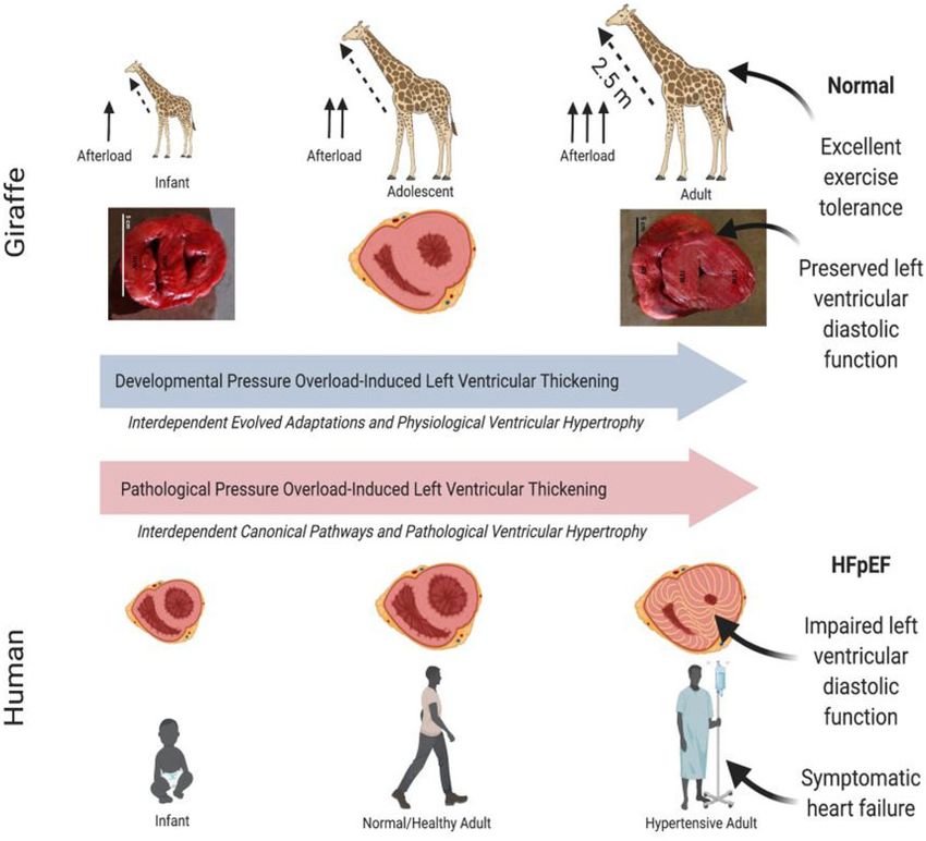
Figure 2. Comparison of the response of the left ventricle of the giraffe to increasing hypertension over the course of development vs. the ventricular response
to chronic hypertension in humans. In both cases, hypertension leads to thickening (i.e. hypertrophy) of the left ventricular wall. Hypertension-induced left
ventricular thickening (LVT) in humans leads to cardiac pathologies such as fibrosis, and commonly heart failure with preserved ejection fraction (HFpEF).
However, developmental pressure-induced LVT in growing giraffes does not compromise exercise capacity, which is an important adaptation this prey spe-
cies. HFpEF, heart failure with preserved ejection fraction.
Are cardiovascular adaptations in giraffes a naturally associated with increased neck length and concentric LV hyper-
occurring model of resistance to heart failure with trophy also induced the myocardial changes, which compro-
preserved ejection fraction (HFpEF)? mised exercise capacity (as is seen in humans with HFpEF), the
While concentric LV hypertrophy in humans is associated with fitness benefits of the increased neck length might be counter-
cellular and subcellular changes leading to increased ventricular balanced by increased risk of predation. In humans with
stiffness, reduced exercise tolerance, and HFpEF, giraffe cardio- HFpEF, increasing heart rates reduce relative diastolic filling
vascular physiology does not follow this pattern. Giraffe appear times which leads to increased pulmonary pressures and HF
to have evolved an adaptation protecting hypertrophic ventricle symptoms. During flight from predators, increased myocardial
from these changes and from progression to HFpEF [25]. oxygen demand contributes to rising heart rates and reduced
Predation risk may be the basis of the selective pressure diastolic filling times without apparent adverse effects on pul-
underlying this adaptation. Giraffe are a prey species and to monary pressures. Although hemodynamic measurements of
evade capture and death they must be able to flee predators at giraffes exercising at maximum capacity are not presently avail-
speeds of up to 60 km h1 [41]. Thus, their survival, and ultim- able, a 1966 study that measured the cardiovascular responses
ately fitness, depend on maintaining maximal exercise capacity of wild East African giraffes running to avoid capture recorded
in order to escape predators. If the increased afterload heart rates of up to 170 bpm via radiotelemetry [37].252 | Natterson-Horowitz et al. Evolution, Medicine, and Public Health
Giraffe cardiovascular physiology has particular salience for these evolved adaptations. For example, cardiac fibrosis—a
human HFpEF. The giraffe heart appears to be an example of a pathological process associated with systemic hypertension
mammalian heart in which pressure-induced concentric ven- and HFpEF in humans, appears to be relatively suppressed in
tricular thickening does not appear to reduce exercise capacity giraffe despite comparable levels of ventricular thickening [34].
as is the case with pressure-induced concentric ventricular Likewise, our preliminary data from reviewing 136 necropsy
thickening and HFpEF in humans [28, 42]. We hypothesize that reports suggests a reduced propensity for myocardial fibrosis in
evolved adaptations in the giraffe myocardia prevent elevation the giraffe relative to humans and other mammalian species
in LV diastolic pressures that are observed in humans with [43].
HFpEF and magnified with exercise-associated tachycardia. Consistent with our observations, reduced fibrosis in giraffe
Figure 3 compares the advanced hemodynamic consequences myocardia may be linked to differences in the amino acid se-
of pathological hypertension-induced hypertrophy seen with quence of the ACE protein [32, 44], as well as recently identified
HFpEF in human with the adaptive physiological concentric LV mutations in fibroblast growth factor receptor-like 1 (FGFRL1)
Downloaded from https://academic.oup.com/emph/article/9/1/248/6296607 by guest on 01 November 2021
hypertrophy that we hypothesize exists in the normal adult gir- [32, 45]. Notably, the FGFRL1 protein sequence in giraffe
affe heart. appears to be highly divergent in comparison to a diverse array
of other mammals, with seven amino acid substitutions in a re-
gion that is crucial for FGF binding. In addition, a comparison
Potential mechanisms
of the giraffe genome to that of its closest living evolutionary
The cellular and subcellular processes that protect giraffe hearts relative, the okapi, identified 70 genes with ‘multiple signs of
from the adverse consequences of systemic hypertension and adaptation’ that were not observed in other eutherian mammals
pressure-induced LV thickening are largely unknown. However, [32], five of which are found within the developmental pathways
several candidate mechanisms are providing new insights in to that lead to cardiac fibrosis [43].
Figure 3. Comparison of gross ventricular anatomy in the healthy human adult heart, human heart failure, and healthy adult giraffe hearts and their relation-
ships to left atrial and ventricular pressure. Despite significant pressure-induced left ventricular thickening (LVT), giraffe cardiac pressures do not display the
elevated left atrial and ventricular pressures observed in humans with severe hypertension-induced LVT. Giraffe ventricular pressures adapted from Smerup
et al. [33]. HFpEF, heart failure with preserved ejection fraction.Did giraffe cardiovascular evolution solve the problem of HFpEF? Natterson-Horowitz et al. | 253
In the recently published study by Liu et al. [45], mouse pathways and related regulatory systems as potential
FGFRL1 was edited to contain the seven amino acid substitu- approaches to HFpEF in humans.
tions of giraffe FGFRL1 using CRISPR-Cas9 technology. The mu- Given the importance of pressure-induced physiological LV
tant mice with giraffe-type FGFRL1 exhibited improved heart thickening in the giraffe and other species-specific cardiovascu-
function and significantly less fibrosis in cardiac and renal tis- lar characteristics for human health, why are these connections
sues than wild-type mice in response to infusion with angioten- relatively unexplored? One factor has been the limited extent to
sin II, indicating a role for FGFRL1 in suppressing fibrosis in which physicians perceive the natural world as a source of in-
the physiological setting of hypertension. Furthermore, the po- spiration for complex human pathophysiology. Veterinarians
tential roles of micro RNAs in the post-transcriptional regula- and wildlife biologists are trained in the core discipline of com-
tion of ACE, ACE2, FGFRL1 and other relevant genes during parative physiology, which seeks to emphasize both differences
cardiac remodeling further underscore the need to elucidate the between species and the importance of elucidating the underly-
underlying mechanisms of different cardiac phenotypes [23, 46]. ing mechanisms of how animals interact with and adapt to their
Downloaded from https://academic.oup.com/emph/article/9/1/248/6296607 by guest on 01 November 2021
Lastly, other genes involved in the regulation of fibrosis, as well environment. Yet, modern medical education does not trad-
as processes contributing to diastolic impairment in humans itionally include broad instruction on the diverse range of high-
(i.e. autonomic regulation, neuroendocrine function, and myo- performance physiologies of other species. Greater collabora-
cardial innervation), could also play a role in the cardiovascular tive interactions between physicians, veterinarians, animal
adaptations and unique exercise capacity of the modern giraffe. physiologists and wildlife biologists would increase the likeli-
Precise characterization of the mechanisms underlying the gir- hood that biomedical investigators could identify ‘solutions’ to
affe heart’s resistance to the adverse effects of chronic pressure challenging human pathophysiologies in the natural world.
overload may yield important insight for preventing and treating Rudolf Virchow, the father of modern pathology, observed
HFpEF in humans. that, ‘Between human and animal medicine there is no dividing
line’ [57]. Despite Virchow’s early insight, the separations be-
tween human, comparative, and veterinary cardiology persist.
RECOGNIZING EVOLUTIONARY ADAPTATIONS AS
The lack of communication between these research fields
A SOURCE OF THERAPEUTIC INNOVATIONS
impedes innovations to the detriment of human CVD. As physi-
The giraffe’s unique physiology has long been a source of fas- cians and investigators increasingly perceive biodiversity in the
cination to biologists and physiologists. Goetz and Keen—two natural world as a source of insight for clinical medicine, bioins-
of the first scientists to gather concrete physiological data on pired solutions to the most challenging cardiovascular issues
the giraffe—noted that giraffes exhibited ‘high’ blood pressures may emerge.
by human standards [47]. The resistance of the giraffe cardio-
vascular system to orthostatic changes via shifts in neck pos-
acknowledgements
ition and the ability of its renal system to withstand high arterial
pressures have also received extensive attention over the last The authors wish to thank Jennifer Head and Susan Kwan for their assist-
65 years [36, 37, 40, 48–56]. While earlier studies established ance in preparing the manuscript for submission.
thick ventricular walls in the giraffe [36], more recent studies on
Conflict of interest: None declared.
the unique cardiac adaptations of the giraffe heart to chronically
high afterload have focused on the physiological and cellular
underpinnings, such as myocardial architecture, cellular struc-
ture, and hemodynamics [19]. Importantly, Smerup et al. [33]
references
demonstrated that ejection fractions, diastolic ventricular pres- 1. Stenvinkel P, Painer J, Johnson RJ et al. Biomimetics—Nature’s road-
sures and measures of LV wall stress remained in the ‘normal’ map to insights and solutions for burden of lifestyle diseases. J Intern
range in comparison to other mammals. Furthermore, normal Med 2020;287:238–51.
diastolic pressures would not be predicted in a morphologically 2. Ralston E, Swain G. Bioinspiration—the solution for biofouling control?
comparable human ventricle. Bioinspir Biomim 2009;4:015007.
3. Theckes B, Langre E. d, Boutillon X. Damping by branching: a bioinspi-
The existence of a mammalian cardiovascular system in
ration from trees. Bioinspir Biomim 2011;6:046010.
which ventricular thickening from pressure-overload does not
4. Benyus JM. Biomimicry: Innovation Inspired by Nature. Morrow New
reduce diastolic relaxation or elevate cardiopulmonary pres-
York: Harper Perennial, 1997.
sures suggests that models of resistance to human cardiovas- 5. Lee BP, Messersmith PB, Israelachvili JN et al. Mussel-inspired adhe-
cular pathologies may have evolved spontaneously in other sives and coatings. Annu Rev Mater Res 2011;41:99–132.
species. Non-pathological cardiac remodeling during somatic 6. Shoffstall A, Srinivasan S, Willis M et al. A mosquito inspired strategy
growth in the giraffe also focuses attention on developmental to implant microprobes into the brain. Sci Rep 2018;8:122.254 | Natterson-Horowitz et al. Evolution, Medicine, and Public Health
7. Ramasubramanian MK, Barham OM, Swaminathan V. Mechanics of a 29. Lalande S, Johnson BD. Diastolic dysfunction: a link between hyperten-
mosquito bite with applications to microneedle design. Bioinspir sion and heart failure. Drugs Today (Barc) 2008;44:503–13.
Biomim 2008;3:046001. 30. Roh J, Houstis N, Rosenzweig A. Why don’t we have proven treatments
8. Stefanini C, Orofino S, Manfredi L et al. A novel autonomous, bioins- for HFPEF? Circ Res 2017;120:1243–5.
pired swimming robot developed by neuroscientists and bioengineers. 31. Letunic I, Bork P. Interactive Tree Of Life (iTOL) v4: recent updates and
Bioinspir Biomim 2012;7:025001. new developments. Nucleic Acids Res 2019;47:W256–9.
9. Martin-Silverstone E, Habib M, Hone D. Volant fossil vertebrates: po- 32. Agaba M, Ishengoma E, Miller WC et al. Giraffe genome sequence
tential for bioinspired flight technology. Trends Ecol Evol 2020;35: reveals clues to its unique morphology and physiology. Nat Commun
618–29. 2016;7:11519.
10. Speck O, Speck T. An overview of bioinspired and biomimetic self- 33. Smerup M, Damkjær M, Brøndum E et al. The thick left ventricular wall
repairing materials. Biomimetics 2019;4:26. of the giraffe heart normalises wall tension, but limits stroke volume
11. Saikawa Y, Hashimoto K, Nakata M et al. The red sweat of the hippo- and cardiac output. J Exp Biol 2016;219:457–63.
potamus. Nature 2004;429:363. 34. Østergaard KH, Baandrup UT, Wang T et al. Left ventricular morph-
Downloaded from https://academic.oup.com/emph/article/9/1/248/6296607 by guest on 01 November 2021
12. Chung KK, Schumacher JF, Sampson EM et al. Impact of engineered ology of the giraffe heart examined by stereological methods: left ven-
surface microtopography on biofilm formation of Staphylococcus aureus. tricular morphology of the giraffe heart. Anat Rec 2013;296:611–21.
Biointerphases 2007;2:89–94. 35. Mitchell G, Skinner JD. An allometric analysis of the giraffe cardiovas-
13. Lurie-Luke E. Product and technology innovation: what can biomimicry cular system. Comp Biochem Physiol A Mol Integr Physiol 2009;154:
inspire? Biotechnol Adv 2014;32:1494–505. 523–9.
14. Tong Q, Qiu N, Ji J et al. Research progress in bioinspired drug delivery 36. Goetz RH, Warren JV, Gauer OH et al. Circulation of the giraffe. Circ
systems. Expert Opin Drug Deliv 2020;17:1269–88. Res 1960;8:1049–58.
15. Lairmore MD, Khanna C. Naturally occurring diseases in animals: con- 37. Van Citters RL, Kemper WS, Franklin DL. Blood pressure responses of
tributions to translational medicine. ILAR J 2014;55:1–3. wild giraffes studied by radio telemetry. Science 1966;152:384–6.
16. Heron M. Deaths: Leading Causes for 2017. National Vital Statistics Reports; 38. Aalkjær C, Wang T. The remarkable cardiovascular system of giraffes.
Vol 68 No 6. Hyattsville: National Center for Health Statistics, 2019. Annu Rev Physiol 2021;83:1–15.
17. Federmann M, Hess OM. Differentiation between systolic and diastolic 39. Meijler FL, Meijler TD. Archetype, adaptation and the mammalian
dysfunction. Eur Heart J 1994;15:2–6. heart. Neth Heart J 2011;19:142–8.
18. Oktay AA, Rich JD, Shah SJ. The emerging epidemic of heart failure 40. Brøndum E, Hasenkam JM, Secher NH et al. Jugular venous pooling
with preserved ejection fraction. Curr Heart Fail Rep 2013;10:401–410. during lowering of the head affects blood pressure of the anesthetized
19. Kitai T, Tang WW. Recent advances in treatment of heart failure. giraffe. Am J Physiol Regul Integr Comp Physiol 2009;297:R1058–65.
F1000Res 2015;4:1475. 41. Hubel TY, Golabek KA, Rafiq K et al. Movement patterns and athletic
20. Shimizu I, Minamino T. Physiological and pathological cardiac hyper- performance of leopards in the Okavango Delta. Proc R Soc B 2018;
trophy. J Mol Cell Cardiol 2016;97:245–62. 285:20172622.
21. Abel ED, Doenst T. Mitochondrial adaptations to physiological vs. 42. Kitzman DW, Groban L. Exercise intolerance. Cardiol Clin 2011;29:
pathological cardiac hypertrophy. Cardiovasc Res 2011;90:234–42. 461–77.
22. Mihl C, Dassen WRM, Kuipers H. Cardiac remodelling: concentric ver- 43. Baccouche BM. (undergraduate thesis) (Barbara N. Horowitz, MD
sus eccentric hypertrophy in strength and endurance athletes. Neth Supervising PI) The Giraffe Paradox: Investigating the Absence of
Heart J 2008;16:129–33. Hypertrophy-Related Exercise Impairment in the Modern Giraffe as a
23. Fernandes T, Soci UPR, Oliveira EM. Eccentric and concentric cardiac Natural Animal Model for Resistance to Diastolic Heart Failure in
hypertrophy induced by exercise training: microRNAs and molecular Humans. 2020.
determinants. Braz J Med Biol Res 2011;44:836–47. 44. Wang D, Deuse T, Ameri K et al. The “giraffe paradox”: what humans
24. Basford JR. The Law of Laplace and its relevance to contemporary can learn from giraffe in heart failure. J Heart Lung Transplant 2017;36:
medicine and rehabilitation. Archiv Phys Med Rehabil 2002;83: S384–5.
1165–70. 45. Liu C, Gao J, Cui X et al. A towering genome: experimentally validated
25. Hirt MN, Sörensen NA, Bartholdt LM et al. Increased afterload induces adaptations to high blood pressure and extreme stature in the giraffe.
pathological cardiac hypertrophy: a new in vitro model. Basic Res Cardiol Sci Adv 2021;7:eabe9459.
2012;107:307. 46. Niu T, Liu N, Zhao M et al. Identification of a novel FGFRL1 MicroRNA
26. Rosen Boaz D, Thor E, Shenghan L et al. Left ventricular concentric target site polymorphism for bone mineral density in meta-analyses of
remodeling is associated with decreased global and regional systolic genome-wide association studies. Hum Mol Genet 2015;24:4710–27.
function. Circulation 2005;112:984–91. 47. Goetz RH, Keen EN. Some aspects of the cardiovascular system in the
27. Rossi MA. Pathologic fibrosis and connective tissue matrix in left ven- giraffe. Angiology 1957;8:542–64.
tricular hypertrophy due to chronic arterial hypertension in humans. J 48. Damkjaer M, Wang T, Brøndum E et al. The giraffe kidney tolerates
Hypertens 1998;16:1031–41. high arterial blood pressure by high renal interstitial pressure and low
28. Nilsson KR, Duscha BD, Hranitzky PM et al. Chronic heart failure and glomerular filtration rate. Acta Physiol 2015;214:497–510.
exercise intolerance: the hemodynamic paradox. Curr Cardiol Rev 2008; 49. Goetz RH. Preliminary observations on the circulation in the giraffe.
4:92–100. Trans Am Coll Cardiol 1955;5:239–48.Did giraffe cardiovascular evolution solve the problem of HFpEF? Natterson-Horowitz et al. | 255
50. Goetz RH, Budtz-Olsen O. Scientific safari-the circulation of the giraffe. 54. ——— et al. Transcapillary fluid balance in giraffe. Interstitial-
S Afr Med J 1955;29:773–6. lymphatic liquid and solute movement. Adv Microcirc 1987;13:195–202.
51. Van Citters RL, Kemper WS, Franklin DL. Blood flow and pressure in 55. Hargens A, Holton E. The gravity of giraffe physiology. 1997. https://
the giraffe carotid artery. Comp Biochem Physiol 1968;24:1035–42. ntrs.nasa.gov/citations/20020042320 (2 April 2021, date last accessed).
52. White CR, Seymour RS. The role of gravity in the evolution of mamma- 56. Mitchell G, Skinner JD, Rickards E et al. Giraffe cerebrovascular resist-
lian blood pressure. Evolution 2014;68:901–8. ance. S Afr J Sci 1990;86: 542.
53. Hargens AR, Millard RW, Pettersson K et al. Gravitational haemo- 57. Reed LD. The important interface between public health and veterinary
dynamics and oedema prevention in the giraffe. Nature 1987;329: medicine for improving human health, animal health, and food safety.
59–60. Public Health Rep 2008;123:257.
Downloaded from https://academic.oup.com/emph/article/9/1/248/6296607 by guest on 01 November 2021You can also read