Discovery of X-rays-Its Impact in India and History of X-ray Research in Colonial India
←
→
Page content transcription
If your browser does not render page correctly, please read the page content below
Review

Discovery of X-rays—Its Impact in India and History of X-ray
Research in Colonial India
Suprakash C. Roy                                   Formerly at Bose Institute, Kolkata 700009, India; suprakash.roy@gmail.com

                                                   Abstract: India holds a respectable position globally in X-ray research, particularly in X-ray crystallography.
                                                   X-ray research in India is as old as the discovery of X-rays and the history of X-ray research in colonial India
                                                   is fascinating. The purpose of this paper is to present how India participated in X-ray research and how X-
                                                   ray research initiated by C.V. Raman, the only Indian Nobel Laureate in physics, at the Indian Association for
                                                   the Cultivation of Science (IACS) paved the way to proliferate X-ray research in all parts of India and acted
                                                   as the foundation stone of modern X-ray research in India. With limited resources under the British rule
                                                   (India became independent in 1947), readers will find that the research work performed by Indians is
                                                   commendable. This article is neither comprehensive nor detailed but will give the readers a flavour of the
                                                   high-quality X-ray research that was performed in India in the early years after the discovery of X-rays.

                                                   Keywords: X-ray; colonial India; history

                                                   1. Introduction
                                                         India entered the international mainstream of scientific research in the late nineteenth and
                                                   early twentieth century. It was Sir Jagadish Chandra Bose (1858–1937) who first showed the world
                                                   that it is possible to do science at par with Western standards in colonial India when he
                                                   demonstrated his millimetre wave experiment with equipment made in India with limited
Citation: Roy, S.C. Discovery of                   resources before the Royal Society of London during his first scientific mission to Europe (1896–
X-rays—Its Impact in India and                     1897). Serious research began in universities in the 1920s and
History of X-ray Research in Colonial
                                                   1930s in Allahabad, Dhaka (now in Bangladesh), Punjab, Delhi, Mysore, Andhra Pradesh, Banaras,
India. https://doi.org/10.3390/
                                                   etc., pursuing the idea of teaching-cum-research institutions in contrast to the purely teaching
qubs6020016
                                                   universities set up by the British [1].
Academic Editor: Akihiro Iwase                           So far as X-ray research in India is concerned, it started immediately after the discovery of X-
Received: 21 March 2022
                                                   rays first by excitation to reproduce X-ray photographs indigenously and then to pursue research
Accepted: 8 April 2022                             more seriously [2,3]. C.V. Raman started his scientific research as an amateur at Indian Association
Published: 22 April 2022                           in Calcutta and later turned into a serious researcher cum teacher and earned the Nobel Prize in
                                                   physics in 1930. After his return from England in 1921, C.V. Raman started serious scientific
                                                   research using X-rays with a host of talented students which paved the way for building X-ray
                                                   crystallography research centres across India.

                                                   2. Discovery of X-rays
Copyright: © 2022 by the author. This                   Wilhelm Conrad Roentgen (1845–1923) has been credited with the discovery of X-rays and
article   is   an   open       access    article   was awarded the first Nobel Prize in Physics in 1901 for this discovery. Roentgen’s discovery is
distributed    under     the     terms     and     known to be the result of an accident while he was experimenting with a discharge tube. The
conditions of the Creative Commons                 objective of the current article is to present a historical account of X-ray discoveries, such as that
Attribution (CC BY) license (https://              X-rays were produced experimentally much before Roentgen’s discovery in 1895 as has been
creativecommons.org/licenses/by/                   found in literature and to study the influence of this discovery on the scientific community of India.
4.0/).                                                  The twenty-eighth of December 1895 is considered as the date of discovery of X-ray, the date
                                                   on which Roentgen submitted his first “provisorial” communication Uebereineneue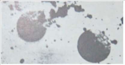
Art von Strahlen (On a New Kind of Rays) which was published in the Proceedings of the
Würzburg Physico-Medical Society (Sitzungber der WürzburgerPhysik.- MedikGesellschaft).
First oral presentation of the discovery was made before the same Würzburg Society on 23
January 1896. The meeting was chaired by the famous anatomist Albert Rudolf von Kolliker (1817–
1905) as reported in the Münchener Medicinische Wochenschrift of 29 January 1896.
After the lecture Roentgen produced an X-ray picture of Kolliker’s hand in a glass plate
(Figure 1). It was Dr. Kolliker who proposed that the new rays henceforth be known as Roentgen
rays [4].

Figure 1. First X-ray picture made in public by Roentgen during his first oral presentation before the Würzburg
Physico-Medical Society on 23 January 1896. The picture shows the hand of Albert Kolliker who chaired the
talk.

      Roentgen’s discovery was shrouded with mystery, stories, and controversies which
sometimes reached a stage of humiliation to the great scientist. It is believed that fluorescence
was first noticed by Roentgen’s laboratory assistant Ludwig Zhender in 1890, who, while working
with the discharge tube covered with black cloth, observed the fluorescence in the fluorescent
screen placed nearby [5]. Zhender was loyal to Roentgen throughout his life and had not claimed
any recognition, but this story was severely distorted to humiliate Roentgen and he was accused
of stealing his Diener’s discovery [4]. It is to be remembered in this connection that X-ray tubes
that we are talking about were known as cold discharge tubes in which ionization of gas by high
voltage was the source of electrons in contrast to modern X-ray tubes which contain a heated
filament for producing electrons.
      According to Roentgen’s will, all his diaries and notes were destroyed after his death which
resulted in a great loss of vital information for historians, such as the exact date when Roentgen
realized the importance of the penetrating power of this new ray. From a letter written by his wife
Anna Bertha Ludwig in March 1896 to one of his cousins Mrs. L.R. Grauel of Indianapolis, it was
found that Roentgen first noticed this new radiation some time in November 1895 [4]. As Bertha
mentioned in her letter, on an evening of November
1895 she became angry with her husband for the quality of food. In order to soothe her, Roentgen
took her downstairs to his laboratory and introduced her to the mysterious new rays. However, it
was not mentioned whether her hand with a ring was exposed to this ray (Figure 2) on the same
day. According to Edgar Ashworth Underwood, Director of Wellcome Historical Museum, the
picture of his wife’s hand was taken on 8 November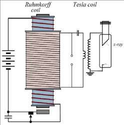
1895. This date was accepted by Konrad Weiss, the historiographer of Austrian Roentgen Society
and is accepted as the date of the discovery.

Figure 2. X-ray picture taken by Roentgen of his wife Bertha Ludwig’s hand. (Courtesy: Prof. Alok Mukherjee,
Jadavpur University, Kolkata, India).

     Like Jagadish Chandra Bose (1858–1937) who refused to take patent of his discovery of
microwave (the discovery for which Guglielimo Marconi was awarded the Nobel Prize) [6], on the
ground that knowledge is for all and should be freely available, Roentgen also refused to take
patent of any part of his discovery and rejected all commercial proposals connected to this
discovery. However, unlike J. C. Bose, he lost all his savings due to post war inflation and suffered
financial and other difficulties in the last years of his life.
Roentgen died in 1923 from colorectal cancer.

3. Production of X-rays before Roentgen
      There is evidence that X-rays had been produced experimentally before Roentgen’s date in
1895. This is not surprising because the basic physical process to generate X-rays is the passage of
electricity through gases, the study of which had been started in eighteenth century. Francis
Hauksbee (estimated to have died in 1713) who was the Curator of Experiments to the Royal
Society, London, described seeing “the shape and figure of all parts of his hand” while working
with electricity and vacuum [5]. Interest in the study of discharge in gases was revived in the
middle of nineteenth century after Faraday’s experiments with “radiant matter” and many
scientists were involved in producing different improvised discharge tubes to study discharge of
gases. Heinrich Geissler, Johann Wilhelm Hittroff, and Sir William Crookes were some of the
people who worked with discharge tubes and they were unknowingly exposed to X-rays while
working.
      In India, Father Lafont (1837–1908), a Jesuit missionary of St. Xavier’s College, Calcutta
brought from Europe a Crooke’s tube at a time (1878–1879) when vigorous research was in
progress in Europe using Crooke’s tube. Father Lafont delivered a lecture in 1880 titled “Crookes
on Radiant Energy” in the Science Association [7]. Lord Lytton, the then Viceroy of India, invited
Dr. Mahendralal Sircar (1833–1904), the founder of Indian Association for the Cultivation of
Science (IACS), to demonstrate the actions of Crookes tube. A contemporary report described: “It
is not possible for any individual to forget the evening. Dr. Sircar had such a wonderful mastery
over the subject that he very easily explained the amazing behaviour of one millionth of
atmosphere to the entire satisfaction to His excellency”.
It has been said that Jagadish Chandra Bose built an X-ray apparatus (presumably used a
discharge tube) a few years before Roentgen and demonstrated “X-rays passing through the hand”
[7]. As reported by A.K. Biswas [8] “we have reports of Jagadish setting up his own X-ray apparatus
in 1887, quite a few years before such a machine was imported to India”. Therefore, we see that
X-ray research in India had its root before the formal announcement of the discovery of X-rays.
The first recorded scholarly evidence to produce Roentgen rays was about hundred years
before Roentgen by William Morgan (1750–1833), the Welsh mathematician and Chief Actuary
of British Equitable Assurance Society. Morgan [9] reported in the Philosophical
Transactions that based on the length of time for which mercury was boiled in vacuum—the
‘electric’ light turned violet, purple, then beautiful green, and finally the light became invisible!
Morgan presented this discovery to the Royal Society in 1784 which was witnessed by Richard
Price and one of Richard’s friends, an American ‘electrician’, the famous Benjamin Franklin
(1706–1790).
It is believed that Philipp Lenard (1862–1947), another contemporary of Roentgen, who
worked on cathode rays came very close to the discovery of X-rays. In fact, both Lenard and
Roentgen were nominated for the Nobel Prize in Physics for the year 1901, and the Committee
recommended that the prize should be divided equally between Roentgen and Lenard. However,
the Royal Academy of Science did not follow this recommendation and decided to award the prize
to Roentgen alone. On 12 October 1892, Lenard had been able to show for the first time that
cathode rays can penetrate the aluminium window and can travel a few inches in air by observing
the fluorescence produced on granules of potassium phosphate placed outside the tube [10]. Four
years later, in 1905, the Nobel Committee decided to award the Nobel Prize to Lenard for his
ingenious work on cathode rays. Lenard, however, considered himself to be “the mother of X-
rays” while Roentgen was “the midwife who happened to deliver the child”. There are other
scientists who are believed to have observed X-rays before Roentgen and we are not going into
the details of those stories.
4. The First X-ray Photograph: Claims and Counter-Claims
The very first terms used for pictures produced by X-rays were the unabbreviated and easily
understandable combination of words such as ‘X-ray photograph’ or ‘shadow photograph’. There
were a lot of debates, controversies, personal choices, and etymological discussions in choosing
the name of the pictures taken by X-rays [3].
Who first produced the first X-ray photograph after the discovery of X-rays is replete with
claims and counter-claims. It is said that the first shadow graph or Roentgenograph of metal
objects [4] was obtained by accident in 1892 by a Philadelphia experimentalist Arthur Willis
Goodspeed (1860–1943). The story goes that Bill Jennings, a friend of Goodspeed, was counting
his coins after the Bill Jennings’ Carfare on the Woodland Avenue trolley when Goodspeed asked
Jennings to help in photographing the spark gap of a Ruhmkorff coil. Jennings put his coins on the
top of a plate while assisting in the positioning of the electrodes. The plate was later found to have
black patches. Rounded shadows shown in Figure 3 are the images of coins. They paid little or no
attention to those circular shadows till the announcement of Roentgen’s discovery. They then
repeated the experiment and understood the reason. However, Goodspeed expressly and
repeatedly denied any claims to priority because at that time he had failed to interpret these
shadows.Figure 3. Goodspeed’s first shadowgraph produced in the University of Pennsylvania on 20 February 1890.
The rounded shadows are coins. (Taken from: The Trail of the Invisible Light).
The most legitimate claim was that of a Scottish engineer Alan Archibald Campbell Swinton
(1863–1930) who tried to replicate X-ray photos in the beginning of 1896 immediately after the
discovery [4]. He conducted his experiment using a homemade tube and his plates were first
reproduced in Nature and subsequently in Industries and Iron and the Electrical Review of London
the next day [11]. From the reconstructed timetable that he maintained, the following were
captured: first (poor) roentgenogram on Tuesday January 7; first satisfactory “metallic” (a razor in
its paperboard casing) roentgenogram on January 8; first roentgenogram of a hand on January 13
(shown to the Prince of Wales); and first exhibit of those two plates on 16 January 1896.
Michael Idvorsky Pupin (1858–1935) of USA, an immigrant from former Serbia, claimed that
he produced the first roentgenogram on 2 January 1896 after Roentgen. In 1924, he wrote: “I
obtained the first X-ray photograph in America on 2 January 1896, two weeks after the discovery
was announced in Germany”. With the aid of a phosphorescent screen, supplied by his friend
Thomas A Edison, superimposed on a photographic plate, he was able to produce a good X-ray
picture in a few seconds of exposure [12]. Literature search shows that Pupin’s first paper on
Roentgen rays (dated Saturday, 1 February 1896) was published on February 5 in Electricity of New
York which was essentially a summary of data from European sources [4]. His first roentgen plate,
as mentioned in his article, was published in Science around February 14.
Thomas Edison (1847–1931) who is famous for his invention of electric bulb, realized the
importance of the Roentgen rays as soon as the news of the discovery reached the USA. According
to a statement made by his secretary William Henry Meadowcraft, “Mr. Edison was the first to
recognize the importance of the cable announcement of Dr. Roentgen’s discovery. The same day
he started to make the apparatus and had it finished the next day. Three of the metropolitan
dailies heard of it and for three weeks more than twenty newspaper reporters were stationed at
the Laboratory”. The first interview of Edison was made on 7 February 1896 and published in Times
on 8 February. Edison strongly believed that some practical (commercial) application would
emerge from Roentgen’s “purely scientific” discovery, and he employed his staff to find out the
most favourable conditions for taking roentgen photograph. A sketch of the design of one of his
earliest apparatuses using Hittrof tube is presented in Figure 4.Figure 4. Sketch of Edison’s early X-ray apparatus. (Courtesy: The Trail of the Invisible Light. Ref. 1).
5. X-ray Discovery and Its Impact in India
It is not known exactly when and how the news of X-ray discovery reached Calcutta
(now Kolkata), but it is fascinating to note that it had produced huge interest among the science
lovers of colonial India. Within a few months of the discovery of X-rays, Dr. Mahendralal Sircar
(MLS) ordered a Roentgen tube from Ducretet Company in Europe and received it in June 1896.
In his diary dated 11 June 1896 we find the following “The cases (3 in number) from Ducretet
containing apparatus for experimenting with Roentgen Rays were brought from the Customs
House to the Association today. When Amrita (son of Mahendralal Sircar) opened them, he found
that they have omitted to send the Flourescent Screen. The cathode disc was slightly bent. The
cells were too big” [8].
The first experiment using X-rays in India was carried out by Mahendralal Sircar
(MLS) on 20 June 1896 by taking the photograph of a hand using the procured Roentgen’s
apparatus and it was noted in his diary that he did not obtain a good picture in his first attempt,
probably due to over exposure. As noted in his diary, he repeated his experiment to obtain a better
photograph and was successful on 23 June. Therefore, according to his diary, the first successful
X-ray photograph was produced in India on 23 June 1896 in Calcutta by Mahendralal Sircar [13].
Experiments with X-rays were continued at the IACS by Amrita Lal Sircar under the guidance
of MLS with blocks of wood and books of different thickness, with sheets of iron, tin foil and zinc
foil as has been recorded in the diary dated 13 December 1899 [4]. This was the beginning of X-
ray research in India after the discovery of X-rays and thus IACS has the distinction of introducing
X-ray research in India.
A spurt of activities continued in India when people were trained in X-ray photography from
England and there is evidence of obtaining an excellent photograph of the right hand of Earl of
Elgin, the then Viceroy of India, wearing rings [13].
6. J. C. Bose and His X-ray Apparatus
Acharya Jagadish Chandra Bose (1858–1937) (Figure 5) is known for his discovery of
microwaves [14] and his epoch-making work on plant physiology demonstrating that plants
respond to external stimuli in a way very similar to animals [15–17]. However, what is less known
is that he was the first person in India who built an improved Roentgen’s apparatus on his return
from England in 1897. Acharya Bose visited Europe in 1896 at a time when the excitement of the
discovery of Roentgen rays in Europe was at its peak. It is possible that Jagadish Chandra, being a
physicist and an exceptional experimentalist, was attracted to this discovery and studied in detail
about the production of Roentgen rays.Figure 5. Jagadish Chandra Bose (1858–1937).
Jagadish Chandra Bose returned to India in April 1897 and started building his apparatus
[3]. D M Bose, his nephew, and former Director of Bose Institute, remarked in one of his articles
that after ‘reading a newspaper account of Roentgen’s discovery’ he built an X-ray apparatus in
Presidency College, Calcutta [18].
The exact date of his building the apparatus is not known but it appears to have been built
sometime around 1897–1898. We found the first mention of his X-ray apparatus in a letter
written to Rabindranath Tagore, the first Asian to receive the Nobel Prize in literature, in
February 1898 [19]. Unfortunately, neither the apparatus nor any sketch of his apparatus is
available either at Presidency College, Calcutta, the place where he built the apparatus or at Bose
Institute, Calcutta, the institute that he built later, to understand the mechanism of the
apparatus. Fortunately, we have been able to find a Press Report published on the 5th of May
1898 edition of the Amrita Bazar Patrika, an erstwhile reputed English daily of India, under the
title ‘Professor Bose and the New Light’ which describes the X-ray apparatus built by him and its
demonstration.
Roentgen used Ruhmkorff’s coil as a source of transient high voltage in discharging the gas
in Crooke’s tube. Ruhmkorff’s coil produces high voltage pulse in the secondary coil by
electromagnetic induction every time when a dc supply in the primary is interrupted by
mechanical means. The secondary is connected with the discharge tube (X-ray tube) and the high
voltage pulse was enough to produce X-rays. By coupling a Tesla coil with the secondary of
Ruhmkorff’s coil (which then acts as the primary of the tesla transformer).
Jagadish Chandra had been able to produce a higher voltage many times more than that can
be produced using a single Ruhmkorff’s coil. On the basis of the published information, we have
presented a schematic diagram (Figure 6) to represent the apparatus he had probably used. The
newspaper reported “We were shown a photograph of human palm taken by the Professor with
the new light, and the ghastly sight will long be vividly imprinted in our memory, for there, in the
photograph, instead of the ordinary fleshy palm is seen depicted a long range of bones presenting
a skeleton-like appearance.” Using Barium Platinocyanide screen prepared by himself with his
assistants in the Presidency College, he took X-ray photographs of different objects.Figure 6. Schematic diagram of the high voltage source J. C. Bose probably used.
Although use of X-rays in India was started within a few months of X-ray discovery by
importing X-ray tube from abroad, commercial production of indigenous machine made in India
started only a few years before India’s independence in 1947 [3].
7. Medical Uses of X-rays
As per available literature, the evidence of first clinical uses of X-rays in India, at least in an
informal setting, was started by Jagadish Chandra Bose in 1898. In a letter (Sen, 1994) written
possibly in February 1898 to Rabindranath Tagore, he expressed his inability to meet Rabindranath
at a time when he will be busy examining a patient with a broken back using Roentgen kal
(machine in Bengali) [19]. The history of application of X-rays by professionals for clinical purposes
in India is not very clearly known. As reported [20], Dr. Kartick Bose (1873–1955), an eminent
doctor, medical scholar and industrialist was “the first clinician in India to start clinical and X-ray
laboratories”.
Installation of the first X-ray machine in hospitals for public use is full of claims, counter-
claims and conflicting reports so much so that it is very difficult to arrive at a definite conclusion
[13]. It was reported by K P Mody in one of his editorials published in the Indian Journal of
Radiology & Imaging that “the first X-ray machine was imported by a chemist in 1902 into India;
that was only 7 years after the discovery” [21]. Madras
Medical College website (www.mmc.ac.in (accessed on 17 June 2021)) claimed that the
“first X-ray outfit was obtained for the general hospital in the year 1900 almost five years after
the discovery of X-rays, the first in South East Asia”. This might be true considering that it is a
very old medical institution started first as a Government General Hospital on 16 November
1664 to treat sick soldiers of the East India Company.
Use of X-rays for therapeutical purposes was also carried out at the beginning of twentieth
century. Successful treatment of leukaemia using X-rays was reported in Calcutta Medical Journal
[22] within five years of the first ever treatment of leukaemia, as reported in a paper published in
the Journal of the American Medical Association in 1902.
Excitement of using X-rays in medical science waned with time and was replaced by Xray
research in physical sciences.
8. C.V. Raman and Initiation of X-ray Research in India
In July 1921, C.V. Raman (Figure 7) on his voyage to England as a delegate of Calcutta and
Banaras University to attend University Congress, was attracted to the “mystery of the blue colour
of the sea”. On his return to India, he undertook a comprehensive programme of research on
molecular scattering of light by solid, liquid and gases which led to the discovery of a new light
effect. The new light effect was later known as ‘Raman effect’ and earned him the Nobel Prize in
physics in 1930. During this time Raman and his students were interested in the study of optical
anisotropy of molecules from scattering of light. Raman and Ramanathan [23] extended theoptical theory of scattering to X-ray diffraction by liquids. Experiments were also carried out to
compare the theory with experimental results. Sogani [24] recorded the X-ray diffraction patterns
of 22 aliphatic and aromatic liquids and estimated the average molecular distance from the
position of the maxima of the diffraction haloes. It became clear from these experiments that the
shape of the molecules and the intermolecular forces had profound influence on the diffraction
patterns. Krishnamurti [25], another student of Raman, initiated small angle X-ray scattering which
enabled us to understand the size and distribution of the particles in a sample. This is reportedly
the first small angle X-ray scattering experiment performed in India [26].
Figure 7. C.V. Raman.
Raman had an encounter with William Bragg and he was of the opinion that Bragg’s first
structure of naphthalene was not consistent with the birefringence, while the second one was. In
order to confirm this, he and his students started extensive studies on the optical and magnetic
anisotropy of organic crystals to understand the arrangements of molecules in crystalline state.
The work on crystal magnetism was initiated by one of his successful students K.S. Krishnan. In
many cases the orientation of the molecules in the unit cell could be calculated with a high degree
of precision from purely magnetic data,
which helped in refining X-ray analysis of the crystal structure [27,28].
Bidhubhusan Ray, another student of Raman, made significant contribution to X-ray
spectroscopy, although he remained unknown to most physicists until a book was published by
Rajinder Singh [29]. Bidhubhushan Ray (B. B. Ray) worked with Manne Siegbahn, a Nobel Laureate
in Physics, at Uppsala and on his return to India established X-ray laboratory at Calcutta University.
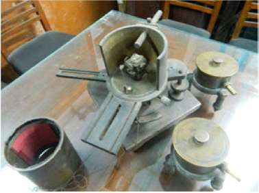
Satyendranath Bose who visited Europe during 1924–1926 met de Broglie brothers and was
attracted to X-ray crystal structure. His experience was utilized both at Dhaka University and later
at the Calcutta University where he took charge of the X-ray laboratory after the premature death
of B. B. Ray and built an X-ray diffraction apparatus in the laboratory (Figure 8).Figure 8. X-ray Diffraction Apparatus built by SN Bose. (Courtesy: Prof. Alok Mukherjee).
Historically the first crystallographic activity in India started with the determination of crystal
structures of napthaelene and anthracene by Kedareswar Banerjee (KB) at IACS [30,31]. In 1930,
in response to KB’s article in “Nature” on the structure of naphthalene, J.M. Robertson of Michigan
University, Ann Arbor, U.S.A., wrote: “I believe Dr. Banerjee’s structure to be essentially correct.
It has been clear to me for some time that the last two sections of my paper to which Dr. Banerjee
refers must be amended as regards the distribution of the scattering centres in the a and b
directions”. His further research on crystallography paved the way for a ‘complete crystallography
structure analysis’ and for understanding statistical relationships in the amplitudes of diffracted
waves. Further details of his life and work are available in a recently published book [32].
With Raman having good contact with the giants in the field of X-ray crystallography, he
was able to send many of his students abroad to let them work with leading scientists of the field
to gain contemporary knowledge of the subject. This made IACS the nucleus of X-ray research in
India.
After independence, there has been a great surge of scientific research in India through
establishment of laboratories by the Govt. of India. X-ray crystallography also flourished and
expanded into areas of biological sciences. Contributions of Kedareswar Banerjee were followed
by equally profound works by other researchers in India such as G. N. Ramachandran (University
of Madras and Indian Institute of Science, Bangalore), A. R. Verma (Banaras Hindu University), S.
Ramashesan (National Aeronautical Laboratory, Bangalore), VR. Chidambaram (Bhabha Atomic
Research Centre, Mumbai) and others [33].
G.N. Ramachandran, a student of C.V. Raman, and most distinguished crystallographer of
independent India worked on fibrous proteins and was recognized for the advancement of triple
helix model of the structure of collagen [34] and the Ramachandran plot for validation of protein
structure.
9. Conclusions
India has a legacy of X-ray research since the discovery of X-rays in 1895. Research in physical
sciences using X-rays was started in India by C V Raman at the IACS and made significant
contribution in colonial India with limited resources available at that time. By virtue of its scientific
activity India made a respectable position in X-ray research globally. IACS acted as the nucleus of
X-ray research in India from where it proliferated to different laboratories such as the Benares
Hindu University, Allahabad University, Madras University, and the Indian Institute of Sciences,
Bangalore.
Funding: This research received no external funding.
Data Availability Statement: Not applicable.Acknowledgments: The author is grateful to Barun Kumar Chatterjee of Bose Institute, Rajinder Singh of
University of Oldenburg, Germany, Gauri Roy, formerly at the Indian Association for the Cultivation of
Science, Alok Mukherjee of Jadavpur University, and many others for providing important information,
valuable discussions and comments. The author also thanks the authorities of Bose Institute and IACS for
giving access to relevant documents.
Conflicts of Interest: The author declares no conflict of interest.
References
1. International Union of Crystallography Newsletter; 2007; Volume 15, No. 4. Available online: https://www.iucr.org/news/
newsletter/volume-15/number-4 (accessed on 17 June 2021).
2. Roy, S.C. Early years of X-ray research in India. Sci. Cult. 2015, 81, 72.
3. Roy, S.C. Discovery of X-rays and its impact in India. Indian J. Hist. Sci. 2017, 52, 66. [CrossRef]
4. William, M. Philosophical Transactions; Royal Society: London, UK, 1784; Volume LXXV, p. 278.
5. Peh, W.C.G. Controversies surrounding and following Roentgen’s discovery. Singap. Med. J. 1995, 36, 54.
6. Emerson, D.T. The Work of Jagadish Chandra Bose: 100 Years of mm Wave Research. IEEE Trans. Microw. Theory Tech. 1997, 45, 2267–
2273. [CrossRef]
7. Biswas, A.K. Father Lafont of St. Xavier’s College, Calcutta and the Contemporary Science Movement; The Asiatic Society: Kolkata, India,
2001; Appendix XXXV.
8. Biswas, A.K. Gleanings of the Past and the Science Movement. In The Diaries of Drs. Mahendralal Sircar and Amritlal Sircar; The Asiatic
Society: Kolkata, India, 2000; pp. 295–296.
9. William, M. Philosophical Transactions; Royal Society: London, UK, 1784; Volume LXXV, p. 278.
10. Glasser, O. The Genealogy of Roentgen Rays II. AJR 1933, 30, 349–3679.
11. Jackson, H. Note on focus tube for X-rays. Electr. Rev. (London) 1896, 38, 340.
12. Davis, B. Biographical Memoir of Michael Idvorky Pupin. Proc. Natl. Acad. Sci. USA 1938, 19, 10.
13. Roy, S.C. History of X-ray research in colonial India. Indian J. Hist. Sci. 2018, 53, T-123. [CrossRef]
14. Bose, J.C. Electromagnetic Radiation and Polarisation of Electric Rays; Collected Physical Papers; Longmans, Green & Co.: London, UK, 1927;
pp. 77–101.
15. Bose, J.C. Responses in the Living and Non-Living; Longmans, Green & Co.: London, UK, 1902.
16. Bose, J.C. Plant Responses as a Means of Physiological Investigations; Longmans, Green & Co.: London, UK, 1906.
17. Bose, J.C. Comparative Electrophysiology; Longmans, Green & Co.: London, UK, 1907.
18. Bose, D.M. The scientific activities of Acharya Jagadish Chandra Bose. Sci. Cult. 1949, 14, 366.
19. Patrabali. Collection of Letters of Acharya Jagadis Chandra Bose to Rabindranath Tagore; Sen, D., Ed.; (Letter No. 1); Bose Institute: Kolkata,
India, 1994; pp. 1–2.
20. Bose, K. Kartick Chandra Bose. Curr. Sci. 2010, 98, 133.
21. Kumar, P.N.; Sharma, S. Study on Cost-Benefit Analysis of Computerised Tomography (CT) Scan. Int. J. Health Sci. Res. 2014, 4, 206.
22. Chatterjee, G.C. A case of Leukaemia successfully treated with X-rays. Calcutta Med. J. 1907, 1, 269.
23. Raman, C.V.; Ramanathan, K.R. The diffraction of X-rays in liquids, liquid mixtures, solutions, fluid crystals amorphous solids. Proc. Indian
Assoc. Cultiv. Sci. 1923, 8, 127.
24. Sogani, C.M. X-ray diffraction in liquids. Indian J. Phys. 1927, 1, 357.
25. Krishnamurti, P. Studies in X-ray diffraction: The structures of Amorphous Carbon. Indian J. Phys. 1930, 5, 473.
26. Chu, B.; Hsiao, S. Small-angle X-ray scattering by polymers. Chem. Rev. 2001, 101, 1727. [CrossRef]
27. Krishnan, K.S.; Banerjee, S. Investigations on magne-crystallic action IV. Magnetic behaviour of paramagnetic ions in the S-state in crystals.
Phil. Trans. Roy. Soc. 1936, A235, 343.
28. Krishnan, K.S.; Mookherji, A. Investigations on magne-crystallic action V. Paramagnetic salts of the rare earth and the ion groups.
Phil. Trans. Roy. Soc. 1938, A237, 135.
29. Singh, R. Bidhu Bhushan Ray-A Pioneer of X-ray Spectroscopy; Shaker Publishers: Maastricht, Germany, 2017.
30. Banerjee, K. Structure of naphthalene and anthracene. Nature 1931, 125, 456. [CrossRef]
31. Banerjee, K. Determination of the signs of the Fourier terms in complete crystal structure. Proc. R. Soc. 1933, 141, 188.
32. Rajinder Singh, C.V. Raman’s Student Kedareswar Banerjee: His Scientific Work in Indian Context; Shaker: Maastricht, Germany, 2021.
33. Mande, S. Early Developments in crystallography. Resonance 2014, 19, 1077. [CrossRef]
34. Venkataraman, V. Quartz India. 15 May 2017. Available online: https://qz.com/india/983439/madras-triple-helix-the-worldhas-nearly-
forgotten-the-indian-scientist-who-cracked-the-structure-of-collagen/ (accessed on 20 March 2022).You can also read