Digital pathology and artificial intelligence in translational medicine and clinical practice
←
→
Page content transcription
If your browser does not render page correctly, please read the page content below
www.nature.com/modpathol

REVIEW ARTICLE                        OPEN

Digital pathology and artificial intelligence in translational
medicine and clinical practice
           ✉
Vipul Baxi1 , Robin Edwards1, Michael Montalto2 and Saurabh Saha1

© The Author(s) 2021

    Traditional pathology approaches have played an integral role in the delivery of diagnosis, semi-quantitative or qualitative
    assessment of protein expression, and classification of disease. Technological advances and the increased focus on precision
    medicine have recently paved the way for the development of digital pathology-based approaches for quantitative pathologic
    assessments, namely whole slide imaging and artificial intelligence (AI)–based solutions, allowing us to explore and extract
    information beyond human visual perception. Within the field of immuno-oncology, the application of such methodologies in drug
    development and translational research have created invaluable opportunities for deciphering complex pathophysiology and the
    discovery of novel biomarkers and drug targets. With an increasing number of treatment options available for any given disease,
    practitioners face the growing challenge of selecting the most appropriate treatment for each patient. The ever-increasing
    utilization of AI-based approaches substantially expands our understanding of the tumor microenvironment, with digital
    approaches to patient stratification and selection for diagnostic assays supporting the identification of the optimal treatment
    regimen based on patient profiles. This review provides an overview of the opportunities and limitations around implementing AI-
    based methods in biomarker discovery and patient selection and discusses how advances in digital pathology and AI should be
    considered in the current landscape of translational medicine, touching on challenges this technology may face if adopted in
    clinical settings. The traditional role of pathologists in delivering accurate diagnoses or assessing biomarkers for companion
    diagnostics may be enhanced in precision, reproducibility, and scale by AI-powered analysis tools.

    Modern Pathology (2022) 35:23–32; https://doi.org/10.1038/s41379-021-00919-2

INTRODUCTION                                                                          breast cancer treated with chemotherapy plus either intravenous
Pathology has historically played a crucial role in the drug                          or subcutaneous trastuzumab14. In the immuno-oncology (I-O)
development process, including preclinical research to facilitate                     arena, immune-related pathologic response criteria have been
target identification, define drug mechanism of action and                              applied retrospectively to surgical specimens from patients
pharmacodynamics, and enable toxicology assessments1,2. More                          treated with immunotherapy in the neoadjuvant or advanced
recently, pathology has formed a bridge between drug discovery,                       disease setting to predict survival in several tumor types15,16.
translational, and clinical research programs that are striving to                       Immunohistochemistry (IHC) has been used to characterize
decipher disease pathophysiology in the context of the mechan-                        biomarkers, such as programmed cell death-ligand 1 (PD-L1), and
ism of action, patient selection, or patient stratification (Fig. 1)3,4.               their association with clinical benefit. Traditional pathology
Such insights form the basis of novel hypotheses that can further                     techniques present several advantages, such as low cost, wide-
be explored in drug discovery programs or applied to inform                           spread availability, and application on formalin-fixed, paraffin-
clinical trial design, thereby improving the probability of technical                 embedded (FFPE) tissue samples17, but challenges pertaining to
and regulatory success.                                                               differences in laboratory methods and subjective interpretation,
   Pathology-based assessments have been used to classify                             particularly with the evaluation of immune cell staining, may lead
disease and determine efficacy in drug development across a                            to inter-observer variability18. This can produce inconsistency in
variety of disease areas5–7. For example, during phase 2 trials for                   diagnoses, which may impact treatment decisions19–23. While the
drug development in non-alcoholic steatohepatitis, the US Food                        use of IHC assays has led to better identification of patients who
and Drug Administration (FDA) considers evidence of efficacy on a                      respond to I-O therapy24–26, there remains a need to more
histological endpoint to support initiation of phase 3 trials7.                       accurately quantify complex immune markers, including cell
Additionally, pathological complete response (pCR) has been                           phenotypes in a spatial context, that require advanced quantita-
studied as a surrogate endpoint in patients with cancer for the                       tive tools to maximize the amount of information yielded from
prediction of long-term clinical benefit and favorable prognosis                       individual samples27,28.
with the administration of neoadjuvant therapy8–13. More recently,                       Artificial intelligence (AI) applications in pathology improve
pCR was associated with improved long-term efficacy in patients                        quantitative accuracy and enable the geographical contextualization
with human epidermal growth factor receptor 2 (HER2)-positive                         of data using spatial algorithms. Adding spatial metrics to IHC can

Bristol Myers Squibb, Princeton, NJ, USA. 2PathAI, Boston, MA, USA. ✉email: Vipul.baxi@bms.com
1

Received: 15 April 2021 Revised: 18 August 2021 Accepted: 30 August 2021
Published online: 5 October 2021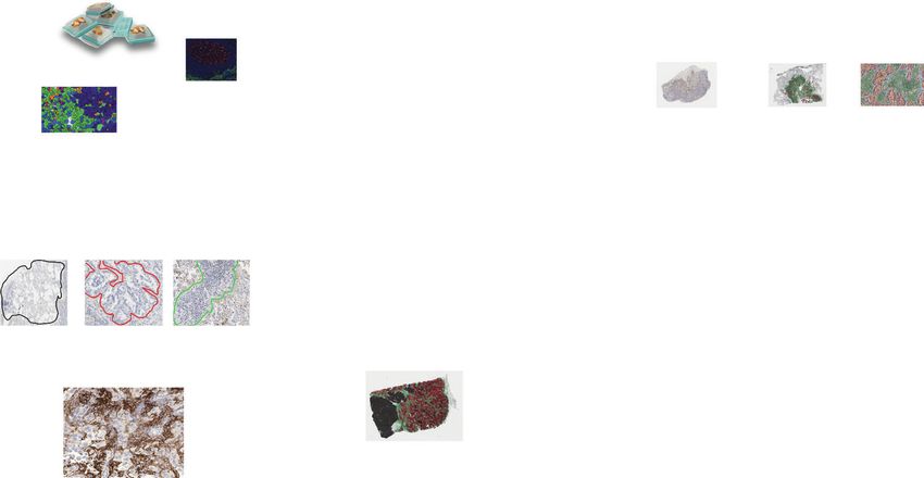
V. Baxi et al.
24
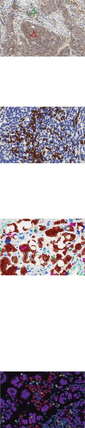
Discovery Translational Clinical IVD
Target identification Indication selection Mechanism of action Pharmacodynamics Patient stratification Companion diagnostics
• Omics approaches • Quantitation of • Cell phenotype in • Relative changes in • Quantitation of single • Guide treatment
(NGS, radiomics) marker across relation to TME marker levels vs baseline markers selection
• Multiplex screening different tumor types • Cell-cell proximity • Biologically relevant • Single markers in • Examples
platforms • Relative levels of • Quantitative association measures (proximity, context of tumor • HER2 (IHC, FISH)
receptor vs ligand to other readouts change in the tumor microenvironment • EGFR (IHC, NGS)
• Relative levels of (eg, genomics) microenvironment) compartments • PD-L1 (IHC)
targets to inform • Prognostics associations • Multiple marker
combination combinations
indications • Proximity
Fig. 1 Digital pathology: from drug discovery to clinical diagnostics. Dx diagnostic, EGFR epidermal growth factor receptor, FISH
fluorescence in situ hybridization, HER2 human epidermal receptor 2, IHC immunohistochemistry, IVD in vitro diagnostic, MOA mechanism of
action, NGS next-generation sequencing, PD pharmacodynamics, PD-L1 programmed death-ligand 1, TME tumor microenvironment.
DIGITAL PATHOLOGY
RAW DATA & SAMPLES
BIOINFORMATICS
Sample Preparation Imaging System
Execute algorithm on sample cohort
Sample set (commercial/clinical)
Imaging System Feature extraction/quantification
QC check/pathologist review
Brightfield Multispectral PD-L1 IHC on slides from CheckMate
017, 026, and 057 (n = 1079)111,d
Chromogenic/fluorescent
1234567890();,:
Monoplex/multiplex
Whole slide images Automated Tumor and
identification of immune cell
tumor region scoring
Gold Standard
Ground truth pathology scores Image Management Analysis
Training annotations Aperio eSlide Managera IHC expression and correlation across indications
Scoring guidelines HALO LinkTM,b Association to response (BOR) and
Concentriq®,c survival (OS/PFS)
Algorithm Development Integration with other -omics data
Composite biomarkers
Classifiers/segmentation
Machine/deep learning
Stain detection/estimation CheckMate 275 1% TC cutoff
Parenchymal CD8+ T-cell
Accuracy, sensitivity, and specificity > 1% by
Normal Tumor Stroma 60 Inflamed 100
manual
abundance (%)
c ed
40 lan and digital
Ba 75 scoring
20 OS (%) 50 < 1% by
10 manual
5 20 and digital
IC 1 scoring
Desert Excluded
0 0 ≥ 1% in
T 0 1 5 10 20 40 60 0 5 10 15 20 25 30 35 40 additional
Stromal CD8+ samples
T-cell abundance (%) identified
by digital
Szabo PM, et al. Poster presentation at the Duan C, et al. Poster presentation scoring only
ASCO Annual Meeting; May 31–June 4, 2019; at the AACR Virtual Annual Meeting;
T = tumor cell IC = immune cell Chicago, IL, USA. Abstract 2594. June 22–24, 2020. Abstract 2017.
Fig. 2 Digital prognostic pathology workflow. BOR best overall response, IHC immunohistochemistry, OS overall survival, PFS progression-
free survival, QC quality control. aLeica Biosystems; bIndica Labs; cProscia; dPD-L1 IHC 28-8 pharmDx. Dako/Agilent Technologies.
improve the clinical value of biomarker identification approaches. scanners capture multiple images of entire tissue sections on the
For example, in a recent meta-analysis, the addition of spatial slide, which are digitally stitched together to generate a WSI that
context to IHC, achieved using multiplex IHC and immunofluores- can be reviewed by a pathologist on a computer monitor
cence (IF), was significantly better at predicting objective response (Fig. 2)30,31. Two scanners, Philips IntelliSite Pathology Solution
to immune checkpoint inhibitors (ICIs) compared with gene (PIPS) (Philips, Amsterdam, Netherlands) and Leica Aperio AT2 DX
expression profiling (GEP) or IHC alone28, indicating the need for System (Leica Biosystems, Buffalo Grove, Illinois, USA), are
more complex computational approaches to decipher the under- approved by the FDA for review and interpretation of digital
lying biology and enhance clinical utility. surgical pathology slides prepared from biopsied tissue32,33.
The development and integration of digital pathology and There are many practical advantages to using these digital
AI–based approaches provide substantive advantages over tradi- pathology image systems and solutions that would bring
tional methods, such as enabling spatial analysis while generating substantial benefits to translational and clinical research. These
highly precise, unbiased, and consistent readouts that can be include the organization and storage of large amounts of data in a
accessed remotely by pathologists29. centralized location, integration of digital workflow software to help
streamline processes and improve efficiency, convenient sharing of
image data to enable cross-specialty worldwide remote commu-
ADVANCING FROM TRADITIONAL PATHOLOGY TO DIGITAL nication, reduced testing turnaround time, and the generation of
PATHOLOGY precise and highly reproducible tissue-derived readouts reducing
Efforts to overcome some of the challenges seen with traditional inter-pathologist variability29,34–37. The increased speed and effi-
pathology methods have led to the development and adoption of ciency gained in image acquisition can enhance the downstream
complex, novel imaging systems and whole slide image (WSI) utilization options of traditional techniques such as hematoxylin
scanners that have enabled the transition of pathology into the and eosin (H&E), IHC, and in situ hybridization. These slides can be
digital era, also known as digital pathology. Within minutes, WSI converted into a remotely available image within minutes and
Modern Pathology (2022) 35:23 – 32V. Baxi et al.
25
centrally reviewed by multiple pathologists from various sites29, A
with applications including education, research, consultation, and Chromogenic Monoplex
diagnostics29. (PD-L1 IHC)
Recently, due to ongoing disruptions in relation to the COVID- Visual assessment by pathologist
19 pandemic, including remote working and restricted travel,
digital pathology has been crucial in the continuation of clinical • % positive cells (semi-quantitative)
and academic research, as well as routine pathology services38. • Tumor vs stromal tissue (qualitative)
Without the need to transport glass slides and the ensuing
Tumor cells Immune cells
logistical and safety concerns, central pathology review enables
secure remote working38. Additionally, the utilization of digital B
images allows the generation of pixel-level pattern information, Chromogenic Monoplex
(CD8 IHC)
leading to expanded use of computational approaches that
enable a quantitative analysis of WSIs39,40.
AI-based quantitative image
analysis
• % positive cells
Improvements gained from digital pathology: quantitative • Density (positive cells/mm2)
analysis of the WSI • Tumor vs stromal tissue
The use of digital image analysis in pathology can identify and CD8+ Negative
quantify specific cell types quickly and accurately and can
quantitatively evaluate histological features, morphological pat- C
terns, and biologically relevant regions of interest (e.g., tumoral or Chromogenic Multiplex
peritumoral areas, relationships between different immune cell (Triplex: FoxP3-GITR-CD8) AI-based quantitative image
populations, areas of expression, presence of metastasis)41,42.
analysis
Quantitative image analysis tools also enable the capturing of data • % positive cells
from tissue slides that may not be accessible during manual • Density (positive cells/mm2)
assessment via routine microscopy. Additionally, performing • Tumor vs stromal tissue
• Co-expression/phenotyping
similar tasks manually can require significant time investment
• Co-localization/proximity
and can be prone to human error, such as counting fatigue43,44.
Unclassified FoxP3– GITR+ CD8+
FoxP3– GITR+ CD8– FoxP3+ GITR+ CD8+
Expanding data capabilities: multiplex and multispectral FoxP3+ GITR+ CD8– Negative in tumor
FoxP3– GITR– CD8+ Negative outside tumor
imaging
Quantitative image analysis can also be used to generate high- D
content data through application to a technique known as Immunofluorescence
multiplexing, which allows co-expression and co-localization Multiplex (6plex: PD-L1/CD8
analysis of multiple markers in situ with respect to the complex /CD68/PD1/FoxP3/CK)
AI-based quantitative image
spatial context of tissue regions, including the stroma, tumor
analysis
parenchyma, and invasive margin45,46. Current imaging metrics • % positive cells
can utilize multispectral unmixing strategies to reveal co- • Density (positive cells/mm2)
expression patterns that define unique cell phenotypes and • Tumor vs stromal tissue
• Complex phenotyping
spatial relationships (Fig. 3)47.
CK+ CD8+/PD–L1+ • Co-localization/proximity
Automated classification of epithelial and immune cells and CD8+/PD1+ FoxP3+
simultaneous marker analysis at the single-cell level has been CD68+/PD–L1
conducted using prostate cancer, pancreatic adenocarcinoma, and Fig. 3 Applications of digital pathology in IHC. a Monoplex slide
melanoma tissue samples46,48,49. Application of this technique stained for PD-L1, as seen on a monitor. b Monoplex slide stained for
allowed identification of distinct T-cell populations and their spatial CD8. c Multiplex stain annotated using AI-based analysis allowing
distributions and underscored the potential of immune markers to multiple marker identification. d Multispectral immunofluorescence.
identify patients who may benefit from immunotherapy48,49. Legend indicates examples of possible phenotypes detected from this
While a highly multiplexed imaging platform can be used to assay; however, many more are possible. CD cluster of differentiation,
understand intra- and inter-cellular signaling pathways by examin- PD-1 programmed death-1, PD-L1 programmed death-ligand 1.
ing how phenotypically distinct cell populations are spatially
distributed relative to one another, it is a time-consuming process diagnostic, or prognostic task using a supervised or unsupervised
applicable to a predefined region of interest50. However, as approach37,41,54. The power of AI to analyze large amounts of data
technology quickly advances, allowing digital evaluation of entire quickly can significantly speed up the discovery of novel
tissue slides, we are no longer confined to a region of interest37,51. histopathology features that may aid our understanding of or
The wealth of new information provided by these techniques has ability to predict how a patient’s disease will progress and how the
created a need for more consistent and reproducible interpretation patient will likely respond to a specific treatment37,39,55. In breast
of large and complex datasets, along with defining the interaction cancer, for example, unsupervised learning models have been
patterns between cell types and spatial context found used to generate histologic scores that can differentiate between
in pathological images that define biological underpinnings37,52,53. low- and high-grade tumors and evaluate prognostically relevant
morphological features from the epithelium and stroma of tissue
Advances in computational approaches: AI and machine samples to provide a score associated with the probability of
learning overall survival56,57. The success of these AI-based approaches
The need for data reproducibility and the increasing complexity of relies on the quality and quantity of the data used to train the
the analyses described above has led to the application of AI in algorithm, limiting the generalizability of these image analysis
pathology37,52,53. AI refers to a broad scientific discipline that algorithms to larger or more complex datasets58.
involves using algorithms to train machines to extract information
or features beyond human visual perception37,41,54. AI approaches Taking it further: deep learning networks. Deep learning takes
are built to initially extract appropriate image representations and machine learning a step further, using sophisticated, multilevel deep
then to train a machine classifier for a particular segmentation, or convolutional neural networks (DNN or CNN) to create systems
Modern Pathology (2022) 35:23 – 32V. Baxi et al.
26
Machine learning Deep learning
Feature
extraction from
images Multilevel feature extraction and classification
Feature
Outcome
Image
Input nodes
Classification
Output
Output
Example outputs:
Example outputs: • Grading of prostate cancer
• Generation of histologic scores • Prediction of disease-specific survival
• Scoring of morphological features in melanoma
• Detection of invasive breast cancer
Hardware requirements CPU for training GPU required for training
Training time Requires less time to train Longer training time required
No operator input. Automated feature extraction is part of
Training input Annotated images
the learning process
Fig. 4 Comparisons between machine learning and deep learning. Deep learning is a subset of machine learning that uses multi-layer
neural networks to analyze data, removing the need for operator input for feature extraction and image annotation37,41,57,59–62,111,145. CPU
central processing units, GPU graphics processing units.
that perform feature classification from large datasets37,40,41,54. is used to generate orthogonal data such as transcriptome or
Figure 4 highlights key differences between machine learning and exome69,70. In a comparison of TP determined using AI (using deep
deep learning. The impact of applications of deep learning learning algorithms generated on the PathAI platform) and manual
algorithms to IHC- and H&E-stained specimens have been well estimates by pathologists, AI-assessed TP was found to be more
documented across many tumor types. These include grading accurate than visual assessment by pathologists71. Previous
prostate cancer59, identifying biomarkers for disease-specific survival evidence has shown that immunosuppressive pathways are
in early-stage melanoma60, detection of invasive breast cancer upregulated in patients with low TP, suggesting that low TP is
regions on WSIs61,62, predicting response to chemoradiotherapy in associated with poor prognosis in some tumor types, including
locally advanced rectal cancer63, and identifying morphological gastric cancer. Therefore, improved methods of evaluating TP may
features (nuclear shape, nuclear orientation, texture, tumor archi- also aid in the identification of patients who may be suitable for
tecture, etc.) to predict recurrence in early-stage non-small cell lung immunotherapy72.
cancer (NSCLC) from H&E slides64. Deep learning has also been used Given the amount of additional detail and insights that can be
to construct entity-graph-based tissue representations, where cell gained from combining WSI with machine learning algorithms, this
morphology and topology are embedded within each node to technology can be readily applied to translational research.
effectively describe the phenotypical and structural properties of However, one major limitation of machine learning is the large
tissues and can be processed by graph neural networks (GNNs). amount of high-quality data required to develop these algorithms58.
GNNs therefore enhance the interpretability of pathological assess- Data used for training need to be accurate and as complete as
ments gleaned from neural networks65,66. possible in order to maximize predictability and utility39. This can be
It is important to compare AI-based interpretations with those of challenging when histological data are obtained from various
the pathologist to define the associated algorithm’s performance laboratories, leading to some variability due to factors such as
characteristics and utility. For example, when a CNN trained to differences in slide preparation (sectioning, fixation, staining, and
classify melanoma samples was compared against manual scoring mounting)73, scoring algorithms18, and inherent inter-observer
by histopathologists, the CNN was significantly superior in classifying variability74. These challenges become more apparent when more
images as malignant melanoma or benign nevi compared with complex computational analytics methods are used for multiplexed
manual assessment by histopathologists67. In the CAMELYON16 imaging. Although AI could be used to overcome inter-reader
challenge, deep learning algorithms to detect breast cancer variability across multiple institutions with the development of
metastases in H&E-stained WSIs of lymph node sections performed robust algorithms that take specific histological features of various
similarly to the best performing pathologists under time constraints tumors and subtypes into account75, further research is needed to
in detecting macrometastases and were better in detecting fully understand the impact of these factors on the quality of AI data.
micrometastases68. However, it should be noted that the perfor-
mance of any algorithm will depend on the task, due to the degree
of accuracy required and the quality of the samples to be assessed37. APPLICATIONS OF DIGITAL PATHOLOGY IN TRANSLATIONAL
Another application of machine learning in the preclinical space is MEDICINE
the assessment of tumor purity (TP). TP estimation, currently Enhancing our understanding of the TME
evaluated visually by pathologists, is used to ensure a signal is Tumor evolution and progression involve many complex cellulars
derived from cancer cells rather than other noncancerous cells that and molecular interactions that are spatially and temporally
may be present in the TME based on tissue morphology when tissue regulated within the TME52. IHC can be used to gain insights into
Modern Pathology (2022) 35:23 – 32V. Baxi et al.
27
the composition of the TME by facilitating the identification of TP53) being predicted from WSIs89. Deep learning has also been
different cell types expressing a protein of interest and assessing used to predict microsatellite instability (MSI) status from tumor
the density and spatial distribution of specific biomarkers50. Digital tissue90. A CNN trained to classify MSI versus microsatellite
pathology approaches, such as quantitative analysis of TILs, stability was able to robustly distinguish features predictive of MSI
present an opportunity to gain greater insight into intra-tumor in gastric and colorectal cancer samples90.
heterogeneity, spatial patterns of cell phenotypes, and the However, there are limitations to using AI for molecular
complex interactions between cancer and the immune system classification. For example, current imaging techniques can only
within the TME52,53. Image-based techniques can be used to identify genetic variants when they directly impact tissue
determine immune cell responses to immunotherapy such as morphology, as described previously91. At the same time, AI
macrophage activation76 or lymphocyte infiltration by regulatory algorithms cannot be applied in cases where actual variant allele
T cells (Tregs) into core tumor regions in solid tumors77, which frequencies of selected mutations can impact the classification
may in turn have value as a predictive indicator for the and prognosis of individual diseases, such as hematologic myeloid
effectiveness of ICIs. Favorable cancer prognosis has also been neoplasms92.
associated with factors in the TME, including high CD8+ TIL
rates78,79. Recently, image analysis and AI methods have
contributed to the development of novel approaches to TRANSLATING DIGITAL PATHOLOGY INTO CLINICAL PRACTICE
concurrently assess multiple biomarkers in preclinical and Potential for patient stratification
exploratory studies, revealing complex interactions within the As a further application in translational medicine, digital pathology
TME and providing the potential to improve cancer diagnosis and approaches have been used to predict response and identify
the selection of treatment regimens. Combining multiple techni- patients most likely to respond to treatment. For example, studies
ques, such as multiplex IF, with image analysis has yielded have used spatial analysis to determine the response of patients
important insights into specific immune cell populations, such as with NSCLC to nivolumab therapy. These included training
those in the TME of classical Hodgkin lymphoma, and their machine learning models to extract morphological details, such
associations with PD-1/CTLA-4+/− T cells80. These studies require as the spatial arrangement of tumor nuclei and variance in shape
multiple large cohorts to add the scale and robustness necessary and chromatin structure93, as well as the area and density of TILs
to gain these important insights, to elucidate relationships that and the proximity of TILs to each other and to tumor cells94. The
may not be apparent to the human eye, and to help overcome features extracted from these models were able to distinguish
observer bias that may mask potential biomarker signals. patients who responded to nivolumab therapy93,94. In another
example, digital image analysis was used to quantify CD8 and
Assessing treatment response: immune cell interactions in the PD-L1 positive cell densities from patients treated with durvalu-
TME mab across multiple tumor types95. Patients defined as positive
Digital pathology can also be used to gain insights into a receptor- for the CD8xPD-L1 composite signature had longer median
ligand binding, as proximity may be indicative of receptor survival compared with signature-negative patients, demonstrat-
engagement and activation. For example, lymphocyte-activation ing the potential predictive value of digitally defined composite
gene 3 (LAG-3), expressed on exhausted T cells, principally biomarkers.
interacts with major histocompatibility-II (MHC-II) molecules, AI and machine learning can also assist in classification and
expressed on the surface of antigen-presenting and tumor staging across various tumor types. A new approach to tumor
cells81,82. Spatial analysis in bladder and gastric cancer tumor subtyping has been developed based on a DNN (MesoNet) to
cells has demonstrated that the density and proximity of LAG-3+ predict OS of patients with mesothelioma from hematoxylin,
were significantly greater when associated with MHC II+ vs. eosin, and saffron stained WSIs, without any pathologist-provided
MHC II− tumor cells, suggesting that LAG-3–expressing TILs may annotations96. Results demonstrated that the model was more
be preferentially located in proximity to MHC II+ tumor cells, accurate in predicting patient survival than using current
allowing for LAG-3 activation and the inhibition of antitumor pathology practices and was able to identify regions contributing
immunity83. The insights provided by digital pathology into the to patient outcomes96, suggesting that deep learning models can
number and location of immune cells relative to tumor cells may identify new features predictive of patient survival and potentially
provide information on immune response37,84, which could guide lead to new biomarker discoveries.
future treatment strategies. AI has also been used to quantify
immune cells within the TME to define T-cell abundance Application of digital pathology and AI algorithms in
and associated geographic localization in the tumor stroma, diagnostics
parenchyma, parenchyma-stromal interface, and invasive margin, Biomarker research has been an area of particular interest in the I-O
which are then associated with transcriptomic factors to define space due to its potential predictive value in some solid
underlying biological associations85. tumors25,26,97–100. ICIs, such as anti–PD-(L)1 and anti–cytotoxic T
lymphocyte antigen-4, have been studied in multiple clinical trials,
Identifying genomic features leading to improved prognosis for patients across various solid
Additionally, AI-based approaches may find applications in tumors101. Evidence has shown that PD-L1 expression may be
translational medicine and clinical practice by predicting gene indicative of response to ICI therapy in some tumor types25,26,97–100,
mutations from routine histopathology slides. With genomic tests while other studies have shown that patients demonstrated durable
being associated with high costs and high rates of failure due to responses to ICIs regardless of PD-L1 expression3,102–108. Given the
stringent sample requirements86,87, AI may be particularly useful widespread clinical use of ICIs, predictive assays are needed to help
for evaluating genomic instability and the mutational landscape, stratify patients to determine who may benefit from such
with the possibility to assess pathologic and genomic features in treatments.
conjunction with one another. A CNN trained with WSIs of H&E- While the use of these assays can help determine whether a
stained hepatocellular carcinoma (HCC) tissue was used to predict patient will benefit from ICI therapy, biomarker identification, such
the ten most common prognostic and mutated genes in HCC, with as PD-L1 status, using tumor biopsies is challenging. Even when
four of these (CTNNB1, FMN2, TP53, and ZFX4) correctly identified used by experienced pathologists, visual interpretation of PD-L1
by the model88. Similar results were obtained when a DNN was using IHC is subjective and prone to error, which may contribute
trained to predict the most commonly mutated genes in lung to inaccurate patient stratification. Digital scoring of PD-L1
adenocarcinoma, with 6 (STK11, EGFR, FAT1, SETBP1, KRAS, and expression can assist pathologists in overcoming these barriers
Modern Pathology (2022) 35:23 – 32V. Baxi et al.
28
by providing standardized metrics for biomarker assessment at real-world environment, sample set size, establishing concordance
single-cell resolution across whole tissue sections36. using intra-observer variability, and documentation, among others30.
Multiple studies have evaluated PD-L1 assessment using digital The performance of AI applications in digital pathology is
scoring and AI algorithms and have shown that digital-based largely dependent on the size and quality of the dataset used to
techniques can perform better than or equal to manual patholo- train an algorithm41. Digital images used for training purposes
gical evaluation across various tumor types. A high correlation should be obtained from multiple staining batches, scanners, and
between AI and manual assessment of PD-L1 expression on tumor institutions to ensure generalizability. Such datasets should be
and immune cells has been observed in multiple CheckMate trials curated by pathologists, ensuring that representative images have
with samples from NSCLC, urothelial carcinoma, melanoma, and been obtained at an appropriate magnification and that all
gastric cancer109–111. Furthermore, similar associations between PD- regions of interest are comprehensively annotated depending on
L1 expression and response to nivolumab have been reported the diagnostic application41. Crucially, the validation of AI
between manual and digital scoring109,110. Using the combined algorithms developed for clinical purposes increases the con-
positive score to assess PD-L1 expression on tumor and immune cordance between manual and digital pathology interpretations.
cells, digital image analyses and pathologists’ interpretations on The role of pathologists in the validation step is equally important
stained slides (using the 22C3 pharmDx assay [Dako, Denmark]) in order to ensure that datasets represent the sample type of
demonstrated 33 (84.6%) of 39 cases had concordant results, and interest (e.g., H&E-stained FFPE section), encompass the entirety of
statistical analyses indicated that PD-L1 expression interpreted by a glass slide, and are big enough to reveal potential interpreta-
pathologists or digital image analysis did not differ significantly for tional discrepancies, as well as to evaluate the accuracy and
predicting responses to pembrolizumab112. Prospective clinical performance of the algorithm30,119.
trials in colorectal cancer113 and NSCLC114 are also using digital
image analyses to identify potential immune cell biomarkers within
the TME. ADOPTION OF DIGITAL PATHOLOGY AND AI: CHALLENGES
The role of AI and machine learning in biomarker identification AND FUTURE CONSIDERATIONS
has been evaluated in studies outside of immunotherapy. For Despite the advantages of incorporating digital pathology into the
example, a DNN model (ConvNets) trained to automatically clinical setting, challenges remain (Table 1). Value determination
recognize cancer cell types were compared with conventional and reimbursement structures for digital pathology are lacking.
machine learning techniques. ConvNets achieved significantly This leaves value interpretation, investment, and cost savings
higher accuracy than conventional algorithms, suggesting a role considerations up to individual laboratories, which is difficult and
for computer-aided diagnosis to facilitate clinical decision- a substantial hinderance to widespread adoption. Image analysis
making115. Beyond oncology, AI and machine learning have been platforms have been shown to provide prognostic value, such as
studied in the context of a morphological assessment of risk classification in patients with colon cancer130. However, these
nonalcoholic steatohepatitis/nonalcoholic fatty liver disease and are offered as single-site, standalone tests, thereby limiting their
liver allograft fibrosis116,117. In these cases, AI-based methods were applicability to the wider pathology community. Studies that have
able to correctly reflect markers of steatotic severity116 and assess evaluated the adoption of complete digital pathology workflows
liver allograft fibrosis progression over time117. have shown increases in efficiency and operational utility131.
Various platforms have been developed for the purpose of Technical concerns related to reproducibility, interpretability,
quantitative image analysis. Several have received FDA approval, the accuracy of competing devices, financial costs of processing
including those used to detect HER2118. The goal of a HER2- hardware, and regulatory approvals that must accompany studies
directed image analysis platform is to detect and quantify HER2 of clinical utility all represent barriers to adoption132. Some level of
membranous IHC staining of invasive breast cancer cells and to error with digital pathology is anticipated to be present at this
provide an accurate, precise, and reproducible quantitative HER2 point, and approaches that combine algorithm performance with
result that can then be used to guide treatment decisions119. manual validation, with margins of error similar to or stricter than
Digital image analysis has also been used to classify biological those used for manual pathology, are likely to be the standard
subtypes beyond HER2, including ER- and progesterone receptor moving forward. This approach has already been tested in routine
(PR)–positive subtypes. Ahern et al demonstrated considerable diagnostics, whereby pathologists interacted directly with an AI
overlap between unsupervised and supervised computational platform to conduct IHC-based intrinsic subtyping of breast
pathology platforms using image analysis to measure ER and PR cancers. The AI platform, both alone and working in consort with
expression in breast tumors between positive and negative pathologists, was significantly more accurate in determining
groups, as classified by a pathologist120. While the supervised subtypes133. Additionally, translation and adoption into clinical
platform had a marginally higher performance than the unsu- practice will depend on algorithms being validated across many
pervised platform, both platforms provided meaningful results patient cohorts utilizing data not included in the training set. This
and may have important roles in future molecular epidemiology will require large amounts of data to be acquired from multiple
studies120. laboratories in order to assure the broad applicability required in a
clinical setting39,134.
Addressing consistency issues for application in clinical While there have been instances of AI being used in the clinical
practice trial setting, most have been observational studies135. Techniques
There are several published resources for pathologists as well as for that take into account variations in real-world practice and can
physicians, including guidelines, position papers, and directives influence decision-making need to be evaluated in interventional
relating to digital pathology30,31,37,121–128. These include detailed studies to ascertain true clinical value134. Although a protocol for
information on the handling of digital images in nonclinical121 and the development of a reporting guideline and risk of bias tool has
clinical122 settings, technical aspects and performance standards for been published136, no official guidelines are available yet on the
WSI devices122–124, validation and quality assurance of digital numbers of annotations, images, and laboratories needed to
pathology systems for nonclinical125 and clinical use30,122,126, capture the variation seen in the real-world. Additional statistical
AI concepts and best practices37,127, tutorials on using deep studies will be required for application to properly determine the
learning frameworks for image analysis128, and reimbursement optimal processes and workflows to ensure full implementation of
considerations129. For example, the College of American Pathologists this technology in clinical practice39. Algorithms would also be
provides comprehensive guidelines to laboratories on validating subject to periodic quality assurance (eg, when a new staining
their own WSI systems for clinical use, including emulation of the protocol is introduced), similar to how assays are revalidated when
Modern Pathology (2022) 35:23 – 32V. Baxi et al.
29
Table 1. Advantages and limitations of digital pathology.
Feature Advantages Limitations
Data use and • Digital images and associated data can be made available to • Success of AI-based approaches relies on the quality
requirements the wider community through electronic medical and quantity of the data used to train the algorithm58
records37,41,146 • Data from DNN can be difficult to extract and
interpret41, and algorithms can be slow to configure
and run39
Clinical utility • Ability to utilize automated algorithms to assist in • Limited access to large, well-annotated datasets, which
identification and diagnosis, thereby reducing user may limit clinical utility39
error37,41,146 • Limited availability of AI-based devices with premarket
• Instantaneous viewing of high-resolution, true-color capture regulatory approval41
of sustained histology slides37,41
Efficiency • Ability to view multiple images at once across different • Algorithm development is a time-consuming
magnifications146,147 endeavor148
• Efficient storage and management of digital slides and
associated clinical information146,147
Cost • Provides opportunities for better management of pathologist • Financial cost of equipment, advanced software, and
workflow147 instrumentation58
• Reimbursement for the cost of AI-based methods is
largely unknown41,58
there is a change in workflow or procedure137. Various quality development of AI-based software for use in prospective clinical
control (QC) techniques can be used to overcome preanalytical trials to evaluate the selected biomarker for patient stratification
issues such as variations in slide preparation, origin, and scanner or selection. The next phase, and the long-term goal of digital
type. One approach is to train individual models of the same pathology, would be to establish deep learning AI models trained
architecture to recognize specific variables73. Other approaches, using large quantities of data39 that can predict patient response
such as combining image metrics in a QC application138, or and stratify patients using only WSIs.
transformation of image patches with synthetically generated
artifacts139, can be used to train an algorithm to recognize
different types of histological artifacts. Other unforeseen hurdles CONCLUSIONS
may exist once these systems are in place, including unfamiliarity The current advances in digital pathology offer practical
with a new system and associated need for training, technical advantages over manual pathology, including enhanced accuracy
support, security, monitoring, and software integration140,141. In and precision, the ability for digital images to be uploaded and
the US, software solutions should be developed under the FDA’s reviewed remotely by multiple pathologists, and the acquisition
Quality System Regulation and Good Machine Learning Practices. and processing of large and complex datasets. Within immuno-
However, artificial neural networks have been described as “black oncology, a deeper understanding of the complexity and under-
boxes”, whereby data can be difficult to interpret, which may lead lying mechanisms of the TME can be achieved with the help of AI
to regulatory concerns, as image features are extracted in ways and machine learning, where datasets can be consistently
that are difficult for a human to understand127,142. Despite the analyzed and validated for application across many large cohorts,
challenges, the efficiency gains, such as faster results and higher which may have implications for drug development and clinical
throughput, are key motivators for pathologists to adopt digital trial design. AI and machine learning can then be utilized within
pathology. the clinic to describe clinical and pathologic features across
The benefits of AI can be seen across all stages of the drug multiple patient samples. These advances will not only facilitate
development process and in the clinical setting143. One of the first the entry of more precise I-O therapies, but also ultimately
applications of AI in the clinical setting is likely to be assessing improve diagnostic, prognostic, and predictive clinical decision-
multiple IHC I-O markers within a single tissue section. Application making in cancer treatment.
of image analysis to multiplexed IHC–stained samples offers
accelerated scan times while increasing accuracy and productivity
by automatically measuring parameters that may be hard to REFERENCES
reliably achieve by eye47. 1. Jubb, A. M., Koeppen, H. & Reis-Filho, J. S. Pathology in drug discovery and
In the evolving field of digital pathology, a strategy towards the development. J. Pathol. 232, 99–102 (2014).
implementation of digital pathology may involve several phases 2. Kramer, J. A. The application of discovery toxicology and pathology towards the
design of safer pharmaceutical lead candidates. Nat. Rev. Drug Discov. 6,
culminating in the adoption of digitized images and AI technology
636–649 (2007).
in the clinic. A first step involves demonstrating the reliability of 3. Carbone, D. P. et al. First-line nivolumab in stage IV or recurrent non-small-cell
digital pathology with a biomarker that has shown clinical utility lung cancer. N. Engl. J. Med. 376, 2415–2426 (2017).
with manual pathology, such as approved complementary 4. Nagtegaal, I. D., West, N. P., van Krieken, J. H. J. M. & Quirke, P. Pathology is a
diagnostics. For example, using PD-L1 expression, which has necessary and informative tool in oncology clinical trials. J. Pathol. 232, 185–189
demonstrated clinical utility across a range of tumor types26,100,144, (2014).
would allow digital pathology readouts to be compared directly 5. Lehmann, B. D. & Pietenpol, J. A. Identification and use of biomarkers in treatment
with manual pathology data and clinical outcomes. In this phase, strategies for triple‐negative breast cancer subtypes. J. Pathol. 232, 142–150 (2013).
pathologists would maintain a role in QC, but with improved 6. Potts, S. J. et al. Evaluating tumor heterogeneity in immunohistochemistry-
stained breast cancer tissue. Lab. Invest. 92, 1342–1357 (2012).
efficiency. Data from the evaluation of such biomarkers with
7. Food and Drug Administration, Center for Drug Evaluation and Research. (2018)
digital pathology could then be used in applications to the FDA Noncirrhotic Nonalcoholic Steatohepatitis With Liver Fibrosis: Developing Drugs
for companion diagnostic status. Subsequent steps would for Treatment Guidance for Industry, U.S. Department of Health and Human
introduce digital pathology as a diagnostic with novel biomarkers, Services (ed) https://www.fda.gov/regulatory-information/search-fda-guidance-
with the aim of demonstrating the clinical utility of the biomarker documents/noncirrhotic-nonalcoholic-steatohepatitis-liver-fibrosis-developing-
with digital quantification. This phase would require the drugs-treatment
Modern Pathology (2022) 35:23 – 32V. Baxi et al.
30
8. Paluch-Shimon, S. et al. High efficacy of pre-operative trastuzumab combined 34. Nam, S. et al. Introduction to digital pathology and computer-aided pathology.
with paclitaxel following doxorubicin & cyclophosphamide in operable breast J. Pathol. Transl. Med. 54, 125–134 (2020).
cancer. Acta Oncol. 47, 1564–1569 (2008). 35. Pell, R. et al. The use of digital pathology and image analysis in clinical trials. J.
9. Coratazar, P. et al. Pathological complete response and long-term clinical ben- Pathol. Clin. Res. 5, 81–90 (2019).
efit in breast cancer: the CTNeoBC pooled analysis. Lancet 384, 164–172 (2014). 36. Koelzer, V. H., Sirinukunwattana, K., Rittscher, J. & Mertz, K. D. Precision immu-
10. Maas, M. et al. Long-term outcome in patients with a pathological complete noprofiling by image analysis and artificial intelligence. Virchows Arch. 474,
response after chemoradiation for rectal cancer: a pooled analysis of individual 511–522 (2019).
patient data. Lancet Oncol. 11, 835–844 (2010). 37. Aeffner, F. et al. Introduction to digital image analysis in whole-slide imaging: a
11. Martin, S. T., Heneghan, H. M. & Winter, D. C. Systematic review and meta- white paper from the digital pathology association. J. Pathol. Inf. 10, 9 (2019).
analysis of outcomes following pathological complete response to neoadjuvant 38. Browning, L. et al. Digital pathology and artificial intelligence will be key to sup-
chemoradiotherapy for rectal cancer. Br. J. Surg. 99, 918–928 (2012). porting clinical and academic cellular pathology through COVID-19 and future crises:
12. Petrillo, M. et al. Prognostic role and predictors of complete pathologic response the PathLAKE consortium perspective. J. Clin. Pathol. 74, 443–447 (2021).
to neoadjuvant chemotherapy in primary unresectable ovarian cancer. Am. J. 39. Serag, A. et al. Translational AI and deep learning in diagnostic pathology. Front.
Obstet. Gynecol. 11, 632.e631–638 (2014). Med. 6, 185 (2019).
13. Food and Drug Administration, Oncology Center of Excellence, Center for Drug 40. Srinidhi, C. L., Ciga, O. & Martel, A. L. Deep neural network models for compu-
Evaluation and Research, Center for Biologics Evaluation and Research. (2020) tational histopathology: a survey. Med. Image Anal. 67, 101813 (2021).
Pathologic complete response in neoadjuvant treatment of high-risk early-stage 41. Bera, K., Schalper, K. A., Rimm, D. L., Velcheti, V. & Madabhushi, A. Artificial
breast cancer: use as an endpoint to support accelerated approval, U.S. intelligence in digital pathology – new tools for diagnosis and precision
Department of Health and Human Services (ed) oncology. Nat. Rev. Clin. Oncol. 16, 703–715 (2019).
14. Jackisch, C. et al. HannaH phase III randomised study: Association of total 42. Tumeh, P. C. et al. Liver metastasis and treatment outcome with anti-PD-1
pathological complete response with event-free survival in HER2-positive early monoclonal antibody in patients with melanoma and NSCLC. Cancer Immunol.
breast cancer treated with neoadjuvant-adjuvant trastuzumab after 2 years of Res. 5, 417–424 (2017).
treatment-free follow-up. Eur. J. Cancer 62, 62–75 (2016). 43. Barisoni, L., Lafata, K. J., Hewitt, S. M., Madabhushi, A. & Balis, U. G. J. Digital
15. Cottrell, T. R. et al. Pathologic features of response to neoadjuvant anti-PD-1 in pathology and computational image analysis in nephropathology. Nat. Rev.
resected non-small-cell lung carcinoma: a proposal for quantitative immune- Nephrol. 16, 669–685 (2020).
related pathologic response criteria (irPRC). Ann. Oncol. 29, 1853–1860 (2018). 44. Neltner, J. H. et al. Digital pathology and image analysis for robust high-
16. Stein, J. E. et al. Pan-tumor pathologic scoring of response to PD-(L)1 blockade. throughput quantitative assessment of Alzheimer disease neuropathologic
Clin. Cancer Res. 26, 545–551 (2020). changes. J. Neuropathol. Exp. Neurol. 71, 1075–1085 (2012).
17. Laurinavicius, A., Plancoulaine, B., Herlin, P. & Laurinaviciene, A. Comprehensive 45. Dixon, A. R. et al. Recent developments in multiplexing techniques for immu-
immunohistochemistry: digital, analytical and integrated. Pathobiology 83, nohistochemistry. Expert Rev. Mol. Diagn. 15, 1171–1186 (2015).
156–163 (2016). 46. Blom, S. et al. Systems pathology by multiplexed immunohistochemistry and
18. Tsao, M. S. et al. PD-L1 immunohistochemistry comparability study in real-life whole-slide digital image analysis. Sci. Rep. 7, 15580 (2017).
clinical samples: results of Blueprint phase 2 project. J. Thorac. Oncol. 13, 1302–1311 47. Stack, E. C., Wang, C., Roman, K. A. & Hoyt, C. C. Multiplexed immunohis-
(2018). tochemistry, imaging, and quantitation: a review, with an assessment of Tyr-
19. de Matos, L. L., Trufelli, D. C., de Matos, M. G. L. & da Silva Pinhal, M. A. amide signal amplification, multispectral imaging and multiplex analysis.
Immunohistochemistry as an important tool in biomarkers detection and clin- Methods 70, 46–58 (2014).
ical practice. Biomark. Insights 5, 9–20 (2010). 48. Carstens, J. L. et al. Spatial computation of intratumoral T cells correlates with
20. Netto, G. J., Eisenberger, M. & Epstein, J. I. Interobserver variability in histologic survival of patients with pancreatic cancer. Nat. Commun. 8, 15095 (2017).
evaluation of radical prostatectomy between central and local pathologists: 49. Feng, Z. et al. Multispectral imaging of formalin-fixed tissue predicts ability to
findings of TAX 3501 multinational clinical trial. Urology 77, 1155–1160 (2011). generate tumor-infiltrating lymphocytes from melanoma. J. Immunother. Cancer
21. Gown, A. M. Diagnostic immunohistochemistry: what can go wrong and how to 3, 47 (2015).
prevent it. Arch. Pathol. Lab. Med. 140, 893–898 (2016). 50. Taube, J. M. et al. The Society for Immunotherapy of Cancer statement on best
22. Brunnström, H. et al. PD-L1 immunohistochemistry in clinical diagnostics of lung practices for multiplex immunohistochemistry (IHC) and immunofluorescence
cancer: inter-pathologist variability is higher than assay variability. Mod. Pathol. (IF) staining and validation. J. Immunother. Cancer 8, e000155 (2020).
30, 1411–1421 (2017). 51. Lopès, A. et al. Deciphering the immune microenvironment of a tissue by digital
23. Buisseret, L. et al. Reliability of tumor-infiltrating lymphocyte and tertiary imaging and cognition network. Sci. Rep. 8, 16692 (2018).
lymphoid structure assessment in human breast cancer. Mod. Pathol. 30, 52. Heindl, A., Nawaz, S. & Yuan, Y. Mapping spatial heterogeneity in the tumor
1204–1212 (2017). microenvironment: a new era for digital pathology. Lab. Invest. 95, 377–384 (2015).
24. Borghaei, H. et al. Nivolumab versus docetaxel in advanced nonsquamous non- 53. Yuan, J. et al. Novel technologies and emerging biomarkers for personalized
small-cell lung cancer. N. Engl. J. Med. 373, 1627–1639 (2015). cancer immunotherapy. J. Immunother. Cancer 4, 3 (2016).
25. Garon, E. B. et al. Pembrolizumab for the treatment of non–small-cell lung 54. Vamathevan, J. et al. Applications of machine learning in drug discovery and
cancer. N. Engl. J. Med. 372, 2018–2028 (2015). development. Nat. Rev. Drug Discov. 18, 463–477 (2019).
26. Schmid, P. et al. Atezolizumab and nab-paclitaxel in advanced triple-negative 55. Barsoum, I., Tawedrous, E., Faragalla, H. & Yousef, G. M. Histo-genomics: digital
breast cancer. N. Engl. J. Med. 379, 2108–2121 (2018). pathology at the forefront of precision medicine. Diagnosis 6, 203–212 (2019).
27. Rimm, D. L. et al. A prospective, multi-institutional, pathologist-based assess- 56. Ganesan S., et al. Computerized histologic image-based risk score (IbRiS) clas-
ment of 4 immunohistochemistry assays for PD-L1 expression in non-small cell sifier for ER+ breast cancer. Cancer Res. 69, 3046 (2009).
lung cancer. JAMA Oncol. 3, 1051–1058 (2017). 57. Beck, A. H. et al. Systematic analysis of breast cancer morphology uncovers
28. Lu, S. et al. Comparison of biomarker modalities for predicting response to PD-1/ stromal features associated with survival. Sci. Transl. Med. 3, 108ra113 (2011).
PD-L1 checkpoint blockade: a systematic review and meta-analysis. JAMA Oncol. 58. Tizhoosh, H. R. & Pantanowitz, L. Artificial intelligence and digital pathology:
5, 1195–1204 (2019). challenges and opportunities. J. Pathol. Inf. 9, 38 (2018).
29. Mroz, P., Parwani, A. V. & Kulesza, P. Central pathology review for phase III 59. Bulten, W. et al. Epithelium segmentation using deep learning in H&E-stained
clinical trials: the enabling effect of virtual microscopy. Arch. Pathol. Lab. Med. prostate specimens with immunohistochemistry as reference standard. Sci. Rep.
137, 492–495 (2013). 9, 864 (2019).
30. Pantanowitz, L. et al. Validating whole slide imaging for diagnostic purposes in 60. Kulkarni, P. M. et al. Deep learning based on standard H&E images of primary
pathology: guideline from the College of American Pathologists Pathology and melanoma tumors identifies patients at risk for visceral recurrence and death.
Laboratory Quality Center. Arch. Pathol. Lab. Med. 137, 1710–1722 (2013). Clin. Cancer Res. 26, 1126–1134 (2020).
31. Zarella, M. D. et al. A practical guide to whole slide imaging: a white paper from 61. Cruz-Roa, A. et al. Accurate and reproducible invasive breast cancer detection in
the Digital Pathology Association. Arch. Pathol. Lab. Med. 143, 222–234 (2019). wholeslide images: a deep learning approach for quantifying tumor extent. Sci.
32. Food and Drug Administration. (2017) IntelliSite Pathology Solution (PIPS, Rep. 7, 46450 (2017).
Philips Medical Systems) https://www.fda.gov/drugs/resources-information- 62. Araújo, T. et al. Classification of breast cancer histology images using Con-
approved-drugs/intellisite-pathology-solution-pips-philips-medical-systems volutional Neural Networks. PLoS One 12, e0177544 (2017).
(accessed 29/9/2020). 63. Zhang, F. et al. Predicting treatment response to neoadjuvant chemor-
33. Food and Drug Administration. (2019) 510(k) Summary Aperio AT2 DX System, adiotherapy in local advanced rectal cancer by biopsy digital pathology image
U.S. Department of Health and Human Services (ed) features. Clin. Transl. Med. 10, e110 (2020).
Modern Pathology (2022) 35:23 – 32You can also read