Deep Learning based Novel Cascaded Approach for Skin Lesion Analysis
←
→
Page content transcription
If your browser does not render page correctly, please read the page content below
Deep Learning based Novel Cascaded Approach
                                                          for Skin Lesion Analysis

                                               Shubham Innani, Prasad Dutande, Bhakti Baheti, Ujjwal Baid, and Sanjay
                                                                             Talbar

                                                Center of Excellence, Signal and Image Prcoessing, Shri Guru Gobind Singhji
                                                          Institute of Engineering and Technology, Nanded, India ?
arXiv:2301.06226v1 [eess.IV] 16 Jan 2023

                                                                           http://www.sggs.ac.in
                                                  {2016bec035,prasad.dutande, bahetibhakti, baidujjwal, sntalbar
                                                                                }@sggs.ac.in

                                                   Abstract. Patients diagnosed with skin cancer like melanoma are prone
                                                   to a high mortality rate. Automatic lesion analysis is critical in skin can-
                                                   cer diagnosis and ensures effective treatment. The computer-aided diag-
                                                   nosis of such skin cancer in dermoscopic images can significantly reduce
                                                   the clinicians’ workload and help improve diagnostic accuracy. Although
                                                   researchers are working extensively to address this problem, early detec-
                                                   tion and accurate identification of skin lesions remain challenging. This
                                                   research focuses on a two-step framework for skin lesion segmentation
                                                   followed by classification for lesion analysis. We explored the effective-
                                                   ness of deep convolutional neural network (CNN) based architectures
                                                   by designing an encoder-decoder architecture for skin lesion segmenta-
                                                   tion and CNN based classification network. The proposed approaches
                                                   are evaluated quantitatively in terms of the Accuracy, mean Intersection
                                                   over Union(mIoU) and Dice Similarity Coefficient. Our cascaded end-
                                                   to-end deep learning-based approach is the first of its kind, where the
                                                   classification accuracy of the lesion is significantly improved because of
                                                   prior segmentation. The code is available at https://www.github.com/
                                                   shubhaminnani/skin/lesion

                                                   Keywords: Skin Lesion · Deep Learning · Classification · Segmentation

                                           1     Introduction and Related Work
                                               Skin cancer is one of the fatal illnesses in today’s world. Even though it
                                           is the least common, the disease is responsible for around 91,000 deaths every
                                           year until now [2]. Regular monitoring and early detection play a vital function
                                           in reducing the mortality rate of skin cancer and can help in precise treatment
                                           planning and improving life. Survival rates decrease significantly if skin cancer is
                                           left to be treated in an advanced stage of the disease [23]. A dermatologist exam-
                                           ines skin images with Dermoscopy, a non-invasive diagnostic tool. This enables
                                           ?
                                               Authors are greatful to Center of Excellence, Signal and Image Processing , SGGS
                                               IET, Nanded, for computing resources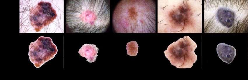
2       S. Innani et al.

     Fig. 1: Classwise sample images from HAM10000 classification dataset

dermatologists to visualize delicate clinical designs of skin lesions and subsurface
skin structures that are generally not visible to the unaided eye. These images
of the skin are studied under a microscope to point out skin abnormalities and
classify them into various types of skin cancers [1]. The enhanced dermoscopic
images are free from any skin surface reflections, which helps the dermatologist
to diagnose skin cancer accurately.
    Skin Cancer Detection, i.e., lesion segmentation, is one of the essential and
primary steps in accurate and precise treatment planning for various skin dis-
eases. Automatic skin cancer lesion segmentation is very challenging because of
the significant variations in lesion characteristics, such as size, location, shape,
color, texture, and skin. The segmentation becomes more arduous due to fuzzy
boundaries of lesions, poor color contrast with the surrounding skin, huge intra-
class variation, and the existence of antiques such as veins and hairs. Various
types of skin cancer images are shown in Fig. 1. Skin lesion segmentation has
drawn researchers for over a decade because of its increased clinical applicabil-
ity and demanding nature. Several image processing and supervised machine
learning-based approaches are presented for accurate lesion segmentation, with
pros and cons. Most studies pointed toward creating computer-aided design
frameworks for skin lesions that would recognize anomalies or skin illnesses.
The methods in literature usually follow a certain analysis pipeline [28]: The
first step is to delineate the lesion area in the image from healthy skin, followed
by automated feature extraction to compute the region of interest. The final
step is to predict the type of skin lesion (classification task). Several conven-
tional methods are available in the literature to handle the segmentation of skin
lesions. The comprehensive review of different lesion segmentation algorithms is
available at [9] [8] [27] [20]. [22].
    In recent times, deep learning techniques have outperformed all the existing
state-of-the-art approaches in various computer vision studies like segmentation
[7] [18] , detection [3], classification [6], etc. [5][4]. The availability of computing
resources and huge annotated data has enabled researchers to develop super-
vised Deep Neural Network models to address these tasks. With the evolution of
DCNN and various challenges in skin lesion latterly [11], multiple effective com-
putational approaches have appeared to solve particular problems in this field.
Regardless, the most current thriving strategies are based on CNN [31], [14],
[32], [13]. Along with lesion segmentation, deep learning approaches have im-
proved classification performance, leading to better diagnosis of diseases in med-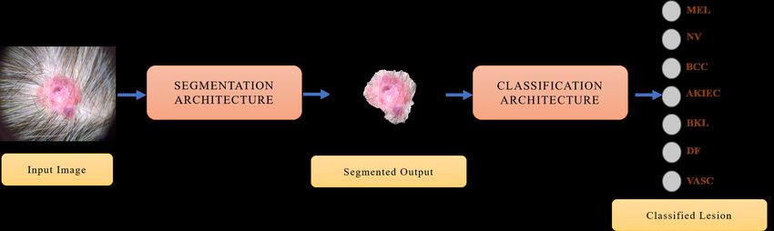
Deep Learning based Novel Cascaded Approach for Skin Lesion Analysis           3

      Fig. 2: Proposed two stage deep learning based framework for lesion
                        segmentation and classification

ical imaging. The methodologies are used to anticipate the existence of illness
and recognize the classes. Recent studies demonstrated remarkable performance
in classifying skin cancer using deep learning algorithms in binary classification
[13] but failed to achieve comparable performance in multi-class classification.
    This research aims to introduce a two-step automated system that will seg-
ment the skin lesion and then classify the disease. After a thorough literature
survey, the proposed approach is an end-to-end deep learning-based approach,
the first for skin lesion segmentation and classification for seven types of lesions.
There is no adequate dataset available having segmentation masks and classifi-
cation labels in a single dataset for seven different types of lesions. To address
this, for segmentation tasks, we work with the International Skin Imaging Col-
laboration (ISIC) 2018 dataset [12] where images with segmentation labels are
available, and for classification HAM10000 dataset [30] which consists of seven
different skin lesion classes. In our two-step proposed approach, the segmentation
task is initially conducted with the ISIC 2018 dataset. With a trained segmen-
tation model, the HAM10000 dataset is segmented, where only classification
labels are available. The Region of Interest(ROI) is extracted from segmented
images of the HAM10000 dataset fed as input to the classification framework.
The two-step framework is shown in Fig. 2.
    The rest of the article is arranged as follows: Database description is given
in Section 2. Presented methods for segmentation and classification of lesions
are described in section 3. Section 4 comprises evaluation metrics, experimental
results, and performance analysis. This article is concluded in Section 5.

2   Dataset
    International Skin Imaging Collaboration (ISIC) 2018 has 2594 images with
corresponding ground truth labels for lesion Segmentation. The images have
different sizes, from hundred to thousand, and varying width and height ratios.
The image lesion has distinct appearances and is located in a different part of the
skin. The HAM10000 [30] dataset is used for the classification task, consisting
of seven types of lesion disease in the dermoscopy images. Fig. 1 provided few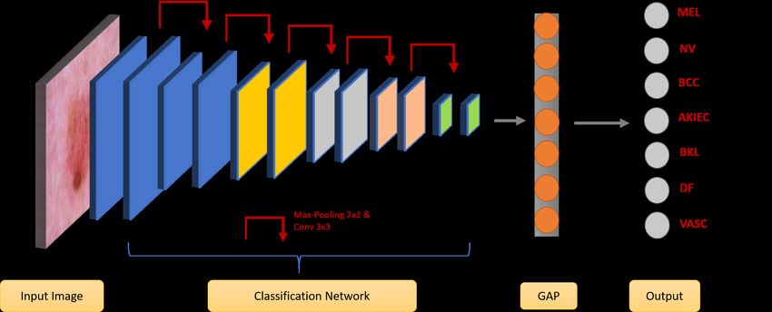
4       S. Innani et al.

                Table 1: Class distribution in HAM10000 dataset
                  Class Number of Images Class Percentage
                  AKIEC       327               3.27
                   BCC        514               5.13
                   BKL       1099              10.97
                    DF        115               1.15
                   MEL       1113              11.11
                   NV        6705              66.95
                  VASC        142               1.42

images from the dataset. The standard pre-processing like scaling the values
between [0,1] or [-1,1] is being implemented on entire dataset.
    The classification dataset consists of around 10015 lesions with Actinic ker-
atosis / Bowen’s disease (intraepithelial carcinoma) (AKIEC), Basal cell carci-
noma (BCC), Benign keratosis (solar lentigo / seborrheic keratosis/lichen planus-
like keratosis) (BKL), Dermatofibroma (DF), Melanoma (MEL), Melanocytic
nevus (NV), Vascular lesion (VASC) diseases. The data distribution is presented
in Table 1, and we observe that high-class imbalance is challenging in the given
datasets, where it is highly skewed towards certain classes. As a result, we ob-
served sparse images for specific groups like DF, VASC, and AKIEC.

3    Proposed Methodology
    We propose a two-step framework to handle the task of segmentation and
classification in skin lesions. In the first step, the images with skin lesions are
segmented to generate coarse-level masks. These segmented masks are multiplied

    Fig. 3: Proposed encoder-decoder architecture for skin lesion segmenation
Deep Learning based Novel Cascaded Approach for Skin Lesion Analysis         5

  Fig. 4: General architecture for skin cancer classifier where Global Average
                        Pooling is abbreviated as GAP.

with the corresponding image to extract the coarse level lesion part in the original
image, as shown in Fig. 2, which removes redundant data in the image, and these
ROI images are input to the classification network that signifies the type of lesion.

3.1    Segmentation Approach

    Encoder-decoder architectures are widely used in computer vision for image
segmentation task [19] [26]. Ronneberger et al. [24] presented U-Net, a break-
through study for medical image segmentation comprising CNN. Generally, a
feature learning block is the encoder module to capture spatial features of the in-
put. It downsamples the input image progressively and decreases feature dimen-
sions to catch high-level patterns of the input image. A decoder block consists
of layers that upsample the feature map obtained from the encoder output with
extracted spatial features. This article’s encoder-decoder module is graphically
presented in Fig. 3. In our approach, we designed three different encoder-decoder
networks by replacing the encoder block in U-Net with popular CNN architec-
tures such as ResNet [15], InceptionResNetV2 [33] and EfficientNets [29]. Our
architecture based on an encoder-decoder module consists of contraction and
expansion paths. The encoder consists of convolutional and max-pooling blocks,
which downsample the image to extract high-level features. This CNN output
contains denser and high-level feature maps. After every block, the number of
feature maps doubles to learn the complex features accurately.
    In the encoder, dense features are extracted with an output stride of 16 in
each variation, where the output stride is the ratio of the input image shape to
the output image shape. These extracted features work well in the classification
task, but performance hampers while rebuilding the fine segmentation map.
Hence, it is challenging to rebuild the segmentation map of the original input
image dimensions from the feature map of the encoder. The decoder module
builds the same U-Net decoder architecture to overcome this problem. This
encoder output expands in the decoder consisting of convolutional and bilinear
upsampling blocks. By concatenating low-level features from the encoder, low-
6 S. Innani et al.
Fig. 5: Basic building blocks for various CNN architecture used for
classification of skin lesion classes.
level feature maps are enhanced to the corresponding block of respective size in
the decoder to generate the segmented output more precisely.
3.2 Classification Approach
Convolutional Neural Network has shown tremendous progress in the task
of image classification. With advancements in computational resources and fine-
tuning methods, CNN fulfills the demand for performance in terms of accuracy.
As shown in Fig. 4, a conventional CNN architecture consists of combination
blocks of convolutional layer and downsampling layers, followed by a fully con-
nected layer (FC) and the output class. For accurate predictions, CNN auto-
matically pulls the patterns known as features from the input image and carries
information at the output block. In the classification step of the dermoscopy, im-
ages of the HAM10000 dataset having seven classes from the are to be predicted
[30]. We propose to use various classification architectures, which are also used
in segmentation encoders like ResNet, Xception, MobileNets, and EfficientNets
for classification tasks with an output stride of 32.
ResNets [15]: Deep neural network have shattered performance due to the prob-
lem of vanishing gradient. To overcome this problem, He et al. proposed the idea
of skip connections or residual networks, as shown in Fig. 5(a). This residual net-
work, known as ResNets, achieved improved performance. ResNet has different
variants formed by increasing the residual blocks, namely ResNet18, ResNet50,
and so on. ResNet consists of 3 × 3 convolutional layers stacked with residual
or skip connection to form the residual block. For denser prediction and deeper
model, maps are periodically doubled. The output of the final layer is 32 times
smaller than the input shape. For an image with input shape 224 × 224, the
output is of shape 7 × 7.
Xception [10]: F. Chollet et al. presented the Xception network as having supe-
rior performance. This architecture is inspired by the Inception [33]. In Xception,Deep Learning based Novel Cascaded Approach for Skin Lesion Analysis 7
the Inception module in the Inception network is replaced by Depthwise sepa-
rable convolution (DSC). Xcpetion architecture consisting of 36 convolutional
layers grouped in 14 blocks extracts features. All the blocks except the first and
last block have skip connections from the previous block. Xception has DSC with
residual or skip connection as the primary building layer, as in Fig. 5(d). The
output stride of the final layer is 32.
MobileNet [16]: Mobilenet architecture is a lightweight architecture having
depthwise separable convolution as the core layer in building this network, as
shown in Fig. 5(c). DSC is a factorized convolution consisting of pointwise 1 × 1
and depthwise convolution. In mobilenet to each input channel, a single filter is
used depthwise followed by pointwise convolution, fed with input from depthwise
convolution for stacking them together. A convolution process combines and
filters the input into output in a single step. The DSC is a two-step process in
which filtering is carried out in separate layers and combing in another. This
division has a significant effect on model size and reduces computation.
EfficientNet [29]: CNNs are developed depending on the resource, and scaling
occurs for influential performance while increasing the resources. e.g., ResNet-
18 [15] can be scaled to ResNet-101 by adding some layers. The traditional
procedure for scaling the network is to increase the CNN depth or depth or
feed with a higher input image size. These methods have proven to improve
performance but with tedious manual tuning. In [29], the author proposed a novel
approach to scaling the model that uses a compound coefficient that is highly
effective for structural scaling of the CNNs. Rather than arbitrarily increasing
network dimensions such as resolution, depth, and width, EfficientNet scales
every parameter in the compound coefficient with a fixed set of scaling factors.
This network is built with mobile inverted bottleneck convolution (MBConv)
[25] and squeeze and excitation optimization [17] as shown in Fig. 5(b).
4 Result and Discussion
We randomly divided the ISIC training dataset into 80% training cohort and
20% testing cohort. The dataset comprises images of varying sizes rescaled to
512×512×3 for the segmentation task. The segmentation network is trained with
a batch size of 8 with a loss function as the sum of cross-entropy and dice loss for
15 epochs setting the parameters for early stopping on Loss. The learning rate
was maintained at 0.001 with the ADAM optimizer. In the classification task, we
fed the network with an input size of 224 × 224 and loss function as categorical
cross-entropy. The model is trained with a batch size of 8 for 30 epochs setting
the parameters for early stopping on Loss. During training, we initialized the
learning rate to 0.001 with the ADAM [21] optimizer. We augmented the data
with various popular augmentation techniques like rotation, shearing, zooming,
brightness, and flipping the original images for segmentation and classification
tasks. The frameworks are designed with Tensorflow 2.0 and Keras open-source
libraries, and the models are trained on NVIDIA P100 GPU with 16 GB memory.8 S. Innani et al.
Fig. 6: Sample results ROI extracted after segmentation for classification task.
Table 2: Performance evaluation of segmentation task on test dataset in terms
of Dice Score and Mean Intersection over Union
Encoder backbone Dice Score mIoU
Original U-Net 71.53 60.58
ResNet50 84.46 73.76
ResNet101 86.30 76.77
MobileNet 83.90 71.32
InceptionResNetV2 87.20 78.03
EfficientNetB4 89.56 81.42
For the segmentation task, U-Net is just a stack of convolutional layers, the
original U-Net underperforms in this task. To experiment, we increase the depth
of the network with various encoders like ResNet, MobileNet, and EffcientNet.
Also, we design an asymmetric decoder, as seen in Fig. 3. Concatenation of
low-level features occurs at some intervals rather than joining each block from
the encoder, as proposed by Ronneberger et al. in U-Net improves performance.
The modification improves performance with the proposed deep encoder with
the asymmetric decoder.
After extracting the ROI by segmenting using an EfficientNet-based encoder,
it is fed as input to the various state-of-the-art networks in classification like
ResNet, MobileNet, EfficientNet, and Xception. As seen in Table 3, there is sig-
nificant performance gain when ROI extracted skin lesion is used. For an in-depth
comparison, classification is performed with different CNNs with and without
ROI obtained from segmentation. The efficacy of the proposed approaches is
evaluated in terms of various popular quantitative evaluation parameters. The
performance of segmentation approaches is assessed in terms of the Dice Simi-
larity Coefficient (DSC) and Mean Intersection over Union (mIoU) and the clas-
sification approach with accuracy. The performance for the segmentation task
of various encoder backbones in DSC and mIoU is given in Table 2. It can be
observed that EfficientNetB4 outperformed other encoders quantitatively. Seg-Deep Learning based Novel Cascaded Approach for Skin Lesion Analysis 9
Table 3: Performance evaluation of classification task on test dataset with and
without considering ROI images
Classification Accuracy
Architecture without ROI with ROI
ResNet50 74.15 78.32
ResNet101 75.65 79.26
MobileNet 78.54 81.54
Xception 78.04 82.41
EfficientNetB0 75.05 79.14
EfficientNetB3 76.65 82.19
mentation outputs predicted by the model for five different images are presented
in Fig. 7.
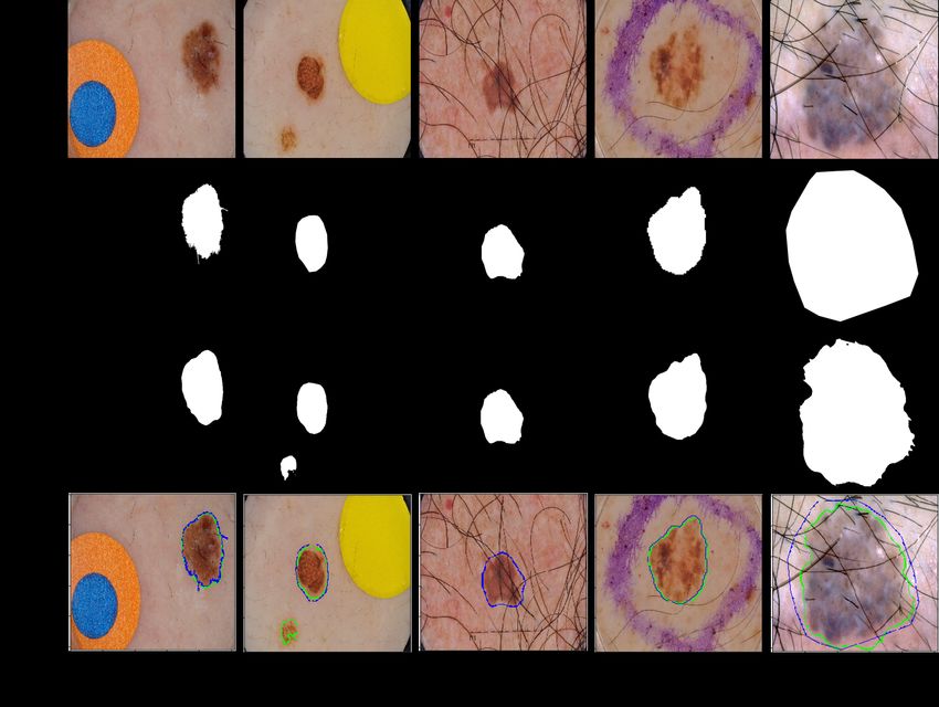
From Fig. 7(a) and 7(b), it can be observed that the proposed approach per-
formed well even if non-skin objects are present in the image. The architecture
could segment lesions, even severe occlusion, because of hairs. These segmen-
tation results are then multiplied with the original image to extract the skin
lesion, as shown in Fig. 6. It can be observed that besides skin lesions, vari-
ous surrounding patterns may hamper the classifier learning. ROI from Fig. 6
(b) and (e) clearly justifies the need of lesion segmentation before classification.
The performance evaluation for the classification task with and without ROI is
given in Table 3. The architectures trained on images containing only lesion ROI
performed better in terms of accuracy, as shown in Table 3.
5 Conclusion
Skin lesion segmentation and classification are the primary steps in design-
ing the Computer-Aided Diagnostic (CAD) Tool and are essential for precise
treatment planning. This study proposed a two-step approach with two distinct
databases for skin lesion segmentation and classification. It was observed that,
except for lesions, various surrounding patterns might hamper the classifier’s
learning. To address this, we proposed a two-step approach where in the first
step, skin lesions are segmented, and in the second step, ROIs are extracted,
which are given input to the classification architecture. Experimentation results
showed that classification accuracy with ROI as input outperformed lesion im-
ages with surrounding patterns and was improved by 5%. We currently have the
performance of the proposed approach on the publicly available dataset.
References
1. Dermoscopy and mole scans in perth and regional wa. https://myskincentre.
com.au/service/dermoscopy/ (2018), [Online accessed on 20-February-2020]10 S. Innani et al.
Fig. 7: Segmentation Output with EfficientNetB7 as encoder. Each column
represents a lesion image, Ground Truth, segmentation predicted by the
network and overlayed image of segmented output(Green) and Ground
Truth(Blue).
2. Melanoma stats, facts, and figures. https://www.aimatmelanoma.org/
about-melanoma/melanoma-stats-facts-and-figures. (2018), [Online ac-
cessed on 20-February-2020]
3. Baheti, B., Gajre, S., Talbar, S.: Detection of distracted driver using convolutional
neural network. In: 2018 IEEE/CVF Conference on Computer Vision and Pattern
Recognition Workshops (CVPRW). pp. 1145–11456 (2018). https://doi.org/10.
1109/CVPRW.2018.00150
4. Baheti, B., Innani, S., Gajre, S., Talbar, S.: Eff-unet: A novel architecture for
semantic segmentation in unstructured environment. In: 2020 IEEE/CVF Con-
ference on Computer Vision and Pattern Recognition Workshops (CVPRW). pp.
1473–1481 (2020). https://doi.org/10.1109/CVPRW50498.2020.00187
5. Baheti, B., Innani, S., Gajre, S., Talbar, S.: Semantic scene segmentation
in unstructured environment with modified deeplabv3+. Pattern Recognition
Letters 138, 223–229 (2020). https://doi.org/https://doi.org/10.1016/j.
patrec.2020.07.029, https://www.sciencedirect.com/science/article/pii/
S0167865520302750
6. Baid, U., Rane, S.U., Talbar, S., Gupta, S., Thakur, M.H., Moiyadi, A., Ma-
hajan, A.: Overall survival prediction in glioblastoma with radiomic features
using machine learning. Frontiers in Computational Neuroscience 14 (2020).
https://doi.org/10.3389/fncom.2020.00061, https://www.frontiersin.org/
articles/10.3389/fncom.2020.00061Deep Learning based Novel Cascaded Approach for Skin Lesion Analysis 11
7. Baid, U., Talbar, S., Rane, S., Gupta, S., Thakur, M.H., Moiyadi, A., Sable, N.,
Akolkar, M., Mahajan, A.: A novel approach for fully automatic intra-tumor seg-
mentation with 3d u-net architecture for gliomas. Frontiers in Computational
Neuroscience 14 (2020). https://doi.org/10.3389/fncom.2020.00010, https:
//www.frontiersin.org/articles/10.3389/fncom.2020.00010
8. Celebi, M.E., Kingravi, H., Iyatomi, H., Aslandogan, Y., Stoecker, W., Moss,
R., Malters, J., Grichnik, J., Marghoob, A., Rabinovitz, H., Menzies, S.: Bor-
der detection in dermoscopy images using statistical region merging. Skin re-
search and technology : official journal of International Society for Bioengineer-
ing and the Skin (ISBS) [and] International Society for Digital Imaging of Skin
(ISDIS) [and] International Society for Skin Imaging (ISSI) 14, 347–53 (09 2008).
https://doi.org/10.1111/j.1600-0846.2008.00301.x
9. Celebi, M.E., Wen, Q., Hwang, S., Iyatomi, H., Schaefer, G.: Lesion border de-
tection in dermoscopy images using ensembles of thresholding methods. CoRR
abs/1312.7345 (2013), http://arxiv.org/abs/1312.7345
10. Chollet, F.: Xception: Deep learning with depthwise separable convolutions. pp.
1800–1807 (07 2017). https://doi.org/10.1109/CVPR.2017.195
11. Codella, N.C.F., Gutman, D., Celebi, M.E., Helba, B., Marchetti, M.A., Dusza,
S.W., Kalloo, A., Liopyris, K., Mishra, N.K., Kittler, H., Halpern, A.: Skin le-
sion analysis toward melanoma detection: A challenge at the 2017 international
symposium on biomedical imaging (isbi), hosted by the international skin imag-
ing collaboration (ISIC). CoRR abs/1710.05006 (2017), http://arxiv.org/abs/
1710.05006
12. Codella, N.C.F., Rotemberg, V., Tschandl, P., Celebi, M.E., Dusza, S.W., Gutman,
D., Helba, B., Kalloo, A., Liopyris, K., Marchetti, M.A., Kittler, H., Halpern, A.:
Skin lesion analysis toward melanoma detection 2018: A challenge hosted by the
international skin imaging collaboration (ISIC). CoRR abs/1902.03368 (2019),
http://arxiv.org/abs/1902.03368
13. Esteva, A., Kuprel, B., Novoa, R., Ko, J., Swetter, S., Blau, H., Thrun, S.:
Dermatologist-level classification of skin cancer with deep neural networks. Na-
ture 542 (01 2017). https://doi.org/10.1038/nature21056
14. González-Díaz, I.: Dermaknet: Incorporating the knowledge of dermatologists to
convolutional neural networks for skin lesion diagnosis. IEEE Journal of Biomedical
and Health Informatics 23(2), 547–559 (March 2019). https://doi.org/10.1109/
JBHI.2018.2806962
15. He, K., Zhang, X., Ren, S., Sun, J.: Deep residual learning for image recognition.
CoRR abs/1512.03385 (2015), http://arxiv.org/abs/1512.03385
16. Howard, A.G., Zhu, M., Chen, B., Kalenichenko, D., Wang, W., Weyand, T., An-
dreetto, M., Adam, H.: Mobilenets: Efficient convolutional neural networks for
mobile vision applications (2017)
17. Hu, J., Shen, L., Sun, G.: Squeeze-and-excitation networks. CoRR
abs/1709.01507 (2017), http://arxiv.org/abs/1709.01507
18. Innani, S., Dutande, P., Baheti, B., Talbar, S., Baid, U.: Fuse-pn: A novel archi-
tecture for anomaly pattern segmentation in aerial agricultural images. In: 2021
IEEE/CVF Conference on Computer Vision and Pattern Recognition Workshops
(CVPRW). pp. 2954–2962 (2021). https://doi.org/10.1109/CVPRW53098.2021.
00331
19. Innani, S., Dutande, P., Baheti, B., Talbar, S., Baid, U.: Fuse-pn: A novel archi-
tecture for anomaly pattern segmentation in aerial agricultural images. In: 2021
IEEE/CVF Conference on Computer Vision and Pattern Recognition Workshops12 S. Innani et al.
(CVPRW). pp. 2954–2962 (2021). https://doi.org/10.1109/CVPRW53098.2021.
00331
20. Jahanifar, M., Zamani Tajeddin, N., Mohammadzadeh Asl, B., Gooya, A.: Su-
pervised saliency map driven segmentation of lesions in dermoscopic images.
IEEE Journal of Biomedical and Health Informatics 23(2), 509–518 (March 2019).
https://doi.org/10.1109/JBHI.2018.2839647
21. Kingma, D., Ba, J.: Adam: A method for stochastic optimization. International
Conference on Learning Representations (12 2014)
22. Oliveira, R.B., Filho, M.E., Ma, Z., Papa, J.P., Pereira, A.S., Tavares, J.M.R.:
Computational methods for the image segmentation of pigmented skin le-
sions: A review. Computer Methods and Programs in Biomedicine 131, 127
– 141 (2016). https://doi.org/https://doi.org/10.1016/j.cmpb.2016.03.032,
http://www.sciencedirect.com/science/article/pii/S0169260716303418
23. Rigel, D.S., Russak, J., Friedman, R.: The evolution of melanoma diagnosis: 25
years beyond the abcds. CA: A Cancer Journal for Clinicians 60(5), 301–316.
https://doi.org/10.3322/caac.20074, https://acsjournals.onlinelibrary.
wiley.com/doi/abs/10.3322/caac.20074
24. Ronneberger, O., Fischer, P., Brox, T.: U-net: Convolutional networks for biomed-
ical image segmentation. In: Navab, N., Hornegger, J., Wells, W.M., Frangi, A.F.
(eds.) Medical Image Computing and Computer-Assisted Intervention – MICCAI
2015. pp. 234–241. Springer International Publishing, Cham (2015)
25. Sandler, M., Howard, A.G., Zhu, M., Zhmoginov, A., Chen, L.: Inverted residuals
and linear bottlenecks: Mobile networks for classification, detection and segmenta-
tion. CoRR abs/1801.04381 (2018), http://arxiv.org/abs/1801.04381
26. Sultana, F., Sufian, A., Dutta, P.: Evolution of image segmentation using deep
convolutional neural network: A survey. arXiv preprint arXiv:2001.04074 (2020)
27. Tajeddin, N.Z., Asl, B.M.: A general algorithm for automatic lesion segmenta-
tion in dermoscopy images. In: 2016 23rd Iranian Conference on Biomedical En-
gineering and 2016 1st International Iranian Conference on Biomedical Engineer-
ing (ICBME). pp. 134–139 (Nov 2016). https://doi.org/10.1109/ICBME.2016.
7890944
28. Tajeddin, N.Z., Asl, B.M.: Melanoma recognition in dermoscopy images us-
ing lesion’s peripheral region information. Computer Methods and Programs
in Biomedicine 163, 143 – 153 (2018). https://doi.org/https://doi.org/10.
1016/j.cmpb.2018.05.005, http://www.sciencedirect.com/science/article/
pii/S0169260717313251
29. Tan, M., Le, Q.V.: Efficientnet: Rethinking model scaling for convolutional neural
networks. CoRR abs/1905.11946 (2019), http://arxiv.org/abs/1905.11946
30. Tschandl, P., Rosendahl, C., Kittler, H.: Data descriptor: The HAM10000 dataset,
a large collection of multi-source dermatoscopic images of common pigmented skin
lesions. Scientific Data 5, 1–9 (2018). https://doi.org/10.1038/sdata.2018.161
31. Yu, L., Chen, H., Dou, Q., Qin, J., Heng, P.: Automated melanoma recognition in
dermoscopy images via very deep residual networks. IEEE Transactions on Medi-
cal Imaging 36(4), 994–1004 (April 2017). https://doi.org/10.1109/TMI.2016.
2642839
32. Yuan, Y., Lo, Y.: Improving dermoscopic image segmentation with enhanced
convolutional-deconvolutional networks. CoRR abs/1709.09780 (2017), http:
//arxiv.org/abs/1709.09780
33. Zhang, X., Huang, S., Zhang, X., Wang, W., Wang, Q., Yang, D.: Residual in-
ception: A new module combining modified residual with inception to improveDeep Learning based Novel Cascaded Approach for Skin Lesion Analysis 13 network performance. In: 2018 25th IEEE International Conference on Image Pro- cessing (ICIP). pp. 3039–3043 (Oct 2018). https://doi.org/10.1109/ICIP.2018. 8451515
You can also read


