Cytotoxic and pro-inflammatory effects of molybdenum and tungsten disulphide on human bronchial cells
←
→
Page content transcription
If your browser does not render page correctly, please read the page content below
Nanotechnology Reviews 2022; 11: 1263–1272
Research Article
Lidia Zapór*, Luiza Chojnacka-Puchta, Dorota Sawicka,
Katarzyna Miranowicz-Dzierżawska, and Jolanta Skowroń
Cytotoxic and pro-inflammatory effects
of molybdenum and tungsten disulphide
on human bronchial cells
https://doi.org/10.1515/ntrev-2022-0073 nanoparticles should be included in the occupational risk
received October 29, 2021; accepted February 9, 2022 assessment.
Abstract: This study aimed to investigate the cytotoxicity Keywords: molybdenum disulphide, tungsten disulphide,
and pro-inflammatory responses induced by tungsten cytotoxicity assay, lung cells, holotomographic microscopy
disulphide (WS2) and molybdenum disulphide (MoS2)
nanoparticles (NPs) in human bronchial cells (BEAS-
2B). For cytotoxicity assessment, the cells were exposed
to different concentrations (2.5–200 µg/mL) of WS2-NPs 1 Introduction
or MoS2-NPs for 24 and 48 h and then the MTT assay was
performed. Afterwards, long-term toxicity was assessed Molybdenum disulphide (MoS2) and tungsten disulphide
by the colony forming efficiency assay (CFEA) during a 10 (WS2) are a class of layered materials well known for their
days’ exposure of the cells. For pro-inflammatory responses, excellent lubrication performance. They are widely used
the expression of interleukin-6 (IL-6) and interleukin-1β both as oil additives as well as dry/solid lubricants. The
(IL-1β) mRNA was estimated by the real-time PCR method. use of molybdenum and tungsten disulphides in dry
Both nanomaterials showed similar cytotoxic effects on lubricants is of particular importance in aviation, auto-
BEAS-2B cells assessed by the MTT assay, i.e. reduction in motive, paper and food industries, in precision mechanics,
cell viability to approx. 60–70% at concentrations of 2.5 electronics, chemical, glass, plastics, and many other indus-
and 5 μg/mL after 24 and 48 h. The percentage viability tries [1–3]. For the last few years, the use of the nano-form
remained relatively constant at this level across all concen- of these compounds has been increasing. Particularly, 2D
trations above 5 μg/mL. In long-term exposure, both nano- nanoplates and fullerene-like structures are widely used in
materials inhibited colony formation in a wide range of many tribological applications [4–6].
concentrations up to 100 µg/mL. MoS2-NPs were slightly At present, there are only a few reports available on
more cytotoxic than WS2-NPs. Additionally, MoS2-NPs caused the effects of MoS2 and WS2 on human bronchial cells.
an increase in mRNA levels of cytokines, IL-1β, and IL-6 at Interestingly, Appel et al. [7] investigated the potential
concentration of 50 µg/mL, while WS2-NPs did not cause any toxicological effects of WS2 nanoparticles prepared by
changes in the level of mRNA for both cytokines. We also several methods such as mechanical exfoliation and che-
visualised the changes in the cells as a result of WS2-NPs or mical vapour deposition (CVD) toward human epithelial
MoS2-NPs exposure (2.5 and 25 µg/mL) via holotomographic kidney cells (HEK293F). More importantly, Pardo’s group
microscopy. This work demonstrates the hazardous potential [8] presented low cytotoxicity of WS2 and MoS2 nano-
of both nanomaterials and indicate that WS2 and MoS2 particles in human bronchial epithelial cells, which are
used in industrial and medical applications to compare
to standard environmental particulate matter. Corazzari
et al. [9] reported evaluation of the impact of commercial
* Corresponding author: Lidia Zapór, Central Institute for Labour 2D WS2 powders on the viability of other human cell
Protection – National Research Institute, Czerniakowska 16, 00-701 lines, such as NL-20, HEPG2, and macrophages. Pro-
Warsaw, Poland, e-mail: lizap@ciop.pl
mising use of MoS2 and WS2 as green components in separa-
Luiza Chojnacka-Puchta, Dorota Sawicka,
Katarzyna Miranowicz-Dzierżawska, Jolanta Skowroń: Central
tion technologies was presented by Köhler’s team [10]. They
Institute for Labour Protection – National Research Institute, explained the possibility of applying MoS2 and WS2 in
Czerniakowska 16, 00-701 Warsaw, Poland modern desalination processes based on 2D membranes,
Open Access. © 2022 Lidia Zapór et al., published by De Gruyter. This work is licensed under the Creative Commons Attribution 4.0
International License.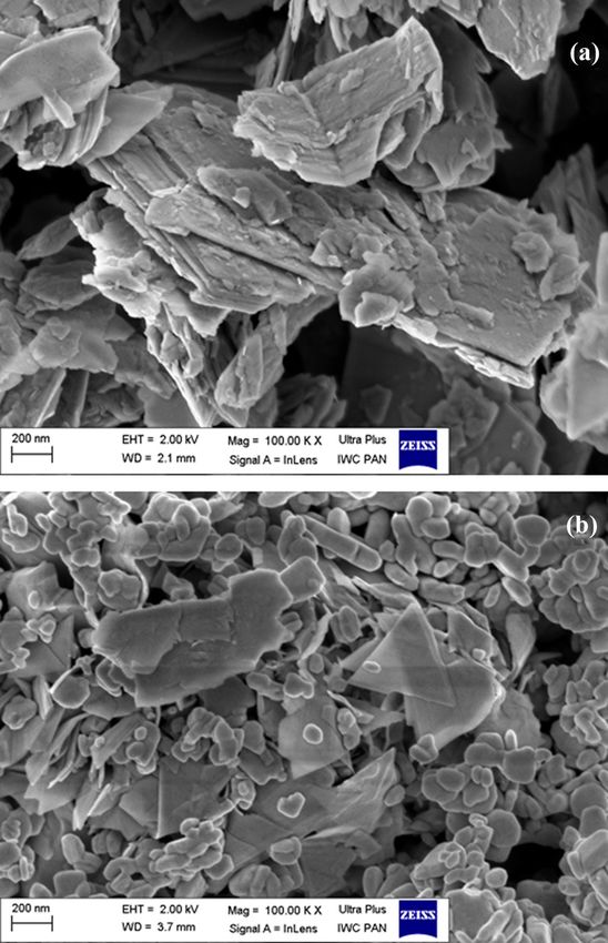
1264 Lidia Zapór et al.
contributing to increase the availability of clean water in 2 Materials and methods
the word. The WS2-NPs and MoS2-NPs are also used for
the synthesis of analogues of such nanomaterials as carbon
2.1 Chemicals and reagents
nanotubes, fullerenes, or graphene, in order to replace them
in many consumer products [11,12]. The 2D layered nano-
WS2-NPs and MoS2-NPs with nominal particle size below
materials including WS2, MoS2 nanosheets and graphene
90 nm were purchased from Sigma-Aldrich. The media
oxide, reduced graphene oxide, and Ti3C2Tx MXene, used
for cell cultures were provided by Gibco BRL (Life
as solid lubricants, ensure low wear and friction over the
Technologies Ltd, Paisley, UK). All reagents for gene expres-
entire component’s lifetime. In addition to being applied as
sion assay were provided by Thermo Fisher Scientific Inc.,
a solid lubricant, MoS2 nanosheets can be used as a reinfor-
Rockford, IL, USA. Other reagents were supplied by Sigma
cement phase in composites and lubricant additive under
Chemical Co. (St Louis, MO, USA), if not indicated
extreme conditions, such as vacuum or dry contacts at
otherwise.
high temperatures [13–15]. Zhang’s team obtained MoS2
nano-balls fabricated in isopropanol by ultrasonication.
This procedure allows to reduce the contact area in steel
contacts through extraordinary macro-scale lubricity [16]. 2.2 Characterisation of WS2-NPs and
The application of WS2-NPs or MoS2-NPs in lubricants MoS2-NPs
which are used in automotive and aviation is also very
beneficial for the environment due to the reduction in par- The morphology of WS2-NPs and MoS2-NPs was deter-
ticulate matter emission, which results in lesser contamina- mined by scanning electron microscope (SEM, Zeiss Ultra
tion of air, water, and soil [3,17]. As a result, the reduction in Plus). The particles were scanned at 2 kV. A specific surface
emissions translate into a reduction in human morbidity, as area was measured by the Brunauer–Emmett–Teller (BET)
solid particles (especially the fraction of ultrafine dust, with technique (Gemini 2360, Micromeritics). The particle prop-
a diameter equivalent to those of nanoparticles) are respon- erties in a liquid environment, such as hydrodynamic dia-
sible for the generation of respiratory, cardiovascular, allergic, meter, particle size distribution, particle size, and zeta
and cancer diseases. On the other hand, dry lubricants are potential were characterised using the Nanoparticle Tracking
used in the form of powders and aerosols, which can lead to Analysis (NTA, NS500, Nanosight Ltd, UK) and Dynamic
their significant emission to the environment, in particular the Light Scattering (DLS, ZetaSizer Nano ZS, Malvern). The mea-
working environment. For this reason, the determination of surements were performed both in the phosphate-buffered
toxicity of the compounds in the nano-form is extremely saline (PBS, Gibco, Invitrogen, Carlsbad, CA, USA) dispersion
important. According to toxicological reports, the use of nano- and in the culture media. The stock solution (1 mg/mL) of
materials, although beneficial for technological reasons, is WS2-NPs and MoS2-NPs was prepared by suspending the
associated with an uncertain health risk [18]. There is a con- powder in cold PBS. Then, suspensions were sonicated on
cern that substances with low toxicity may adversely affect ice at a high energy level of 420 J/cm3 (for 5 min with 90%
the body if used in the form of nanoparticles [19]. Despite the amplitude) (Sonica Q 700, Qsonica LLC, USA). Working solu-
still emerging applications of WS2-NPs and MoS2-NPs, only a tions were prepared immediately before the toxicity tests in a
few investigations into their biocompatibility and toxicity serum-free culture medium and vortexed for approx. 1 min to
have been performed. It is important to note that their mod- ensure homogeneity prior to use with the cells. A new stock
ified forms were mainly evaluated [7,20,21]. Considering the solution was prepared before each experiment.
increased use of WS2-NPs and MoS2-NPs in nanoscale, there is
the necessity to study their unfunctionalised forms, especially
in terms of potential health effects after inhalation. Most of the
toxicity studies of nanomaterials are conducted using in vitro 2.3 Cell culture and treatment
methods.
The aim of this study was to assess the cytotoxic and pro- The BEAS-2B cells (CRL-9609) were purchased from American
inflammatory effects after exposure of human bronchial cells Type Culture Collection (LGC Standards Sp. z o.o.). The cells
(BEAS-2B cell line) to unmodified WS2-NPs and MoS2-NPs. were grown in the LHC-9 serum-free medium in the culture
Application of relatively new holotomographic technique flasks coated with collagen type 1 (Greiner). The cells were
allowed to observe the morphological alterations in cells maintained at 37°C in a humidified atmosphere with 5% CO2.
exposed to low doses of WS2-NPs and MoS2-NPs (2.5 and To detach cells from the culture plates, 0.25% trypsin-EDTA
25 µg/mL), which testified to adverse processes in the cells. (Gibco) was used. Cell number and viability were determinedCytotoxic and pro-inflammatory effects of MoS2 and WS2 on human bronchial cells 1265
in a Bürker chamber by the trypan blue exclusion method. PE ratio for BEAS-2B cells calculated from three inde-
Additionally, the cells were screened for Mycoplasma sp. pendent experiments was above 0.6.
infection using MycoAlert™ PLUS Mycoplasma Detection
Kit (Lonza, Walkersville, Inc.).
2.6 Cytokine real-time PCR assays
2.4 Cytotoxicity studies BEAS-2B cells were seeded into tissue culture 6-well
plates (TPP, Techno Plastic Products AG) at a density of
To detect cytotoxic effects of WS2-NPs or MoS2-NPs, we per- 300,000 cells/well and allowed to adhere and establish
formed the MTT-tetrazolium salt reduction assay (MTT), overnight at 37°C in a humidified atmosphere (5% CO2).
which measures mitochondrial dehydrogenase activity Cells were treated with WS2-NPs or MoS2-NPs at different
living cells. The BEAS-2B cells were seeded in 96-well concentrations (5, 25, and 50 µg/mL) for 24 h. Next total
microplates at a density of 2 × 10⁴/well and cultured over- cellular RNA was extracted using the ReliaPrep RNA Cell
night. Then, cells were exposed to WS2-NPs or MoS2-NPs MiniPrep System (Promega), the quantity of RNA obtained
(2.5–200 µg/mL) for 24 or 48 h. The toxicity was calculated from each sample was measured using Quantus fluorometer
as a ratio of absorbencies of treated and untreated cells. The with the QuantiFluor RNA System (Promega) according to
absorbencies were measured at 570/620 nm using a Synergy the manufacturer’s guidelines.
2 microplate reader (BioTek, USA). Cytotoxicity tests were The 0.5 μg total RNA were reverse transcribed to obtain
conducted in at least three independent replications. cDNA using the High-Capacity cDNA Reverse Transcription
Kit (Thermo Fisher Scientific, Inc.) according to manufac-
turer’s instructions.
All genes were amplified using TaqMan® Gene Expression
2.5 Colony forming efficiency assay (CFEA) assays (Thermo Fisher Scientific, Inc.) for interleukin IL-1β
(assay ID. Hs01555410_m1) and IL-6 (assay ID. Hs00174131_m1).
The CFEA (also referred to as clonogenic assay) was con- Data were normalised to glucuronidase beta (GUSB) mRNA
ducted according to the procedure described by Franken levels (assay ID. Hs00939627_m1) as an endogenous con-
et al. [22] and adapted from Kruszewski et al. [23]. The trol. Quantitative real-time PCR was performed on samples
colony forming efficiency assay is based on the ability of using the 7500 real-time PCR detector (Thermo Fisher
a single cell to grow into a colony. A colony is defined to Scientific, Inc.).
consist of at least 50 clones of one cell, which corresponds
to 6 mitotic divisions. This test is used to detect cells that
retained the capacity for producing a large number of pro-
genies after treatments that can cause reproductive death as 2.7 Holotomographic visualisation of
a result of damage to chromosomes, apoptosis, etc. Expo- morphological changes
nentially growing cells were harvested and seeded in a Petri
dish of 60 × 15 mm (21 cm2) (Iwaki Cell Biology, Japan) at a The BEAS-2B cells were seeded into 35 mm culture dishes
density of 500 cells/dish together with a tested compound. (IBIDI, Gräfelfing, Germany) at a density of 20,000 cells/
Each dish finally contained 5 mL of a cell culture medium dish and incubated for 24 h. After this period, cells were
with WS2-NPs or MoS2-NPs in appropriate concentrations treated with WS2-NPs or MoS2-NPs (2.5 or 25 µg/mL) and
(25, 50, and 100 µg/mL) at least in three replicates for incubated for 24 h. Then, the medium was replaced with
each treatment. The cells were exposed to nanoparticles MitoView dye (Biotium) at a concentration of 100 nM and
for 10 days. After this period, particle solutions were incubated for 15 min at 37°C to visualise mitochondrial
removed, cells were washed with PBS, fixed with ethanol structures. The cells were also counterstained for DNA
(Sigma-Aldrich), stained (0.4% Giemza, Sigma-Aldrich), with 0.1 μg/mL Hoechst 33258 (Thermo Fisher Scientific).
and colonies were counted using a stereomicroscope (IUL, As a positive control we used cells treated with stauros-
Spain). Then, plating efficiency (PE) and surviving fraction porine (37 nM, Sigma-Aldrich). Then, dishes of control
(SF) were calculated as below: (untreated cells) or exposed cells were placed on the
PE = number of colonies formed/number of cells tomographic microscope (3D Cell Explorer, Nanolive
seeded SF = number of colonies formed after treatment/ S.A., Lausanne, Switzerland) and 3D tomographic images
number of cells seeded × PE. (z-stacks) were created. Further post processing steps such1266 Lidia Zapór et al.
as background reduction and contrast enhancement
were applied to the final figures using STEVE software
(Nanolive).
2.8 Statistical analyses
Three separate in vitro cytotoxicity experiments were con-
ducted in which all samples of nanomaterials were tested
simultaneously. At least three independent experiments
were performed for cytotoxicity endpoint. The CFEA results
were presented as surviving fraction ratio (SF) ± standard
deviation (SD). SF = 1 was set for the control. CFEA data were
analysed by Student’s test for comparison between two
groups. A value of p < 0.05 indicates a statistically significant
difference. The software employed for statistical analysis was
Statistica, version 7.1. The results of RT-qPCR are presented
as mean values obtained from three experiments. The signif-
icance of differences between the mean values was assessed
using the paired Student’s t-test using 7500 Software v2.0.6.
(Thermo Fisher Scientific, Inc.). The data were presented as
the mean value ± standard deviation (SD).
3 Results
3.1 Characterisation of WS2-NPs and
MoS2-NPs
Figure 1: SEM image of MoS2-NPs (a) and WS2-NPs (b).
Microscopic analysis (SEM) showed that both nanomater- Magnification and scale are presented on the picture.
ials had the structure of multilayer plates, whose length
and width was on the order of a few micrometres, while
the thickness was on the nano scale (Figure 1a and b). or 48 h. Afterwards, the MTT assays were performed. The
WS2-NPs were characterised by a larger fraction of parti- results of the MTT assays are summarised in Figure 2.
cles of smaller dimensions in length, width, and thick- Both nanomaterials caused a reduction in cell viability
ness. Specific surface area for MoS2-NPs and WS2-NPs to approx. 60–70% at concentrations of 2.5 and 5 μg/mL
was 7.8 and 6.2 m2/g, respectively. after 24 or 48 h. The percentage viability remained rela-
The particle size distribution analysis by DLS showed tively constant at this level across all concentrations
that the suspensions of WS2-NPs were more homogeneous above 5 μg/mL.
(PdI ≈ 0.3) and more stable (ζ ≈ −30) in comparison with As shown in Figure 3, both nanomaterials were able
MoS2-NPs (PdI ≈ 0.6 and ζ ≈ −10) (Table 1). Both DLS and to significantly decrease clonogenic survival and cell pro-
NTA methods showed that MoS2-NPs and WS2-NPs existed liferation in a dose-dependent manner in a wider range
both as single and as aggregate/agglomerate particles. of concentrations up to 100 µg/mL, when exposed con-
stantly over 10 days. MoS2-NPs in the lowest concentra-
tions (25 and 50 μg/mL) decreased the proliferation
3.2 Effects of MoS2-NPs and WS2-NPs on cell of BEAS-2B cells to about 80%. Higher concentration
viability and proliferation (100 μg/mL) resulted in inhibition of growth of the cul-
ture by approx. 30%. In case of WS2-NPs, the concen-
BEAS-2B cells were exposed to a wide range of concen- tration of 100 μg/mL inhibited the colony formation by
trations of MoS2-NPs or WS2-NPs (2.5–200 µg/mL) for 24 about 50%.Cytotoxic and pro-inflammatory effects of MoS2 and WS2 on human bronchial cells 1267
Table 1: Physicochemical characteristics of MoS2-NPs and WS2-NPs
Particles MoS2-NPs WS2-NPs
Specific surface area [m /g] [BET method]
2
7.80 6.20
Hydrodynamic diameter [nm] [DLS method] 616.17 ± 2.92 226.57 ± 4.84
Polydispersity index [PdI] 0.59 ± 0.08 0.320 ± 0.03
Zeta potential ζ [mV] −10.56 ± 0.78 −29.4 ± 2.18
Particle diameter [nm] [NTA method] Mean value = 294.25 ± 8.7 Mean value = 218.03 ± 5.2
Mode = 139.9 ± 8.5 Mode = 190.37 ± 3.5
3.3 Cellular cytokine response did not observe significant changes in the IL-1β or IL-6
gene expression after treatment of cells with WS2-NPs.
The expression of mRNAs for cytokines IL-1β and IL-6
was used as a marker of the pro-inflammatory response
induced in BEAS-2B cells exposed to different concentra- 3.4 Holotomographic visualisation of
tions (5, 25, or 50 µg/mL) of WS2-NPs or MoS2-NPs for morphological changes
24 hours. The gene expression of IL-1β and IL-6 increased
after treatment with the MoS2-NPs in a dose-dependent After exposure of BEAS-2B cells to MoS2-NPs or WS2-NPs at
manner (Figure 4). However, the level of mRNA for both low doses of 2.5 and 25 µg/mL, we observed morphological
cytokines significantly differed compared to control only changes after 24 and 48 h. The untreated cells (negative con-
in the highest concentration of MoS2-NPs (50 µg/mL). We trol) had no alteration in cell morphology or any membrane
Figure 2: Cytotoxic effect of MoS2-NPs or WS2-NPs in BEAS-2B cells after 24 and 48 h exposure assessed by MTT assay. Data are expressed
as the percentage viability of cells exposed to compounds relative to control cells. Each point represents an average value ± SD from at least
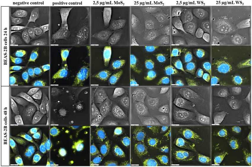
three experiments; *p < 0.05 vs control.1268 Lidia Zapór et al. Figure 3: Colony forming ability of BEAS-2B cells treated with different concentrations of WS2-NPs or MoS2-NPs. Each bar represents the mean value ± SD of three independent experiments. *p < 0.05 vs control. damages. In treated cells, we noticed changes in the size and were observed as a result of exposure of cells to NPs. In shape of cells and loss of cell–cell contact compared with untreated cells, mitochondria showed a typical cytoplasmic negative control. During the cells’ treatment with MoS2-NPs distribution. We noticed that the intensity of the described or WS2-NPs and time of exposure, the degree of cytoplasmic changes increases in a time – and dose – dependent manner. vacuolisation was increased. Changes in the shape of nucleus Morphological changes occurring in cells after exposure are and intracellular location of mitochondria and their density presented in Figure 5. Figure 4: The expressions of cytokine mRNA in BEAS-2B cells exposed to MoS2-NPs or WS2-NPs. Cells were exposed to 5, 25, or 50 µg/mL for 24 h. The relative mRNA expressions of IL6 and IL-1β were measured by real-time PCR. The expressions were normalised against GUSB. The data represent the mean value ± SD of three independent experiments. * significantly different from control [p < 0.005], using Student’s t-test.
Cytotoxic and pro-inflammatory effects of MoS2 and WS2 on human bronchial cells 1269
Figure 5: Representative images after fluorescence staining to visualise mitochondrial and nucleus location and morphology of living BEAS-
2B cells. Negative control (untreated cells) and positive control (Staurosporine 37 nM) cells after exposure to MoS2-NPs or WS2-NPs (2.5 and
25 µg/mL). Images are presented in bright field and after excitation of FITC fluorescence. Description of changes is presented in the text.
Pictures were taken under a magnification of 60×. Scale bar: 20 µm.
4 Discussion distribution, and formation of aggregates or agglomer-
ates in culture media. These parameters may be a signifi-
The determination of MoS2-NPs and WS2-NPs toxicity has cance to the interpretation of results obtained in toxico-
gained importance due to their expanding application in logical studies [26]. In the present work, the MoS2-NPs
many fields of industry or biomedicine and the associated and WS2-NPs formed polydisperse suspensions of dif-
increase in the potential health risk in the work environ- ferent particle sizes in the LHC-9 medium. Both single
ment. Our research focused on two areas. First, we eval- particles and agglomerates, which were about 2–4 times
uated the ability of WS2-NPs and MoS2-NPs to induce larger than the nominal particle size, were determined. It
cytotoxicity and cytokine response in the human lung has to be taken into consideration that aggregation is a
epithelial cell model – BEAS-2B. These cells are often natural process for nanoparticles and is included in the
used to evaluate the toxicity of dust pollution and pre- definition of nanomaterials accepted by the European
ferred as an experimental model of the respiratory system Union [27].
due to the normal phenotype of the cells and their ability The cytotoxic activity of MoS2-NPs and WS2-NPs was
to release pro-inflammatory cytokines [24,25]. Second, we evaluated with the use of MTT test. The cells were exposed
documented the morphological alterations and changes in to different concentrations of NPs (2.5–200 μg/mL) for 24
organelles after exposure of cells to low doses (2.5 and and 48 h. We found little trend in their cytotoxicity profiles:
25 µg/mL) of NPs. nanomaterials in concentrations of 2.5 and 5 μg/mL reduced
The crucial role in the in vitro toxicity assessment of the viability of the cells to the level of about 70%, after
nanomaterials is played by their proper characterisation, which there was no further decrease in cell viability, despite
in particular, changes in particle size and particle size being exposed to higher concentrations. We might conclude1270 Lidia Zapór et al.
that particle aggregation in higher concentrations causes assay. This assay is a suitable and robust in vitro method
lower availability to the cells. Very similar results of MoS2- to assess cytotoxicity of nanomaterials, which was con-
NPs cytotoxicity were achieved by Chng et al. [28] in MTT firmed by interlaboratory comparison study performed in
assay using A549 cells. Pristine MoS2-NPs decreased viabi- the frame of OECD’s Working Party of Manufactured
lity of the cells at concentrations below 6.25 µg/mL, whereas Nanomaterials [31]. This test does not require any dyes,
higher concentrations did not affect cells viability. In turn, which can interact with nanomaterials and lead to invalid
Wang et al. [20] showed a slight (up to 80%) decrease in the results [32,33]. In this experiment, both MoS2-NPs and
viability of THP-1 cells and no cytotoxic effect in BEAS-2B WS2-NPs were able to significantly decrease clonogenic
cells exposed to various structural forms including aggre- survival and cell proliferation in a dose-dependent manner
gates of MoS2-NPs in concentrations of 10–50 μg/mL. Impor- after exposure at doses of 25–100 µg/mL for 10 days. The
tantly, the authors demonstrated that MoS2-NPs, despite the MoS2-NPs were slightly more cytotoxic than WS2-NPs.
lack of cytotoxic effects, caused the release of pro-inflamma- According to the concept of grouping nanomaterials
tory mediators in cells, in vivo inflammation, and fibrosis. in terms of risk that they can pose for health, they can be
Also, Qureshi et al. [29] observed a minimal decrease in the classified as High Aspect Ratio Nanomaterials (HARNs),
survival rate of HeLa cells with the increase in MoS2-NPs so they potentially cause the induction pro-inflammatory
concentration, but they used higher doses, i.e. from 32.5 to reaction in the body tissues [34]. Inflammatory reactions
300 μg/mL. Likewise, Corazzari et al. [9] in studies on A549 induced in lungs under the influence of nanoparticles,
cells demonstrated cytotoxic activity of MoS2-NPs and WS2- especially HARNs, were considered as one of the main
NPs in high concentrations only. reason of their adverse health effects [35,36]. The induc-
In the literature, there are few reports on the toxicity tion of cytokines by airway cells in vitro has been widely
of pristine MoS2-NPs and even fewer of those about WS2- used to assess the pro-inflammatory effects of ambient
NPs. Their functionalised or modified forms, such as and occupational nanoparticles, e.g. particulate matter
exfoliated, fullerene-like ones or nanotubes were mainly PM2,5, diesel particles, pollution-related metals, and many
evaluated. The results of some studies indicate that mod- others [37]. In the present study, the mRNA for cytokine IL-6
ifications may change toxicity of MoS2-NPs or WS2-NPs. and IL-1β was evaluated after exposure of BEAS-2B cells to
Such a tendency was confirmed by Wang et al. [20], the MoS2-NPs or WS2-NPs. IL-6 is a pleiotropic cytokine which
data obtained on THP-1 and BEAS-2B cells suggested that plays an important role in acute inflammation in the lungs.
exfoliation attenuates the toxicity of MoS2-NPs. Other stu- The activation of IL-6 transcription has been observed
dies [7] also confirm rather low toxicity of a modified form in response to exposure to organic extracts from diesel
of MoS2-NPs or WS2-NPs. Exfoliated MoS2-NPs and WS2- exhaust particles [38] and transitions metals [37]. In the
NPs at concentrations of 1–100 μg/mL caused weak cyto- present study, there were differences with regard to inflam-
toxic and prooxidative activity in HEK293F cells. matory potential of MoS2-NPs and WS2-NPs. The MoS2-NPs
MoS2-NPs functionalised with polyethylene glycol caused an increase in the level of both IL-6 and IL-1β mRNA
caused a slight reduction in viability (up to 80–90%) in at a concentration of 50 µg/mL, while in the case of WS2-
the culture of RAW264.7 mouse macrophages and human NPs, mRNA expression was at control level.
embryonic HEK293F cells at a high concentration of Treatment of cells with NPs may also affect changes
200 μg/mL [21]. A similar effect, that is a slight decrease in their morphology [39]. In previous report [40], we showed
in viability to the level 80%, was observed by Liu et al. that holotomographic microscopy (HTM) is an excellent tool
[30] in HeLa cells exposed to exfoliated MoS2-NPs at a for visualisation of the morphological alterations in cells
concentration of 160 μg/mL. Fullerene-like forms (IF-MoS2) after exposure to NPs. In this work, we also used HTM to
and nanotubes (INT-WS2) remained nontoxic for nontrans- document changes in morphology and organelles of cells
formed human bronchial cells (NL-20) in concentrations treated with MoS2-NPs or WS2-NPs (2.5 and 25 µg/mL) for
up to 100 μg/mL. However, they induced a relatively low 24 and 48 h (Figure 5). After exposure of cells to nanoparti-
release of the pro-inflammatory cytokines IL-1β, IL-6, IL-8, cles and staurosporine, we observed cell contraction due to
and TNF-α [8]. The weak cytotoxic effect of IF-MoS2 and the loss of intracellular waters and electrolytes compared to
IF-WS2 nanoplaques was observed by Teo et al. [11]. After negative control. The vacuolisation of cytoplasm in both,
a 24 h exposure of A549 cells, they observed a reduction in positive control and treatment cells, reflected the effect of
cell viabilities to approx. 30% at a high concentration NPs. The characteristic impact of NPs was time dependent
(400 µg/mL). loss of cell-cell contact and changes in the shape of nucleus.
In order to study long-term toxic effects of MoS2-NPs We noticed swelling of mitochondria, changes in their intra-
and WS2-NPs, we used the colony forming efficiency cellular location, their density, and dense granules in theCytotoxic and pro-inflammatory effects of MoS2 and WS2 on human bronchial cells 1271
matrix as a result of cells’ exposure to staurosporine or NPs. [4] Rapoport L, Fleischer N, Tenne R. Applications of WS2 (MoS2)
We might conclude that even low dose (2.5 and 25 µg/mL) of inorganic nanotubes and fullerene-like nanoparticles for solid
NPs can cause morphological alterations and induce disrup- lubrication and for structural nanocomposites. J Mater Chem.
2005;15:1782–8. doi: 10.1039/B417488G.
tion of cells.
[5] Zhmud B, Pasalskiy B. Nanomaterials in lubricants: an indus-
Summarising, cytotoxic and pro-inflammatory effects trial perspective on current research. Lubricants.
of MoS2-NPs and WS2-NPs suggest their hazardous potency. 2013;1:95–101. doi: 10.3390/lubricants1040095.
The inclusion of WS2 and MoS2 nanoparticles in the occu- [6] Feng PF, Cao WC. Properties, application and synthesis
pational risk assessment is advisable. Holotomographic methods of nano-molybdenum powder. J Mater Sci Chem Eng.
2016;4:36–44.
images show the morphological changes and alterations
[7] Appel JH, Li DO, Podlevsky JD, Debnath A, Green AA, Wang QH,
of organelles in BEAS-2B cells as a result of exposure to et al. Low cytotoxicity and genotoxicity of two-dimensional
low concentrations of MoS2-NPs or WS2-NPs. Our findings MoS2 and WS2. ACS Biomater Sci Eng. 2016;2:361–7.
highlight the importance of cytotoxicity of MoS2-NPs and doi: 10.1021/acsbiomaterials.5b00467.
WS2-NPs investigations, which in the future allow to improve [8] Pardo M, Shuster-Meiseles T, Levin-Zaidman S, Rudich A,
Rudich Y. Low cytotoxicity of inorganic nanotubes and full-
the safety in the work environment.
erene-like nanostructures in human bronchial epithelial cells:
relation to inflammatory gene induction and antioxidant
Acknowledgements: The authors thank Lilianna Marciniak response. Env Sci Technol. 2014;48(6):3457–66. doi: 10.1021/
for her assistance, time and effort. es500065z.
[9] Corazzari I, Deorsola FA, Gulino G, Aldieri E, Bensaid S, Turci F,
Funding information: This article has been based on the et al. Hazard assessment of W and Mo sulphide nanomaterials
for automotive use. J Nanopart Res. 2014;16:2401.
results of a research task carried out within the scope of
doi: 10.1007/s11051-014-2401-7.
the fourth stage of the National Programme “Improvement [10] Köhler MH, Abal JPK, Soares GV, Barbosa MC. Molybdenum
of safety and working conditions” partly supported in disulfide and tungsten disulfide as novel two-dimensional nano-
2017–2019 – within the scope of research and development – materials in separation science. In: Das R, eds. Two-Dimensional
by the Ministry of Science and Higher Education/National (2D) Nanomaterials in Separation Science. 2021. Springer
Centre for Research and Development. The Central Institute Series on Polymer and Composite Materials. Cham: Springer.
doi: 10.1007/978-3-030-72457-3_8. ISBN 978-3-030-72456-6.
for Labour Protection – National Research Institute is the
[11] Teo WZ, Chang EL, Sofer Z, Pumera M. Cytotoxicity of exfo-
Programme’s main co-ordinator. liated transition-metal dichalcogenides (MoS2, WS2,
and WSe2) is lower than that of graphene and its analogues.
Author contributions: Conceived and designed the experi- Chem – Eur J. 2014;20(31):9627–32. doi: 10.1002/
ments: L.Z., L.Ch.P., D.S., K.M.D., and J.S. Performed the chem.201402680.
[12] Jana MK, Rao CNR. Two-dimensional inorganic analogues of
experiments: L.Z., L.C.h.P., and D.S. Analysed the data:
graphene: transition metal dichalcogenides. Phil Trans R Soc
L.Z., L.C.hP., and D.S. Wrote the paper: L.Z., L.Ch.P., and A. 2016;374:20150318. doi: 10.1098/rsta.2015.0318.
D.S. Corrected and edited the manuscript: L.Ch.P and D.S. [13] Rosenkranz A, Liu Y, Yang L, Chen L. 2D nano-materials beyond
All authors have accepted responsibility for the entire con- graphene: from synthesis to tribological studies. Appl
tent of this manuscript and approved its submission. Nanosci. 2020;10:3353–88. doi: 10.1007/s13204-020-
01466-z.
[14] Rosenkranz A, Perini G, Aguilar-Hurtado JY, Zambrano DF,
Conflict of interest: The authors state no conflict of interest.
Wang B, Niccolini B, et al. Laser-mediated antibacterial effects
of few- and multi-layer Ti3C2Tx MXenes. Appl Surf Sci.
2021;567:150795. doi: 10.1016/j.apsusc.2021.150795.
[15] Vazirisereshk MR, Martini A, Strubbe DA, Baykara MZ. Solid
Lubrication with MoS2: A Review. Lubricants. 2019;7(7):57.
References doi: 10.3390/lubricants7070057.
[16] Zhang R, Yang X, Pu J, He Z, Xiong L. Extraordinary macroscale
[1] Alazemi AA, Dysart AD, Phuah XL, Pol VG, Sadeghi AF. MoS2 lubricity of sonication-assisted fabrication of MoS2 nano-ball
nanolayer-coated carbon spheres as an oil additive for and investigation of in situ formation mechanism of graphene
enhanced tribological performance. Carbon. 2016;110:367–77. induced by tribochemical reactions. Appl Surf Sci.
doi: 10.1016/j.carbon.2016.09.047. 2020;20:30212–19.
[2] Österle W, Dmitriev AI. The role of solid lubricants for brake [17] Koppula SB, Sudheer NVVS. A review on effect of adding
friction materials. Lubricants. 2016;4(1):5. doi: 10.3390/ additives and nano additives on thermal properties of gear box
lubricants4010005. lubricants. Int J Appl Eng Res. 2016;11(5):3509–26.
[3] Vidal-Abarca Garrido C, Kaps R, Wolf O, Escamilla M, Josa J [18] Drew R, Hagen T Engineered nanomaterials: an update on the
et al. Revision of European ecolabel criteria for Lubricants. JRC toxicology and work health hazards. Safe Work Australia 2015,
Technical Report; 2016. ISBN 978-1-76028-042-0.1272 Lidia Zapór et al.
[19] ECETOC Technical Report No. 122. 2014. Poorly Soluble and chemotherapy of cancer. Adv Mater. 2014;26:3433–40.
Particles/Lung Overload ISSN-2079-1526-122 [online]. doi: 10.1002/adma.201305256.
[20] Wang X, Mansukhani ND, Guiney LM, Ji Z, Chang CH, Wang M, [31] JRC Science and Policy Report. Interlaboratory comparison
et al. Differences in the toxicological potential of two-dimen- study of the Colony Forming Efficiency assay for assessing
sional versus aggregated molybdenum disulfide in the lung. cytotoxicity of nanomaterials. 2014 Report EUR 27009 EN.
Small. 2015;11(38):5079–87. doi: 10.1002/smll.201500906. 10.2788/406937.
[21] Hao J, Song G, Liu T, Yi X, Yang K, Cheng L, et al. In vivo long‐ [32] Casey A, Herzog E, Davoren M, Lyng FM, Byrne HJ, Chambers G.
term biodistribution, excretion, and toxicology of PEGylated Spectroscopic analysis confirms the interactions between
transition‐metal dichalcogenides MS2 (M = Mo, W, Ti) single walled carbon nanotubes and various dyes commonly
nanosheets. Adv Sci (Weinh). 2017;4(1):1600160. used to assess cytotoxicity. Carbon. 2007;45:1425–32.
doi: 10.1002/advs.201600160. doi: 10.1016/j.carbon.2007.03.033.
[22] Franken N, Rodermond HM, Stap J, Haverman , van Bree Ch. [33] Ponti J, Colognato R, Rauscher H, Gioria S, Broggi F,
Clonogenic assay of cells in vitro. Nat Protoc. Franchini F, et al. Colony forming efficiency and microscopy
2006;1(5):2315–9. analysis of multi-wall carbon nanotubes cell interaction.
[23] Kruszewski M, Grądzka I, Bartłomiejczyk T, Chwastowska J, Toxicol Lett. 2010;197:29–37. doi: 10.1016/
Sommer S, Grzelak A, et al. Oxidative DNA damage corre- j.toxlet.2010.04.018.
sponds to the long term survival of human cells treated with [34] Braakhuis HM, Oomen AG, Casse FR. Grouping nanomaterials
silver nanoparticles. Toxicol Lett. 2013;219:151–9. to predict their potential to induce pulmonary inflammation.
[24] Garcia-Canton C, Minet E, Anadon A, Meredith C. Metabolic Toxicol Appl Pharmacology. 2016;299:3–7. doi: 10.1016/
characterization of cell systems used in in vitro toxicology j.taap.2015.11.009.
testing: lung cell system BEAS-2B as a working example. [35] Gebel T, Foth H, Damm G, Freyberger A, Kramer PJ,
Toxicol Vitro. 2013;27:1719–27. Lilienblum W, et al. Manufactured nanomaterials: categoriza-
[25] Tripathi P, Deng F, Scruggs AM, Chen Y, Huang SK. Variation in tion and approaches to hazard assessment. Arch Toxicol.
doses and duration of particulate matter exposure in bronchial 2014;88:2191–211. doi: 10.1007/s00204-014-1383-7.
epithelial cells results in upregulation of different genes [36] Xia T, Hamilton Jr, RF, Bonner JC, Crandall ED. Interlaboratory
associated with airway disorders. Toxicol Vitro. evaluation of in vitro cytotoxicity and inflammatory responses
2018;51:95–105. to engineered nanomaterials: the NIEHS NanoGo consortium.
[26] Lankoff A, Sandberg WJ, Wegierek-Ciuk A, Lisowska H, Environ Health Perspect. 2013;121:683–90. doi: 10.1289/
Refsnes M, Sartowska B, et al. The effect of agglomeration ehp.1306561.
state of silver and titanium dioxide nanoparticles on cellular [37] Låg M, Øvrevik J, Totlandsdal AI, Lilleaas EM, Thormodsæter A,
response of HepG2, A549 and THP-1 cells. Toxicol Lett. Holme JA, et al. Air pollution-related metals induce differentia
2012;208:197–213. doi: 10.1016/j.toxlet.2011.11.006. cytokine responses in bronchial epithelial cells. Toxicol Vitro.
[27] ISO/TR 19601:2017. Nanotechnologies — Aerosol generation 2016;36:53–65. doi: 10.1016/j.tiv.2016.07.004.
for air exposure studies of nano-objects and their aggregates [38] Fuentes-Mattei E, Rivera E, Gioda A, Sanchez-Rivera D. Use of
and agglomerates (NOAA). human bronchial epithelial cells (BEAS-2B) to study immuno-
[28] Chng ELK, Sofer Z, Pumera M. MoS2 exhibits stronger toxicity logical markers resulting from exposure to PM2.5 organic
with increased exfoliation. Nanoscale. 2014;6(23):14412–18. extract from puerto rico. Toxicol Appl Pharmacol.
doi: 10.1039/C4NR04907A. 2010;243(3):81–9. doi: 10.1016/j.taap.2009.12.009.
[29] Qureshi N, Patil R, Shinde M, Umarji G. Innovative biofilm [39] Liu HY, Du L, Zhao YT, Tian WQ. In vitro hemocompatibility and
inhibition and anti-microbial behaviour of molybdenum sul- cytotoxicity evaluation of halloysite nanotubes for biomedical
fide nanostructures generated by microwave-assisted sol- application. J Nanomater. 2015;2:1–9. doi: 10.1155/2015/68532.
vothermal route. Appl Nanosci. 2015;5:331–41. doi: 10.1007/ [40] Sawicka D, Zapor L, Chojnacka-Puchta L, Miranowicz-
s13204-014-0322-5. Dzierzawska K. The in vitro toxicity evaluation of halloysite
[30] Liu T, Wang C, Gu X, Gong H, Cheng L, Shi X, et al. Drug delivery nanotubes (HNTs) in human lung cells. Toxicol Res.
with PEGylated MoS2 nano-sheets for combined photothermal 2021;37:301–10. doi: 10.1007/s43188-020-00062-1.You can also read