Cutaneous Involvement in Systemic Lupus Erythematosus: A Review for the Rheumatologist
←
→
Page content transcription
If your browser does not render page correctly, please read the page content below
The Journal of Rheumatology 2023;50:27–35
doi:10.3899/jrheum.220089
First Release November 15 2022
Expert Review
Cutaneous Involvement in Systemic Lupus Erythematosus:
A Review for the Rheumatologist
Courtney Stull1, Grant Sprow2, and Victoria P. Werth2
ABSTRACT. The majority of patients with systemic lupus erythematosus (SLE) have cutaneous manifestations at some
point in their disease course. The skin findings in SLE are classified as SLE-specific or SLE-nonspecific
based on histopathologic findings. SLE-specific skin diseases include chronic cutaneous lupus erythema-
tosus (CLE), subacute CLE, and acute CLE. There are subsets of skin lesions within each group and the
likelihood of associated SLE varies among them. SLE-nonspecific lesions are more common in patients with
SLE and tend to coincide with active systemic disease. SLE-nonspecific lesions may be seen as a feature of
another disease process, including other connective tissue diseases. It is important for the rheumatologist to
be familiar with the spectrum of cutaneous diseases in SLE to help prognosticate the likelihood of systemic
disease and to ensure patients receive timely dermatologic care with the goal of controlling disease activity to
prevent damage.
Key Indexing Terms: autoimmune, skin, systemic lupus erythematosus
Lupus erythematosus (LE) is a complex autoimmune disease with a prevalence of 70 per 100,000, whereas the incidence of
entity with heterogeneous cutaneous and systemic manifesta- discoid lupus erythematosus (DLE) is estimated at 0.8 to 3.7 per
tions that can evolve over the course of disease. The skin is the 100,000.4-9 These numbers are comparable to recent incidence
second most frequently affected organ system in systemic lupus and prevalence rates for SLE in the US.10
erythematosus (SLE), with cutaneous manifestations occurring
in 70% to 85% of individuals over the course of the disease and Classification of SLE and CLE
as a presenting symptom in up to 25% of patients.1 In the 1960s A brief review of the classification criteria of SLE and CLE is
before autoimmune serology became generally available, skin included to frame the discussion of cutaneous involvement
changes were said to be the second most common presenting in SLE. Importantly, these criteria are designed for research
clinical manifestation of SLE.2 Skin disease carries a signifi- purposes and not intended to diagnose individual patients.
cant burden in terms of psychosocial well-being and medical Four of the 11 criteria in the 1997 American College of
costs. Patients with cutaneous lupus erythematosus (CLE) have Rheumatology (ACR) diagnostic criteria for SLE are cutaneous
similar or worse emotional components of quality of life than features of disease, including malar rash, discoid rash, photosen-
patients with hypertension, congestive heart failure, and type 2 sitivity, and oral ulcers.11,12 Based on these criteria, patients can
diabetes mellitus.3 Population-based studies in the United States be classified as having SLE with only skin manifestations that
and Europe report an incidence of CLE of 3 to 4 per 100,000, are not exclusive for SLE (photosensitivity is a typical feature
of dermatomyositis [DM]); therefore, these diagnostic criteria
may skew diagnosis and fail to distinguish CLE from SLE.13
This work was supported by the US Department of Veterans Affairs (Veterans The 2019 European Alliance of Associations for Rheumatology
Health Administration, Office of Research and Development and Biomedical (EULAR)/ACR classification criteria for SLE include positive
Laboratory Research and Development) and the National Institutes of Health
antinuclear antibodies (ANA) followed by additive weighted
(R01AR071653).
criteria in 7 clinical and 3 immunologic domains; patients accu-
1
C. Stull, MD, Corporal Michael J. Crescenz VAMC, and Department of
mulating > 10 points are classified as having SLE. Mucocutaneous
Dermatology, University of Pennsylvania, Philadelphia, and Department of
Rheumatology, University of Pittsburgh Medical Center, Pittsburgh; is one of the 7 clinical realms, and includes alopecia (2 points),
2
G. Sprow, BA, V.P. Werth, MD, Corporal Michael J. Crescenz VAMC, and oral ulcers (2 points), subacute CLE (SCLE) or DLE (4 points),
Department of Dermatology, University of Pennsylvania, Philadelphia, and acute CLE (ACLE; 6 points).14 One study, requiring ANA
Pennsylvania, USA. positivity according to the EULAR/ACR criteria, excluded
The authors declare no conflicts of interest relevant to this article. 7.5% of patients with CLE previously diagnosed with SLE, some
Address correspondence to Dr. V.P. Werth, Department of Dermatology, Perelman of whom had internal organ involvement including cytopenia,
Center for Advanced Medicine, Suite 1-330A, 3400 Civic Center Boulevard, proteinuria, and/or inflammatory arthritis.15 The Systemic
Philadelphia, PA 19104, USA. Email: werth@pennmedicine.upenn.edu. Lupus Collaborating Clinics (SLICC) criteria classify a patient
Accepted for publication August 25, 2022. as having SLE if they have biopsy-proven lupus nephritis with
© 2023 The Journal of Rheumatology. This is an Open Access article, which
permits use, distribution, and reproduction, without modification, provided
Stull et al the original article is correctly cited and is not used for commercial purposes. 27
Downloaded on April 5, 2023 from www.jrheum.org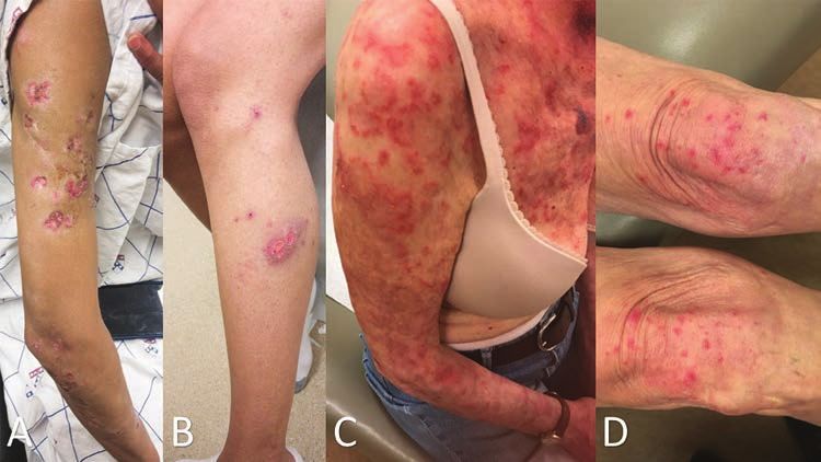
positive ANA or anti-dsDNA antibodies or at least 4 out of 17                    LE-specific skin disease
criteria including at least 1 immunologic criterion and 1 clinical               As mentioned above, CLE is divided into the following primary
criterion.16 Four of the clinical criteria are mucocutaneous in                  subsets: ACLE, SCLE, CCLE, as well as ICLE in certain clas-
nature including ACLE, chronic CLE (CCLE), oral ulcers, and                      sification systems (Table 1). It is possible for patients to have
nonscarring alopecia.16                                                          more than one form of CLE. A study of 191 patients with CLE
    No universally accepted classification criteria exist for CLE.               showed that 68% had 1 type, 29% had 2 types, and 3% had 3
Skin lesions in patients with SLE are divided according to the                   types.24 A US population-based study showed that 12% of
most widely used criteria suitable for rheumatologists in everyday               patients with CLE had disease progression to SLE, with a mean
clinical practice proposed by Gilliam and Sontheimer, which                      time to progression of 8 years.7 The cumulative incidence of
divides CLE into LE-specific and LE-nonspecific skin condi-                      SLE among patients with a diagnosis of CLE in the same study
tions.17,18 Other classification criteria, such as the Duesseldorf               was 5% at 5 years, 10% at 10 years, 15% at 15 years, 19% at 20
Classification, have been developed but have not gained universal                years, and 23% at 25 years.7 Early recognition of patients with
acceptance.19 LE-specific skin conditions include CCLE,                          CLE who are at risk for developing SLE is important. Signs of
SCLE, and ACLE as well as their various subtypes (Figure 1).                     nephropathy, elevated ANA titers, serositis, and arthralgias/
Although lupus erythematosus tumidus (LET) is considered by                      arthritis or other new symptoms of systemic disease may suggest
some as a form of CCLE, it is recognized by the Duesseldorf                      transition into SLE and should be closely followed. Patients
Classification and European S2k guidelines as a fourth primary                   with localized DLE, hypertrophic LE, LEP, and LET are more
subset of CLE known as intermittent CLE (ICLE).20,21 These                       likely to have skin-limited LE; those with generalized DLE or
LE-specific conditions have distinct clinical morphologies, but                  SCLE often meet ACR criteria for SLE; and those with ACLE
similar histopathologic features on routine H&E staining. These                  or LE-nonspecific skin lesions are most likely to have systemic
histologic features include lichenoid interface dermatitis with                  disease.25
basal layer vacuolization, apoptotic keratinocytes, periadnexal                  CCLE. CCLE has several subtypes, including DLE (Figure 1A),
and perivascular mononuclear cell infiltrate, epidermal atrophy,                 LEP, LET, and chilblain LE.26 CCLE is notable for demon-
and basement membrane thickening (Figure 2).15 In DLE, there                     strating a chronic, recurrent disease course which typically
is a tendency for more hyperkeratosis, follicular plugging, and                  requires long-term treatment with potential for progression to
thickening of the basement membrane relative to ACLE or                          involve internal organs.27,28 DLE is the most common subtype
SCLE. However, not all these features are found in all forms of                  of CCLE, representing 50% of cases.28 DLE is considered local-
LE-specific variants, and they can be found in conditions other                  ized if it involves exclusively the head and neck area and gener-
than CLE. Interface dermatitis, which consists of liquefactive                   alized if it extends below the neck with a predilection for the
degeneration of the epidermal basal layers, is not typically asso-               upper extremity extensor surfaces.28 Generalized DLE is more
ciated with LET or lupus erythematosus panniculitis (LEP) but                    often associated with SLE, and patients with generalized DLE or
is often seen in DM.22,23 A biopsy is recommended to confirm                     progressive localized DLE should be reevaluated for progressive
the diagnosis of CLE, as there are a variety of other diseases that              systemic disease.25 Both localized and generalized DLE consist
mimic its variants (Figure 3).                                                   of erythematous and sometimes scaly plaques in sun-exposed

                    Figure 1. Typical CLE lesions. (A) Active DLE lesions with erythema and scale are shown along with areas of
                    damage (ie, dyspigmentation and scarring) from prior active lesions. (B) Erythematous DLE lesions are shown on
                    the leg. (C) Annular SCLE lesions are seen on the arm and chest as well as (D) the legs. CLE: cutaneous lupus ery-
                    thematosus; DLE: discoid lupus erythematosus; SCLE: subacute cutaneous lupus erythematosus.

28                                                                                                                                       Skin in SLE

                                                                            Downloaded on April 5, 2023 from www.jrheum.org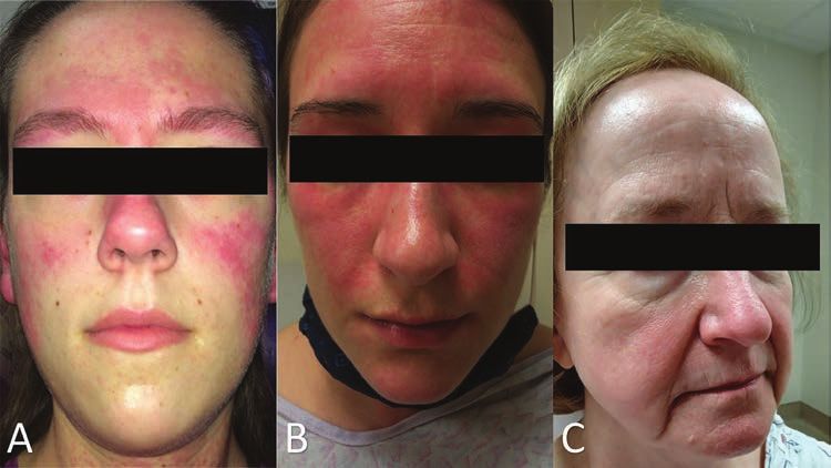
mentioned above, LET is considered by European S2k guidelines
                                                                                     to be a fourth primary subset of CLE, that is, ICLE.21 LET lesions
                                                                                     tend to occur on the face, neck, upper chest, and shoulders, and
                                                                                     consist of erythematous macules, papules, and plaques, normally
                                                                                     with smooth surfaces and no scale.31 Compared to other variants
                                                                                     of CCLE, LET is particularly photosensitive and less likely to be
                                                                                     associated with SLE.25 However, there is a mucinous form of SLE
                                                                                     with a skin biopsy identical to LET that can be seen in patients
                                                                                     with SLE.32 Chilblain LE affects cold-exposed areas, particularly
                                                                                     the acral surfaces, with painful, violaceous plaques and nodules
                                                                                     that may progress to erosions or ulcerations.31 At some point in
                                                                                     their disease course, 20% of patients with chilblain LE develop
         Figure 2. This biopsy demonstrates the vacuolar interface
                                                                                     features of SLE.33
         dermatitis found in most LE-specific skin conditions.
         LE: lupus erythematosus.                                                    SCLE. SCLE is believed to occur in 10% to 15% of patients with
                                                                                     SLE.1 Up to 50% of patients with SCLE meet diagnostic criteria
areas that progress to dyspigmentation and scarring.28 A recent                      for SLE, but systemic symptoms are typically arthritis/arthral-
study to develop classification criteria for DLE determined                          gias, malaise, and myalgias, with internal organ involvement
that clinical variables including atrophic scarring, location in                     such as renal or nervous system disease occurring in less than
the conchal bowl, and preference for the head and neck were                          10%.4,27 Seventy percent of patients with SCLE are anti-Ro/
most important, with lower importance given to dyspigmenta-                          SSA positive and 70% to 80% are ANA positive.34 Children
tion, follicular hyperkeratosis and/or plugging, and erythema-                       of women who have SSA or SSB antibodies during pregnancy
tous to violaceous color.29 Early in the disease course, prior to                    should be carefully monitored as they are at increased risk of
the development of damage, it can be difficult to differentiate                      neonatal LE.35 Histologically, SCLE is frequently characterized
DLE from SCLE since SCLE can also cause dyspigmentation                              by a less dense infiltrate than in DLE, but a denser perivascular
that can mimic scarring. Careful attention to loss of skin mark-                     infiltrate than found in ACLE. Other histologic features include
ings including follicular openings is required to establish that                     notable atrophy of the epithelium, and more significant vacu-
scarring is present. Hypertrophic or verrucous DLE is character-                     olization at the dermal-epidermal junction than in ACLE.28
ized by papular lesions that tend to occur on the face, extensor                     Dust-like particles representing IgG binding to keratinocytes
surfaces, or palms and soles.28 Mucosal DLE typically presents as                    are a specific, but not sensitive, finding on direct immunofluo-
erosions or macules that can have radiating striae located in the                    rescence.36 The 2 forms of SCLE include the annular and papu-
lips, palate, gingiva, or other mucosal surfaces.30 LEP, also known                  losquamous subtypes, both of which are notable for a recurrent
as lupus profundus, presents as indurated subcutaneous nodules                       course of widespread, highly photosensitive lesions.28 Lesions
or plaques that tend to occur in the face, scalp, upper torso,                       tend to be distributed symmetrically in sun-exposed regions,
buttocks, and proximal extremities. These lesions can progress                       though the central face, scalp, and skin below the waist are typi-
to ulceration or subcutaneous atrophy.30 A biopsy shows lobular                      cally spared.28,31 Lesions usually resolve without scarring, though
panniculitis, and it is important to confirm the diagnosis as                        dyspigmentation may occur.31 Some patients exhibit features
the differential includes subcutaneous panniculitic-like T-cell                      of both subtypes.31 Annular SCLE presents with scaly annular
lymphoma.25 Approximately 50% of patients with LEP will also                         erythematous plaques, which often merge to form a polycyclic
have DLE skin lesions visible at the overlying skin surface. As                      morphology.31 Papulosquamous SCLE can resemble psoriasis or

                       Figure 3. Careful clinical examination is often required to distinguish CLE from dermatomyositis. (A)
                       Dermatomyositis of the hands often shows confluent erythema of the skin overlying the MCP and IP joints and
                       the extensor tendons while (B) DLE lesions are less likely to be localized to these areas and can resolve with scar-
                       ring. Involvement of the v-area of the neck can appear very similar in (C) dermatomyositis and (D) CLE and
                       requires clinical correlation with other areas of involved skin to arrive at the correct diagnosis. CLE: cutaneous
                       lupus erythematosus; DLE: discoid lupus erythematosus; IP: interphalangeal; MCP: metacarpophalangeal.

Stull et al                                                                                                                                          29

                                                                              Downloaded on April 5, 2023 from www.jrheum.org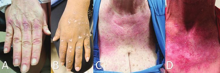
Table 1. LE-specific skin disease.

CLE Subtype                                       Clinical Characteristics

ACLE                                              Occurs in 30-50% patients with SLE. Flares often parallel systemic disease activity. Can see positive
		                                                ANA, anti-dsDNA, and anti-Sm antibodies.
    Localized                                     Raised or flat malar rash. Photosensitive, nonscarring, transient.
    Generalized                                   Widespread maculopapular rash above and below the neck. Dorsum of hands sparing MCP and IP
		                                                joints. Photosensitive, pruritic.
    TEN-like                                      Widespread denudation and blistering on sun-exposed areas.
SCLE                                              Recurrent course of widespread, highly photosensitive lesions that resolve without scarring, though
		                                                dyspigmentation may occur. 10-15% patients have SLE with arthralgias/myalgias; rare internal organ
		                                                involvement. Often positive ANA, SSA. 1 in 3 cases are drug-induced.
    Annular                                       Scaly annular erythematous plaques often merge to polycyclic morphology.
    Papulosquamous                                Resembles psoriasis or eczema.
    Erythrodermic                                 Generalized exfoliative erythroderma.
CCLE                                              Chronic, recurrent disease course. Rates of SLE vary between subtypes.
    DLE                                           Erythematous, sometimes scaly plaques exacerbated by sun exposure and trauma that progress to
		                                                dyspigmentation and atrophic scarring. Localized if confined to head and neck. Generalized if extends
		                                                below neck.
    Hypertrophic                                  Papular lesions on face, extensor surfaces, palms/soles.
    Mucosal                                       Erosions and macules on mucosal surfaces.
    LEP                                           Indurated subcutaneous nodules or plaques in face, scalp, upper torso, buttocks, proximal extremities.
		                                                Atrophic scars.
    CHLE                                          Painful violaceous plaques and nodules in cold-exposed areas, may progress to erosions or ulcerations on
		                                                acral surfaces.
    LET                                           Erythematous macules, papules, plaques with smooth surfaces and no scale, sharp raised borders. Very
		                                                photosensitive.

ANA: antinuclear antibody; ACLE: acute cutaneous lupus erythematosus; CCLE: chronic cutaneous lupus erythematosus; CHLE: chilblain lupus erythe-
matosus; CLE: cutaneous lupus erythematosus; DLE: discoid lupus erythematosus; IP: interphalangeal; LE: lupus erythematosus; LEP: lupus erythematosus
panniculitis; LET: lupus erythematosus tumidus; MCP: metacarpophalangeal; SCLE: subacute cutaneous lupus erythematosus; SLE: systemic lupus erythe-
matosus; TEN: toxic epidermal necrolysis.

eczema.31 Erythrodermic LE and LE gyrates repens are consid-                   found in patients with SCLE and is more commonly associated
ered rare variants of SCLE.28 Erythrodermic LE presents with                   with SCLE than other CLE subtypes.43
generalized exfoliative erythroderma that may represent a flare of             ACLE. ACLE is believed to occur in 30% to 50% of patients
papulosquamous SCLE after sun exposure.28 Only a few cases of                  with SLE.1 Systemic involvement is typical and ACLE rashes
LE gyratum repens have been discussed in the literature, and they              often flare in parallel with other organ disease activity.31 Ninety-
typically manifest as widely distributed chronic and recurrent                 five percent of patients with ACLE have positive ANA.34
figurate erythematous plaques.28 Although most cases of SCLE                   Histologically, ACLE lesions show liquefactive degeneration
are idiopathic, up to one-third of cases are believed to be induced            of the basal layer, an interface dermatitis, with perivascular
by exposure to drugs. The most common causes of drug-induced                   and periadnexal lymphocytic infiltrate. There are localized and
SCLE are proton pump inhibitors (PPIs), antihypertensives                      generalized forms of ACLE. The localized form of ACLE is the
(especially thiazide diuretics and calcium channel blockers), anti-            malar rash, characterized by butterfly-shaped erythema over the
convulsants, and antibiotics.37,38 Recently, cases of patients with            cheeks and nasal bridge that tends to spare the nasolabial folds,
preexisting SLE who subsequently developed SCLE after expo-                    as opposed to DM which typically involves nasolabial folds
sure to antihypertensives or PPIs have been described.37 It should             (Figure 4). The malar rash can be raised or flat, may be associ-
be noted that patients with SLE on systemic corticosteroids are                ated with a fine scale, and is classically sun-induced, nonscarring,
often placed on PPIs prophylactically to prevent gastrointes-                  and transient. The less common generalized form of ACLE,
tinal side effects. Adding to the risk of drug-induced SCLE are                sometimes called a maculopapular lupus rash or photosensitive
several over-the-counter forms of PPIs that are now available to               lupus dermatitis, occurs above and below the neck and presents
the public in the US. Particularly relevant to the rheumatologist              as a widespread eruption of macules and papules that is photo-
is the potential for biologic therapies including TNF-α inhib-                 sensitive and often pruritic. The pattern of involvement on the
itors, interleukin (IL)-17 inhibitors, IL-12/23 inhibitors, and                dorsum of hands can help distinguish generalized ACLE from
cytotoxic T-lymphocyte associated protein 4 therapy to induce                  DM; the metacarpophalangeal and interphalangeal joints are
SCLE.39-42 Rowell syndrome is an entity that can be associated                 normally spared in ACLE.31 Less common presentations of
with SLE, DLE, or SCLE, in which patients develop erythema                     ACLE include involvement of the lips and periorbital edema.
multiforme-like lesions and have a speckled ANA pattern.25                     Rare cases of toxic epidermal necrolysis (TEN)-like ACLE or
Sjögren syndrome is often a concomitant autoimmune disorder                    hyperacute CLE have been reported, which encompasses clinical

30                                                                                                                                            Skin in SLE

                                                                             Downloaded on April 5, 2023 from www.jrheum.org
Figure 4. A “butterfly rash” may be due to a variety of dermatological conditions. (A) The malar rash of ACLE
                   refers to erythema over the nasal bridge and cheeks that spares the nasolabial folds. Erythema of ACLE can be
                   found in other areas of the face, such as the forehead here. (B) Facial erythema in dermatomyositis tends to involve
                   the nasolabial folds. (C) Rosacea can mimic the facial erythema of ACLE but tends to worsen with specific triggers
                   such as alcohol, heat, and spicy foods. ACLE: acute cutaneous lupus erythematosus.

                             Figure 5. Alopecia due to (A) CLE can be difficult to distinguish from mimickers such as alo-
                             pecia because of (B) lichen planopilaris; correlation between clinical and histologic findings may
                             be required to correctly identify the cause of hair loss. CLE: cutaneous lupus erythematosus.

and histological findings of both ACLE and toxic epidermal                        nonspecific lesions tend to have increased SLE disease activity.47
necrolysis together without an inciting drug or infection.44,45 The               In addition to vascular disease, which will be discussed in more
majority of patients have a previously confirmed or new diag-                     depth below, other nonspecific cutaneous findings can occur
nosis of SLE or SCLE at the time of TEN-like ACLE.46                              in SLE including sclerodactyly, calcinosis cutis, rheumatoid
                                                                                  nodules, urticaria, cutis laxa/anetoderma, acanthosis nigrans,
LE-nonspecific skin disease                                                       lichen planus, and erythema multiforme. Bullous LE is consid-
LE-nonspecific skin disease includes skin changes that are                        ered a LE-nonspecific entity. Diagnosis of bullous LE requires
frequently associated with LE but are not specific to the disease                 an existing SLE diagnosis and patients frequently have increased
itself (Table 2). LE-nonspecific skin lesions are common in                       SLE disease activity. Unlike the lymphocytic inflammation seen
patients with SLE and often occur during the active phase of                      in SLE-specific lesions, the inflammation in bullous LE is neutro-
disease. Compared to those with LE-specific lesions, those with                   philic, the blister is subepidermal, and an antibody against type

Stull et al                                                                                                                                      31

                                                                          Downloaded on April 5, 2023 from www.jrheum.org
Table 2. LE-nonspecific skin disease. disease such as scleroderma, DM, and mixed connective tissue
disease, occurs in many patients with SLE and is characterized by
Cutaneous vascular disease cold-induced blanching followed by livedoid and erythematous
• Leukocytoclastic vasculitis
color change of fingers and other acral skin. Nonspecific changes
o Palpable purpura
o Urticarial vasculitis on nailfold capillaroscopy, including tortuous and dilated capil-
• Vasculopathy laries and hemorrhage, are more prevalent in SLE compared to
o Degos disease-like lesions healthy controls.51
o Secondary atrophie blanche Hair loss is frequent in SLE, occurring in more than half
• Periungual telangiectasias of patients at some point in the disease course (Figure 5).52,53
• Livedo reticularis Nonscarring alopecia, defined as diffuse thinning and fragility
• Thrombophlebitis
of the hair in the absence of other causes, is seen in 40% to 70%
• Raynaud phenomenon
• Erythromelalgia
of patients with SLE.16 It is important to rule out other poten-
Sclerodactyly tial causes of nonscarring alopecia before attributing it to SLE.
Calcinosis cutis Lupus hair refers to breakage of hair that typically occurs in the
Rheumatoid nodules frontal scalp. It commonly occurs during disease flares and may
Neutrophilic urticarial dermatosis be a form of telogen effluvium.
Cutis laxa/anetoderma Finally, returning to the cutaneous features included in the
Acanthosis nigrans 1997 ACR diagnostic criteria for SLE,12 2 of the 4 criteria that
Papulonodular mucinosis
have not yet been discussed are photosensitivity and oral ulcers.
Lichen planus
Erythema multiforme (Rowell syndrome) Photosensitivity is a phenomenon whereby exposure to ultra-
Bullous LE violet light causes skin rash in sun-exposed areas and/or other
Nonscarring alopecia systemic symptoms of SLE flares. It is a clinical observation and
occurs in a variety of other conditions, including DM, polymor-
LE: lupus erythematosus. phous light eruption, photoallergic contact dermatitis, solar
urticaria, and porphyrias. Oral or nasopharyngeal ulcers occur in
VII collagen is seen in the blood. Skin biopsy shows linear IgG at more than 40% of patients with SLE.54,55 Lesions can be painful
the dermal-epidermal junction on direct immunofluorescence. or painless and while palatal ulcers are the most specific for SLE,
Thus, a biopsy for direct immunofluorescence is helpful, and ulcers can also occur on buccal mucosa, hard palate, and the
findings are distinguished from epidermolysis bullosa acquisita vermilion border.
because of the diagnosis of SLE.48
Cutaneous vascular disease is a subtype of LE-nonspecific skin Disease monitoring
disease that includes vasculitis, vasculopathy, periungual telangi- The differentiation between disease activity and damage is
ectasias, livedo reticularis, thrombophlebitis, Raynaud phenom- important in SLE and CLE, given the chronic nature of these
enon (RP), and erythromelalgia. Cutaneous vasculitis has been diseases with periods of flares. The goal in managing SLE
reported in 10% to 20% of patients with SLE. It is a small vessel and CLE is to prevent and control activity in order to avoid
leukocytoclastic vasculitis that manifests as palpable purpura or damage, which is frequently irreversible. The Systemic Lupus
urticarial vasculitis. Occasionally vessels in the deeper dermis Erythematosus Disease Activity Index (SLEDAI) and SLEDAI
and subcutaneous tissues can be involved, resulting in nodules or 2000 (SLEDAI-2K), tools used to assess disease activity and
ulceration in a polyarteritis nodosa-like presentation. Cutaneous guide decisions to increase therapy, use a cut-off score of 3 to 4 to
vasculitis is most common with increased SLE activity and is define active disease and include several cutaneous features. Both
often associated with circulating immune complexes and hypo- versions include alopecia (2 points), oral or nasal mucosal ulcers
complementemia. While vasculitic lesions are due to a primary (2 points), vasculitis including ulceration, gangrene, tender
inflammatory attack on the vessel wall, other vascular skin finger nodules, periungual infarction, and splinter hemorrhages
manifestations associated with SLE are the result of vasculop- (8 points); the SLEDAI also includes “new rash,” defined as
athy secondary to coagulation abnormalities, including, but not new onset or recurrence of inflammatory rash (2 points); and
limited to, antiphospholipid antibody syndrome.49 Sometimes the SLEDAI-2K includes “rash,” defined as inflammatory rash
grouped as livedoid vasculopathy, these entities likely represent (2 points).56,57 The SLICC/ACR Damage Index, used to assess
an inflammatory response because of hypercoagulability. Livedo damage over the course of disease, incorporates scarring chronic
reticularis, a bluish net-like pattern typically most prominent on alopecia, extensive scarring of panniculum other than scalp and
the skin of buttocks, legs, and arms, results from reduced arterial pulp space, and skin ulceration.58 Damage in the SLICC criteria
blood flow and hypo-oxygenation and is common with cold expo- is defined as an irreversible change not related to active inflam-
sure. Livedo racemosa, with an irregular, broken net-like pattern mation that has occurred since the onset of disease and has been
occurs with an underlying focal skin pathology such as thrombi present for at least 6 months.
or calcification.50 Other vascular related phenomena that occur The Cutaneous Lupus Erythematosus Disease Area and
in SLE include periungual telangiectasia and erythema in 10% Severity Index (CLASI) is a validated instrument that has sepa-
to 15% of patients. RP, also common in other connective tissue rate scores to measure activity and damage of CLE. Activity
32 Skin in SLE
Downloaded on April 5, 2023 from www.jrheum.orgscores are based on the extent of erythema, scale/hypertrophy, methotrexate (MTX), mycophenolate mofetil (MMF), or
mucous membrane involvement, acute hair loss, and nonscar- azathioprine (AZA), although AZA is frequently less effective.67
ring alopecia. Damage scores are based on dyspigmentation and Though the algorithm suggested in the European guidelines lists
scarring, including scarring alopecia.59,60 Since the CLASI was MTX as a second-line therapy and MMF as a third-line, there
developed and validated, it has been used in two-thirds of clin- have been limited controlled trials in treating refractory CLE;
ical studies and trials with CLE outcomes.61,62 Additional vali- therefore, the European guidelines were based on a consensus
dated activity and damage scores for CLE have been developed. conference of dermatologists rather than being an evidence-
The revised CLASI includes adjustments to the original CLASI, based statement.67 A recent cohort study suggests similar efficacy
such as new variables like edema/infiltration and subcutaneous for MTX and MMF between subtypes of CLE, suggesting that
nodules/plaques.61 A working core outcome set for CLE trials other factors such as side-effect profile and comorbid condi-
was recently developed to guide future clinical and outcomes tions may influence medication selection.68 Thalidomide is
research in CLE, recommending the CLASI as a primary recommended only in treatment-refractory CLE with careful
endpoint and the Cutaneous Lupus Activity IGA (CLA-IGA) monitoring for the development of polyneuropathy, a potential
as a secondary endpoint for CLE physician-reported outcome adverse effect.63 Another therapeutic option for patients refrac-
measures.62 tory to antimalarials is lenalidomide, a thalidomide analog with
a superior adverse effect profile to thalidomide.69 Dapsone can
Treatment be effective in the treatment of bullous LE, LEP, and in some
As previously mentioned, the goal in the management of cuta- cases of SCLE and DLE; use requires close monitoring for
neous manifestations of SLE is to prevent and treat skin activity hematologic toxicities and the drug should not be used with
to minimize damage. A treatment algorithm for CLE has been patients who have a glucose-6-phosphate dehydrogenase defi-
put forth in the European S2k guidelines.63 An essential compo- ciency. Three case series showed significant improvement in
nent to managing cutaneous disease in SLE is prevention, CLASI activity scores with belimumab.70-72 ACLE can respond
with aggressive sun-protective measures including protective favorably to rituximab; however, no beneficial effects and some
clothing, avoiding exposure during peak sunlight hours, and exacerbations or new-onset disease have been seen in patients
daily use of SPF 70 or higher broad-spectrum ultraviolet A/B with DLE or SCLE.73-75 Anifrolumab, a drug recently approved
sunscreens. Vitamin D supplementation should be considered in by the US Food and Drug Administration for SLE, was shown
all patients, especially when serum levels are below normal range. in a phase III trial to be superior to placebo in improving skin
Patients who use tobacco should be counseled on smoking cessa- disease measures in patients with at least moderately active
tion, as it has been identified as a risk factor for widespread CLE, skin disease.76 Multiple agents are also under investigation as
it can increase disease severity, and it can decrease the efficacy of alternative therapies for CLE. Iberdomide has been shown in
antimalarial therapy.64,65 a recent phase II trial to have beneficial effects on skin disease
Topical and intralesional corticosteroids can be used in in patients with SCLE and CCLE, but not ACLE. BIIB059,
limited cutaneous disease or as adjunctive therapy along with a humanized monoclonal antibody targeting BDCA2 on plas-
systemic agents. As with systemic steroid use, the goal is to use macytoid dendritic cells (pDCs), was shown to improve skin
the least potent formula for the shortest amount of time to disease activity in patients with CLE in a recent phase II trial.77,78
lower the risk of local complications such as steroid atrophy and Similarly, VIB7734, a monoclonal antibody that targets pDCs
telangiectasia. An initial regimen of a medium-strength (class for antibody-dependent cellular cytotoxicity, showed clinically
III) topical corticosteroid such a triamcinolone acetonide 0.1% significant improvement in measures of skin disease activity in
applied daily to lesional skin can be tried, especially on areas off patients with CLE in a phase I trial.79
the face. If this does not provide sufficient relief, a more potent
topical steroid such as clobetasol propionate 0.05% or betameth- Conclusion
asone dipropionate 0.05% (class I) should be considered. When The spectrum of cutaneous disease in SLE is extremely broad
class I to III topical corticosteroids are providing clinical benefit and can occur at any point in the disease. Collaboration between
in sensitive areas such as the face, one can minimize the chances dermatology and rheumatology specialists is essential to properly
for developing cutaneous atrophy from longer-term therapy by diagnose and manage affected patients. Skin biopsy is important
rotating the topical corticosteroid every 2 weeks with a topical to differentiate CLE from other skin conditions and must be
calcineurin inhibitor such as pimecrolimus cream or tacrolimus considered in clinical context to reach a diagnosis. Timely and
ointment.5 Calcineurin inhibitors are recommended as alterna- appropriate therapy to control activity and minimize damage is
tive first-line or second-line topical therapeutic options, espe- the goal of treatment.
cially for the face, on the basis of randomized clinical trials.63
Antimalarials (hydroxychloroquine, chloroquine, and quin- REFERENCES
1. Yell JA, Mbuagbaw J, Burge SM. Cutaneous manifestations
acrine) are first-line therapies for cutaneous disease in SLE.
of systemic lupus erythematosus. Br J Dermatol 1996;
Seventy-five percent of patients respond to antimalarials with or 135:355-62.
without the addition of topical glucocorticoids.66 Disease refrac- 2. Dubois EL, Tuffanelli DL. Clinical manifestations of systemic
tory to antimalarials can be treated with immunosuppressives lupus erythematosus: computer analysis of 520 cases. JAMA
common in the rheumatologists’ armamentarium, including 1964;190:104-11.
Stull et al 33
Downloaded on April 5, 2023 from www.jrheum.org3. Klein R, Moghadam-Kia S, Taylor L, et al. Quality of life in 22. Bailey EE, Fiorentino DF. Amyopathic dermatomyositis: definitions,
cutaneous lupus erythematosus. J Am Acad Dermatol 2011; diagnosis, and management. Curr Rheumatol Rep 2014;16:465.
64:849-58. 23. Yell J, Allen J, Wojnarowska F, Kirtschig G, Burge SM. Bullous
4. Sontheimer R. The lexicon of cutaneous lupus systemic lupus erythematosus: revised criteria for diagnosis. Br J
erythematosus--a review and personal perspective on the Dermatol 1995;132:921-8.
nomenclature and classification of the cutaneous manifestations of 24. Watanabe T, Tsuchida T. Classification of lupus erythematosus
lupus erythematosus. Lupus 1997;6:84-95. based upon cutaneous manifestations. Dermatol 1995;190:277-83.
5. Rothfield N, Sontheimer RD, Bernstein M. Lupus erythematosus: 25. Werth VP. Clinical manifestations of cutaneous lupus
systemic and cutaneous manifestations. Clin Dermatol erythematosus. Autoimmun Rev 2005;4:296-302.
2006;24:348-62. 26. O’Brien JC, Chong BF. Not just skin deep: Systemic disease
6. Jarukitsopa S, Hoganson DD, Crowson CS, et al. Epidemiology of involvement in patients with cutaneous lupus. J Invest Dermatol
systemic lupus erythematosus and cutaneous lupus erythematosus Symp Proc 2017;18:S69-74.
in a predominantly white population in the United States. Arthritis 27. Lu Q, Long H, Chow S, et al. Guideline for the diagnosis, treatment
Care Res 2015;67:817-28. and long-term management of cutaneous lupus erythematosus.
7. Durosaro O, Davis MD, Reed KB, Rohlinger AL. Incidence of J Autoimmun 2021;123:102707.
cutaneous lupus erythematosus, 1965-2005: a population-based 28. Herzum A, Gasparini G, Cozzani E, Burlando M, Parodi A. Atypical
study. Arch Dermatol 2009;145:249-53. and rare forms of cutaneous lupus erythematosus: The importance
8. Izmirly P, Buyon J, Belmont HM, et al. Population-based prevalence of the diagnosis for the best management of patients. Dermatol
and incidence estimates of primary discoid lupus erythematosus 2022;238:195-204.
from the Manhattan Lupus Surveillance Program. Lupus Sci Med 29. Elman SA, Joyce C, Braudis K, et al. Creation and validation of
2019;6:e000344. classification criteria for discoid lupus erythematosus. JAMA
9. Drenkard C, Parker S, Aspey LD, et al. Racial disparities in the Dermatol 2020;156:901-6.
incidence of primary chronic cutaneous lupus erythematosus in the 30. Cooper EE, Pisano CE, Shapiro SC. Cutaneous manifestations of
Southeastern US: The Georgia Lupus Registry. Arthritis Care Res “lupus”: Systemic lupus erythematosus and beyond. Int J Rheumatol
2019;71:95-103. 2021;2021:6610509.
10. Stojan G, Petri M. Epidemiology of systemic lupus erythematosus: 31. Okon LG, Werth VP. Cutaneous lupus erythematosus: diagnosis
an update. Curr Opin Rheumatol 2018;30:144-50. and treatment. Best Pract Res Clin Rheumatol 2013;27:391-404.
11. Tan EM, Cohen AS, Fries JF, et al. The 1982 revised criteria for the 32. Stead J, Headley C, Ioffreda M, Kovarik C, Werth V. Coexistence of
classification of systemic lupus erythematosus. Arthritis Rheum tumid lupus erythematosus with systemic lupus erythematosus and
1982;25:1271-7. discoid lupus erythematosus: a report of 2 cases. J Clin Rheumatol
12. Hochberg MC. Updating the American College of Rheumatology 2008;14:338-41.
revised criteria for the classification of systemic lupus erythematosus. 33. Hedrich C, Fiebig B, Hauck F, et al. Chilblain lupus
Arthritis Rheum 1997;40:1725. erythematosus--a review of literature. Clin Rheumatol 2008;
13. Biazar C, Sigges J, Patsinakidis N, et al. Cutaneous lupus 27:949-54.
erythematosus: first multicenter database analysis of 1002 patients 34. Tebbe B, Mansmann U, Wollina U, et al. Markers in cutaneous lupus
from the European Society of Cutaneous Lupus Erythematosus erythematosus indicating systemic involvement. A multicenter study
(EUSCLE). Autoimmun Rev 2013;12:444-54. on 296 patients. Acta Derm Venereol 1997;77:305-8.
14. Aringer M, Costenbader K, Daikh D, et al. 2019 European 35. Dao KH, Bermas BL. Systemic lupus erythematosus management in
League Against Rheumatism/American College of Rheumatology pregnancy. Int J Womens Health 2022;14:199-211.
classification criteria for systemic lupus erythematosus. Arthritis 36. Nieboer C, Tak-Diamand Z, Van Leeuwen-Wallau HE. Dust-like
Rheumatol 2019;71:1400-12. particles: a specific direct immunofluorescence pattern in sub-acute
15. Tarazi M, Gaffney RG, Kushner CJ, Chakka S, Werth VP. cutaneous lupus erythematosus. Br J Dermatol 1988;118:725-9.
Cutaneous lupus erythematosus patients with a negative antinuclear 37. Keyes E, Grinnell M, Vazquez T, Diaz D, Thomas P, Werth VP.
antibody meeting the American College of Rheumatology and/ Drug-induced subacute cutaneous lupus erythematosus in previously
or Systemic Lupus International Collaborating Clinics criteria for diagnosed systemic lupus erythematosus patients: a case series.
systemic lupus erythematosus. Arthritis Care Res 2019;71:1404-9. JAAD Case Rep 2021;12:18-21.
16. Petri M, Orbai AM, Alarcón GS, et al. Derivation and validation of 38. Laurinaviciene R, Sandholdt LH, Bygum A. Drug-induced
the Systemic Lupus International Collaborating Clinics classification cutaneous lupus erythematosus: 88 new cases. Eur J Dermatol
criteria for systemic lupus erythematosus. Arthritis Rheum 2017;27:28-33.
2012;64:2677-86. 39. Borucki R, Werth VP. Cutaneous lupus erythematosus induced by
17. Gilliam JN, Sontheimer RD. Distinctive cutaneous subsets in the drugs - novel insights. Expert Rev Clin Pharmacol 2020;13:35-42.
spectrum of lupus erythematosus. J Am Acad Dermatol 1981; 40. Conforti C, Retrosi C, Giuffrida R, et al. Secukinumab-induced
4:471-5. subacute cutaneous lupus erythematosus. Dermatol Ther
18. Gilliam JN, Sontheimer RD. Skin manifestations of SLE. Clin 2020;33:e13417.
Rheum Dis 1982;8:207-18. 41. Tierney E, Kirthi S, Ramsay B, Ahmad K. Ustekinumab-induced
19. Kuhn A, Landmann A. The classification and diagnosis of cutaneous subacute cutaneous lupus. JAAD Case Rep 2019;5:271-3.
lupus erythematosus. J Autoimmun 2014;48:14-9. 42. Tarazi M, Aiempanakit K, Werth VP. Subacute cutaneous lupus
20. Kuhn A, Ruzicka T. Classification of cutaneous lupus erythematosus and systemic lupus erythematosus associated with
erythematosus. In: Kuhn A, Lehmann P, Ruzicka T, editors. abatacept. JAAD Case Rep 2018;4:698-700.
Cutaneous lupus erythematosus. Berlin: Springer-Verlag; 2005:53-7. 43. Koskenmies S, Järvinen TM, Onkamo P, et al. Clinical and
21. Worm M, Zidane M, Eisert L, et al. S2k guideline: Diagnosis laboratory characteristics of Finnish lupus erythematosus patients
and management of cutaneous lupus erythematosus - Part 1: with cutaneous manifestations. Lupus 2008;17:337-47.
Classification, diagnosis, prevention, activity scores. J Dtsch 44. Marija S, Ivana B, Nina R, et al. Toxic epidermal necrolysis in a child
Dermatol Ges 2021;19:1236-47. with lupus-associated pancreatitis. Rheumatol Int 2017;37:1221-6.
34 Skin in SLE
Downloaded on April 5, 2023 from www.jrheum.org45. Mir TH, Bhat IA, Jabeen B, Haji MLI. Toxic epidermal Academy of Dermatology and Venereology (EADV). J Eur Acad
necrolysis-like acute cutaneous lupus/acute syndrome of apoptotic Dermatol Venereol 2017;31:389-404.
pan-epidermolysis. Rheumatol 2021;60:5876-7. 64. Kuhn A, Sigges J, Biazar C, et al. Influence of smoking on disease
46. Romero LS, Bari O, Forbess CJ, Schneider JA, Cohen PR. Toxic severity and antimalarial therapy in cutaneous lupus erythematosus:
epidermal necrolysis-like acute cutaneous lupus erythematosus: analysis of 1002 patients from the EUSCLE database. Br J Dermatol
report of a case and review of the literature. Dermatol Online J 2014;171:571-9.
2018;24:13030. 65. Bourré-Tessier J, Peschken CA, Bernatsky S, et al. Association
47. Zecević RD, Vojvodić D, Ristić B, Pavlović MD, Stefanović D, of smoking with cutaneous manifestations in systemic lupus
Karadaglić D. Skin lesions-an indicator of disease activity in systemic erythematosus. Arthritis Care Res 2013;65:1275-80.
lupus erythematosus? Lupus 2001;10:364-7. 66. Callen JP. Management of skin disease in patients with lupus
48. Contestable JJ, Edhegard KD, Meyerle JH. Bullous systemic lupus erythematosus. Best Pract Res Clin Rheumatol 2002;16:245-64.
erythematosus: a review and update to diagnosis and treatment. Am 67. Borucki R, Werth VP. Expert Perspective: An evidence-based
J Clin Dermatol 2014;15:517-24. approach to refractory cutaneous lupus erythematosus. Arthritis
49. Diógenes MJ, Diógenes PC, de Morais Carneiro RM, Neto CC, Rheumatol 2020;72:1777-85.
Duarte FB, Holanda RR. Cutaneous manifestations associated with 68. Keyes E, Jobanputra A, Feng R, et al. Comparative responsiveness
antiphospholipid antibodies. Int J Dermatol 2004;43:632-7. of cutaneous lupus erythematosus patients to methotrexate and
50. Obermoser G, Sontheimer RD, Zelger B. Overview of common, rare mycophenolate mofetil: A cohort study. J Am Acad Dermatol
and atypical manifestations of cutaneous lupus erythematosus and 2022;87:447-8.
histopathological correlates. Lupus 2010;19:1050-70. 69. Yuki EFN, Silva CA, Aikawa NE, et al. Thalidomide and
51. Cutolo M, Melsens K, Wijnant S, et al. Nailfold capillaroscopy lenalidomide for refractory systemic/cutaneous lupus erythematosus
in systemic lupus erythematosus: a systematic review and critical treatment: a narrative review of literature for clinical practice. J Clin
appraisal. Autoimmun Rev 2018;17:344-52. Rheumatol 2021;27:248-59.
52. Moghadam-Kia S, Franks AG Jr. Autoimmune disease and hair loss. 70. Iaccarino L, Bettio S, Reggia R, et al. Effects of belimumab on
Dermatol Clin 2013;31:75-91. flare rate and expected damage progression in patients with active
53. Concha JSS, Werth VP. Alopecias in lupus erythematosus. Lupus Sci systemic lupus erythematosus. Arthritis Care Res 2017;69:115-23.
Med 2018;5:e000291. 71. Vashisht P, Borghoff K, O’Dell JR, Hearth-Holmes M. Belimumab
54. Urman JD, Lowenstein MB, Abeles M, Weinstein A. Oral mucosal for the treatment of recalcitrant cutaneous lupus. Lupus
ulceration in systemic lupus erythematosus. Arthritis Rheum 2017;26:857-64.
1978;21:58-61. 72. Parodis I, Sjöwall C, Jönsen A, et al. Smoking and pre-existing
55. Kudsi M, Nahas LD, Alsawah R, Hamsho A, Omar A. The organ damage reduce the efficacy of belimumab in systemic lupus
prevalence of oral mucosal lesions and related factors in systemic erythematosus. Autoimmun Rev 2017;16:343-51.
lupus erythematosus patients. Arthritis Res Ther 2021;23:229. 73. Hofmann S, Leandro M, Morris SD, Isenberg D. Effects of
56. Bombardier C, Gladman DD, Urowitz MB, Caron D, Chang rituximab-based B-cell depletion therapy on skin manifestations of
CH. Derivation of the SLEDAI. A disease activity index for lupus lupus erythematosus--report of 17 cases and review of the literature.
patients. Arthritis Rheum 1992;35:630-40. Lupus 2013;22:932-9.
57. Gladman DD, Ibañez D, Urowitz MB. Systemic lupus 74. Quelhas da Costa RQ, Aguirre-Alastuey ME, Isenberg DA, Saracino
erythematosus disease activity index 2000. J Rheumatol AM. Assessment of response to B-cell depletion using rituximab in
2002;29:288-91. cutaneous lupus erythematosus. JAMA Dermatol 2018;
58. Gladman D, Ginzler E, Goldsmith C, et al. The development and 154:1432-40.
initial validation of the Systemic Lupus International Collaborating 75. Vital EM, Wittmann M, Edward S, et al. Brief report: responses to
Clinics/American College of Rheumatology damage index for rituximab suggest B cell-independent inflammation in cutaneous
systemic lupus erythematosus. Arthritis Rheum 1996;39:363-9. systemic lupus erythematosus. Arthritis Rheumatol 2015;
59. Albrecht J, Taylor L, Berlin JA, et al. The CLASI (Cutaneous Lupus 67:1586-91.
Erythematosus Disease Area and Severity Index): an outcome 76. Morand EF, Furie R, Tanaka Y, et al. Trial of anifrolumab in active
instrument for cutaneous lupus erythematosus. J Invest Dermatol systemic lupus erythematosus. N Engl J Med 2020;382:211-21.
2005;125:889-94. 77. Werth V, Merrill J, Furie R, et al. OP0132 Effect of Iberdomide on
60. Chong BF, Werth V. Cutaneous lupus erythematosus and cutaneous manifestations in systemic lupus erythematosus: Results
dermatomyositis: Utilizing assessment tools for treatment efficacy. of a 24-week, placebo controlled, phase 2 study. Ann Rheum Dis
J Invest Dermatol 2022;142:936-43. 2021;80:76-7.
61. Kuhn A, Meuth AM, Bein D, et al. Revised Cutaneous Lupus 78. Werth V, Furie R, Romero-Diaz J on behalf of the LILAC
Erythematosus Disease Area and Severity Index (RCLASI): A investigators, et al. OP0193 BIIB059, a humanized monoclonal
modified outcome instrument for cutaneous lupus erythematosus. antibody targeting BDCA2 on plasmacytoid dendritic cells (pDC),
Br J Dermatol 2010;163:83-92. shows dose-related efficacy in the phase 2 LILAC study in patients
62. Guo LN, Perez-Chada LM, Borucki R, Nambudiri VE, Werth (pts) with active cutaneous lupus erythematosus (CLE). Ann
VP, Merola JF. Development of a working core outcome set for Rheum Dis 2020;79:120-1.
cutaneous lupus erythematosus: a practical approach to an urgent 79. Karnell JL, Wu Y, Mittereder N, et al. Depleting plasmacytoid
unmet need. Lupus Sci Med 2021;8:e000529. dendritic cells reduces local type I interferon responses and
63. Kuhn A, Aberer E, Bata-Csörgő Z, et al. S2k guideline for treatment disease activity in patients with cutaneous lupus. Sci Transl Med
of cutaneous lupus erythematosus - guided by the European 2021;13:eabf8442.
Dermatology Forum (EDF) in cooperation with the European
Stull et al 35
Downloaded on April 5, 2023 from www.jrheum.orgYou can also read