Correction of Vertebral Bone Development in Ectodysplasin A1-Deficient Mice by Prenatal Treatment With a Replacement Protein
←
→
Page content transcription
If your browser does not render page correctly, please read the page content below
ORIGINAL RESEARCH
                                                                                                                                             published: 11 August 2021
                                                                                                                                       doi: 10.3389/fgene.2021.709736

                                              Correction of Vertebral Bone
                                              Development in Ectodysplasin
                                              A1-Deficient Mice by Prenatal
                                              Treatment With a Replacement
                                              Protein
                                              Clara-Sophie Kossel 1,2 , Mandy Wahlbuhl 1,2 , Sonia Schuepbach-Mallepell 3 , Jung Park 1,2 ,
                                              Christine Kowalczyk-Quintas 3 , Michaela Seeling 4 , Klaus von der Mark 5 ,
                                              Pascal Schneider 3 and Holm Schneider 1,2*
                                              1
                                                Department of Pediatrics, Friedrich-Alexander University Erlangen-Nürnberg, Erlangen, Germany, 2 Center for Ectodermal
                                              Dysplasias, University Hospital Erlangen, Erlangen, Germany, 3 Department of Biochemistry, University of Lausanne,
                                              Epalinges, Switzerland, 4 Department of Biology, Friedrich-Alexander University Erlangen-Nürnberg, Erlangen, Germany,
                            Edited by:        5
                                                Department of Experimental Medicine I, Friedrich-Alexander University Erlangen-Nürnberg, Erlangen, Germany
                     Corrado Romano,
     Oasi Research Institute, Scientific
 Institute for Research, Hospitalization      X-linked hypohidrotic ectodermal dysplasia with the cardinal symptoms hypodontia,
          and Healthcare (IRCCS), Italy
                                              hypotrichosis and hypohidrosis is caused by a genetic deficiency of ectodysplasin A1
                        Reviewed by:
                                              (EDA1). Prenatal EDA1 replacement can rescue the development of skin appendages
                        Marja Mikkola,
         University of Helsinki, Finland      and teeth. Tabby mice, a natural animal model of EDA1 deficiency, additionally feature a
                            Izzet Yavuz,      striking kink of the tail, the cause of which has remained unclear. We studied the origin
               Dicle University, Turkey
                                              of this phenomenon and its response to prenatal therapy. Alterations in the distal spine
                 *Correspondence:
                    Holm Schneider            could be noticed soon after birth, and kinks were present in all Tabby mice by the age of
      holm.schneider@uk-erlangen.de           4 months. Although their vertebral bones frequently had a disorganized epiphyseal zone
                                              possibly predisposing to fractures, cortical bone density was only reduced in vertebrae
                   Specialty section:
         This article was submitted to        of older Tabby mice and even increased in their tibiae. Different availability of osteoclasts
       Genetics of Common and Rare            in the spine, which may affect bone density, was ruled out by osteoclast staining. The
                              Diseases,
               a section of the journal
                                              absence of hair follicles, a well-known niche of epidermal stem cells, and much lower
                  Frontiers in Genetics       bromodeoxyuridine uptake in the tail skin of 9-day-old Tabby mice rather suggest the
            Received: 14 May 2021             kink being due to a skin proliferation defect that prevents the skin from growing as fast
            Accepted: 19 July 2021            as the skeleton, so that caudal vertebrae may be squeezed and bent by a lack of skin.
          Published: 11 August 2021
                                              Early postnatal treatment with EDA1 leading to delayed hair follicle formation attenuated
                             Citation:
              Kossel C-S, Wahlbuhl M,         the kink, but did not prevent it. Tabby mice born after prenatal administration of EDA1,
      Schuepbach-Mallepell S, Park J,         however, showed normal tail skin proliferation, no signs of kinking and, interestingly, a
     Kowalczyk-Quintas C, Seeling M,
     von der Mark K, Schneider P and
                                              normalized vertebral bone density. Thus, our data prove the causal relationship between
        Schneider H (2021) Correction         EDA1 deficiency and kinky tails and indicate that hair follicles are required for murine tail
       of Vertebral Bone Development          skin to grow fast enough. Disturbed bone development appears to be partially pre-
in Ectodysplasin A1-Deficient Mice by
               Prenatal Treatment With        determined in utero and can be counteracted by timely EDA1 replacement, pointing to
               a Replacement Protein.         a role of EDA1 also in osteogenesis.
             Front. Genet. 12:709736.
    doi: 10.3389/fgene.2021.709736            Keywords: bone, development, ectodysplasin A1, ectodermal dysplasia, fetal therapy, NF-κB

Frontiers in Genetics | www.frontiersin.org                                         1                                          August 2021 | Volume 12 | Article 709736
Kossel et al.                                                                                                   Correction of Murine Bone Development

INTRODUCTION                                                                 deficiency might have impact on bone development not only in
                                                                             Tabby mice.
X-linked hypohidrotic ectodermal dysplasia (XLHED; #MIM                         Various strategies to treat XLHED with an EDA1 replacement
305100), a rare hereditary disease characterized by missing or               protein have been investigated (Gaide and Schneider, 2003; Casal
dysplastic skin appendages and teeth, is caused by deficiency                et al., 2007; Hermes et al., 2014). Postnatal protein replacement
of the signaling protein ectodysplasin A1 (EDA1) during early                was partially effective in animal models but not in human
development (Mikkola and Thesleff, 2003). Numerous mutations                 patients (Körber et al., 2020), whereas prenatal intra-amniotic
in the X-chromosomal gene EDA which encodes EDA1 have been                   administration of a recombinant EDA1 molecule to infants with
reported (Cluzeau et al., 2011; Schneider et al., 2011; Trzeciak             XLHED resulted in normal sweat gland endowment and sweating
and Koczorowski, 2016; Wohlfart et al., 2016), and most of them              ability, development of more teeth, and normalized function
result in a complete absence of functional EDA1.                             of salivary glands (Schneider et al., 2018; Körber et al., 2020)
    Mutations in the genes EDA1 receptor (EDAR) or                           and, thus, showed the potential to permanently resolve the most
EDARADD which underlie genetic deficiencies of the                           relevant clinical problems associated with XLHED (Wohlfart
EDAR and its associated death domain-containing adaptor                      et al., 2020). This recombinant protein, composed of the Fc part
protein, respectively, lead to autosomal recessive or dominant               of human IgG and the receptor-binding domain of EDA1, was
hypohidrotic ectodermal dysplasia. Since all three proteins are              named Fc-EDA (Schneider et al., 2018). When swallowed by
part of the EDA1 pathway to nuclear factor (NF)-κB activation,               a fetus in the third trimester of pregnancy, the Fc component
the phenotypic features are similar (Cluzeau et al., 2011).                  facilitates intestinal uptake into the fetal circulation via specific
They include hypotrichosis, oligo- or anodontia, and hypo-                   Fc receptors (Schneider et al., 2018).
or anhidrosis. The latter is the most severe disability as it can               In this study, we focused again on Tabby mice to elucidate
lead to life-threatening hyperthermia on summer days, during                 the pathogenesis of their kinky tail, the peculiarities of bone
physical activity or febrile illness. This significantly increases the       development in this animal model, and their response to
childhood mortality (Blüschke et al., 2010).                                 prenatal therapy with Fc-EDA. We show that intra-amniotic
    Although knowledge about NF-κB signaling has been                        administration of the replacement protein rescues tail skin as well
increasing constantly (Smahi et al., 2002; Zhang et al., 2017),              as vertebral bone development and discuss a yet unexplored role
the exact mechanisms by which EDA1 regulates the development                 of EDA1 in osteogenesis.
of ectodermal structures are still subject to research. After
binding of EDA1 to its cognate receptor, the EDA1–EDAR
complex recruits the receptor-associated adaptor molecule which              MATERIALS AND METHODS
activates several tumor necrosis factor receptor-associated factors
(TRAFs). TRAF6 builds a complex with TAK1 binding protein 2                  Animal Model
(TAB2) and transforming growth factor beta-activated kinase 1                C57BL/6 wild-type mice (Charles River, Sulzfeld, Germany),
(TAK1) that activates the NF-κB essential modulator (NEMO),                  white-bellied agouti B6CBAa Aw-J/A-EdaTa/J Tabby mice (a
leading to phosphorylation and dissociation of the inhibitory                mouse model for XLHED; Jackson Laboratory, Bar Harbor, ME,
complex IκBα from NF-κB. Upon its translocation into the                     United States), and appropriate control mice were housed in
nucleus NF-κB acts as a transcription factor for numerous target             individually ventilated cages under standard conditions with
genes, e.g., sonic hedgehog (shh) and members of the Wnt                     a light/dark cycle of 12 h and free access to standard chow
protein family (Kowalczyk-Quintas and Schneider, 2014).                      and tap water. All experimental procedures on animals were
    The EDA1 pathway is highly conserved among vertebrates,                  conducted in accordance with German or Swiss regulations and
which provides the opportunity of valid research in a mouse                  legal requirements (depending on the site of investigation) and
model (Pantalacci et al., 2008). The Tabby mouse (Ferguson                   had been approved by the local government authorities.
et al., 1997; Srivastava et al., 1997) is a well-established animal
model for EDA1 deficiency. Human and murine EDA1 show an                     Therapeutic Interventions
amino acid sequence homology of 94% (Monreal et al., 1998).                  Three litters of newborn Tabby mice were treated within 24 h
In general, the Tabby phenotype reflects the characteristics of              after birth by intraperitoneal injection of Fc-EDA, a recombinant
human XLHED patients, meaning that hair, teeth, and eccrine                  fusion protein consisting of the receptor-binding tumor necrosis
glands are affected by the developmental disorder. Different and             factor (TNF) homology domain of EDA1 and the Fc domain of
particularly striking, however, is a characteristic kink in the tail         human IgG1 (single dose: 2 mg/kg body weight).
tip of Tabby mice which has been reported to result from fractures              Prenatal administration of Fc-EDA to mouse fetuses was done
of vertebral bodies (Hill et al., 2002). Structural abnormalities of         as described before (Hermes et al., 2014; Schneider et al., 2018).
both vertebral and tibial bones of Tabby mice were described by              Briefly, pregnant time-mated Tabby mice were anesthetized
the same authors (Hill et al., 2002).                                        with isoflurane (2%) and analgized during and after surgery
    In the human embryo, EDA is expressed by osteoblasts                     on gestational day 15 (E15) with metamizole (subcutaneous
between gestational weeks 12 and 16 (Montonen et al., 1998).                 injection of 5 mg). A laparotomy was performed, followed by
The NF-κB pathway is also known to play an essential role in                 complete exposure of both uterine horns. Fc-EDA was injected
the differentiation of osteoclasts via RANK-RANKL signaling                  into the amniotic sacs of individual fetuses at doses of 100 µg/g
that involves TRAF6 activation (Clauss et al., 2008). Thus, EDA1             fetal body weight (16 µl of Fc-EDA at 2.2 mg/ml, assuming a

Frontiers in Genetics | www.frontiersin.org                              2                                    August 2021 | Volume 12 | Article 709736
Kossel et al.                                                                                                                        Correction of Murine Bone Development

  FIGURE 1 | Tabby mice develop vertebral dislocations soon after birth. Macroscopic comparison of representative tail tips from (A) wild-type and (B) untreated
  Tabby mice at embryonic day E18.5 and postnatal days P0, P4, and P21. Lower panels in panels (A,B) depict whole-mount staining of cartilaginous parts of the
  same tails with alcian blue. In nearly half of the Tabby mice, first alterations of the distal spine could be detected already on day P4; kinks were evident in 21 of 23
  animals investigated at day P21 and in all Tabby mice by day P120.

fetal weight of approximately 0.35 g) using a glass syringe with 33                       detached to facilitate permeabilization of the target tissue. The
gauge needle (Hamilton Robotics, Bonaduz, Switzerland). After                             embryos/pups were fixed in 70% ethanol at 4◦ C overnight,
repositioning of the uterine horns, the abdominal cavity was                              transferred to 95% ethanol for 1 h, followed by incubation in
closed by suturing peritoneum and skin separately (Schuepbach-                            acetone at room temperature overnight and subsequent staining
Mallepell et al., 2020). Treated animals were sacrificed at the age                       with Alcian blue solution for 1–4 h (embryos) or overnight
of 2 months or above by cervical dislocation under isoflurane                             (pups), respectively. Embryos were cleared in KOH (1%) and
anesthesia. Their tails were collected and investigated.                                  glycerol-KOH (1%) solution (equal parts) for 12 h each and
                                                                                          finally transferred to 100% glycerol for long-term storage. In
Micro-Computed Tomography                                                                 newborn pups, unspecific Alcian blue staining was removed
After removing all muscle tissue, tibial, and vertebral bones                             by washing initially with 70% ethanol (two changes), followed
were fixed in paraformaldehyde solution (4%) for 24 h. Images                             by incubation in 95% ethanol overnight prior to the KOH
of these bones at 50 µm-resolution were then acquired on an                               clearing described above. Stained whole-mount skeletal tissue
Inveon Micro-PET/CT-Scanner (Siemens, Erlangen, Germany)                                  sections were investigated using a stereomicroscope MZ 10F
at the Preclinical Imaging Platform Erlangen. Cross-sections                              (Leica Microsystems, Wetzlar, Germany) with Zeiss Axiocam Typ
were evaluated with respect to the volume of the medullary                                MRc and Axiovision 4.7 software (Carl Zeiss Microscopy GmbH,
cavity and cortical bone density, measured in Hounsfield                                  Göttingen, Germany).
units, using the DICOM viewer Osirix (Würzburg, Germany).
Macroscopic images shown in this paper are based on virtual                               Histological Assessments and
three-dimensional reconstructions.                                                        Trabecular Bone Histomorphometry
                                                                                          Vertebral and tibial bones of older pups or adult mice were
Whole-Mount Skeletal Staining of                                                          decalcified for 7 days in 14% (wt/vol) EDTA (pH adjusted to
Embryos and Newborn Pups                                                                  7.2 by addition of ammonium hydroxide) and then embedded
Embryos of untreated Tabby and wild-type mice at gestational                              in paraffin. Vertebral bones (tails) were cut into 5-µm sections,
days 16.5 (E16.5) and 18.5 (E18.5) and pups at postnatal days                             tibiae into 8-µm sections, respectively. For morphological
0 (P0), 4 (P4), 7 (P7), and 21 (P21) were investigated for                                assessment, tissue sections were stained with hematoxylin
tail axis deviations and sacrificed by decapitation at the time                           and eosin. Alcian blue staining was used to visualize the
points indicated. Skin, internal organs and adipose tissue were                           cartilaginous components of the epiphyseal growth plate. In

Frontiers in Genetics | www.frontiersin.org                                           3                                            August 2021 | Volume 12 | Article 709736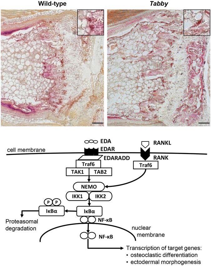
Kossel et al.                                                                                                                     Correction of Murine Bone Development

  FIGURE 2 | Impact of ectodysplasin A1 (EDA1) deficiency on osteogenesis and bone density. (A,B) Representative three-dimensional reconstructions of (A) the
  distal tail portion and (B) tibiae of wild-type and Tabby mice. (C–F) Vertebral and tibial bone sections stained with hematoxylin-eosin (C,D) or alcian blue (E,F): The
  epiphyseal region appears less well organized and contains more cartilage islands (arrowed) in the tails of Tabby mice than in wild-type mice. (G) Although computed
  tomography scanning (micro-CT) revealed a tendency toward reduced cortical bone density already in tails of Tabby mice at an age of 2–4 months (mean of 7–10
  distal vertebrae where the kink is formed; indicated in Hounsfield units), this difference became significant only in Tabby mice older than 6 months (n = 6; white bars)
  compared with wild-type animals (n = 6; black bars). (H) Tibial bones, in contrast, showed a higher cortical density in Tabby mice of both age groups (n = 6 and
  n = 7, respectively; white bars) than in wild-type animals (black bars) as determined by micro-CT measurements. Scale bar in panels (C–F): 100 µm. Data are shown
  as mean ± SD; ∗ p < 0.05; ∗∗∗∗ p < 0.0001.

order to evaluate trabecular bone parameters and the amount of                           0.025% protease (Type V; Sigma-Aldrich) and then subjected to
osteoclasts, tissue sections were stained for tartrate-resistant acid                    immunofluorescence staining using a rat monoclonal antibody
phosphatase using the leukocyte acid phosphatase (TRAP) kit                              against BrdU (Abcam, Cambridge, United Kingdom). A donkey
387A (Sigma-Aldrich, St. Louis, MO, United States) according to                          anti-rat IgG antibody conjugated to Alexa Fluor 568 (Abcam)
the manufacturer’s instructions. All parameters were quantified                          was applied for BrdU detection. Cell nuclei were counter-stained
by digital image analysis (OsteoMeasure; OsteoMetrics, Decatur,                          with 40 ,6-diamidino-2-phenylindole dihydrochloride (DAPI;
GA, United States).                                                                      Sigma-Aldrich). The resulting images were investigated and
                                                                                         photographed under a fluorescence microscope (Axio Observer;
Bromodeoxyuridine Incorporation                                                          Zeiss, Jena, Germany). BrdU-positive cells were counted in
Assays                                                                                   epidermis and dermis except for the hair bulb region. Data were
Wild-type and Tabby mouse pups at day P9 were treated by                                 obtained from six independent sections of each mouse.
intraperitoneal bromodeoxyuridine (BrdU) injection (40 mg/kg
body weight) 2 h before sacrificing them by decapitation
(BrdU stock of 5 mg/ml; Sigma-Aldrich, Taufkirchen, Germany).                            Statistical Analyses
The tails were fixed in 4% paraformaldehyde solution and                                 Datasets were analyzed with unpaired t-tests using the
embedded in paraffin. Sections were deparaffinized, rehydrated,                          GraphPad Prism software 7 (GraphPad Software Inc., La
subjected to antigen retrieval in citrate buffer and digestion with                      Jolla, CA, United States).

Frontiers in Genetics | www.frontiersin.org                                          4                                           August 2021 | Volume 12 | Article 709736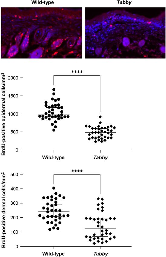
Kossel et al.                                                                                                                     Correction of Murine Bone Development

  FIGURE 3 | Possible role of EDA1 in the differentiation of osteoclasts. (A) Representative tartrate-resistant acid phosphatase (TRAP)-stained tail sections of wild-type
  and Tabby mice indicating similar number and distribution of osteoclasts in the metaphyseal trabecular part of vertebral bones. The boxes in the upper right corner
  show magnified osteoclasts (dark red). (B) Overview of the molecular link between the EDA1 pathway and the RANK–TRAF6–NFκB pathway and its possible
  involvement in osteoclastic differentiation. Scale bar: 100 µm.

RESULTS                                                                                  of vertebrae in the caudal spine of Tabby mice until birth.
                                                                                         Tail tips of Tabby mouse embryos at days E16.5 (not shown)
Embryonic and Postnatal Development                                                      and E18.5 (Figure 1B), were indistinguishable from those of
                                                                                         wild-type animals (Figure 1A). Deviations from the otherwise
of the Caudal Spine in Tabby Mice
                                                                                         linear tail axis were observed in 13 of 29 Tabby mice (45%)
As the kinky tail tip, combined with the absence of tail and guard
                                                                                         at day P4. Their frequency and extent increased during the
hair, is a characteristic feature of adult Tabby mice (Kowalczyk-                        next 3 weeks (Figure 1B). Kinks were present in the tails of
Quintas et al., 2014), we first determined the age range in which                        all Tabby mice by the age of 4 months (data not shown). The
the kink appears. Macroscopic assessment (Figures 1A,B, upper                            vertebral bodies at the kink position seemed to be shortened
panels) and whole-mount Alcian blue staining (Figures 1A,B,                              compared with the surrounding ones, yet until day P21 no
lower panels) of the tails did not indicate abnormal development                         obvious fracture was noticed.

Frontiers in Genetics | www.frontiersin.org                                          5                                           August 2021 | Volume 12 | Article 709736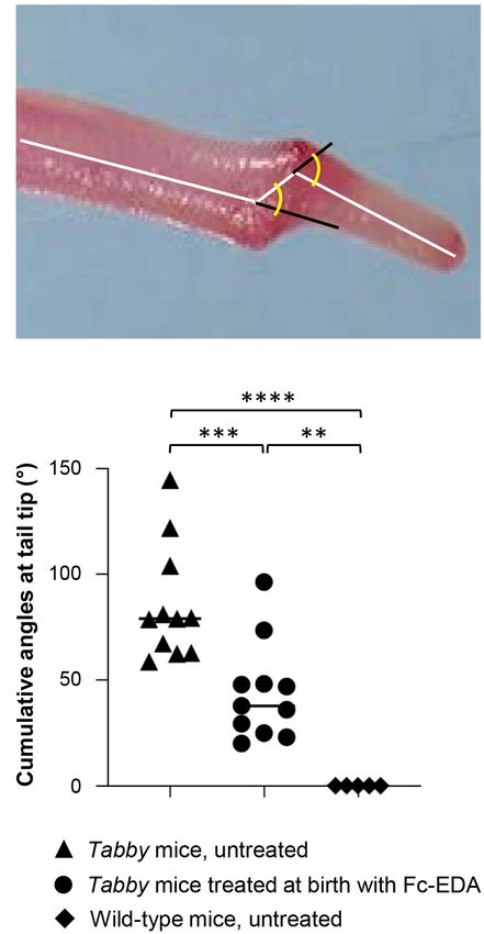
Kossel et al.                                                                                                                      Correction of Murine Bone Development

  FIGURE 4 | Diminished proliferation of tail skin in Tabby mice. (A) Immunofluorescence staining of tail tip cross-sections from wild-type (n = 6; left panel) and Tabby
  mice (n = 6; right panel) after intraperitoneal injection of bromodeoxyuridine (BrdU) at day P9 to label replicating cells. The amount of BrdU-positive cells in the skin
  (red; nuclei counter-stained blue with DAPI) was assessed. (B,C) Quantitation of replicating cells visualized by BrdU incorporation. BrdU-positive nuclei were counted
  in the epidermis and the upper dermis (depth ≤ 0.15 mm) except for the hair bulb region. Data from six independent skin sections of each Tabby and wild-type
  mouse are shown. Scale bar: 50 µm. ∗∗∗∗ p < 0.0001.

Cortical Bone Densities of Tabby Mice                                                    sometimes even two or more fractures in one vertebral body.
                                                                                         Usually the third to sixth vertebrae, counted from the tail tip,
and Osteoclastogenesis
                                                                                         were affected. These fractures which most likely occurred at some
Tails of adult Tabby and control mice (2–12 months old)
                                                                                         point between day P21 and the time of tomography could have
were investigated by micro-computed tomography (CT). Three-                              been due to structural abnormalities of the bone. The tibiae of
dimensional reconstructions of the caudal spine showed                                   wild-type and Tabby mice (Figure 2B) did not show macroscopic
fractured vertebral bodies in most of the Tabby mice (Figure 2A),                        differences. Histological examination of vertebral and tibial bone

Frontiers in Genetics | www.frontiersin.org                                          6                                            August 2021 | Volume 12 | Article 709736
Kossel et al. Correction of Murine Bone Development
0.290 ± 0.0274 mm3 , p = 0.004), while cortical bone density
appeared to be comparatively low in Tabby mice aged 2–
4 months and was significantly reduced in older Tabby mice
(Figure 2G). Tibial bones, in contrast, were found to have
a higher cortical density in Tabby than in wild-type animals
(Figure 2H), irrespective of their age. Thus, EDA1 deficiency had
an impact on bone density.
Microscopic assessment of TRAP-stained sections of wild-
type and Tabby mouse vertebrae indicated a similar amount of
osteoclasts in the metaphyseal trabecular bone (Figure 3A). We
focused on osteoclasts, because the EDA1 pathway is known to
interact with the RANKL/RANK signaling pathway (Figure 3B)
that plays an important role in osteoclastogenesis (Wada et al.,
2006). Trabecular bone histomorphometry using digital image
analysis software was performed: The number of osteoclasts
normalized to the trabecular bone perimeter in the metaphyseal
part of vertebrae was comparable between Tabby and wild-type
mice (3.41 ± 1.28 vs. 3.19 ± 0.82, respectively; five animals per
group investigated). Although trabecular separation appeared to
be larger in the vertebral bones of some Tabby mice, in agreement
with the higher volume of the medullary cavity revealed by micro-
CT measurements, neither trabecular number nor separation
differed significantly between Tabby and wild-type mice.
As the absence of significant differences in vertebral cortical
bone density between wild-type and Tabby mice at the time when
the kink appears did not support an important role of bone
density in the formation of the kink, an alternative hypothesis
needed to be investigated: the possibility that the tail skin does
not grow fast enough and the pressure on the vertebral bodies at
the tip of the tail bends them, until this is “fixed” by some kind of
sclerosis. Kinks would then form close to the tail tip, because the
pressure there will be higher and the vertebral bones smaller and
easier to deform.
Insufficient Growth of the Tail Skin in
Tabby Mice
FIGURE 5 | Attenuation of the tail kink in Tabby mice upon postnatal
In order to assess the skin growth at the tails of newborn animals,
treatment with EDA1. (A) Tabby mice were treated at birth with EDA1 we performed a BrdU incorporation experiment at day P9 and
(2 mg/kg body weight, intraperitoneal injection) and sacrificed at weaning. compared the numbers of BrdU-positive cells in tail skin sections
Pictures of the tails were taken for measurements. The amplitude of kink was of wild-type and Tabby mice (Figure 4). Our data clearly show
quantified by the cumulative angles at the tail tip using ImageJ. (B) Cumulative
a diminished keratinocyte proliferation in the epidermis of Tabby
angles (◦ ) of the tail tips of untreated Tabby mice (n = 11; black triangles),
Tabby mice treated at birth with EDA1 (n = 11; black circles) and untreated mice and an absence of hair follicles (Figure 4A), the main source
wild-type mice (n = 5; black squares). Data are shown as mean ± SD; of epidermal stem cells. The number of BrdU-positive cells in
∗∗ p = 0.001–0.01; ∗∗∗ p = 0.0001–0.001; ∗∗∗∗ p < 0.0001.
the epidermis of wild-type mice was more than two-fold higher
(p < 0.0001; Figure 4B). A similar difference was observed with
respect to BrdU-positive cells in the upper dermis of Tabby and
sections of the same animals revealed an apparent loss of the wild-type mice except for the hair bulb region (Figure 4C).
longitudinal orientation of vertebral bodies (Figure 2C). Their When Tabby mice were treated by intraperitoneal injection
epiphyseal growth plate was still recognizable, but smaller and of Fc-EDA (2 mg/kg body weight) within 24 h after birth, kink
less well organized with respect to the chondrocytes’ zonal formation–quantified by measuring cumulative angles at the tail
arrangement (Figures 2C,E) than in wild-type animals or in the tip (Figure 5A)–was diminished but not completely prevented
tibia of Tabby mice (Figures 2D,F) and contained numerous (Figure 5B), while delayed normal tail hair development
cartilage islands. occurred. As several days are probably required for the
Micro-CT measurements of 7–10 vertebral bodies indicated development of hair follicles after a triggering signal and as tail
a significantly higher volume of the medullary cavity in Tabby kinks had been observed as early as on day P4 in untreated Tabby
mice compared with wild-type animals (0.405 ± 0.028 vs. mice, these data fit well with the hypothesis that such kinks form
Frontiers in Genetics | www.frontiersin.org 7 August 2021 | Volume 12 | Article 709736Kossel et al. Correction of Murine Bone Development
FIGURE 6 | EDA1 replacement in utero corrects vertebral bone development in Tabby mice. (A) Representative tails of wild-type, untreated, and prenatally treated
Tabby mice. (B) Tail tip sections from the region of the kink stained with alcian blue (scale bar: 100 µm). (C) Vertebral bone density (Hounsfield units) of wild-type
mice (n = 6; black bar), untreated Tabby mice (n = 6; white bar) and Tabby mice treated in utero (n = 7; gray bar). All animals were 6–12 months old. Data are shown
as mean ± SD; ∗ p < 0.05; ∗∗ p = 0.001–0.01.
because a skin devoid of hair follicles does not grow fast enough (unpublished own observation) rather than by defective EDA1
to accommodate the incessantly growing caudal vertebral bodies signaling. In any case, osteoporosis does not explain the kinky
in the first weeks of life. tails regularly observed during the first weeks or months of life.
Tail hair development can, however, be rescued even at an On the other side, the results of our prenatal rescue
earlier stage by administration of Fc-EDA in utero (Hermes et al., experiment indicate that although reduced bone density is only
2014). Such prenatal treatment of Tabby mice on day E15 indeed seen in aged EDA1-deficient mice, it may already be pre-
resulted in a completely corrected phenotype (Figures 6A,B) with determined in utero and, if not, is at least counteracted by
a kink-free tail resembling that of wild-type animals. This is in something that can be induced by timely EDA1 replacement.
full agreement with the findings described above and confirms NF-κB, the transcription factor activated by the EDA1 signaling
our hypothesis that hair follicles are required for a sufficient cascade, is a major regulator of skeletal development and
growth of murine tail skin. Furthermore, and perhaps most osteoclastic differentiation, requiring activation of the RANKL–
interestingly, prenatal EDA1 replacement also led to a normal RANK–TRAF6 complex (Franzoso et al., 1997; Smahi et al.,
cortical bone density in the caudal vertebral bodies of Tabby mice 2002; Asagiri and Takayanagi, 2007). Impairment of this pathway
aged 6–12 months (Figure 6C). in Tabby mice might interfere with the EDA1–TRAF6–RANK
molecular interaction and, thus, with osteoclastic differentiation
(Clauss et al., 2008). The EDAR was found to be expressed
DISCUSSION in osteoclasts (unpublished own data). Osteoclasts in Tabby
and control mice did not differ in number, but their activity
The results of our study confirm previously reported observations might be different. NF-κB is known to play a critical role also
by another group (Hill et al., 2002) that EDA1 deficiency entails in the maturation of osteoclasts (Clauss et al., 2008). As TNF
bone defects in mice. In particular, adult Tabby mice display can synergize with minute amounts of RANKL to promote
fractures of vertebral bodies in the caudal spine. They seem to osteoclastogenesis (Lam et al., 2000), EDA1 might also be
develop osteoporotic vertebral bodies – characterized by reduced able to synergize with RANKL, which could be a subject of
cortical bone density – earlier than wild-type mice. This, however, further studies. In addition, future research may use relevant
may be caused by a reduced mobility of older Tabby mice mesenchymal precursor cells, such as bone marrow stromal cells,
Frontiers in Genetics | www.frontiersin.org 8 August 2021 | Volume 12 | Article 709736Kossel et al. Correction of Murine Bone Development
from Tabby mice to investigate their osteogenic differentiation understand a direct or indirect contribution of EDAR signaling
in vitro (Gelse et al., 2003), aiming at a careful characterization to bone development and which cell types are involved.
and evaluation of the activity of osteoblasts and osteoclasts.
Kinky tails have been observed in various mouse models
(Wright, 1947; Schrick et al., 1995; Farkas and Chapman, 2009) DATA AVAILABILITY STATEMENT
and may be related to a second, independent mutation in genes
involved in the development of the spine. The latter is highly The raw data supporting the conclusions of this article will be
unlikely in the case of Tabby mice, because the effect of an made available by the authors, without undue reservation.
independent mutation would probably not be correctable by
treatment with Fc-EDA and should not be sensitive to the time
of its administration. ETHICS STATEMENT
Previous work of our group demonstrating the generation of
Tabby-like mice by treatment of wild-type animals with anti- Animal experiments performed in this study were approved
EDA1 antibodies (Kowalczyk-Quintas et al., 2014) suggested either by Regierungspräsidium Karlsruhe and Regierung von
restricted skin growth in Tabby mice to be at least part of the Unterfranken (authorization 55.2-2532-2-222 to HS) or by the
explanation for the kinky tail. Also, in a related mouse model, local institutional Animal Care and Use Committee and by the
hair follicles were found to be required for optimal growth during Office Vétérinaire Cantonal du Canton de Vaud (authorization
lateral skin expansion and their absence was shown to lead to 1370.7 to PS).
shortening and kinking of the caudal spine (Heath et al., 2009).
The authors concluded that skin growth was unable to keep pace
with the rapidly elongating axial skeleton of the tail. We focused
AUTHOR CONTRIBUTIONS
on this hypothesis, because wild-type mice treated perinatally PS and HS conceived the experiments and supervised the work.
with the anti-EDA1 antibody EctoD2 got a kinky tail (Kowalczyk- C-SK, MW, SS-M, JP, CK-Q, and MS performed laboratory
Quintas et al., 2014), albeit late appearance of tail hair indicated investigations. KM curated data. C-SK and HS wrote the first
that the intermittent treatment was not inhibiting EDA1 all the draft of the manuscript. All authors critically reviewed the
time, so that hair follicles could develop with some delay. manuscript and approved its final version.
Early postnatal treatment of Tabby mice with Fc-EDA
attenuated but did not completely prevent the kink. This can also
be explained by the delayed rescue of hair follicles in the tail FUNDING
skin. Only prenatal administration of Fc-EDA may ensure full
correction of the skin’s capacity to grow fast enough. Induction This study was supported by grants from the Interdisciplinary
of tail hair at a later stage does not solve the problem at the initial Center for Clinical Research (IZKF) Erlangen (to C-SK),
step of kink formation, but thereafter allows the skin to grow the German-Swiss-Austrian ectodermal dysplasia patient
faster, leading to a smaller amplitude of the kink. organization (to HS), and by the Swiss National Science
Thus, our prenatal rescue experiment demonstrates clearly the Foundation (Grant 310030A_176256 to PS).
causal relationship between EDA1 deficiency and kinky tails. To
what extent the partially disturbed bone development evident in
aged Tabby mice is pre-determined in the embryo, pointing to ACKNOWLEDGMENTS
a role of EDA1 also in osteogenesis, needs to be investigated in
further studies. As the bone phenotype in Tabby mice appears to Most of the work was performed by C-SK in fulfillment of
be due to the lack of EDA1, not EDA2, it is most likely EDAR- the requirements for obtaining the degree “Dr. med.” from
dependent. The implication of this receptor could be assessed the Friedrich-Alexander-Universität Erlangen-Nürnberg. The
using conditional EDAR-knockout mice (Vial et al., 2019) crossed authors thank Ida Allabauer, Elisabeth Koppmann, and Vanessa
with bone-specific Cre deleter mice (Dallas et al., 2018) to better Endres for continuous technical support.
REFERENCES Clauss, F., Manière, M. C., Obry, F., Waltmann, E., Hadj-Rabia, S.,
Bodemer, C., et al. (2008). Dento-craniofacial phenotypes and underlying
Asagiri, M., and Takayanagi, H. (2007). The molecular understanding of molecular mechanisms in hypohidrotic ectodermal dysplasia (HED):
osteoclast differentiation. Bone 40, 251–264. doi: 10.1016/j.bone.2006.0 a review. J. Dent. Res. 87, 1089–1099. doi: 10.1177/15440591080870
9.023 1205
Blüschke, G., Nüsken, K.-D., and Schneider, H. (2010). Prevalence and prevention Cluzeau, C., Hadj-Rabia, S., Jambou, M., Mansour, S., Guigue, P., Masmoudi,
of severe complications of hypohidrotic ectodermal dysplasia in infancy. Early S., et al. (2011). Only four genes (EDA1, EDAR, EDARADD, and WNT10A)
Hum. Dev. 86, 397–399. doi: 10.1016/j.earlhumdev.2010.04.008 account for 90% of hypohidrotic/anhidrotic ectodermal dysplasia cases. Hum.
Casal, M. L., Lewis, J. R., Mauldin, E. A., Tardivel, A., Ingold, K., Favre, M., Mutat. 32, 70–72. doi: 10.1002/humu.21384
et al. (2007). Significant correction of disease after postnatal administration of Dallas, S. L., Xie, Y., Shiflett, L. A., and Ueki, Y. (2018). Mouse Cre models for the
recombinant ectodysplasin A in canine X-linked ectodermal dysplasia. Am. J. study of bone diseases. Curr. Osteoporos. Rep. 16, 466–477. doi: 10.1007/s11914-
Hum. Genet. 81, 1050–1056. doi: 10.1086/521988 018-0455-7
Frontiers in Genetics | www.frontiersin.org 9 August 2021 | Volume 12 | Article 709736Kossel et al. Correction of Murine Bone Development Farkas, D. R., and Chapman, D. L. (2009). Kinked tail mutation results in Schrick, J. J., Dickinson, M. E., Hogan, B. L. M., Selby, P. B., and Woychik, R. P. notochord defects in heterozygotes and distal visceral endoderm defects in (1995). Molecular and phenotypic characterization of a new mouse insertional homozygotes. Dev. Dyn. 238, 3237–3247. doi: 10.1002/dvdy.22141 mutation that causes a defect in the distal vertebrae of the spine. Genetics 140, Ferguson, B. M., Brockdorff, N., Formstone, E., Nguyen, T., Kronmiller, J. E., and 1061–1067. doi: 10.1093/genetics/140.3.1061 Zonana, J. (1997). Cloning of Tabby, the murine homolog of the human EDA Schuepbach-Mallepell, S., Kowalczyk-Quintas, C., Dick, A., Eslami, M., Vigolo, M., gene: evidence for a membrane-associated protein with a short collagenous Headon, D. J., et al. (2020). Method for the administration of EDAR pathway domain. Hum. Mol. Genet. 6, 1589–1594. doi: 10.1093/hmg/6.9.1589 modulators in mice. Methods Mol. Biol. 2248, 167–183. doi: 10.1007/978-1- Franzoso, G., Carlson, L., Xing, L., Poljac, L., Shores, E., Brown, K., et al. (1997). 0716-1130-2_12 Requirement for NF-κB in osteoclast and B-cell development. Genes Dev. 11, Smahi, A., Courtois, G., Hadj-Rabia, S., Döffinger, R., Bodemer, C., Munnich, 3482–3496. doi: 10.1101/gad.11.24.3482 A., et al. (2002). The NF-kappaB signalling pathway in human diseases: Gaide, O., and Schneider, P. (2003). Permanent correction of an inherited from incontinentia pigmenti to ectodermal dysplasias and immune-deficiency ectodermal dysplasia with recombinant EDA. Nat. Med. 9, 614–618. doi: 10. syndromes. Hum. Mol. Genet. 11, 2371–2375. doi: 10.1093/hmg/11.20. 1038/nm861 2371 Gelse, K., von der Mark, K., and Schneider, H. (2003). Cartilage regeneration by Srivastava, A. K., Pispa, J., Hartung, A. J., Du, Y., Ezer, S., Jenks, T., et al. (1997). gene therapy. Curr. Gene Ther. 3, 305–317. doi: 10.2174/1566523034578276 The Tabby phenotype is caused by mutation in a mouse homologue of the Heath, J., Langton, A. K., Hammond, N. L., Overbeek, P. A., Dixon, M. J., and EDA gene that reveals novel mouse and human exons and encodes a protein Headon, D. J. (2009). Hair follicles are required for optimal growth during (ectodysplasin-A) with collagenous domains. Proc. Natl. Acad. Sci. U.S.A. 94, lateral skin expansion. J. Invest. Dermatol. 129, 2358–2364. doi: 10.1038/jid. 13069–13074. doi: 10.1073/pnas.94.24.13069 2009.102 Trzeciak, W. H., and Koczorowski, R. (2016). Molecular basis of hypohidrotic Hermes, K., Schneider, P., Krieg, P., Dang, A., Huttner, K., and Schneider, ectodermal dysplasia: an update. J. Appl. Genet. 57, 51–61. doi: 10.1007/s13353- H. (2014). Prenatal therapy in developmental disorders: drug targeting via 015-0307-4 intra-amniotic injection to treat X-linked hypohidrotic ectodermal dysplasia. Vial, J., Royet, A., Cassier, P., Tortereau, A., Dinvaut, S., Maillet, D., et al. (2019). J. Invest. Dermatol. 134, 2985–2987. doi: 10.1038/jid.2014.264 The Ectodysplasin receptor EDAR acts as a tumor suppressor in melanoma by Hill, N. L., Laib, A., and Duncan, M. K. (2002). Mutation of the ectodysplasin- conditionally inducing cell death. Cell Death Differ. 26, 443–454. doi: 10.1038/ a gene results in bone defects in mice. J. Comp. Pathol. 126, 220–225. doi: s41418-018-0128-1 10.1053/jcpa.2001.0531 Wada, T., Nakashima, T., Hiroshi, N., and Penninger, J. M. (2006). RANKL–RANK Körber, I., Klein, O., Morhart, P., Faschingbauer, F., Grange, D., Clarke, A., et al. signaling in osteoclastogenesis and bone disease. Trends Mol. Med. 12, 17–25. (2020). Safety and immunogenicity of Fc-EDA, a recombinant ectodysplasin A1 doi: 10.1016/j.molmed.2005.11.007 replacement protein, in human subjects. Br. J. Clin. Pharmacol. 86, 2063–2069. Wohlfart, S., Hammersen, J., and Schneider, H. (2016). Mutational spectrum in doi: 10.1111/bcp.14301 101 patients with hypohidrotic ectodermal dysplasia and breakpoint mapping Kowalczyk-Quintas, C., and Schneider, P. (2014). Ectodysplasin a (EDA)–EDA in four independent cases of rare genomic rearrangements. J. Hum. Genet. 61, receptor signalling and its pharmacological modulation. Cytokine Growth 891–897. doi: 10.1038/jhg.2016.75 Factor Rev. 25, 195–203. doi: 10.1016/j.cytogfr.2014.01.004 Wohlfart, S., Meiller, R., Hammersen, J., Park, J., Menzel-Severing, J., Melichar, V., Kowalczyk-Quintas, C., Willen, L., Dang, A., Sarrasin, H., Tardivel, A., Hermes, et al. (2020). Natural history of X-linked hypohidrotic ectodermal dysplasia: a K., et al. (2014). Generation and characterization of function-blocking 5-year follow-up study. Orphanet J. Rare Dis. 15:7. doi: 10.1186/s13023-019- anti-ectodysplasin A (EDA) monoclonal antibodies that induce ectodermal 1288-x dysplasia. J. Biol. Chem. 289, 4273–4285. doi: 10.1074/jbc.m113.535740 Wright, M. E. (1947). Undulated: a new genetic factor in Mus musculus affecting Lam, J., Takeshita, S., Barker, J. E., Kanagawa, O., Ross, F. P., and Teitelbaum, the spine and tail. Heredity 1, 137–141. doi: 10.1038/hdy.1947.10 S. L. (2000). TNF-alpha induces osteoclastogenesis by direct stimulation of Zhang, Q., Lenardo, M. J., and Baltimore, D. (2017). 30 Years of NF-kappaB: a macrophages exposed to permissive levels of RANK ligand. J. Clin. Invest. 106, blossoming of relevance to human pathobiology. Cell 168, 37–57. doi: 10.1016/ 1481–1488. doi: 10.1172/JCI11176 j.cell.2016.12.012 Mikkola, M. L., and Thesleff, I. (2003). Ectodysplasin signaling in development. Cytokine Growth Factor Rev. 14, 211–224. doi: 10.1016/s1359-6101(03)00020-0 Conflict of Interest: PS, CK-Q, and HS are inventors on patents relevant to this Monreal, A. W., Zonana, J., and Ferguson, B. (1998). Identification of a new splice publication. form of the EDA1 gene permits detection of nearly all X-linked hypohidrotic ectodermal dysplasia mutations. Am. J. Hum. Genet. 63, 380–389. doi: 10.1086/ The remaining authors declare that the research was conducted in the absence of 301984 any commercial or financial relationships that could be construed as a potential Montonen, O., Ezer, S., Saarialho-Kere, U. K., Herva, R., Karjalainen- conflict of interest. Lindsberg, M. L., Kaitila, I., et al. (1998). The gene defective in anhidrotic ectodermal dysplasia is expressed in the developing epithelium, neuroectoderm, Publisher’s Note: All claims expressed in this article are solely those of the authors thymus, and bone. J. Histochem. Cytochem. 46, 281–289. doi: 10.1177/ and do not necessarily represent those of their affiliated organizations, or those of 002215549804600301 the publisher, the editors and the reviewers. Any product that may be evaluated in Pantalacci, S., Chaumot, A., Benoît, G., Sadier, A., Delsuc, F., Douzery, E. J. P., this article, or claim that may be made by its manufacturer, is not guaranteed or et al. (2008). Conserved features and evolutionary shifts of the EDA signaling endorsed by the publisher. pathway involved in vertebrate skin appendage development. Mol. Biol. Evol. 25, 912–928. doi: 10.1093/molbev/msn038 Copyright © 2021 Kossel, Wahlbuhl, Schuepbach-Mallepell, Park, Kowalczyk- Schneider, H., Faschingbauer, F., Schuepbach-Mallepell, S., Körber, I., Wohlfart, S., Quintas, Seeling, von der Mark, Schneider and Schneider. This is an open-access Dick, A., et al. (2018). Prenatal correction of X-linked hypohidrotic ectodermal article distributed under the terms of the Creative Commons Attribution License dysplasia. N. Engl. J. Med. 378, 1604–1610. doi: 10.1056/NEJMoa1714322 (CC BY). The use, distribution or reproduction in other forums is permitted, provided Schneider, H., Hammersen, J., Preisler-Adams, S., Huttner, K., Rascher, W., and the original author(s) and the copyright owner(s) are credited and that the original Bohring, A. (2011). Sweating ability and genotype in individuals with X-linked publication in this journal is cited, in accordance with accepted academic practice. hypohidrotic ectodermal dysplasia. J. Med. Genet. 48, 426–432. doi: 10.1136/ No use, distribution or reproduction is permitted which does not comply with jmg.2010.084012 these terms. Frontiers in Genetics | www.frontiersin.org 10 August 2021 | Volume 12 | Article 709736
You can also read