Conjoined Twins-Cephalo-Thoraco-Omphalopagus: A Case Report - Bahrain Medical Bulletin
←
→
Page content transcription
If your browser does not render page correctly, please read the page content below
Bahrain Medical Bulletin, Vol. 43, No. 2, June 2021

        Conjoined Twins- Cephalo-Thoraco-Omphalopagus: A Case Report
    Basma Alansari, MB, BCh, BAO* Wassan Al ani, M.B.ch.B, Arab Board, RCPI Membership** Maha Ghorabah, FRCOG***

              ABSTRACT
              A thirty-one-years old woman with spontaneous conjoined twin pregnancy, Gravida 3 Para 2 with history of
              previous 2 cesarean sections was referred to the obstetrics and gynecology clinic in King Hamad University
              Hospital at 18 weeks of gestation for further management. She was diagnosed as cephalo-thoraco-omphalopagus
              female conjoined twins in private clinic, which was confirmed in our institute. Thus, surgical termination by
              hysterotomy was performed.

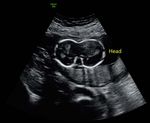
INTRODUCTION                                                                adjacent to each other. Two brains were fused along the temporal
                                                                            region, which appeared grossly normal (Figure 1).
Conjoined twins (CT) also known as Siamese twins are defined as
monochorionic monoamniotic twins that are anatomically fused
together in utero1. It is an extraordinary phenomenon of uncertain
etiology that occurs due to an abnormality during embryological
development. The incidence rate varies between 1 in 50,000 to 1 in
100,000 births2 and it has a 1:3 male to female ratio with female fetuses
being most commonly affected3. Several types of conjoined twins were
described in the literature with thoracopagus being the most common
type and omphalopagus the least common4.

THE CASE
A thirty-one-years old Egyptian woman with spontaneous twin
pregnancy, Gravida 3 Para 2 with history of previous 2 cesarean
sections was referred to the obstetrics and gynecology clinic in King
Hamad University Hospital as a tertiary center at 18 weeks of gestation
seeking for a second opinion regarding an abnormal antenatal scan
report from a private clinic. The patient did not have any family           Figure 1: Trans-abdominal scan showing conjoined twin with two
history of twins and no history of consanguinity. No medical history        brains fused along the temporal region
or allergies were reported.
                                                                            Only one stomach and heart were seen. Ventricular septal defect was
The ultrasound that was performed at the private clinic at 17+weeks         noted however the septum primum was not visualized, as it would be
gestation revealed a diagnosis of conjoined viable female twins             in a complete atrio-ventriculum canal defect. (Figure 2) Spine of fetus
(cephalo-thoraco-omphalopagus) with 2 faces fused laterally and four        B appears scoliotic (Figure 3); Spine of fetus A appears grossly normal.
eyes with two central globes lying adjacent to each other. Two brains
were fused along the temporal region, which appeared grossly normal.

A single fused chest was noted along with fused abdomens. A single
heart lying along the left side of the thoracic region and a possible
atrioventricular canal defect were noted.

 The lung volumes appeared reduced with a single stomach seen at the
level of the heart. A single liver was seen, and the kidneys were not
clearly visualized however 2 urinary bladders were present. Extremities
appeared grossly intact with four arms and legs. Amniotic fluid seemed
normal, and the placenta was located in the upper posterior uterine
segment. The cervix appeared long and closed on transabdominal scan.

An ultrasound scan performed in our hospital confirmed a diagnosis
of conjoined viable female twins (cephalo-thoraco-omphalopagus)
with 2 faces fused laterally and four eyes with two central globes lying    Figure 2: Trans-abdominal scan showing ventricular septal defect

*   Senior House officer
    Department of Obstetrics and Gynecology
    King Hamad University Hospital,
    Bahrain, E-mail: basma.alansari@khuh.org.bh
** Registrar
*** Consultant & Training program director
    Jordanian Board, FRCOG, Postgraduate Diploma in Health Professions Education at Royal College of Surgeons in Ireland

531
Bahrain Medical Bulletin, Vol. 43, No. 2, June 2021
occur resulting in cell signaling and finally conjoined anomalies6. Most
of the authors accepted the fusion theory as it can explain all types of
conjoined twins7.
The cause of conjoined twins is still not fully understood. There are
some risk factors that were thought to have a possible effect on the
development of this phenomenon such as a positive genetic history of
twins, delivery abnormality, ovulation inducing medications, fertility
treatment and radiation exposure. In our case we could not identify any
obvious risk factors that may have caused this abnormality8.
The prognosis of this condition is poor. Fourteen cases were included in
a study of prenatally diagnosed conjoined twins. The study found that
28% of the cases died antenatally and 54% died immediately following
delivery and18% survived; of which 50% died postoperatively 3,9,10.
There are different classification systems of conjoined twins. (Spencer
et al 1996) 4 classified them into 8 main types based on the degree and
site of fusion (Figure 5). Thoracophagus, omphalophagus and thoraco-
Figure 3: Spine of fetus B appears scoliotic omphalophagus are the most common types in this classification system
accounting for about 56% of conjoined twins. The rarest type according
Both the patient and her husband were counseled in detail about to Spencer is Cephalophagus accounting for 11% of all cases. Fusion
the results of the ultrasound report and were advised for surgical of the head, thorax and upper abdominal cavities are characteristics
termination, as both fetuses are incompatible with life. The couple of Cephalophagus twins. Cephalopagus twins are further divided into
agreed so consent was taken from the patient and signed by 3 two types: Janiceps (two faces are on either side of the head) or non-
consultants and the pediatrics team was informed about the case. Janiceps (with a relatively normal head and face)11. To the best of our
knowledge, the conjoined twins reported in our case are one of the
Following the discussion, the patient was booked for admission and rarest types of cephalophagus in which both heads are fused laterally
hyesterotomy was performed at 18 weeks of gestation without any (Janiceps type).
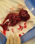
complications to the mother. The twins exhibited no signs of life
at the time of delivery. Gross examination of the conjoined twins
confirmed that both heads were fused laterally along with thorax and
upper abdomen while the four upper limbs and 4 lower limbs appeared
normal. Both twins were females weighting a total of 345 grams
(Figure 4). Post-operative recovery was uneventful, and the mother
was discharged in stable condition.
Figure 5: Classification of conjoined twins
Early diagnosis of conjoined twins with trans-abdominal or trans-
vaginal ultrasonography has a vital role in the management so early
termination of pregnancy can be done however usually it cannot be
detected before 10 weeks of gestation12.
Once conjoined twins are confirmed three-dimensional ultrasound,
Figure 4: Conjoined twins post hysterotomy computed tomography, or magnetic resonance imaging are used to
identify the type and the severity of the conjoined twins regarding
anatomical anomalies13. This emphasized the important role of the
DISCUSSION
radiologist and obstetricians in early detection of conjoined twins to
Conjoint twins are thought to be the result of a faulty division of an avoid any problems in the later half of pregnancy and to determine the
embryo at 13-15 days of conception5. There are various theories that mode of delivery3.
were proposed to explain this phenomenon. The most common theories
behind the basis of development of conjoined twins are the “fission
Since most conjoined twins are diagnosed antenatally, they are
theory “and “fusion theory”5. The former suggests that an incomplete
split of a single fertilized egg occurs, and the forming 2 embryos delivered electively by cesarean section, as it is the safest option for
remain fused at the unseparated parts. Whereas the fusion theory both mother and babies and to avoid complications such as stillbirth,
suggests that there is complete separation of the fertilized egg but as uterine rupture, labour dystocia, shoulder dystocia, retained second
they are close in proximity, an interaction between both twin cells may twin and hysterectomy14-16.
532Conjoined Twins- Cephalo-Thoraco-Omphalopagus: A Case Report
In cases where the gestational period is between 18-20 weeks, the 2. Mutchinick OM, Luna Muñoz L, Amar E, et al. Conjoined twins: a
pregnancy can be terminated medically and delivered vaginally using worldwide collaborative epidemiological study of the International
labor-inducting medication17. As there is an increased risk of uterine Clearinghouse for Birth Defects Surveillance and Research. Am J
rupture and shoulder dystocia with vaginal delivery, especially with Med Genet C Semin Med Genet 2011; 157C (4):274-87.
a history of multiple previous cesarean sections like in our case, we 3. Graham G, Gaddipati S. Diagnosis and Management of Obstetrical
opted for surgical intervention after full counseling of the patient Complications Unique to Multiple Gestations. Semin Perinatol
about the risks and benefits of each mode of delivery. The patient was 2005;29(5):282-95.
booked for an elective hysterotomy at 18 weeks of gestation without 4. Spencer R. Anatomic description of conjoined twins: A plea for
any complications to the mother and was discharged in good condition. standardized terminology. J Pediatr Surg 1996;31(7):941-4.
5. Sabih D, Ahmad E, Sabih A, et al. Ultrasound diagnosis of
cephalopagus conjoined twin pregnancy at 29 weeks. Biomed
CONCLUSION Imaging Interv J 2010;6(4): e38.
6. DeRuiter, Corinne, "Conjoined Twins". Embryo Project
To conclude, antenatal care, early ultrasound scan and multi- Encyclopedia, 2011.
disciplinary team management plays a vital role in the early 7. Spencer R. Theoretical and analytical embryology of conjoined
detection of conjoined twins in order to avoid any complications that twins: Part I: Embryogenesis. Clin Anat 2000;13(1):36-53.
result from undiagnosed conjoined twins at a later gestation and to 8. Kamalian N, Shirani S, Soleymanzadeh M. Thoraco- Omphalo-
involve multi- disciplinary team earlier on in the management plan Ischiopagus Tripus Conjoined Twins: Report of a Case. J Forensic
to give optimal care to both the patient and babies. Res 2011;2(1): 1000117.
________________________________________________________ 9. Stone J, Goodrich J. The craniopagus malformation: classification
and implications for surgical separation. Brain 2006;129(5):1084-95.
Authorship Contribution: All authors share equal effort contribution 10. Hill L. The sonographic detection of early first-trimester conjoined
towards (1) substantial contributions to conception and design, twins. Prenat Diagn 1997;17(10):961-3.
acquisition, analysis and interpretation of data; (2) drafting the article 11. Chen c, Lee c, Liu f, et al. Prenatal diagnosis of cephalothoracopagus
and revising it critically for important intellectual content; and (3) final janiceps monosymmetros. Prenat Diagn 1997;17(4):384-8.
approval of the manuscript version to be published. Yes.
12. Hubinont C, Kollmann P, Malvaux V, et al. First-Trimester
Diagnosis of Conjoined Twins. Fetal Diagn Ther 1997;12(3):185-7.
13. Kuroda K, Kamei Y, Kozuma S, et al. Prenatal evaluation of
Potential Conflict of Interest: None.
cephalopagus conjoined twins by means of three-dimensional
ultrasound at 13 weeks of pregnancy. Ultrasound Obstet Gynecol
Competing Interest: None.
2000;16(3):264-6.
14. RCOG 2016- Monochorionic Twin Pregnancy, Management
Sponsorship: None. (Green-top Guideline No. 51)
15. Woldeyes SW. Delivery of Retained Second Twin in Case of
Acceptance Date: 19 March 2021 Omphalopagus Conjoined Twins: Abdominovaginal Approach.
Case Rep Obstet Gynecol 2018; 9319721.
Ethical Approval: Approved by the Research and Ethics committee, 16. Leigh MB, John-Cole V, Kamara M, et al. A Triple Obstetric
King Hamad University Hospital, Bahrain. Challenge of Thoracopagus-Type Conjoined Twins, Eclampsia,
and Obstructed Labor: A Case Report from Sub-Saharan Africa.
REFERENCES Case Rep Obstet Gynecol 2017; 6815748.
17. Yılmaz-Semerci S, Güzelbey T, Kurnaz D, et al. A rare case of
1. Mathew RP, Francis S, Basti RS, et al. Conjoined twins–role of cephalothoracopagus janiceps conjoined twins. Turk J Pediatr
imaging and recent advances. J Ultrason 2017; 71:259-66. 2018;60(6):751-4.
533You can also read


