Concurrent Infection With the Filarial Helminth Litomosoides sigmodontis Attenuates or Worsens Influenza A Virus Pathogenesis in a Stage-Dependent ...
←
→
Page content transcription
If your browser does not render page correctly, please read the page content below
ORIGINAL RESEARCH
                                                                                                                                                published: 24 January 2022
                                                                                                                                          doi: 10.3389/fimmu.2021.819560

                                           Concurrent Infection With the Filarial
                                           Helminth Litomosoides sigmodontis
                                           Attenuates or Worsens Influenza
                                           A Virus Pathogenesis in a
                                           Stage-Dependent Manner
                                           Gareth R. Hardisty 1, Johanna A. Knipper 2, Alison Fulton 2, John Hopkins 1,
                                           Bernadette M. Dutia 1 and Matthew D. Taylor 2*
                                           1 The Roslin Institute, University of Edinburgh, Edinburgh, United Kingdom, 2 Institute of Immunology and Infection Research,

                         Edited by:        Ashworth Laboratories, University of Edinburgh, Edinburgh, United Kingdom
                       Manuel Ritter,
  University Hospital Bonn, Germany
                                           Filarial helminths infect approximately 120 million people worldwide initiating a type 2
                       Reviewed by:
                        Lubna Pinky,       immune response in the host. Influenza A viruses stimulate a virulent type 1 pro-
      University of Tennessee Health       inflammatory immune response that in some individuals can cause uncontrolled
           Science Center (UTHSC),
                        United States
                                           immunopathology and fatality. Although coinfection with filariasis and influenza is a
                       Minka Breloer,      common occurrence, the impact of filarial infection on respiratory viral infection is
 Bernhard Nocht Institute for Tropical
                                           unknown. The aim of this study was to determine the impact of pre-existing filarial
        Medicine (BNITM), Germany
                                           infection on concurrent infection with influenza A virus. A murine model of co-infection
                 *Correspondence:
                  Matthew D. Taylor        was established using the filarial helminth Litomosoides sigmodontis and the H1N1 (A/
            Matthew.Taylor@ed.ac.uk        WSN/33) influenza A virus (IAV). Co-infection was performed at 3 different stages of
                                           L. sigmodontis infection (larval, juvenile adult, and patency), and the impact of co-
                   Specialty section:
         This article was submitted to     infection was determined by IAV induced weight loss and clinical signs, quantification of
                 Parasite Immunology,      viral titres, and helminth counts. Significant alterations of IAV pathogenesis, dependent upon
               a section of the journal
              Frontiers in Immunology
                                           stage of infection, was observed on co-infection with L. sigmodontis. Larval stage
      Received: 21 November 2021
                                           L. sigmodontis infection alleviated clinical signs of IAV co-infection, whilst more
      Accepted: 29 December 2021           established juvenile adult infection also significantly delayed weight loss. Viral titres
       Published: 24 January 2022          remained unaltered at either infection stage. In contrast, patent L. sigmdodontis infection
                               Citation:   led to a reversal of age-related resistance to IAV infection, significantly increasing weight loss
   Hardisty GR, Knipper JA, Fulton A,
 Hopkins J, Dutia BM and Taylor MD         and clinical signs of infection as well as increasing IAV titre. These data demonstrate that the
 (2022) Concurrent Infection With the      progression of influenza infection can be ameliorated or worsened by pre-existing filarial
         Filarial Helminth Litomosoides
  sigmodontis Attenuates or Worsens
                                           infection, with the outcome dependent upon the stage of filarial infection.
   Influenza A Virus Pathogenesis in a
                                           Keywords: helminth, coinfection, mouse, respiratory virus, filariasis, influenza A virus
             Stage-Dependent Manner.
           Front. Immunol. 12:819560.
    doi: 10.3389/fimmu.2021.819560         Abbreviations: IAV, Influenza A virus; Mf, microfilaria; L3, third larval stage; d, day; pi, post infection; L4, fourth larval stage.

Frontiers in Immunology | www.frontiersin.org                                        1                                         January 2022 | Volume 12 | Article 819560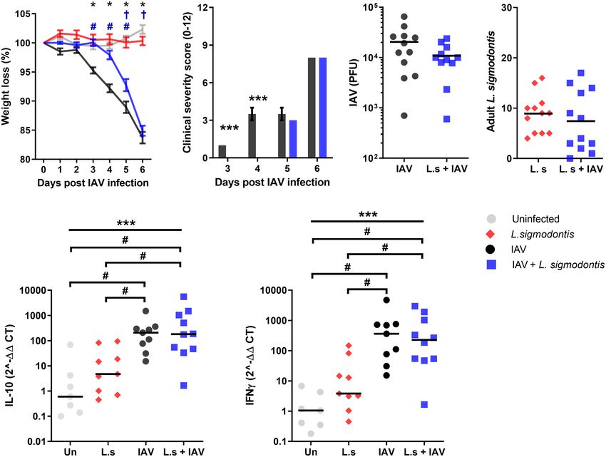
Hardisty et al.                                                                                                  Influenza and Filariasis Coinfection

INTRODUCTION                                                             MATERIALS AND METHODS
Filarial helminths infect approximately 120 million people               Ethics Statement
worldwide, and remain commonplace in many low and middle                 Experiments were in undertaken accordance with the United
income nations despite the existence of effective treatments and         Kingdom Animals (Scientific Procedures) Act of 1986 (PPL 60/
detection methods (1). The filarial helminths Wuchereria                  4479), and approved by the University of Edinburgh Animal
bancrofti, Brugia malayi and Brugia timori are referred to as            Welfare and Ethical Review Body.
lymphatic filariasis and are a significant global health concern
(1). It is common for humans to be infected with multiple                Animals and Infections
microbes at any given time, including commensal organisms                Female BALB/c mice were purchased from Charles River and
and chronic or persistent infections. The incidence of concurrent        maintained under specific pathogen free conditions at the
infection with filarial helminths is highly prevalent (2), and            University of Edinburgh. Mice were used at 6–8 weeks of age. To
therefore it is important to understand their impact on                  maintain the L. sigmodontis lifecycle, L. sigmodontis infected jirds
other infections.                                                        (Meriones unguiculatus) were used to infect haematophagous tick
    Filarial helminths predominantly stimulate Type 2 immune             parasites (Ornithonyssus bacoti) in order to generate L3 stage larvae
responses in their host, although mixed Type 1 and 2 responses           (23). Mice were infected s.c. on the upper back with 30 L.
can develop (3). As with other helminth infections, filarial              sigmodontis L3 stage larvae. L. sigmodontis larvae or adults were
parasites secrete immunosuppressive molecules that impair                recovered from the pleural cavity by lavage and counted using a
host immunity in order to maintain a persistent infection (4),           dissection microscope. To quantify blood Mf, 30 mL of tail blood
and combined with this, the host downregulates its immune                was collected in FACS lysing solution (Becton-Dickinson) and the
responses during chronic infection to avoid severe disease (5).          Mf counted using a haemocytometer. IAV infections were
Thus, hosts tend to develop regulatory and modified Type 2                performed intranasally with 5x10³ PFU A/WSN/33, a mouse
response during chronicity (6, 7). This development of                   H1N1 influenza A strain (Dr D. Jackson, University of St
regulatory and Type 2 immunity during chronic filariasis and              Andrews, St Andrews, UK), either 12, 34 or 68 days (d) post L.
other helminth infections can influence systemic immunity,                sigmodontis or mock infection. A/WSN/33 was grown in MDCK
including immune responses to third-party antigens such as               cells as described previously (24). Mice were weighed daily and
allergens and concurrent infections (8, 9).                              assessed for visual signs of clinical disease as described previously
    Litomosoides sigmodontis infection of inbred mice is used as a       (25, 26). Briefly, signs of infection were scored as follows, reduced
model of human lymphatic filariasis (10), and provides the                mobility/activity (0-3), ruffled fur/piloerection (0-3), hunched
opportunity to test the impact of filariasis on coinfection. L.           posture (0-3) and increased or laboured breathing (0-3). The
sigmodontis infection initially stimulates a Type 2 immune               severity score presented is a sum of these criteria.
response during the larval and juvenile adult stages. However,
similar to human infections, it develops a mixed Type 1 and 2            Influenza Viral Plaque Assay
response as the adult parasites become fully mature and release          MDCK cells were grown to confluence in 6 well plates (Corning) in
the transmission stage microfilaria (Mf) into the blood stream, at        DMEM (Gibco) containing 5% heat inactivated foetal calf serum
which point the infection is referred to as patent. L. sigmodontis       (Gibco), 1% Penicillin and Streptomycin (Gibco) and 1% L-
coinfection has been shown to both protect against and worsen            glutamine (Gibco). The left lobe of the lungs was recovered and
malaria (11–14), protect against Leishmania major (15), increase         mechanically homogenised with a TH homogeniser (OMNI) in 1.5
the severity of LPS-mediated inflammation (16), but has no                ml serum free DMEM before supernatants were recovered following
apparent effect on Mycobacterium tuberculosis infection (17).            centrifugation at 3000 rpm for 5 mins at 4°C. 10-fold serial dilutions
    Influenza A virus (IAV) infections cause virulent pro-                of supernatants were placed onto MDCK cells in duplicate for 1
inflammatory immune responses (18) hallmarked by ‘type I’                 hour at 37°C. After removal and washing with DPBS (Gibco), a
immunity, interferon production and generation of pro-                   layer of 1% agarose containing 1 x EMEM (Invitrogen), 7.5%
inflammatory cytokines such as IL-6, TNF-a and IL-1b. These,              fraction V BSA (Sigma), 1% L-glutamine, 7.5 NaHCO 2
can lead to extensive airway pathology within days of infection,         (Invitrogen) 1M Hepes (Sigma) 1% Dextran (Sigma) 1%
increasing the severity of disease and incidence of mortality (19–       Penicillin and Streptomycin and 2mg/ml N-acetyl trypsin from
21). Whilst L. sigmodontis can suppress vaccine-induced                  bovine pancreas type V-S (Sigma) was added, plates were inverted
antibody responses to IAV (22), the impact of filarial co-                and cultured for 3 days at 37°C, 5% CO2. Cells were fixed in 10%
infection on viral infections, including acute IAV infection, is         neutral buffered formalin (Sigma) and stained with 0.1% toluidine
unknown. We therefore tested whether infection with L.                   blue O (Sigma) for 20 minutes before plaques were quantified.
sigmodontis, which resides in the pleural cavity of mice, could
affect an acute respiratory challenge with IAV. In particular, we        IFN-g and IL-10 qPCR
tested the hypothesis that the immune regulatory pathways                A segment of the left lung lobe was homogenised in Trizol reagent
associated with L. sigmodontis infection would protect against           (Thermo) in a tissue homogeniser (Precellys) with ceramic beads.
IAV-induced pathology, and that the strongest protection would           RNA was then isolated with phenol/chloroform extraction
be seen during patent L. sigmodontis infection.                          according to manufacturer’s instructions and quantified with a

Frontiers in Immunology | www.frontiersin.org                        2                                  January 2022 | Volume 12 | Article 819560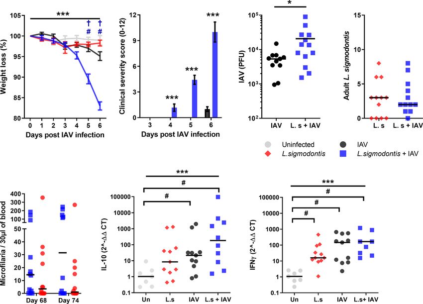
Hardisty et al. Influenza and Filariasis Coinfection
NanoDrop (Thermo). cDNA first strand synthesis was performed Thirty L3 stage L sigmodontis larvae were given by subcutaneous
with MultiScribe Reverse Transcriptase (Thermo) in a c1000 touch, injection into the back of 8-week-old female BALB/c mice to
thermal cycler (Bio-Rad). Murine 18s, IFN-g and IL-10 were mimic the physiological route of infection. L. sigmodontis L3
detected with SsoAdvanced Universal SYBR Green Supermix larvae migrate to the pleural cavity via the lymphatics over the
(Bio-Rad) on a StepOnePlus Real-Time PCR System (Thermo). first 3-4 days of infection. They moult to the fourth larval (L4)
Results are shown as 2^-(DDCT) values. stage between 8 -12 (d) post infection (pi) (27), and then to the
adult stage between 25 – 30 d pi. Mice were challenged with 3x10⁶
Statistical Tests PFU influenza A infection intranasally, or mock infected, on d 12
Weight loss data were analysed by General Linear Model and of L. sigmodontis infection so that the 6-day course of influenza
Tukey’s method for pairwise comparisons in Minitab 20 infection would take place during the L4 stage (Figure 1A), which
(Minitab LLC). Clinical severity rank scoring data was is associated with a low-level Type 2 immune response (28, 29).
analysed by Mann-Whitney non-parametric analysis in Prism Weight loss was monitored over the 6-d course of co-
9 (Graphpad). Influenza viral titre was analysed by parametric, infection as a clinical sign of IAV severity. This was not
two tailed, unpaired T test in Prism 9. QPCR data for IL-10 and significantly altered in mice co-infected with larval stage L.
IFN-g mRNA was analysed in JMP by two-way analysis of sigmodontis compared with mice infected with IAV alone
variance and Tukey’s method for pairwise comparisons. (Figure 1B). In contrast, other clinical signs associated with
IAV infection including reduced movement, hunching and
piloerection were significantly reduced in mice co-infected with
RESULTS larval stage L. sigmodontis (Figure 1C). At d 5 post IAV infection
the average clinical score in IAV infected mice was 5.2 ± 0.75
Larval-Stage L. sigmodontis Infection while co-infected mice were 1.0 ± 0.45, with some mice not yet
Reduces the Severity of Influenza A showing clinical signs and thereby scored as 0. To determine
Clinical Signs whether the reduced clinical signs correlated with lower viral
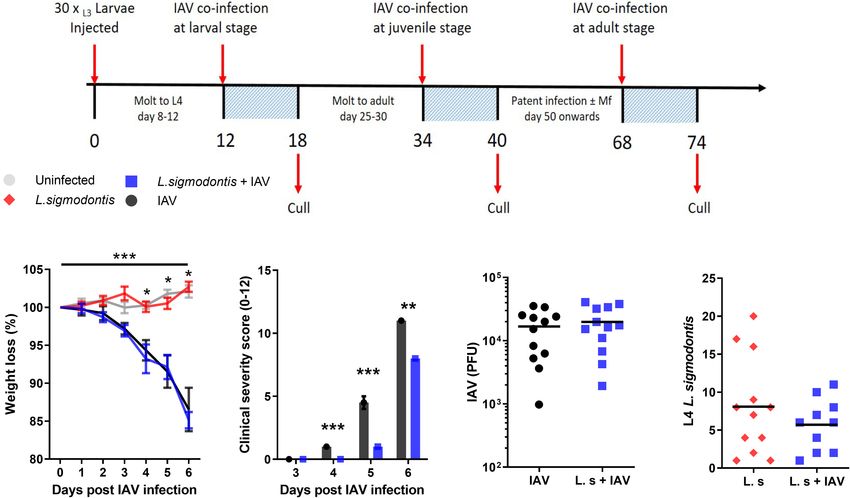
We first tested whether early larval stage L. sigmodontis infection replication, we measured the IAV lung titre 6 d post-IAV
could alter the progression of an acute IAV infection in the lung. infection. IAV lung titres were not significantly altered in co-
A
B C D E
FIGURE 1 | Larval stage L. sigmodontis infection reduces the severity of influenza A clinical signs. (A) Co-infection timeline. Mice were infected with 30 L3 L.
sigmodontis larvae subcutaneously. 5x10³ PFU IAV was then given intranasally at either d 12 (larval stage), 34 (juvenile adult stage) or 68 (patent infection) pi. Mice
were culled 6 d after IAV infection. (B) Weight loss over time (d post IAV infection). Mean ± SEM shown. ***Significant effect between groups dependent upon time
point, p < 0.001 (GLM). *p < 0.05 between IAV single infection and uninfected group, and co-infected and uninfected groups (Tukey’s HSD). (C) Combined IAV
clinical severity score. Median ± MAD shown ***p < 0.001, **p < 0.01 (Mann Witney U test). (D) IAV titre (PFU) in lung homogenates 6 d post IAV infection. Mean and
individual mice shown. (E) Numbers of L. sigmodontis (L4 larvae) recovered in pleural cavity lavage 6 d post IAV infection. Mean and individual mice shown. All
panels show combined data from two independent experiments, (n=12 for all groups).
Frontiers in Immunology | www.frontiersin.org 3 January 2022 | Volume 12 | Article 819560Hardisty et al. Influenza and Filariasis Coinfection
infected mice at 6 d post IAV infection (Figure 1D). There was delayed in the mice co-infected with juvenile L. sigmodontis
also no difference in the number of L. sigmodontis L4 larvae parasites 3-5 d post IAV infection (Figure 2A). However, these
recovered from co-infected mice and those infected with L. mice reached the same maximum weight loss 6 d post IAV. In
sigmodontis alone (Figure 1E). concordance, clinical signs of infection were also significantly
reduced in co-infected mice 3 and 4 d post IAV infection, yet
Juvenile Adult-Stage L. sigmodontis reached the same severity at 5 and 6 d post IAV infection
Infection Delays Weight Loss and (Figure 2B). These findings were independent of changes in viral
Progression of Influenza A Clinical Signs lung titre (Figure 2C). Similar to the larval stage of L. sigmodontis
Type 2 and immune regulatory responses increase as L. sigmodontis infection, IAV co-infection had no impact on the number of L.
infection progresses (30). Thus, we hypothesised that co-infection sigmodontis parasites recovered (Figure 2D).
with L. sigmodontis would have a more profound protective effect To determine whether the reduced clinical signs were associated
on IAV co-infection at later time points. L. sigmodontis L4 larvae with a change in Type 1 or regulatory cytokines, quantitative PCR
molt towards the juvenile adult stage around 25 - 30 d pi, becoming was used to measure IFN-g and IL-10 within the lung. Single IAV
reproductively mature adults around 55 d pi when they start infection resulted in significantly increased mRNA expression of IL-
producing Mf and infection becomes patent. To test whether the 10 and IFN-g compared to the naïve controls (Figures 2E, F).
juvenile adult stage has a stronger protective effect on IAV co- However, expression of IL-10 and IFN-g mRNA was unaltered upon
infection, mice were co-infected with 5x10³ IAV on d 34 of L. co-infection. L. sigmodontis infection alone did not result in
sigmodontis infection (Figure 1A). Weight loss was significantly increased levels of IL-10 and IFN-g mRNA within the lung.
A B C D
E F
FIGURE 2 | Juvenile adult stage of L. sigmodontis infection delays weight loss and progression of influenza A clinical signs. All panels show combined data from
two independent experiments, uninfected n=8, IAV n=12, L. sigmodontis n=12, IAV + L. sigmodontis n=12. (A) Weight loss over time (d post IAV infection).
Displayed as mean ± SEM. ***significant effect between groups dependent upon time point, p < 0.001 (GLM). *p < 0.05 significant effect between IAV and uninfected
group (Tukey’s HSD). #p < 0.05 significant effect between IAV infected and coinfected group (Tukey’s HSD). †p < 0.05 significant effect between uninfected and
coinfected group, (Tukey’s HSD). (B) Combined clinical severity score. Median ± MAD shown, ***p < 0.001 (Mann Whitney U test). (C) IAV titre (PFU) in lung
homogenates 6 d post IAV infection. Mean and individual mice shown. (D) Numbers of adult L. sigmodontis recovered from the pleural cavity 6 d post IAV infection.
Median and individual mice shown. (E, F) Levels of IL-10 (E) and IFN-g (F) mRNA in lung tissue, normalised to 18s RNA expression (n=7-12). Mean and individual
mice shown. ***Significant effect between groups (p < 0.001, ANOVA), #p < 0.05 (Tukey’s HSD).
Frontiers in Immunology | www.frontiersin.org 4 January 2022 | Volume 12 | Article 819560Hardisty et al. Influenza and Filariasis Coinfection
Co-Infection During Patent L. sigmodontis in IAV infected mice at d 6 (average score of 1.0 ± 0.27), co-infected
Infection Increases Susceptibility to IAV, animals demonstrated clinical symptoms by d 4 and progressed to
and Blocks Age-Related Resistance show increasingly severe clinical signs at 6 d post IAV infection with
to IAV Clinical Signs an average score of 10.0 ± 1.17 (Figure 3B). Co-infected mice also had
L. sigmodontis infection becomes patent at around 55 d pi, at a significantly increased IAV lung titre, with a 3.9-fold increase in
which point the adult parasites are fully mature and are releasing PFU compared with IAV infection alone (Figure 3C).
Mf into the blood. Patency associates with a switch from a As with co-infection at earlier stages of L. sigmodontis infection,
predominant Th2 response towards a mixed Th2/Th1 immune IAV did not alter susceptibility to L. sigmodontis. The total numbers
response, and the development of additional layers of immune of adult L. sigmodontis remained similar between co-infected and
regulation (16). To determine whether patent L. sigmodontis L. sigmodontis infected mice (Figure 3D). Co-infection also had no
infection alters susceptibility to IAV, mice were co-infected with impact on the numbers of circulating Mf in the blood, with
IAV at d 68 of L. sigmodontis infection (Figure 1). L. sigmodontis singly and co-infected mice showing similar levels
BALB/c mice show increasing resistance to IAV (A/WSN/33) of Mf immediately prior to, and after, IAV infection (Figure 3E).
infection with age (31), and as patent L. sigmodontis infection takes Similarly, the incidence of mice developing blood Mf remained the
over 2 months to develop, the mice were 4-5 months of age at the time same between groups both before (co-infected 8/14, L. sigmodontis
of IAV challenge. Consistent with their age, the weight loss due to only 10/14) and at d 6 of IAV infection (co-infected 9/14,
IAV infection alone was reduced compared to previous time points, L. sigmodontis only 7/14).
with a mean weight loss of 4.9% at 6 d post IAV infection (Figure 3A). Quantitative PCR was used to determine whether the
However, co-infected mice demonstrated significantly higher weight increased lung pathology was associated with alterations in
loss in response to IAV infection, with a mean weight loss of 17.0% by IFN-g or IL-10 mRNA expression within the lung. There were
d 6. Furthermore, whilst clinical signs of infection were only detected significant increases in mRNA expression of IL-10 and IFN-g in
A B C D
E F G
FIGURE 3 | Co-infection during patent L. sigmodontis infection increases susceptibility to IAV, and blocks age-related resistance to IAV clinical signs. All panels
show combined data from two independent experiments. Uninfected n=12, IAV n=11, L. sigmodontis n=12, IAV + L. sigmodontis n=13 (A) Weight loss over time
(d post IAV infection) shown as mean ± SEM, ***significant effect between groups over time, p < 0.001 (GLM). #p < 0.05 significant effect between IAV infected
and coinfected group (Tukey’s HSD). † p < 0.05 significant effect between uninfected and coinfected group (Tukey’s HSD). (B) Combined clinical severity score.
Median ± MAD shown. ***p < 0.001 (Mann Whitney U test) (C) IAV titre (PFU) in lung homogenates 6 d post IAV infection. Mean ± SEM. *p < 0.05 (unpaired T test).
(D) Numbers of adult L. sigmodontis recovered 6 d post IAV infection. Median and individual mice shown (L.sigmodontis n=12, IAV + L. sigmodontis n=13).
(E) Mf count in 30 ml blood before and after IAV co-infection. Median and individual mice shown. (F, G) Expression of IL-10 (F) and IFN-g (G) mRNA in the lung.
***Significant effect between groups, p < 0.001 (ANOVA), #p < 0.05 (Tukey’s HSD).
Frontiers in Immunology | www.frontiersin.org 5 January 2022 | Volume 12 | Article 819560Hardisty et al. Influenza and Filariasis Coinfection
the lung as a result of IAV infection compared with uninfected In contrast to the protective effect of pre-patent L. sigmodontis
mice, and significantly increased IFN-g due to L. sigmodontis infection, patent L. sigmodontis infection worsened the progression of
infection alone (Figures 3F, G). However, co-infected mice did IAV infection. Mice develop age-related resistance to IAV (31),
not display differences in expression of mRNA of either cytokine displaying reduced clinical signs and weight loss as they age. Patent
compared to the single infections. L. sigmodontis infection countered this age-related resistance and
significantly worsened weight loss and clinical signs of IAV infection,
as well as increasing IAV titre. The impaired protection is similar to
DISCUSSION studies demonstrating that T. spiralis infection impairs immunity to
murine norovirus (37), but contrasts with Heligmosomoides polygyrus
This study demonstrates that pre-existing L. sigmodontis and respiratory syncytial virus coinfection where H. polygyrus
infection can significantly ameliorate or worsen the enhanced resistance to the virus through type 1 interferons (38).
progression of acute respiratory infection with Influenza A An increasing number of immune regulatory pathways develop as
virus (IAV), with the outcome dependent upon the stage of L. infection progresses to patency, including Tregs (28, 39), alternatively
sigmodontis infection. Co-infection with IAV during the larval activated macrophages (40), and T cell-intrinsic dysfunction (10, 29,
and juvenile adult stages of L. sigmodontis infection delayed the 41), and increased suppression of third-party immune responses are
onset of clinical signs, whilst co-infection during patent L. seen as infection matures (42). An increased immune-regulatory
sigmodontis infection increased IAV clinical signs, weight loss, response may have hindered the immune system’s ability to control
and viral loads. IAV replication, with the resultant increase in viral burden
Mice infected with the intestinal parasite Trichinella spiralis at exacerbating immune pathology. However, as overspill of immune
an early enteric stage were found to undergo faster recovery from regulation could also protect against pathology, the immune
IAV induced weight loss (32). Infection with pre-patent L. regulatory mechanism would need to inhibit protective, but not
sigmodontis also significantly decreased the severity of IAV co- pathogenic, immune responses to IAV.
infection, although the protective effect presented as a reduction An alternative reason for the increased susceptibility to IAV
in the initial severity of infection. This protection was more infection could relate to perturbations in cytokine production
pronounced during the juvenile adult stage of L. sigmodontis during infection. The induction of Type 2 responses by helminths
infection than the larval L4 stage, with the juvenile adult stage are found to be sufficient to reactivate latent murine g-herpesvirus
delaying weight loss as well as clinical signs. This contrasts to infection (43). However, pre-patent L. sigmodontis infection
coinfection with L. sigmodontis and Friend retrovirus where L4 stimulates a Type 2 response and protected against influenza
stage L. sigmodontis infection caused significantly increased signs and weight loss, suggesting that the Type 2 response at
splenomegaly and viral loads (33). Decreased IAV severity was patency is unlikely to have worsened the progression of IAV
independent of viral titre and helminth burden, which were infection. In contrast with pre-patent infection, patent L.
unaltered by co-infection at either of the pre-patent life stages. sigmodontis infection does associate with the development of a
The dose of IAV was chosen to cause an infection of moderate mixed Th1/Th2 response (10). Patent L. sigmodontis singly infected
severity that could modulated up or down by coinfection without mice showed increased levels of IFN-g in their lung homogenates,
mortality. As the initial dose of IAV can instruct early interferon, that were not present during pre-patent infection. IFN-g, along with
cytokine and chemokine expression (34), different IAV doses IL-5, is part of the protective response against microfilaria (44), and
could result in different coinfection outcomes. In particular, L. microfilaremia associates with increased severity of LPS-induced
sigmodontis only delayed the onset of clinical signs and weight inflammation due to elevated IFN-g, TNF-a, IL-6 and IL-12
loss, raising a question of whether the protective effect of L. expression (16). Similarly, increased IFN-g correlates with more
sigmodontis infection would still be sufficient to ameliorate severe disease in L. sigmodontis and Plasmodium chabaudi chabaudi
severity during high dose, more pathogenic, IAV co-infection. co-infected mice. However, the effect of IFN-g levels on concurrent
IL-10 plays an important regulatory role during L. sigmodontis infections is context dependent. During Leishmania major
infection (35), and is involved in suppressing cerebral malaria coinfection, L. sigmodontis-enhanced IFN-g responses were
during L. sigmodontis co-infection (11). Similarly, concurrent associated with a delay in disease onset rather than enhancement
pre-patent L. sigmodontis infection suppresses IAV immunisation of pathology (15), whilst L. sigmodontis infection does not affect the
in an IL-10 dependent manner (22). Administration of the generation of Th1 IFN-g driven responses and susceptibility during
immunomodulatory filarial cystatin (AvCystatin/Av17), which concurrent infection with Mycobacterium tuberculosis (17).
stimulates IL-10 producing Foxp3+ Tregs, can also protect against Although L. sigmodontis infection did not increase IFN-g mRNA
inflammation and weight loss in a model of respiratory syncytial levels in IAV coinfected mice, IFN-g is a key factor determining the
virus inflammation (36). However, at the endpoint, IL-10 extent of pathology (26), and future studies should explore IFN-g as
expression in the lung homogenates of L. sigmodontis and IAV a potential mechanism.
co-infected mice did not correlate with the increased protection. Malaria models have also highlighted the importance of L.
Whilst measuring IL-10 protein production at earlier time points sigmodontis infection stage on the outcome of coinfection. Similar
and in specific cell populations would give a more accurate to IAV coinfection, larval stage L. sigmodontis infection protects
representation of IL-10 activity, this data could suggest an IL-10 against pathology in P. yoelii and P. chaboudi infections. Although it
independent effect. also increases resistance to P. yoelii, and co-infection with either
Frontiers in Immunology | www.frontiersin.org 6 January 2022 | Volume 12 | Article 819560Hardisty et al. Influenza and Filariasis Coinfection
malaria species increases resistance to L. sigmodontis (12). Patent ETHICS STATEMENT
L. sigmodontis infection also worsened pathology in P. chaboudi
coinfected mice, again mirroring IAV coinfection (13, 14). The animal study was reviewed and approved by University of
However, contrasting with IAV and P. chaboudi, patent Edinburgh Animal Welfare and Ethical Review Body.
L. sigmodontis infection protected 30% of co-infected mice from
P. berghei infection again indicating that the effects of coinfection
are context dependent (34). Not all mice develop patent
L. sigmodontis infection (defined by Mf circulating in the blood), AUTHOR CONTRIBUTIONS
and the extent of P. chaboudi pathology correlated with the presence BD, JH, GH, and MT designed the experiments. GH, JK, AF and
or absence of blood Mf, with more severe pathology in Mf negative MT conducted the experiments and collected the data. GH, BD,
mice (13, 14). Similarly, Mf negative mice showed lower protection and MT analysed the data and wrote the manuscript. All authors
against P. berghei (13, 14). All but 2 mice developed Mf between d contributed to the article and approved the submitted version.
68 and 74 post-L. sigmodontis infection in our study, and so it was
not possible to determine whether Mf status impacted the outcome
of IAV. As Mf do stimulate IFN-g production (45), it would be
interesting to determine whether the exacerbation of pathology by FUNDING
patent L. sigmodontis infection is due to the release of Mf.
This study demonstrates that there are interactions between the This project was funded by the Biotechnology and Biological
filarial helminth and acute respiratory viral infections, and that the Sciences Research Council (BBSRC) Institute Strategic Program
presence of a helminth infection can both ameliorate and worsen Grant BB/J004324/1 to The Roslin Institute. GH was funded by a
IAV severity with the outcome dependent upon the stage of BBSRC Doctoral Training Grant to the Centre for Infectious
helminth infection. Further research is required to elucidate the Diseases, University of Edinburgh. MT, AF, and JK were funded
mechanisms by which this interaction occurs. This suggests that by the Medical Research Council (MRC) UK grant number MR/
consideration of concomitant infection with filarial helminths may K020196/1, and the Wellcome Trust grant number 095831.
be a significant factor in the treatment and outcome of IAV and Open access publication costs were provided by the UKRI
other respiratory infections such as SARS-CoV-2, where the Open Access Fund. The funders had no role in study design,
expression of type 2 cytokine responses is associated with data collection and analysis, decision to publish, or preparation
increased disease severity (46). of the manuscript.
DATA AVAILABILITY STATEMENT ACKNOWLEDGMENTS
The original contributions presented in the study are included in We would like to gratefully acknowledge the contributions of The
the article/supplementary material. Further inquiries can be Bioimaging and Flow Cytometry Facility (Roslin Institute) and
directed to the corresponding author. Bioresearch & Veterinary Services (University of Edinburgh).
8. Mishra PK, Palma M, Bleich D, Loke P, Gause WC. Systemic Impact of
REFERENCES Intestinal Helminth Infections. Mucosal Immunol (2014) 7(4):753–62. doi:
1. Cano J, Rebollo MP, Golding N, Pullan RL, Crellen T, Soler A, et al. The Global 10.1038/mi.2014.23
Distribution and Transmission Limits of Lymphatic Filariasis: Past and 9. Moreau E, Chauvin A. Immunity Against Helminths: Interactions With the
Present. Parasit Vectors (2014) 7(1):1–19. doi: 10.1186/s13071-014-0466-x Host and the Intercurrent Infections. J BioMed Biotechnol (2010) 2010:1–9.
2. Donohue RE, Cross ZK, Michael E. The Extent, Nature, and Pathogenic Consequences doi: 10.1155/2010/428593
of Helminth Polyparasitism in Humans: A Meta-Analysis. Akullian A, Editor. PloS 10. Finlay CM, Allen JE. The Immune Response of Inbred Laboratory Mice to
Negl Trop Dis (2019) 13(6):e0007455. doi: 10.1371/journal.pntd.0007455 Litomosoides Sigmodontis: A Route to Discovery in Myeloid Cell Biology.
3. Babu S, Nutman TB. Immunology of Lymphatic Filariasis. Parasite Immunol Parasite Immunol (2020) (7):1–17. doi: 10.1111/pim.12708
(2014) 36(8):338–46. doi: 10.1111/pim.12081 11. Specht S, Ruiz DF, Dubben B, Deininger S, Hoerauf A. Filaria-Induced IL-10
4. Maizels RM, Smits HH, McSorley HJ. Modulation of Host Immunity by Suppresses Murine Cerebral Malaria. Microbes Infect (2010) 12(8–9):635–42.
Helminths: The Expanding Repertoire of Parasite Effector Molecules. doi: 10.1016/j.micinf.2010.04.006
Immunity (2018) 49(5):801–18. doi: 10.1016/j.immuni.2018.10.016 12. Karadjian G, Berrebi D, Dogna N, Vallarino-Lhermitte N, Bain O, Landau I,
5. Maizels RM, McSorley HJ. Regulation of the Host Immune System by et al. Co-Infection Restrains Litomosoides Sigmodontis Filarial Load and
Helminth Parasites. J Allergy Clin Immunol (2016) 138(3):666–75. doi: Plasmodial P. Yoelii But Not P. Chabaudi Parasitaemia in Mice. Parasite
10.1016/j.jaci.2016.07.007 (2014) 21:16. doi: 10.1051/parasite/2014017
6. Nutman TB. Looking Beyond the Induction of Th2 Responses to Explain 13. Ferná ndez Ruiz D, Dubben B, Saeftel M, Endl E, Deininger S, Hoerauf A, et al.
Immunomodulation by Helminths. Parasite Immunol (2015) 37(6):304–13. Filarial Infection Induces Protection Against P. Berghei Liver Stages in Mice.
doi: 10.1111/pim.12194 Microbes Infect (2009) 11(2):172–80. doi: 10.1016/j.micinf.2008.11.003
7. Maizels RM, Yazdanbakhsh M. Immune Regulation by Helminth Parasites: 14. Graham AL, Lamb TJ, Read AF, Allen JE. Malaria-Filaria Coinfection in Mice
Cellular and Molecular Mechanisms. Nat Rev Immunol (2003) 3(9):733–44. Makes Malarial Disease More Severe Unless Filarial Infection Achieves
doi: 10.1038/nri1183 Patency. J Infect Dis (2005) 191(3):410–21. doi: 10.1086/426871
Frontiers in Immunology | www.frontiersin.org 7 January 2022 | Volume 12 | Article 819560Hardisty et al. Influenza and Filariasis Coinfection
15. Lamb TJ, Graham AL, Le Goff L, Allen JE. Co-Infected C57BL/6 Mice Mount Editor. PloS Negl Trop Dis (2016) 10(12):e0005170. doi: 10.1371/journal.
Appropriately Polarized and Compartmentalized Cytokine Responses to pntd.0005170
Litomosoides Sigmodontis and Leishmania Major But Disease Progression Is 34. Marois I, Cloutier A, Garneau É , Richter MV. Initial Infectious Dose Dictates
Altered.ParasiteImmunol(2005)27(9):317–24.doi:10.1111/j.1365-3024.2005.00779.x the Innate, Adaptive, and Memory Responses to Influenza in the Respiratory
16. Hubner MP, Pasche B, Kalaydjiev S, Soboslay PT, Lengeling A, Schulz-Key H, Tract. J Leukoc Biol (2012) 92(1):107–21. doi: 10.1189/jlb.1011490
et al. Microfilariae of the Filarial Nematode Litomosoides Sigmodontis 35. Hartmann W, Schramm C, Breloer M. Litomosoides Sigmodontis Induces
Exacerbate the Course of Lipopolysaccharide-Induced Sepsis in Mice. Infect TGF-b Receptor Responsive, IL-10-Producing T Cells That Suppress
Immun (2008) 76(4):1668–77. doi: 10.1128/IAI.01042-07 Bystander T-Cell Proliferation in Mice: Immunomodulation. Eur J Immunol
17. Hübner MP, Killoran KE, Rajnik M, Wilson S, Yim KC, Torrero MN, et al. (2015) 45(9):2568–81. doi: 10.1002/eji.201545503
Chronic Helminth Infection Does Not Exacerbate Mycobacterium 36. Schuijs MJ, Hartmann S, Selkirk ME, Roberts LB, Openshaw PJM, Schnoeller
Tuberculosis Infection. MacDonald AS, Editor. PloS Negl Trop Dis (2012) 6 C. The Helminth-Derived Immunomodulator AvCystatin Reduces Virus
(12):e1970. doi: 10.1371/journal.pntd.0001970 Enhanced Inflammation by Induction of Regulatory IL-10+ T Cells. Sun J,
18. Chen X, Liu S, Goraya MU, Maarouf M, Huang S, Chen J-L. Host Immune Editor. PloS One (2016) 11(8):e0161885. doi: 10.1371/journal.pone.0161885
Response to Influenza A Virus Infection. Front Immunol (2018) 9:320/full. 37. Osborne LC, Monticelli LA, Nice TJ, Sutherland TE, Siracusa MC, Hepworth
doi: 10.3389/fimmu.2018.00320/full MR, et al. Virus-Helminth Coinfection Reveals a Microbiota-Independent
19. Liu Q, Zhou Y, Yang Z. The Cytokine Storm of Severe Influenza and Mechanism of Immunomodulation. Science (2014) 345(6196):578–82. doi:
Development of Immunomodulatory Therapy. Cell Mol Immunol (2016) 13 10.1126/science.1256942
(1):3–10. doi: 10.1038/cmi.2015.74 38. McFarlane AJ, McSorley HJ, Davidson DJ, Fitch PM, Errington C, Mackenzie KJ,
20. Tisoncik JR, Korth MJ, Simmons CP, Farrar J, Martin TR, Katze MG. Into the et al. Enteric Helminth-Induced Type I Interferon Signaling Protects Against
Eye of the Cytokine Storm. Microbiol Mol Biol Rev (2012) 76(1):16–32. doi: Pulmonary Virus Infection Through Interaction With the Microbiota. J Allergy
10.1128/MMBR.05015-11 Clin Immunol (2017) 140(4):1068–1078.e6. doi: 10.1016/j.jaci.2017.01.016
21. Price I, Mochan-Keef ED, Swigon D, Ermentrout GB, Lukens S, Toapanta FR, 39. Taylor MD, LeGoff L, Harris A, Malone E, Allen JE, Maizels RM. Removal of
et al. The Inflammatory Response to Influenza A Virus (H1N1): An Regulatory T Cell Activity Reverses Hyporesponsiveness and Leads to Filarial
Experimental and Mathematical Study. J Theor Biol (2015) 374:83–93. doi: Parasite Clearance In Vivo. J Immunol (2005) 174(8):4924–33. doi: 10.4049/
10.1016/j.jtbi.2015.03.017 jimmunol.174.8.4924
22. Hartmann W, Brunn M-L, Stetter N, Gagliani N, Muscate F, Stanelle-Bertram 40. Taylor MD, Harris A, Nair MG, Maizels RM, Allen JE. F4/80 + Alternatively
S, et al. Helminth Infections Suppress the Efficacy of Vaccination Against Activated Macrophages Control CD4 + T Cell Hyporesponsiveness at Sites
Seasonal Influenza. Cell Rep (2019) 29(8):2243–56. doi: 10.1016/j.celrep. Peripheral to Filarial Infection. J Immunol (2006) 176(11):6918–27. doi:
2019.10.051 10.4049/jimmunol.176.11.6918
23. Fulton A, Babayan SA, Taylor MD. Use of the Litomosoides Sigmodontis 41. Knipper JA, Ivens A, Taylor MD. Helminth-Induced Th2 Cell Dysfunction is
Infection Model of Filariasis to Study Type 2 Immunity, in: Type 2 Immunity Distinct From Exhaustion and Is Maintained in the Absence of Antigen.
(2018). New York, NY: Springer New York. Available at: http://link.springer. Makepeace BL, Editor. PloS Negl Trop Dis (2019) 13(12):e0007908. doi:
com/10.1007/978-1-4939-7896-0_2 (Accessed cited 2019 Jul 18). 10.1371/journal.pntd.0007908
24. Nicol MQ, Ligertwood Y, Bacon MN, Dutia BM, Nash AA. A Novel Family of 42. Haben I, Hartmann W, Breloer M. Nematode-Induced Interference With
Peptides With Potent Activity Against Influenza A Viruses. J Gen Virol (2012) Vaccination Efficacy Targets Follicular T Helper Cell Induction and Is
93(Pt_5):980–6. doi: 10.1099/vir.0.038679-0 Preserved After Termination of Infection. Mitre E, Editor. PloS Negl Trop
25. Bouvier NM, Lowen AC. Animal Models for Influenza Virus Pathogenesis Dis (2014) 8(9):e3170. doi: 10.1371/journal.pntd.0003170
and Transmission. Viruses (2010) 2(8):1530–63. doi: 10.3390/v20801530 43. Reese TA, Wakeman BS, Choi HS, Hufford MM, Huang SC, Zhang X, et al.
26. Nicol MQ, Campbell GM, Shaw DJ, Dransfield I, Ligertwood Y, Beard PM, Helminth Infection Reactivates Latent g-Herpesvirus via Cytokine Competition at
et al. Lack of Ifng Signaling Attenuates Spread of Influenza A Virus In Vivo a Viral Promoter. Science (2014) 345(6196):573–7. doi: 10.1126/science.1254517
and Leads to Reduced Pathogenesis. Virology (2019) 526:155–64. doi: 10.1016/ 44. Saeftel M, Arndt M, Specht S, Volkmann L, Hoerauf A. Synergism of Gamma
j.virol.2018.10.017 Interferon and Interleukin-5 in the Control of Murine Filariasis. Infect Immun
27. Karadjian G, Fercoq F, Pionnier N, Vallarino-Lhermitte N, Lefoulon E, (2003) 71(12):6978–85. doi: 10.1128/IAI.71.12.6978-6985.2003
Nieguitsila A, et al. Migratory Phase of Litomosoides Sigmodontis Filarial 45. Lawrence RA, Allen JE, Osborne J, Maizels RM. Adult and Microfilarial Stages
Infective Larvae Is Associated With Pathology and Transient Increase of of the Filarial Parasite Brugia Malayi Stimulate Contrasting Cytokine and Ig
S100A9 Expressing Neutrophils in the Lung. Brehm K, Editor. PloS Negl Trop Isotype Responses in BALB/c Mice. J Immunol Baltim Md 1950 (1994) 153
Dis (2017) 11(5):e0005596. doi: 10.1371/journal.pntd.0005596 (3):1216–24.
28. Taylor MD, van der Werf N, Harris A, Graham AL, Bain O, Allen JE, et al. 46. Lucas C, Wong P, Klein J, Castro TBR, Silva J, Sundaram M, et al.
Early Recruitment of Natural CD4+Foxp3+ Treg Cells by Infective Larvae Longitudinal Analyses Reveal Immunological Misfiring in Severe COVID-
Determines the Outcome of Filarial Infection. Eur J Immunol (2009) 39 19. Nature (2020) 584(7821):463–9. doi: 10.1038/s41586-020-2588-y
(1):192–206. doi: 10.1002/eji.200838727
29. van der Werf N, Redpath SA, Azuma M, Yagita H, Taylor MD. Th2 Cell- Conflict of Interest: The authors declare that the research was conducted in the
Intrinsic Hypo-Responsiveness Determines Susceptibility to Helminth absence of any commercial or financial relationships that could be construed as a
Infection. PloS Pathog (2013) 9(3):e1003215. doi: 10.1371/journal.ppat.1003215 potential conflict of interest.
30. Babayan S, Ungeheuer M-N, Martin C, Attout T, Belnoue E, Snounou G, et al.
Resistance and Susceptibility to Filarial Infectionwith Litomosoides Publisher’s Note: All claims expressed in this article are solely those of the authors
Sigmodontis Are Associated With EarlyDifferences in Parasite Development and do not necessarily represent those of their affiliated organizations, or those of
and in Localized ImmuneReactions. Infect Immun (2003) 71(12):6820–9. doi: the publisher, the editors and the reviewers. Any product that may be evaluated in
10.1128/IAI.71.12.6820-6829.2003 this article, or claim that may be made by its manufacturer, is not guaranteed or
31. Lu J, Duan X, Zhao W, Wang J, Wang H, Zhou K, et al. Aged Mice Are More endorsed by the publisher.
Resistant to Influenza Virus Infection Due to Reduced Inflammation and
Lung Pathology. Aging Dis (2018) 9(3):358. doi: 10.14336/AD.2017.0701 Copyright © 2022 Hardisty, Knipper, Fulton, Hopkins, Dutia and Taylor. This is an
32. Furze RC, Hussell T, Selkirk ME. Amelioration of Influenza-Induced open-access article distributed under the terms of the Creative Commons Attribution
Pathology in Mice by Coinfection With Trichinella Spiralis. Infect Immun License (CC BY). The use, distribution or reproduction in other forums is permitted,
(2006) 74(3):1924–32. doi: 10.1128/IAI.74.3.1924-1932.2006 provided the original author(s) and the copyright owner(s) are credited and that the
33. Dietze KK, Dittmer U, Koudaimi DK, Schimmer S, Reitz M, Breloer M, et al. original publication in this journal is cited, in accordance with accepted academic
Filariae-Retrovirus Co-Infection in Mice Is Associated With Suppressed practice. No use, distribution or reproduction is permitted which does not comply with
Virus-Specific IgG Immune Response and Higher Viral Loads. Hsieh MH, these terms.
Frontiers in Immunology | www.frontiersin.org 8 January 2022 | Volume 12 | Article 819560You can also read