Concomitance or consequence? Stevens Johnson syndrome in COVID 19: A case report
←
→
Page content transcription
If your browser does not render page correctly, please read the page content below
EXPERIMENTAL AND THERAPEUTIC MEDICINE 23: 257, 2022

         Concomitance or consequence? Stevens‑Johnson syndrome
                     in COVID‑19: A case report
      CARMEN MANCIUC1, GEORGIANA ALEXANDRA LACATUSU2, ANDREI VATA1, CRISTINA SAPANIUC2,
                  CARMEN MIHAELA ARTENI2 and FLORIN DUMITRU PETRARIU3

          1
              Department of Infectious Diseases, Grigore T. Popa University of Medicine and Pharmacy, 700115 Iasi;
                2
                 Department of Infectious Diseases, Sf. Parascheva Clinical Hospital of Infectious Diseases, 700116 Iasi;
                                       3
                                        Department of Preventive Medicine and Interdisciplinary,
                          Grigore T. Popa University of Medicine and Pharmacy, 700115 Iasi, Romania

                                     Received November 17, 2021; Accepted December 17, 2021

                                                    DOI: 10.3892/etm.2022.11182

Abstract. The novel coronavirus infection has been, and still         Introduction
is, a pressing medical problem with a catastrophic effect, not
only from a medical point of view, but also from an economic          The pandemic created by SARS CoV‑2 infection still repre‑
and social one. The cutaneous manifestations of the disease           sents a pressing medical problem considering the multitude
have a diverse morphology and can signal the presence of the          of risk factors for severe disease and the lack of specific
infection. The present article reports the case of a 77‑year‑old      symptoms (1‑3). Sanitary education of the population
male patient admitted at The Sf. Parascheva Clinical Hospital         and vaccination have served an essential role in prophy‑
of Infectious Diseases in Iasi (Romania) after testing positive       laxis by helping individuals understand the risks they are
for SARS CoV‑2 infection. Initially, the patient presented            exposed to (4‑7).
a pruriginous generalized maculopapular‑erythematous                      Literature highlights that cutaneous manifestation of SARS
eruption with a tendency towards confluence, peri‑oro‑nasal           CoV‑2 infection presents as lesions with varying morphology
meliceric crusts and desquamation of the skin on the third            that could be classified in four categories: Acro‑papular
anterosuperior and posterior thorax, scalp and forehead, which        lesions, urticarial eruption, vascular (chilblain‑like lesions,
was accompanied by low back pain, headache and orbital pain.          commonly known as COVID‑19 toes, livedoid and purpuric
The suspicion of Stevens‑Johnson syndrome (SJS) was raised,           lesions) and exanthema (morbilliform and papulo‑vesicular
and treatment was given according to the recommendation of            rash and varicella‑like eruption) (8‑13).
the hospital dermatologist. This association raises multiple              Stevens‑Johnson syndrome (SJS) has the potential to be a
questions regarding whether SJS is a cutaneous manifestation          lethal skin reaction that has a mortality rate of up to 30%, which
of COVID‑19 or if there was a concomitance between the viral          is caused by an immune‑complex‑mediated hypersensitivity
infection and the immune reaction. The combination of SJS             reaction. The clinical presentation appears as mucosal and
and COVID‑19 can have a fatal outcome if not recognized and           cutaneous tenderness accompanied by erythema, hemorrhagic
promptly treated. To our knowledge, this is the first case of SJS     erosions, and epidermal detachment that can be described as
in a patient diagnosed with SARS CoV‑2 infection in Romania.          blisters and areas of denuded skin, accompanied by systemic
                                                                      symptoms (14,15). This disease is a dermatological emergency.
                                                                      The recognition in association with prompt and appropriate
                                                                      management can save the patient (16). The present study is
                                                                      a case report of a 77‑year‑old male with a metabolic, cardio‑
                                                                      logic and neurological history diagnosed with SARS CoV‑2
Correspondence       to: Dr Georgiana Alexandra Lacatusu,             infection associated with SJS. Few cases have been reported
Department of Infectious Diseases, Sf. Parascheva Clinical Hospital   concerning this association, which raises the question of
of Infectious Diseases, Str. Octav Botez 2, 700116 Iasi, Romania      whether, in the case of our patient, SJS appeared independently
E‑mail: alexandra.lacatusu@gmail.com                                  from COVID‑19 or was the primary manifestation of the
                                                                      disease (17‑23).
Dr Andrei Vata, Department of Infectious Diseases, Grigore T.
Popa University of Medicine and Pharmacy, Str. Universitatii 16,
700115 Iasi, Romania                                                  Case report
E‑mail: andreiandrei@yahoo.com
                                                                      The present article reports the case of a 77‑year‑old male
Key words: SARS CoV‑2, Stevens‑Johnson syndrome, emergency,           patient with a history of stroke, stage‑2 arterial hypertension,
cutaneous manifestation, pandemic                                     dyslipidemia, obesity and gout, together with an underlying
                                                                      treatment: Aspirin, 75 mg; bisoprolol, 2.5 mg bidaily; atorvas‑
                                                                      tatin, 10 mg/day; vinopectine, 10 mg bidaily; and allopurinol,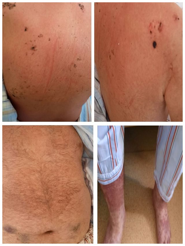
2 MANCIUC et al: CONCOMITANCE OR CONSEQUENCE? STEVENS-JOHNSON SYNDROME IN COVID-19
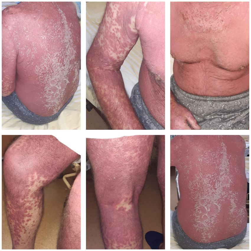
Figure 1. Clinical presentation of the dermatological lesions at hospital admission. The images demonstrate disseminated maculopapular‑erythematous erup‑
tion with a tendency towards confluence with desquamation of the skin on the third anterosuperior and posterior thorax.
100 mg bidaily. The gout medication was prescribed 14 days At admission, the patient had a general fair status and was
before admission to our hospital. conscious. He was experiencing bradylalia, but stable both
Initially, the patient presented to the Emergency Room of hemodynamically and in terms of respiration (blood pressure,
Sf. Spiridon County Hospital for a non‑pruriginous general‑ 106/67 mmHg; heart rate, 95 beats/min; oxygen saturation,
ized maculopapular‑erythematous eruption with a tendency 98% ambient air). This was associated with the aforemen‑
towards confluence, accompanied by low back pain, headache tioned lesions, as well as peri‑oronasal meliceric crusts and
and orbital pain. Considering the epidemiological context, a desquamation of the skin on the third anterosuperior and
reverse transcription PCR for SARS CoV‑2 virus and a CT scan posterior thorax, scalp and forehead (Fig. 1).
were performed. The result of the molecular test was positive, Considering the clinical and paraclinical evidence
and the CT examination demonstrated bilateral centrilobular (Table I), the suspicion of SJS was raised, and a dermatological
emphysema and bilateral apical pachypleuritis. In the inferior consultation was requested, which confirmed the diagnosis.
two‑thirds of the lungs, bilateral, extensive areas of pulmonary The recommendations were to stop the administration of allo‑
condensation were observed that were predominantly located purinol and administer methylpredinisolone at 250 mg/day,
subpleurally, heterogeneous and imprecise. Based on these 20 mg bilastine bidaily, vitamin C intravenously at 500 mg
results, the patient was directed to Sf. Parascheva Clinical bidaily, gluconic calcium at 10 ml/day (94 mg/ml), vitonal and
Hospital of Infectious Diseases, which was a designated gentamicin cream (applied bidaily on the lesions located on
first‑line COVID‑19 hospital. the peri‑oronasal area) and a cream consisting of 5 g urea, 1 gEXPERIMENTAL AND THERAPEUTIC MEDICINE 23: 257, 2022 3
Table I. Laboratory data.
Date of measurement
‑‑‑‑‑‑‑‑‑‑‑‑‑‑‑‑‑‑‑‑‑‑‑‑‑‑‑‑‑‑‑‑‑‑‑‑‑‑‑‑‑‑‑‑‑‑‑‑‑‑‑‑‑‑‑‑‑‑‑‑‑‑‑‑‑‑‑‑‑‑‑‑‑‑‑‑‑‑‑‑‑‑‑‑‑‑‑‑‑‑‑‑‑‑‑‑‑‑‑‑‑‑‑‑‑‑‑‑‑‑‑‑‑‑‑‑‑‑‑‑‑‑‑‑‑‑‑‑‑‑‑‑‑‑‑‑‑‑‑‑‑‑‑‑‑‑‑‑‑‑‑‑‑‑‑‑‑‑‑‑‑‑‑‑‑‑‑‑‑‑‑‑‑‑‑‑‑‑‑
Parameter 20.11 23.11 24.11 26.11 30.11 3.12
Leukocytes (per mm3) 27,840 NA 12,820 12,320 9,690 10,210
Neutrophil (%) 70.70 NA 79.2 82.3 87.3 83.40
Lymphocytes (%) 11.20 NA 14.3 11 9.2 13.50
Platelets (per mm3) 349,000 NA 274,000 233,000 79,000 117,000
C‑reactive protein (mg/l) 27.3 NA 31.57 NA 58.66 53.46
ESR (mm/h) 18 NA 20 NA 40 85
INR 1.33 NA NA NA 2.42
Fibrinogen (g/l) 1.8 NA NA NA 3.29 3.29
IL‑6 (pg/ml) 27.19 NA NA NA NA
D‑dimer 1235 NA NA NA NA NA
Urea (mg/dl) 172 NA 86 85 102 118
Creatinine (mg/dl) 1.75 NA 1.2 0.95 0.96 1.11
Glucose (mg/dl) 140 NA 111 NA 103 103
Na (mmol/l) 141 NA 146.1 146.6 146.7 146.7
K (mmol/l) 4.37 NA 3.99 4.05 4.80 4.58
Cl (mmol/l) 97.7 NA 102.6 103.1 105.2 105.4
HCO3 (mmol/l) NA 21.4 NA 13.6 NA NA
ALT(U/l) 37 NA 62 NA 63 58
AST(U/l) 39 NA 80 NA 88 88
Bilirubin (mg/dl) 1.25 NA 1.31 1.60 1.15 2.64
Ionic calcium (mg/dl) NA NA NA 4.72 NA 4.40
HIV serology NA Negative NA NA NA NA
LDH (U/l) NA NA NA NA 609 NA
Total protein (g/l) 60.78 NA NA NA 75.21 NA
Ferritin (ng/ml) NA 511 NA NA NA NA
ESR, erythrocyte sedimentation rate; ALT, alanine transaminase; AST, aspartate transaminase; HIV, human immunodeficiency virus; LDH,
lactate dehydrogenase; INR, international normalized ratio.
hydrocortisone and 100 g Vaseline® (applied bidaily over all intensive care unit consultation was requested, arterial gases
affected areas). In addition, antibiotic (meropenem, 4 g/day; were measured, which suggested that the patient was in meta‑
linezolid, 1.2 g/day), anticoagulant (enoxaparine sodium, bolic acidosis (low PaCO2 and HCO3), and recommendations
0.6 mg bidaily), acetaminophen (500 mg) and acetylcysteine for intensive care therapy were given.
(600 mg/daily) were administered. After 20 days of hospitalization, the patient had a fatal
The algorithm of drug causality for epidermal necrol‑ outcome.
ysis (ALDEN) and the severity‑of‑illness score for toxic
epidermal necrolysis (SCORTEN) were calculated. The Discussion
ALDEN score for the patient was 5, corresponding to a
‘probable’ causal link, suggesting that the implicated drug SJS/toxic epidermal necrolysis (TEN) represents a significant
in our case could be allopurinol (Table II). In addition, the dermatological emergency, being one of the most severe
SCORTEN was 3 for this patient, indicating a mortality rate cutaneous adverse reactions and associated with a high risk
of 35.3% (Table III). of mortality. SJS/TEN, due to an immune‑complex‑mediated
After five days of treatment, the dermatological aspects hypersensitivity reaction, involves the mucous membranes and
had a favorable evolution, with healing of most of the lesions skin (24,25). Initial symptoms can be unspecific and include
but persistence of those located on the inferior limbs (Fig. 2). fever, cough, sore throat or eye discomfort, which are followed
However, the general condition of the patient started to dete‑ by the cutaneous manifestations (26).
riorate. On the 7th day of admission, the patient desaturated to SJS/TEN is drug‑induced in 70‑80% of cases. Graft
76% ambient air, requiring an oxygen supplement that corrected versus‑host disease is another well‑established but rare cause,
saturation to 93% with 15 l of additional oxygen. Therefore, an independent of drugs (27). A few cases are related to infections
IL‑1 inhibitor was added to his treatment (200 mg on day 1, (such as with Mycoplasma pneumoniae), while others remain
then 100 mg/day for four days). Considering his status, an unexplained (idiopathic forms) (28).4 MANCIUC et al: CONCOMITANCE OR CONSEQUENCE? STEVENS-JOHNSON SYNDROME IN COVID-19
Table II. ALDEN results for allopurinol.
Score Value
Delay from initial drug intake to index day +3
Drug present in the body (on index day) 0
Pre‑challenge/Re‑challenge 0
De‑challenge 0
Type of drug (notoriety) +3
Other cause ‑1
Total ALDEN score 5a
a
This value corresponds to a ‘probable’ causal link. ALDEN, algo‑
rithm of drug causality for epidermal necrolysis.
Table III. SCORTEN score.
Prognostic factor Score
Age >40 years 1
Associated cancer 0
Heart rate >120 bpm 0
Serum blood urea >28 mg/dl 1
Detached or compromised body surface >10% 1
Serum bicarbonate 250 mg/dl 0
treatment.
Total SCORTEN 3
Mortality rate according to score: 0‑1, 3.2%; 2, 12.1%; 3, 35.3%;
4, 58.3% and ≥5, ≥90%. SCORTEN, severity‑of‑illness score for virus and cytomegalovirus) (36‑39), suggesting this could also
toxic epidermal necrolysis; bpm, beats per minute. be possible in SARS CoV‑2 infection in our case. Therefore,
it may be hypothesized that allopurinol was the causative
agent of SJS/TEN, although the fact that SJS/TEN could be
a cutaneous manifestation of SARS CoV‑2 infection and or
This pathology represents a delayed reaction that usually represent a consequence in this type of viral infection should
occurs 4‑28 days from the moment of exposure to a drug (29); not be discounted.
thus, it is of utmost importance to conduct an in‑depth The severity of SJS/TEN can be assessed using SCORTEN,
anamnesis and a thorough retrospective pharmacological which is a severity‑of‑illness scale that was defined in 2000
investigation for an extended period of time preceding the and is a specific predictor of mortality. The score includes the
onset of skin manifestations. following variables: Age >40, the presence of neoplasia, heart
The drugs that are associated with SJS/TEN include rate >120 bpm, serum blood urea >28 mg/dl, serum glucose
anticonvulsants, allopurinol, sulfonamides, antibiotics >250 mg/dl, serum HCO3 10% detached
(such as penicillin, cephalosporins, quinolones and minocy‑ body surface. Each constant receives one point, and the final
cline), acetaminophen and nonsteroidal anti‑inflammatory score ranges from 0 to 7. (40). In the presented case, the patient
drugs (30‑33). had an initial score of 3, which indicated mortality of 35.3%
The ALDEN score is one of the most valuable tools in the As the status of the patient started to deteriorate, and the HCO3
assessment of SJS/TEN, which helps identify the possible drug level decreased to 13.6 mmol/l, the prognosis of mortality
associated with the severe cutaneous adverse reaction, as well grew to 58.3%.
as the drugs that can still be administered to the patient (34). The In our case, although correct dermatological treatment led
algorithm gives the suspected causal drug taken by the patient to a favorable evolution of the skin lesions (41,42), the patient's
a score that sums between ‑12 and 10, which corresponds to condition was ultimately influenced by the complication
the probability of having caused the reaction. The total score of SARS CoV‑2 infection, which progressed to respiratory
corresponds to ‘causal links’ that range from ‘very unlikely’ failure associated with major hydro‑electrolytic and acid‑base
to ‘very probable’ (35). SJS can occur as a rare side effect of imbalance. Together with the negative prognostic factors
allopurinol, which, in this case, could have been favored by that the patient presented (hypertension, obesity and
the immune stimulation induced by the SARS CoV‑2 virus. dyslipidemia) (43‑45), this led to a fatal outcome.
This idea is supported by the fact that SJS/TEN has been The SCORTEN in our patient led to an estimated mortality
associated with viral replication (human immunodeficiency rate of 35.3% that later grew to 58.3% as a result of bicarbonateEXPERIMENTAL AND THERAPEUTIC MEDICINE 23: 257, 2022 5 levels being
6                  MANCIUC et al: CONCOMITANCE OR CONSEQUENCE? STEVENS-JOHNSON SYNDROME IN COVID-19

25. Chung WH, Hung SI, Hong HS, Hsih MS, Yang LC, Ho HC,                  36. Peter J, Choshi P and Lehloenya RJ: Drug hypersensitivity in
    Wu JY and Chen YT: Medical genetics: A marker for                         HIV infection. Curr Opin Allergy Clin Immunol 19: 272‑282,
    Stevens‑Johnson syndrome. Nature 428: 486, 2004.                          2019.
26. Harr T and French LE: Toxic epidermal necrolysis and                  37. Yang CW, Cho YT, Hsieh YC, Hsu SH, Chen KL and Chu CY:
    Stevens‑Johnson syndrome. Orphanet J Rare Dis 5: 39, 2010.                The interferon‑γ‑induced protein 10/CXCR3 axis is associated
27. Hazin R, Ibrahimi OA, Hazin MI and Kimyai‑Asadi A:                        with human herpesvirus‑6 reactivation and the development of
    Stevens‑Johnson syndrome: Pathogenesis, diagnosis, and                    sequelae in drug reaction with eosinophilia and systemic symp‑
    management. Ann Med 40: 129‑138, 2008.                                    toms. Br J Dermatol 183: 909‑919, 2020.
28. Levy M and Shear NH: Mycoplasma pneumoniae infections and             38. Tagajdid MR, Doblali T, Elannaz H, Hammi S, Belfequih B
    Stevens‑Johnson syndrome. Report of eight cases and review of             and Mrani S: Reactivation of cytomegalovirus in a patient with
    the literature. Clin Pediatr (Phila) 30: 42‑49, 1991.                     Stevens‑Johnson syndrome‑toxic epidermal necrolysis. Iran
29. Jawaro T, Kumar A, Pistun O and Dixit D: Stevens‑Johnson                  J Med Sci 38 (2 Suppl): S195‑S197, 2013.
    syndrome associated with chlordiazepoxide. J Pharm Technol 34:        39. Richard EB, Hamer D, Musso MW, Short T and O'Neal HR Jr:
    82‑85, 2018.                                                              Variability in management of patients with SJS/TEN: A survey
30. De Luca F, Losappio LM, Mirone C, Schroeder JW, Citterio A,               of burn unit directors. J Burn Care Res 39: 585‑592, 2018.
    Aversano MG, Scibilia J and Pastorello EA: Tolerated drugs            40. Bastuji‑Garin S, Fouchard N, Bertocchi M, Roujeau JC, Revuz J
    in subjects with severe cutaneous adverse reactions (SCARs)               and Wolkenstein P: SCORTEN: A severity‑of‑illness score for
    induced by anticonvulsants and review of the literature. Clin Mol         toxic epidermal necrolysis. J Invest Dermatol 115: 149‑153,
    Allergy 15: 16, 2017.                                                     2000.
31. Techasatian L, Panombualert S, Uppala R and Jetsrisuparb C:           41. Papp A, Sikora S, Evans M, Song D, Kirchhof M, Miliszewski M
    Drug‑induced Stevens‑Johnson syndrome and toxic epidermal                 and Dutz J: Treatment of toxic epidermal necrolysis by a multi‑
    necrolysis in children: 20 years study in a tertiary care hospital.       disciplinary team. A review of literature and treatment results.
    World J Pediatr 13: 255‑260, 2017.                                        Burns 44: 807‑815, 2018.
32. Frey N, Bodmer M, Bircher A, Jick SS, Meier CR and                    42. Miliszewski MA, Kirchhof MG, Sikora S, Papp A and Dutz JP:
    Spoendlin J: Stevens‑Johnson syndrome and toxic epidermal                 Stevens‑Johnson syndrome and toxic epidermal necrolysis: An
    necrolysis in association with commonly prescribed drugs in               analysis of triggers and implications for improving prevention.
    outpatient care other than anti‑epileptic drugs and antibiotics:          Am J Med 129: 1221‑1225, 2016.
    A population‑based case‑control study. Drug Saf 42: 55‑66, 2019.      43. Di Stadio A, Ricci G, Greco A, de Vincentis M and Ralli M:
33. Diphoorn J, Cazzaniga S, Gamba C, Schroeder J, Citterio A,                Mortality rate and gender differences in COVID‑19 patients
    Rivolta AL, Vighi GD and Naldi L; REACT‑Lombardia study                   dying in Italy a comparison with other countries. Eur Rev Med
    group: REACT‑lombardia study group: Incidence, causative                  Pharmacol Sci 24: 4066‑4067, 2020.
    factors and mortality rates of Stevens‑Johnson syndrome (SJS)         44. Onder G, Rezza G and Brusaferro S: Case‑fatality rate and
    and toxic epidermal necrolysis (TEN) in northern Italy: Data              characteristics of patients dying in relation to COVID‑19 in Italy.
    from the REACT registry. Pharmacoepidemiol Drug Saf 25:                   JAMA 323: 1775‑1776, 2020.
    196‑203, 2016.                                                        45. Guan WJ, Ni ZY, Hu Y, Liang WH, Ou CQ, He JX, Liu L, Shan H,
34. Lerch M, Mainetti C, Beretta‑Piccoli BT and Harr T: Current               Lei CL, Hui DSC, et al: China medical treatment expert group
    perspectives on Stevens‑Johnson syndrome and toxic epidermal              for Covid‑19: Clinical characteristics of coronavirus disease
    necrolysis. Clin Rev Allergy Immunol 54: 147‑176, 2018.                   2019 in China. N Engl J Med 382: 1708‑1720, 2020.
35. Honma M, Tobisawa S, Iinuma S, Shibuya T, Komatsu S,
    Takahashi I, Ishida‑Yamamoto A and Iizuka H: Toxic epidermal                           This work is licensed under a Creative Commons
    necrolysis with prominent facial pustules: A case with reactiva‑                       Attribution-NonCommercial-NoDerivatives 4.0
    tion of human herpesvirus 7. Dermatology 221: 306‑308, 2010.                           International (CC BY-NC-ND 4.0) License.
You can also read


