Comminuted navicular fracture treated with an internal fixation plate - Technical Tip
←
→
Page content transcription
If your browser does not render page correctly, please read the page content below
DOI: https://doi.org/10.30795/jfootankle.2022.v16.1609

Technical Tips

Comminuted navicular fracture treated with an
internal fixation plate – Technical Tip
Henrique Mansur1,2 , Landweherle de Lucena da Silva2 , Daniel Augusto Maranho3
1. Department of Orthopaedics Surgery, Hospital Santa Helena, Brasília, DF, Brazil.
2. Department of Orthopaedics Surgery Hospital Regional do Gama, Gama, DF, Brazil.
3. Department of Orthopaedics Surgery, Hospital Sírio-Libanês, Brasília, DF, Brazil.

Abstract
Fractures of the navicular are relatively uncommon lesions. Comminuted fractures of the navicular body may be associated with focal
bone collapse and loss of integrity of the medial column of the midfoot with gross instability, leading to deformities and potential early
osteoarthritis. The treatment is challenging due to the difficulties to maintain the length and stability of the midfoot medial column
and restoring the anatomy without damaging the vascular supply. Here we present an option for the surgical treatment of comminuted
fractures of the navicular using a low-profile locking plate with the “bridge” principle as a temporary internal fixator. The technique con-
sists of restoring the navicular anatomy by fixing the larger fragments with screws and applying indirect reduction of minor fragments.
The bone length is maintained by a bridge-plating system with screws inserted into the talus and the medial cuneiform, crossing the
talonavicular and naviculocuneiform joints. Implants are removed after fracture healing.
Level of Evidence V; Therapeutic Study; Expert Opinion.
Keywords: Tarsal bones; Fractures, bone; Foot injuries; Diagnostic techniques, surgical.

Introduction                                                                    >2 mm, shortening of the medial column >3 mm, subluxation
                                                                                or dislocation, open fracture, compartment syndrome, and
  Fractures of the navicular are relatively uncommon lesions
                                                                                risk of skin lesion due to an overlying bone fragment(1,5,6). Sim-
resulting from acute injuries or chronic overloading(1). The
                                                                                ple and noncomminuted displaced fractures may be treated
talus and the midfoot cuneiform bones compress the navi-
                                                                                with open or closed reduction followed by fixation with iso-
cular with a combination of forces that may result in trans-
                                                                                lated screws or plate and screw systems. However, comminu-
verse fractures towards the plantar navicular aspect and su-
                                                                                ted fractures are challenging, requiring additional stability to
perior displacement of the dorsal fragment(2). The fractures
of the navicular body with comminution or joint displace-                       maintain the length of the medial column, including external
ment may be associated with bone collapse and/or loss of                        or internal fixation, Kirschner wires, and, in some cases, bone
integrity of the medial midfoot column. Moreover, they are                      grafting(1,2,5). As these lesions are uncommon, the literature re-
frequently associated with gross instability, progressing to                    porting treatment details is relatively scarce.
potentially severe deformities with flattening of the midfoot                         This study aims to present an option for the surgical treat-
arch(1,3). Even following adequate treatment, patients may                          ment of comminuted fractures of the navicular based on the
experience chronic pain, disability, early osteoarthritis, and                      use of a low-profile locking plate with the “bridge” principle
loss of quality of life(1-5).                                                       as a temporary internal fixator. The technique consists of (1)
  Surgical treatment is recommended for the displaced and                           restoring the navicular morphology by open reduction, (2)
comminuted fractures of the navicular to restore the articu-                        fixing the larger articular fragments with lag screws, and (3)
lar surface, the length of the medial column, and the anato-                        positioning a plate that crosses the talonavicular and navi-
mical relationship between the hindfoot and the forefoot(1-5).                      culocuneiform joints, with proximal screws inserted into the
The decision-making criteria include articular displacement                         talus and distal screws into the medial cuneiform.

Study performed at the Hospital Santa Helena, Brasilia, DF, Brazil.                                 How to cite this article: Mansur H, Silva LL,
Correspondence: Henrique Mansur. SMHN Quadra 2 - Centro Clínico Cléo Octávio                    Maranho DA. Comminuted navicular fracture treated
- Asa Norte, 70710-146, Brasília, DF, Brazil. E-mail: drhenriquemansur@gmail.com.                  with an internal fixator plate – Technical Tip.
Conflicts of Interest: none. Source of funding: none. Date received: February                            J Foot Ankle. 2022;16(1):103-7.
08, 2022. Date accepted: March 23, 2022. Online: April 30, 2022.

Copyright © 2022 - Journal of the Foot&Ankle                                                                      J Foot Ankle. 2022;16(1):103-7     103
Mansur et al. Comminuted navicular fracture treated with an internal fixation plate – Technical Tip
Case presentation 4. The anterior tibial tendon was identified and retracted
medially, while the extensor hallucis longus tendon was re-
The technique is indicated for comminuted and displaced
tracted laterally (Figure 3A);
fractures of the navicular body (type 3 in the classification of
Sangeorzan et al.(7)) with subluxation of the dorsal fragment 5. The articular capsules were partially disrupted, and their
of the navicular or comminution of the plantar fragment re- dorsal aspect was sectioned to widely expose the talonavicu-
sulting in loss of integrity of the medial column. lar and naviculocuneiform joints (Figure 3B);
To illustrate the technique, we describe a case of a patient
who had a motorcycle fall injury and sustained a comminuted
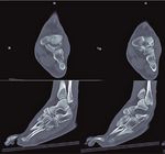
and displaced fracture of the navicular. The dorsomedial frag-
ment was displaced dorsally, with impaction of the dorsome-
dial articular surface of the talonavicular joint. The plantar-la-
teral fragment of the navicular was displaced with shortening
of the medial column of the midfoot (Figures 1 and 2). The
surgical strategy was to reduce major fragments through a
direct dorsomedial approach, restoring articular talonavicular
and naviculocuneiform surfaces with interfragmentary com-
pression screws. Subsequently, a dorsal bridge plate crossing
the navicular fracture was inserted to maintain the length of
the medial column and indirectly reduce the comminuted
fragments (Figures 3 and 4).
Surgical technique
1. The surgical procedure was performed with the patient in
the supine position under spinal anesthesia in combination
with peripheral sciatic and popliteal nerve block;
2. A pneumatic tourniquet was positioned proximally at
the thigh and inflated to the pressure of 300 mm Hg (after
Esmarch exsanguination);
3. A direct dorsomedial straight incision of 8 cm was perfor-
med from the talus neck to the medial cuneiform;
Figure 2. Computed tomography scans on the coronal, axial, and
sagittal views showing the dorsomedial fragment displaced dor-
sally, with impaction of the dorsomedial articular surface of the
talonavicular joint, and the plantar-lateral fragment comminu-
Figure 1. Initial radiographs of the foot and the ankle showing ted and displaced, with shortening of the medial column of the
comminuted and displaced fracture of the navicular. midfoot.
104 J Foot Ankle. 2022;16(1):103-7Mansur et al. Comminuted navicular fracture treated with an internal fixation plate – Technical Tip
6. The fracture of the navicular was noted with the presence
of two major fragments, one dorsomedial, and the anatomi-
cal reduction of the articular surfaces. Such fragments were
fixed with two 3.0 mm HCS™ lag screws (DePuy Synthes, J&J
Company, USA) (Figure 3C);
7. Appropriate length of the medial column of the foot
was restored and confirmed by radioscopy, and a 2.7-mm
low-profile locking plate (Medartis AG, Switzerland) was po-
sitioned at the dorsal aspect of the midfoot. Then, 2.7-mm
locking screws were inserted proximally into the talus head
and distally into the medial cuneiform, sparing the navicular
and maintaining the anatomy of the medial longitudinal arch
(Figure 3D);
Figure 4. Postoperative radiographs with navicular fracture re-
duction and restoration of the talonavicular and naviculocunei-
form joint congruence and medial column length.
A B
C D
Figure 3. A) Dorsomedial incision from the talus neck to the me-
dial cuneiform, with retraction of the anterior tibial and extensor
hallucis longus tendons. B) Intraoperative image of the fracture
of the navicular with the presence of two major fragments, one
dorsomedial and the other plantar-lateral, and naviculocuneiform
joint luxation. C) Reduction of the main articular fragments. D)
Positioning of the bridge plate at the dorsal aspect of the midfoot Figure 5. Radiographic views of the foot 10 months following the
after screw fixation of the main fragments. injury, 6 months after plate removal.
J Foot Ankle. 2022;16(1):103-7 105Mansur et al. Comminuted navicular fracture treated with an internal fixation plate – Technical Tip
Discussion
Fractures of the tarsal navicular bone are uncommon. Only
5% of foot fractures affect the midfoot, and 35% of those
affect the navicular(5). The most serious fractures may pro-
gress with midfoot collapse and/or early osteoarthritis, cau-
sing important functional limitations(1,5,6,8).
In 1989, Sangeorzan et al.(7) classified fractures of the navi-
cular body into (1) fractures with coronal line without angu-
lation, with dorsal or tuberosity avulsion; (2) body fractures
with dorsolateral to plantar-medial line with loss of the medial
arch (most common type); and (3) navicular body fractures
with central or lateral comminution. The classification system
was based on the direction of the fracture line, the direction
of displacement of the foot, and the pattern of disruption of
the surrounding joints, and was intended to assist in the surgi-
cal treatment of fractures(1,7). Type 3 fractures are challenging
injuries because a large amount of energy usually promotes
joint fragmentation and displacement. The treatment aims to
restore the medial longitudinal arch and the anatomical rela-
tionship between the hindfoot and the forefoot, promoting
stability of the midfoot(1). In this injury pattern with small-si-
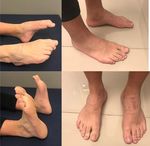
Figure 6. Clinical aspect of the feet and ankle 10 months following zed comminuted fragments, conventional fixation techniques
the initial trauma and surgery. may not be feasible or sufficiently stable(1,3,5,9). Kirschner wires
may not provide sufficient stability to maintain the length and
shape of the medial midfoot arch, and plate and screw sys-
tems may cause excessive vascular damage(9-11). Furthermore,
late complications and poor outcomes have been associa-
8. After the correct positioning of the implants and the
ted with comminuted navicular fractures, including avascu-
reduction of the fracture and joints were confirmed with
lar necrosis, delayed union, osteoarthritis, joint stiffness, and
radioscopy, the wound was irrigated with 0.9% saline and
chronic pain, especially when the medial column is not resto-
sutured with 2.0 absorbable sutures subcutaneously and 4.0
red(3,10,11). Therefore, some authors recommend the association
mononylon sutures on the skin;
of an external fixator with primary osteosynthesis or primary
9. An occlusive dressing was applied with sterile gauze arthrodesis as a salvage procedure(1,5,12).
protecting the surgical incision. The pneumatic tourniquet For comminuted fracture patterns, surgical attempts to
was released with consequent restoration of normal periphe- obtain optimal open anatomical reduction and compression
ral perfusion. fixation may be counterproductive if the vascular bone su-
10. The limb was immobilized with a removable ankle/foot ortho pply is not respected and when multiple small fragments are
pedic orthosis for 3 weeks, when the suture was removed. present. Important fundaments are to carefully plan surgical
approaches, use indirect reduction techniques when possible,
During the first 6 weeks, we recommended avoiding bearing
and consider biologic fixation(6). For example, in a series(11) of
weight on the affected foot, allowing only toe-touch weight-
10 types 2 and 3 navicular fractures treated with a navicular
bearing. Stiffness of the talonavicular joint is expected while
locking plate, the authors reported the presence of healed
the bridge plate is present. After week 6, the patient was re-
fractures in all patients and overall good functional outcomes
commended to progressively bear weight with the orthosis of the feet after 20 months of follow-up. However, one pa-
until week 9, when the orthosis was discontinued. At week tient had a compartment syndrome requiring fasciotomy, and
16, we confirmed by clinical and radiographic evaluation the another developed partial necrosis of the navicular(11).
presence of fracture healing (Figure 4), preservation of the
Bridge plating has been used more recently in trauma-
medial longitudinal arch and joint congruence, and mainte-
tic foot injuries, mainly in severe and comminuted fractures
nance of the length of the medial column of the foot. At that associated with subluxation and bone shortening or impac-
moment, the patient underwent a second surgical procedure, tion. Biomechanical cadaveric studies(13) and case series stu-
and the bridge plate was removed. Ten months following the dies(14) have suggested that outcomes following surgical Lis-
fracture, new radiographs showed a healed navicular fractu- franc joint injuries might be superior when fixed with dorsal
re with joint congruence and without avascular necrosis. The bridge plating compared with screw fixation only. A previous
patient presented with a range of motion similar to the con- study(10) reported the outcomes of 7 patients with comminu-
tralateral side, had no complaints of pain, and had resumed ted and displaced type 3 fractures of the navicular treated
sports activities (Figures 5 and 6). surgically with bridge plating. The authors used a 2.7-mm re-
106 J Foot Ankle. 2022;16(1):103-7Mansur et al. Comminuted navicular fracture treated with an internal fixation plate – Technical Tip
construction plate with 8 to 10 holes at the medial column of and the medial column is stabilized by the bridge plate, ru-
the foot, from the talus to the first metatarsal. The 2 proxi- ling out the need for external fixators and avoiding complica-
mal and 2 distal holes were fixed with screws, the distal ones tions and multiple incisions. A disadvantage of the technique
being long enough to reach the lateral column(15). Apostle and is the need for a new surgery to remove the plate. Previous
Younger(15) described a technique consisting of double bridge authors(10,15) have recommended removal after 4–9 months to
plating to the medial and middle cuneiforms. In the current allow restoration of function in the transverse tarsal and sub-
technical report, we performed bridge plating fixation using talar joints. Because of the possibility of delaying early osteo-
a 2.7 mm locking plate, inserting two screws into the talus arthritis and arthrodesis, we consider the present technique
and the medial cuneiform. Therefore, in addition to a single to be a good alternative in selected cases. The case presen-
and smaller incision, the Lisfranc joint and the bones of the ted to illustrate this technical tip was followed up only for 10
lateral column were not included in the fixation, avoiding joint months, which is an important limitation. Therefore, mid- and
stiffness and symptoms in the lateral column. long-term follow-up studies are warranted to evaluate the
In our opinion, the bridge plating technique is indicated for maintenance of anatomical alignment, functional scores, and
severely comminuted navicular fracture–dislocations. In these development of post-traumatic osteoarthritis.
fractures, the forefoot assumes an abducted position, and In sum, the technique described herein has the following ad-
comminution of the medial midfoot results in shortening of vantages: (a) maintenance of the medial longitudinal column
the medial column, which may lead to a midfoot collapse(1). in comminuted navicular fractures; (b) indirect reduction of
Direct open reduction and fixation of small bony fragments the small bone fragments; (c) maintenance of the congruen-
may not be achieved, in addition to leading to excessive vas- ce of the talonavicular and naviculocuneiform joints; (d) lon-
cular damage. Furthermore, medial column instability may ger protection in potential cases of vascular damage of the
require the addition of an external fixator. In the technique bone tissue or delayed union; and (e) lower morbidity with
described, the small fragments, especially the plantar ones, limited surgical incisions, saving the Lisfranc joint and the la-
which are difficult to achieve, are reduced by ligamentotaxis, teral column of the foot.
Authors’ contributions: Each author contributed individually and significantly to the development of this article: HM *(https://orcid.org/0000-0001-7527-
969X) Data collection, interpreted the results of the study, wrote the article; LLS *(https://orcid.org/0000-0003-3006-7831) Wrote the article; DAM
*(https://orcid.org/0000-0002-3893-0292) Wrote the article, participated in the review process. All authors read and approved the final manuscript.
*ORCID (Open Researcher and Contributor ID) .
References
1. Ramadorai MU, Beuchel MW, Sangeorzan BJ. Fractures and 9. Karmali S, Ramos JT, Almeida J, Barros A, Campos P, Costa DS.
dislocations of the tarsal navicular. J Am Acad Orthop Surg. Tarsal navicular fracture in a parkour practitioner, a rare injury –
2016;24(6):379-89. Case report and literature review. Rev Bras Ortop. 2019;54(6):739-45.
2. Burne SG, Mahoney CM, Forster BB, Koehle MS, Taunton JE, 10. Schildhauer TA, Nork SE, Sangeorzan BJ. Temporary bridge
Khan KM. Tarsal navicular stress injury: long-term outcome and plating of the medial column in severe midfoot injuries. J Orthop
clinicoradiological correlation using both computed tomography Trauma 2003;17(7):513-20.
and magnetic resonance imaging. Am J Sports Med 2005;33(12): 11. Cronier P, Frin JM, Steiger V, Bigorre N, Talha A. Internal fixation
1875-81. of complex fractures of the tarsal navicular with locking plates.
3. Schmid T, Krause F, Gebel P, Weber M. Operative treatment of A report of 10 cases. Orthop Traumatol Surg Res. 2013;99(4
acute fractures of the tarsal navicular body: midterm results with Suppl):S241-9.
a new classification. Foot Ankle Int. 2016;37(5):501-7. 12. Moreira ET, Andrade CA, Maluf Neto J, Lovisotto LA. Navicular
4. Pinney SJ, Sangeorzan BJ. Fractures of the tarsal bones. Orthop bone comminuted fracture: medial plate fixation. Case report and
Clin North Am. 2001;32(1):21-33. literature review. Rev ABTPé. 2014;8(1):39-44.
5. Marshall D, MacFarlane RJ, Molloy A, Mason L. A review of the 13. Alberta FG, Aronow MS, Barrero M, Diaz-Doran V, Sullivan RJ,
management and outcomes of tarsal navicular fracture. Foot Adams DJ. Ligamentous Lisfranc joint injuries: a biomechanical
Ankle Surg. 2020;26(5):480-6. comparison of dorsal plate and transarticular screw fixation. Foot
6. DiGiovanni CW. Fractures of the navicular. Foot Ankle Clin. 2004; Ankle Int. 2005;26(6):462-73.
9(1):25-63. 14. Hu SJ, Chang SM, Li XH, Yu GR. Outcome comparison of Lisfranc
7. Sangeorzan BJ, Benirschke SK, Mosca V, Mayo KA, Hansen ST Jr. injuries treated through dorsal plate fixation versus screw fixation.
Displaced intra-articular fractures of the tarsal navicular. J Bone Acta Ortop Bras. 2014;22(6):315-20.
Joint Surg Am. 1989;71(10):1504-10. 15. Apostle KL, Younger AS. Technique tip: open reduction internal
8. Coulibaly MO, Jones CB, Sietsema DL, Schildhauer TA. Results and fixation of comminuted fractures of the navicular with bridge
complications of operative and non-operative navicular fracture plating to the medial and middle cuneiforms. Foot Ankle Int.
treatment. Injury. 2015;46(8):1669-77. 2008;29(7):739-41.
J Foot Ankle. 2022;16(1):103-7 107You can also read


