Cold shock induces a terminal investment reproductive response in C. elegans
←
→
Page content transcription
If your browser does not render page correctly, please read the page content below
www.nature.com/scientificreports
OPEN Cold shock induces a terminal
investment reproductive response
in C. elegans
Leah Gulyas1,2 & Jennifer R. Powell1*
Challenges from environmental stressors have a profound impact on many life-history traits of an
organism, including reproductive strategy. Examples across multiple taxa have demonstrated that
maternal reproductive investment resulting from stress can improve offspring survival; a form of
matricidal provisioning when death appears imminent is known as terminal investment. Here we
report a reproductive response in the nematode Caenorhabditis elegans upon exposure to acute cold
shock at 2 °C, whereby vitellogenic lipid movement from the soma to the germline appears to be
massively upregulated at the expense of parental survival. This response is dependent on functional
TAX-2; TAX-4 cGMP-gated channels that are part of canonical thermosensory mechanisms in worms
and can be prevented in the presence of activated SKN-1/Nrf2, the master stress regulator. Increased
maternal provisioning promotes improved embryonic cold shock survival, which is notably suppressed
in animals with impaired vitellogenesis. These findings suggest that cold shock in C. elegans triggers
terminal investment to promote progeny fitness at the expense of parental survival and may serve as a
tractable model for future studies of stress-induced progeny plasticity.
Environmental stressors can severely jeopardize the ability of an organism and its progeny to survive and repro-
duce, endangering organismal fitness. Consequently, organisms must launch effective stress response mecha-
nisms to mitigate damage, promote recovery, and/or ensure progeny survival. A traditional view of life-history
trade-offs assumes that challenge from a stressor should invoke a response in which resources are shuttled
away from reproduction to support organismal defense and repair1. While this approach temporarily decreases
reproductive fitness, improved long-term survival may allow for greater reproduction post-recovery. However,
in a stressful environment where survival is perceived to be unlikely, an alternate strategy may instead be to
funnel resources into reproduction, ensuring maximal output prior to parental death and subsequent survival
of the F1 g eneration1.
The benefit of this strategy has been demonstrated in a number of organisms in different environmental
conditions2. Both biotic (i.e. pathogen exposure) and abiotic (thermal, osmotic) stress conditions may potenti-
ate molecular changes to reproduction. Alterations ranging from epigenetic modifications to simple maternal
cytosolic investments of macromolecules and nucleic acids have been associated with increased offspring resil-
ience to ensuing t hreats2,3. Yet despite myriad examples, the precise mechanisms by which stressors induce these
responses are still widely under exploration.
The nematode Caenhorhabditis elegans recently arose as an ideal model for investigating stress-induced
parental progeny investment. Several studies have detailed molecular tactics by which worms modify and provi-
sion gametes and embryos in response to dietary restriction, infection by pathogenic pseudomonads and micro-
sporidia, and osmotic stress4–7. Among an array of environmentally-relevant stressors, though, the reproductive
impacts of thermal stress (specifically cold stress) on C. elegans have not been closely assessed. Both flies and
mice employ reproductive strategies to respond to cold, with flies inducing a more general stress response of
reproductive dormancy and mice depositing epigenetic modifications in sperm to promote retention of brown
adipose tissue favoring increased metabolic rates in offspring8,9. Given the likelihood of C. elegans regularly
encountering diurnal and seasonal temperature fluctuations, it seems probable that worms may also have evolved
rigorous reproductive programs to deal with cold exposure.
Several aspects of cold response in C. elegans have been well-characterized in the literature, including ther-
motaxis and h abituation10–12. Neurons and other tissues rely on a multitude of signaling mechanisms for these
physiological responses to occur, which are in turn supported by a bevy of channels and other proteins. These
processes are described in detail in a recent review from Takeishi et al.12. Only lately, though, have links between
1
Department of Biology, Gettysburg College, Gettysburg, PA 17325, USA. 2Present address: Plant and Microbial
Biology Department, University of California Berkeley, Berkeley, CA 94702, USA. *email: jpowell@gettysburg.edu
Scientific Reports | (2022) 12:1338 | https://doi.org/10.1038/s41598-022-05340-6 1
Vol.:(0123456789)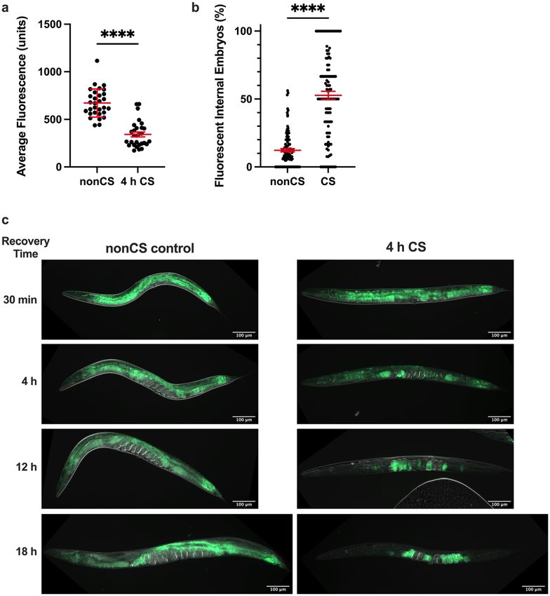
www.nature.com/scientificreports/
cold and reproduction begun to emerge. Sonoda et al.13 demonstrate that the presence and integrity of sperm is
required for normal cold tolerance by exerting an effect through multiple tissues, including ASJ neuronal activity.
At the level of population dynamics, it has also been reported that C. elegans warming from a 2-h cold exposure
enter a state of programmed death which is thought to result in kin selection for the survival of younger (and
presumably more reproductively fit) a nimals14. However, the extent to which acute cold stress directly impacts
reproductive capacity in these animals remains unknown. As alternating periods of cold and moderate tem-
peratures preceding a winter season may comprise a substantial portion of the short reproductive window in C.
elegans, this necessitates further study.
We have previously shown that acute cold shock at 2 °C causes loss of intestinal pigmentation, immobility,
and reproductive disruption in wild-type hermaphrodite worms. In many cases, this phenotypic program results
in lethality15. Here we extend this work by characterizing the induction of these phenotypes via the neuronal
TAX-2/TAX-4 thermotransduction channel, mediated by the canonical stress response regulator SKN-1/Nrf2.
We further describe a role for the normal vitellogenesis machinery in reallocating pigmented intestinal lipid sup-
plies to the germline, which appears to come at the expense of parental survival following cold exposure. Overall,
our findings suggest that acute cold shock provokes a terminal investment reproductive response in C. elegans,
making this discovery an exciting opportunity to explore the phenomenon in a genetically tractable system.
Results
Acute cold shock causes drastic phenotypic alterations. The duration of cold exposure for young
adult hermaphrodite C. elegans at 2 °C is negatively correlated to post-shock survival rates15. Wild-type her-
maphrodite worms exposed to a 4-h cold shock (CS) do not initially display high mortality rates (Fig. 1a); this
allows observation of a range of phenotypic transitions as they recover from the limited-duration cold stress at
their preferred temperature of 20℃. One of the most striking phenotypes exhibited in post-cold shock (post-CS)
animals during the recovery period is a dramatic decrease in pigmentation in the normally highly pigmented
intestine, so that the body becomes almost entirely clear (Fig. 1b, c)15. This is often accompanied by motor
and reproductive disruptions such as mobility loss, withering of the gonad arms, decreased number of internal
embryos, and the eventual death of about 30% of the population (Fig. 1a–d)15. It should be noted that these
phenotypic responses do not appear to be due to any relative heat shock following the transition from 2 to
20 °C as the expression of GFP-tagged HSP-4 (heat shock protein) is not induced following cold shock (Fig. 1e).
Neither is the reduced pigmentation following cold shock due to a period of starvation presumably experienced
by the worms while they are at 2 °C. At this extreme cold temperature, the worms enter a “chill coma” in which
pharyngeal pumping and virtually all other movement c eases15,16; however, a total absence of food for a similar
time period does not induce a comparable clearing phenotype (Supplemental Fig. S1). Interestingly, some CS
wild-type animals regain pigmentation after clearing; these worms do not die and display a general reversal of
the other negative impacts of cold shock (Fig. 1b)15. We sought to better understand the factors regulating the
post-CS recovery program in wild-type worms, focusing particularly on the functional role of pigmentation loss
and the genetic components involved in producing it.
Cold shock induces lipid reallocation from somatic tissues to the germline. Since pigmentation
in the C. elegans intestine is due in part to the presence of lipid-storing fat d
roplets17, we wondered whether
the decrease in pigmentation in cold-shocked worms corresponds to a depletion of intestinal lipid supplies,
potentially as a result of increased metabolism meant to fuel post-cold shock recovery. Using Nile Red, which
accumulates and fluoresces in hydrophobic e nvironments18, as an indicator of lipid content, we therefore ana-
lyzed the fat stores of worms 12 h following CS (note that all cold shocks were performed for 4 h at 2℃). We
indeed observed a significant decrease (P < 0.0001) in the average lipid content per worm (Fig. 2a); visually, this
presents as an overall reduction in average fluorescence that is most striking in the intestine (Fig. 2c). However,
we also unexpectedly noted that the lipid content of the gonad appears to concomitantly increase, marked by
the presence of fluorescent (and therefore lipid-rich) internal embryos in the gonads of many cold-shocked
worms that were mostly absent in their non-cold shocked counterparts (Fig. 2b, c). These embryos are some-
what sporadic, usually accounting only for a proportion of all embryos in the germline and are interspersed with
non-fluorescent embryos. Importantly, the percentage of fluorescent internal embryos per worm was found to
be elevated two-fold in CS worms relative to that of nonCS controls (Fig. 2b). These observations suggest that
rather than just metabolically depleting lipid supplies, C. elegans may also reallocate lipids from the intestine to
the germline. To further analyze this process spatiotemporally, we performed a time course of Nile Red staining
following cold shock (Fig. 2c). After exit from cold shock and the corresponding chill coma15,16, most worms
gradually resume movement over the first 30 min of recovery at 20℃. At this point, the distribution of Nile Red-
staining lipids is indistinguishable from non-cold shocked controls, indicating that lipid reallocation occurs
after, rather than during, the cold shock. By 4 h post-cold shock, we observed visually decreased but rarely absent
fluorescence in the intestine; at this time, many worms have brightly fluorescing proximal oocytes, suggesting
that lipids are beginning to relocate from the intestine into the germline. This process of reallocation appears
to continue over the next 14 + hours, with many 12 h-recovered worms containing fluorescent embryos and
oocytes but only slight intestinal fluorescence, and most 18 h-recovered worms fluorescing almost exclusively in
the embryos. Though it is unclear what other factors (e.g. metabolism) may contribute to the overall pigmenta-
tion loss, these observations suggest that the dramatic decrease in intestinal pigmentation results in large part
from the reallocation of lipids from somatic tissues to the germline during the recovery phase following a severe
cold shock.
Scientific Reports | (2022) 12:1338 | https://doi.org/10.1038/s41598-022-05340-6 2
Vol:.(1234567890)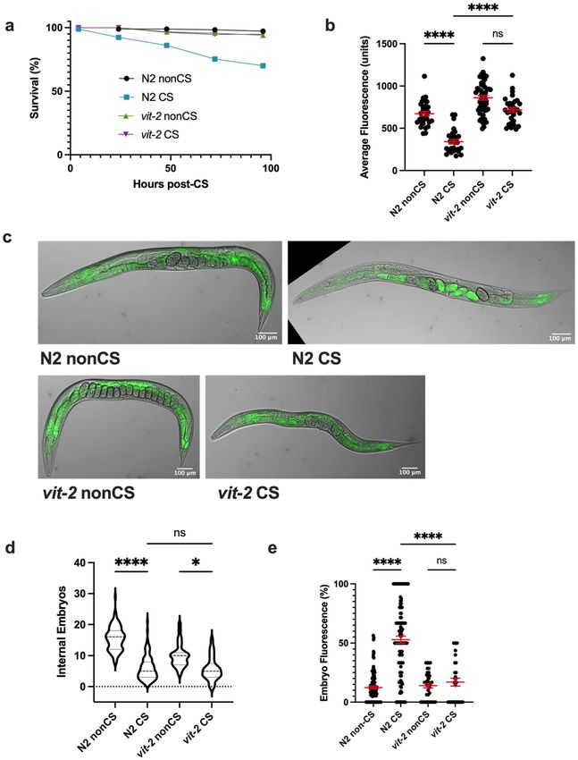
www.nature.com/scientificreports/
Figure 1. Cold-shocked worms show decrease in survival and characteristic phenotypic alterations. N2 young
adult hermaphrodites were shifted from 20 to 2 °C for a 4 h cold shock (CS) and thereafter recovered at 20 °C
for 96 h with assessment of (a) survival and (b) phenotypic alterations (n = 177). Death and immobility were
assayed by nose tap; worms were considered to be immobile if the tap elicited slight movement in the head
region but no other body movement, and dead worms were completely unresponsive (Chi-squared Test for
Homogeneity: P < 0.0001 at 24 h). (c) Representative images of young adult N2 hermaphrodites following cold
shock. (d) Average number of internal embryos per worm in CS versus nonCS conditions (Mann–Whitney U
test: U = 604, two-tailed P < 0.0001). (e) Induction of heat shock protein HSP-4::GFP 12 h after either 2 h heat
shock at 35 °C, 4 h cold shock at 2 °C, or no treatment (n ≥ 26 per condition; Kruskal–Wallis test: H = 33.86,
P < 0.0001; Dunn’s Multiple Comparison Test: ****P < 0.0001). Error bars are mean ± s.e.m.
Thermosensation is required to initiate the cold shock response. We next asked how wild-type
C. elegans perceive and interpret cold shock as a signal for lipid reallocation. The cGMP-gated channel subunits
TAX-2 and TAX-4 are important for thermosensation and acquired cold tolerance in worms, acting specifically
in the ASJ neurons11,19,20. We reasoned that worms might also rely on a neuronal mechanism of sensation that
acts through the TAX-2/TAX-4 channel to induce the cold shock response in the absence of prior cold exposure.
tax-2(p671); tax-4(p678) double loss-of-function mutants were therefore tested for sensitivity to cold shock. Not
only do these mutants show 100% survival following cold shock (Fig. 3a), but, unlike wild-type worms, they
also do not display any significant difference (P > 0.9999) in overall lipid content 12 h after CS, nor the striking
lipid reallocation phenotype characterized by large numbers of fluorescent embryos (Fig. 3b–d). Interestingly,
despite this resilience to shock, tax-2(p671); tax-4(p678) mutants still show decreased internal embryo quanti-
ties, though not to the same degree as N2 worms (Fig. 3e), suggesting that cold exposure has some additional
Scientific Reports | (2022) 12:1338 | https://doi.org/10.1038/s41598-022-05340-6 3
Vol.:(0123456789)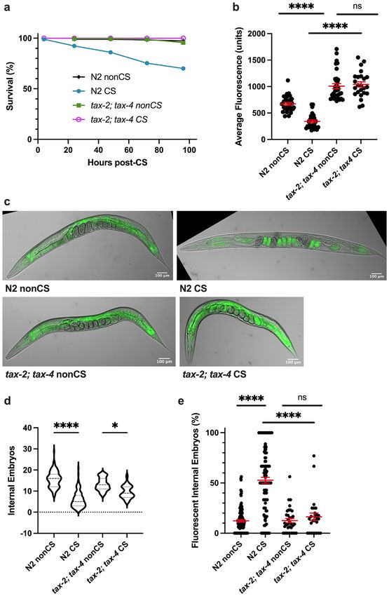
www.nature.com/scientificreports/

                                            Figure 2.  Cold shock recovery is associated with lipid localization shifts from the intestine to the germline.
                                            Young adult N2 hermaphrodites were exposed to 2 °C cold shock or control nonCS conditions (20 °C) for 4 h
                                            and recovered before Nile Red staining and imaging for lipid content. (a) Average fluorescence per worm (n ≥ 29
                                            per condition; Mann–Whitney U test: U = 48; two-tailed P < 0.0001) and (b) percent of internal fluorescent
                                            embryos per worm (n ≥ 111; Mann–Whitney U test: U = 1885; two-tailed P < 0.0001) were quantified 12 h
                                            post-CS. Error bars are mean ± s.e.m. (c) Representative images of Nile Red-stained worms at indicated time
                                            points post-CS show progression of intestinal lipid loss and relocalization to the embryos.

                                            impact on fertility independent of this thermosensation pathway. Taken together, though, these data support
                                            the hypothesis that fat reallocation as a stress response following CS may depend on a neuronal mechanism for
                                            induction. Since TAX-2/TAX-4 plays a role in temperature sensation, we speculate that canonical cold ther-
                                            mosensation is required for lipid reallocation to take place.

          Scientific Reports |   (2022) 12:1338 |               https://doi.org/10.1038/s41598-022-05340-6                                                4

Vol:.(1234567890)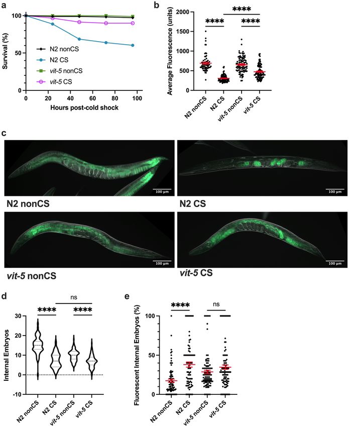
www.nature.com/scientificreports/

                                  Figure 3.  TAX-2; TAX-4-mediated thermosensation is required for lipid relocalization following cold
                                  shock. (a) tax-2(p671); tax-4(p678) double loss-of-function mutants were cold shocked and recovered while
                                  monitoring survival rates (n ≥ 120 worms per condition). (b) At 12 h post-CS or nonCS control, tax-2(p671);
                                  tax-4(p678) were Nile Red lipid-stained and the average fluorescence per worm was quantified (Kruskal–Wallis
                                  test: H = 83.83, P < 0.0001; Dunn’s Multiple Comparison test: ****P < 0.0001). (c) Representative images of Nile
                                  Red staining show retention of fluorescence in the intestines of tax-2; tax-4 mutants. (d) Number of embryos
                                  (Kruskal–Wallis test: H = 171.1, P < 0.0001; Dunn’s Multiple Comparison test: *P = 0.0484, ****P < 0.0001) and (e)
                                  percent fluorescent internal embryos (Kruskal–Wallis test: H = 100.1, P < 0.0001; Dunn’s Multiple Comparison
                                  test: ****P < 0.0001) were quantified per worm from Nile Red images (n ≥ 24 worms per condition for b-e; error
                                  bars are mean ± s.e.m.).

Scientific Reports |   (2022) 12:1338 |                https://doi.org/10.1038/s41598-022-05340-6                                                 5

                                                                                                                                             Vol.:(0123456789)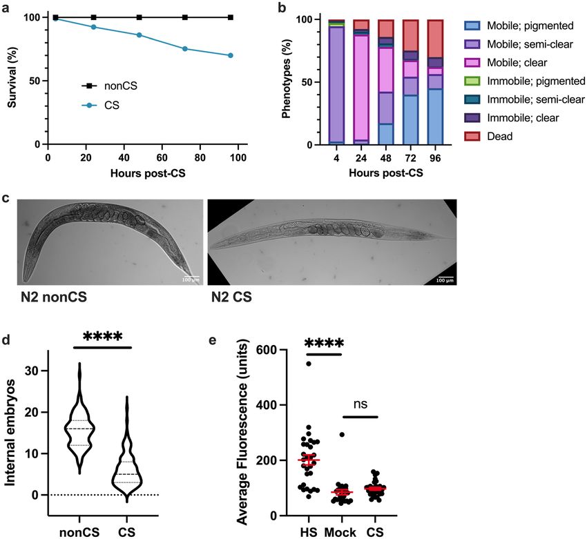
www.nature.com/scientificreports/
SKN‑1 promotes cold stress resistance. After determining a requirement for neuronal signaling in
cold-shock-induced lipid reallocation, we next wondered what intermediate factors were needed to translate the
cold stimulus into a signal to mobilize resources. The master regulatory transcription factor SKN-1/Nrf2 coordi-
nates a return to homeostasis following a variety of stresses, including oxidative stress4, pathogen infection21,22,
and osmotic stress23. We therefore predicted that skn-1 would perform a similar function during cold stress
recovery. Indeed, skn-1(lax188) gain-of-function mutants24 are highly resistant to cold shock, with nearly 100%
survival rates 96 h post-CS (Fig. 4a). Consistent with a retention of overall lipid stores by Nile Red Staining
(Fig. 4b,d), these mutants did not contain significantly greater numbers of fluorescing internal embryos fol-
lowing cold shock (P > 0.9999), though they did have a marginal reduction in the number of internal embryos
(Fig. 4d–f). Conversely, skn-1(mg570) loss-of-function mutants displayed a wild-type cold shock response, with
significant reallocation (P < 0.00001) of lipids to the germline and loss of somatic fats (Fig. 4c–f). Because mg570
eliminates only the skn-1a mRNA i soform25, but skn-1(lax188gf) activates both skn-1a and skn-1c, we addition-
ally tested the CS response of skn-1(zj15) loss-of-function mutants, which are reported to have a 76% reduction
in both skn-1a and skn-1c mRNA levels26. We observed a similar clearing phenotype (Supplemental Fig. S2);
however, these animals appeared slow-growing and less robust, which may have contributed to more substantial
CS-induced lethality. Taken together, these data suggest that the skn-1a and perhaps also the skn-1c isoforms
are not required for lipid mobilization in response to cold shock. However, SKN-1a/c activation protects cold-
shocked worms from recovery phase lethality and prevents lipid reallocation.
Lipid reallocation results from upregulated vitellogenesis following cold shock. The process
governing the normal movement of lipids from the soma to the germline is vitellogenesis, requiring the vitel-
logenin proteins coded for by vit-1–6 genes. Once coupled to somatic lipid supplies, the resulting lipoprotein
complexes shuttle to the germline27. We hypothesized that the lipid mobilization induced by cold shock is a
result of the normal vitellogenesis machinery being commandeered as part of a stress response.
One of the major vitellogenins is VIT-2, which generates a 170 kD yolk protein product known as YP170B28.
We predicted that loss of vit-2 would reduce lipid reallocation following CS. Since preventing reallocation in our
previous mutants also increased post-CS survival, we expected that vit-2 loss of function would enhance protec-
tion from CS. Consistent with these hypotheses, impairment of vitellogenesis in vit-2(ok3211) loss-of-function
mutants produced phenotypes similar to tax-2(p671); tax-4(p678) animals. In addition to maintaining high
survival rates, CS vit-2(ok3211) mutants did not undergo a substantial loss of intestinal lipid supplies, nor did
they exhibit a marked increase in internal embryo fluorescence (despite a modest decrease in internal embryo
counts) (Fig. 5a–e).
Since other vitellogenin transcripts produce different lipoprotein forms for movement to the germline, we
decided to additionally test whether a member of the YP170A-generating class of vitellogenins would reca-
pitulate our results with vit-2. To this end, we analyzed the CS phenotypes of vit-5(ok3239) loss-of-function
animals. Indeed, we found that as in vit-2 mutants, survival was rescued in these animals following CS, cor-
responding with a retention of somatic lipids and inhibition of embryonic lipid reallocation (Fig. 6a–c,e). As
with other mutants that prevent lipid reallocation, there was still an effect of cold in reducing overall embryo
output 12 h post-CS, hinting that temperature impacts fertility independent of the other reproductive alterations
exhibited by wild-type worms (Fig. 6d).
Cold shock response is a form of a terminal investment. Our data thus far suggest that upon recov-
ery from acute CS, wild-type C. elegans induce a reproductive phenotype whereby somatic lipid supplies are
massively relocalized to the germline using the normal vitellogenesis machinery. This appears to come at the
expense of the parent’s own mortality, as retaining somatic lipids by preventing thermosensation or vitellogen-
esis is sufficient to rescue survival. Such a trade-off between survival and reproduction is redolent of the terminal
investment hypothesis (reviewed in Gulyas and Powell, 20192), which predicts that in some instances of acute
stress where the likelihood of death is high, organisms can preferentially funnel resources to reproduction to
maximize reproductive fitness at the cost of survival.
To determine whether cold stress-associated phenotypes in C. elegans are an example of such a process, we
eliminated all potential for reproductive investment by assaying sterile glp-1(e2141) and glp-1(q231) worms
to ask whether these worms still exhibited lipid loss or death upon cold shock. Strikingly, the absence of a
germline in these animals completely prevented both intestinal clearing and death, again confirming that lipid
movement from the soma to the germline is associated with parental lethality (Fig. 7a). If resource realloca-
tion to the progeny is indeed meant to increase reproductive fitness in inclement conditions, there should be
some benefit to the progeny of CS worms that offspring of nonCS parents do not receive. We speculated that
in the case of environmental CS, a sudden, seasonal, cold to warm cycle might signal likelihood of future cold
conditions that would impede the ability of embryos to hatch and survive to reproductive age. We therefore
devised an assay to test for the relative fitness of embryos in cold conditions depending on whether they came
from nonCS or CS parents and were thus more likely to have received lipid provisioning. To do this, embryos
were collected from nonCS and CS parents within a 2 h time window when post-CS lipid reallocation seems
to peak and subsequently exposed them to a cold stress of 24 h. We then assessed hatching rates 24 h following
the embryonic cold shock. Excitingly, embryos coming from parents that had experienced cold shock prior
to reproduction exhibited a small but significant increase in hatching rates relative to their counterparts from
nonCS hermaphrodite parents. Furthermore, preventing lipid reallocation by impairment of vitellogenesis in
either vit-2(lf) or vit-5(lf) was able to substantially ablate this effect, suggesting that the improved survival is
attributable specifically to lipid investment in the F1 generation (Fig. 7b, c). Altogether, it appears that while
preventing embryonic lipid endowment may promote adult survival post-CS, offspring that go on to experience
Scientific Reports | (2022) 12:1338 | https://doi.org/10.1038/s41598-022-05340-6 6
Vol:.(1234567890)www.nature.com/scientificreports/
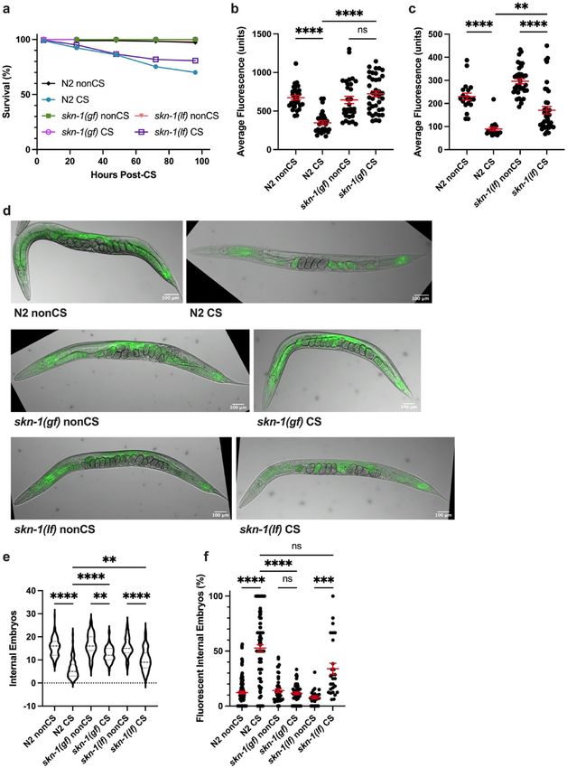
Figure 4. Activated SKN-1 prevents lipid relocalization following cold shock (a) skn-1(lax188) gain-of-function mutants and skn-
1(mg570) loss-of-function mutants were cold shocked and recovered while monitoring survival rates (n ≥ 120 worms per condition).
(b-c) At 12 h post-CS or control nonCS, (b) skn-1(lax188) and (c) skn-1(mg570) were Nile Red lipid-stained and average fluorescence
per worm quantified (N2 v. skn-1(lax188)- Kruskal–Wallis test: H = 47.46, P < 0.0001; Dunn’s Multiple Comparison test: ****P < 0.0001;
N2 v. skn-1(mg570)- Kruskal–Wallis test: H = 64.55, P < 0.0001; Dunn’s Multiple Comparison test: **P = 0.0064, ****P < 0.0001). (d)
Relative to wild-type N2s, representative images of skn-1(lax188) mutants show maintenance of intestinal fluorescence contrary to
skn-1(mg570), which show lipid relocalization. (e) Number of embryos (Kruskal–Wallis test: H = 223.1, P < 0.0001; Dunn’s Multiple
Comparison test: **Pskn-1(gf) nonCS vs. CS = 0.0020, **PN2 CS vs. skn-1(lf) CS = 0.0014, **** P < 0.0001) and (f) percent of fluorescent internal
embryos (Kruskal–Wallis test: H = 132.0, P < 0.0001; Dunn’s Multiple Comparison test: ***P = 0.0002, ****P < 0.0001) were quantified
per worm from Nile Red images (n ≥ 26 worms per condition for b-e; error bars are mean ± s.e.m.).
Scientific Reports | (2022) 12:1338 | https://doi.org/10.1038/s41598-022-05340-6 7
Vol.:(0123456789)www.nature.com/scientificreports/
Figure 5. VIT-2-regulated vitellogenesis during recovery promotes lipid relocalization and impairs survival.
(a) vit-2(ok3211) loss-of-function mutants were cold shocked and recovered while monitoring survival rates
(n ≥ 130 worms per condition). (b) At 12 h post-CS or nonCS, vit-2(ok3211) were Nile Red lipid-stained
and average fluorescence per worm quantified (n ≥ 31 worms per condition; Kruskal–Wallis test: H = 75.18,
P < 0.0001; Dunn’s Multiple Comparison test: ****P < 0.0001). Error bars are mean ± s.e.m. (c) Images of Nile Red
Staining show intestinal lipid retention in vit-2 mutants. (d) Number of embryos (Kruskal–Wallis test: H = 169.9,
P < 0.0001; Dunn’s Multiple Comparison test: *P = 0.0149, ****P < 0.0001) and (e) percent of fluorescent internal
embryos (Kruskal–Wallis test: H = 101.5, P < 0.0001; Dunn’s Multiple Comparison test: ****P < 0.0001) were
quantified per worm from Nile Red images (n ≥ 31 for b–e; error bars are mean ± s.e.m.).
Scientific Reports | (2022) 12:1338 | https://doi.org/10.1038/s41598-022-05340-6 8
Vol:.(1234567890)www.nature.com/scientificreports/
Figure 6. VIT-5 vitellogenin family also promotes embryo lipid reallocation during cold shock recovery.
(a) vit-5(ok3239) loss-of-function mutants were cold shocked and recovered while monitoring survival rates
(n ≥ 170 worms per condition). At 12 h post-CS or control nonCS, vit-5(ok3239) were Nile Red lipid-stained
and average fluorescence per worm quantified (n ≥ 67 worms per condition; Kruskal–Wallis test: H = 172.0,
P < 0.0001; Dunn’s Multiple Comparison test: *****P < 0.0001. Error bars are mean ± s.e.m. (c) Representative
Nile Red staining shows intestinal fluorescence in vit-5(ok3239) following cold shock. (d) Number of embryos
(Kruskal–Wallis test: H = 137.2, P < 0.0001; Dunn’s Multiple Comparison test: ****P < 0.0001) and (e) percent
of fluorescent internal embryos (Kruskal–Wallis test: H = 37.58, P < 0.0001; Dunn’s Multiple Comparison test:
H = 37.58, ****P < 0.0001) were quantified per worm from Nile Red images (n ≥ 67 worms per condition; error
bars are mean ± s.e.m.).
future inclement conditions suffer diminished survival rates, underscoring the evolutionary advantage of terminal
investment as a reproductive strategy.
Scientific Reports | (2022) 12:1338 | https://doi.org/10.1038/s41598-022-05340-6 9
Vol.:(0123456789)www.nature.com/scientificreports/
a
100
Population Phenotypes (%)
Mobile, pigmented
Immobile; pigmented
Semi-clear
50 Mobile; clear
Immobile; clear
Dead
0
q2 ) no S
S
S
31 S
)n S
S
C
q2 C
C
31 nC
gl e21 C
C
p- 41 n
on
1( N2
1( 41)
)
gl 21 no
1( N2
p-
p-
e
1(
gl
p-
gl
ns
b c
40 40 **** ****
ns
Hatching rates (%)
Hatching rates (%)
30 30
**** ****
20 20
10 10
0 0
N2 vit-2 N2 vit-2 N2 vit-5 N2 vit-5
NonCS P0 CS P0 NonCS P0 CS P0
Figure 7. Cold shock response is a form of terminal investment. (a) glp-1(e2141) and glp-1(q231) were grown
at the restrictive temperature (25 °C) to induce sterility and then habituated at 20 °C for 4 h. Worms were then
cold shocked or mock shocked (nonCS) and recovered at 20 °C for 24 h and phenotypes were scored (n ≥ 197
worms for all conditions; Chi-Squared Test for Homogeneity, P < 0.0001 for N2 CS vs. e2141 CS or q231 CS).
(b) Young adult hermaphrodite N2 and vit-2(ok3211) (P0) were cold shocked or mock shocked (nonCS) for 4 h
and recovered for 16 h. Embryos from P0 treatments were collected from 16 to 18 h and cold shocked for 24 h.
Embryos were allowed to recover 24 h at 20 °C and then the number of hatched embryos was quantified. Data
points represent rates as percent hatched for a plate containing 50–100 embryos (n ≥ 10 plates per condition;
2-way ANOVA (F(3,32) = 20.87, P < 0.0001) with Tukey multiple comparison test (****P < 0.0001). Bars are
mean ± s.e.m. (c) Performed as in b with N2 and vit-5 (ok3239) worms (n = 10 plates per condition; 2-way
ANOVA (F(3,27) = 35.28, P < 0.0001) and Tukey multiple comparison test (****P < 0.0001).
Discussion
Cold shock response overlaps with other stress response phenotypes. We have provided here
the first evidence that C. elegans, upon acute cold shock (CS), is able to improve the success of progeny survival
during future cold exposure by mobilizing lipids to the germline as reproductive provisioning. This process
seems to depend on neuronal sensation of the cold temperature, which permits a shift of lipid localization from
the soma to the germline. Lipid reallocation is mediated by vitellogenins, and despite an overall decrease in
embryo output, the embryos in these worms are more resilient to future cold exposure and display more robust
hatching. The switch to a sudden last reproductive event and resulting parental mortality is consistent with the
adoption of a more semelparous lifestyle29. In this instance, potentiation by a severe environmental stress to
induce a higher quality (rather than quantity) final reproductive investment suggests that C. elegans has evolved
a terminal investment response to deal with acute cold exposure.
Scientific Reports | (2022) 12:1338 | https://doi.org/10.1038/s41598-022-05340-6 10
Vol:.(1234567890)www.nature.com/scientificreports/
Though, to our knowledge, no previous study has linked terminal investment to C. elegans, there are other
documented phenomena characterized by a similar trade-off. For example, the “age-dependent somatic deple-
tion of fat (ASDF)” phenotype is characterized by age-dependent changes in lipid homeostasis that are induced
during both starvation and oxidative stress and are highly reminiscent of post-cold-shock reallocation of lipids
from the intestine to the germline24. As is also the case with cold shock-induced terminal investment, reduction
of vitellogenic transcripts (including vit-2) suppresses lipid mobilization. Lipid reallocation during ASDF does
appear to negatively impact parental survival, but it is unclear whether this phenotype also holds functional
significance for offspring survival. Surprisingly, while the process governing the ASDF phenotype and cold
shock recovery are both regulated by the master stress response transcription factor SKN-1/Nrf2, activating
SKN-1 has opposite effects. When measuring ASDF, skn-1(gf) stimulates the shift of lipids from the intestine
to the g ermline24; in contrast, skn-1(gf) prevents lipid mobilization and promotes intestinal lipid sequestration
following cold shock. It is possible that the role of SKN-1 varies with age since ASDF occurs in much older adults
than those analyzed following cold stress. This potential will be analyzed in future work, in addition to elaborat-
ing the roles of specific skn-1 alleles during CS. The major outstanding question of how stress-altered metabolic
state impacts lipid governance and the nature of the lipids involved also merits further exploration. Although
regulation of lipid homeostasis may be via distinct pathways, the existence of mechanistically similar responses
to starvation, oxidative stress, and cold stress suggests that terminal investment may be a more generalized
response to severe stressors in C. elegans.
Despite the fact that terminal investment requires extremely high parental resource investment, there are other
similar and less severe forms of maternal provisioning in C. elegans. For example, osmotic stress shifts embryonic
contents to include less glycogen and more glycerol, a cryoprotectant5. It is particularly relevant to our study that
mild nutrient deprivation in hermaphrodites leads to intergenerational plasticity in which embryos tend to be
slightly larger and offspring are somewhat protected from the effects of larval s tarvation4. Remarkably, this corre-
sponds to decreased maternal insulin signaling and upregulated vitellogenin provisioning in the germline, much
as we report during cold exposure30. It will be interesting in future experiments to dissect how thermosensory
signaling may hyperactivate vitellogenesis upon acute cold stress, especially as it relates to different vitellogenin
yolk protein products. Additionally, analyzing how various stressors impact and modify embryonic composition
will be important for understanding the extent to which maternal investments are conserved. Taken together, it
is clear that stressors in the parental generation can have remarkable consequences on reproductive and survival
trade-offs as well as offspring physiology and plasticity.
Terminal investment in response to cold may have evolved in regularly fluctuating environ-
mental conditions. Since the preeminent goal of survival is reproduction, terminal investment is sensible
from a fitness standpoint. But why would cold shock elicit this phenotype? Experimental evolution in C. elegans
reveals that deterministic maternal effects (those that attempt to successfully invest offspring for survival in
a particular environment) evolve in response to predictably fluctuating e nvironments31. Thus, we conjecture
that predictably encountered cold environments, such as those during seasonal freeze–thaw cycles in temperate
regions, are likely to have given rise to the reallocation phenotype. This could happen by one of the two possible
mechanisms. First, cold shock-induced damage may signal to the worm that death is imminent and reproductive
timespan is limited. Worms may then funnel all their resources into embryos which are then better capable of
surviving cold stress. However, this seems unlikely since these data suggest that C. elegans that avoid reallocation
(or are genetically prevented from doing so) survive CS extremely well, which is inconsistent with CS causing
large amounts of damage. It is possible that these worms are simply more resistant to CS-induced damage or are
better able to repair damage in the absence of reallocation, though this would need to be tested more rigorously.
A second, more plausible scenario is that seasonal cold cycles signal a coming winter season with a low chance
of offspring survival. At specific larval stages, worms can enter a highly stress-resistant state (called the dauer
stage) which may enable them to survive the winter season and resume development when conditions are more
favorable. If the better-provisioned embryos of worms that have recently experienced a cold cycle survive suc-
cessive cold shock, this could favor a larger percentage of offspring that are able to successfully enter the dauer
state. Long-term, this would translate to a higher probability that offspring survive to reproduce when winter
conditions are alleviated, continuing the parent’s lineage. This model accounts for the selection of the realloca-
tion trait in evolutionary history. It further suggests that rather than being induced as a result of damage that
signals impending death (and thus a final chance to reproduce), reallocation may instead be prompted by a more
general prediction of future damage to both the parent and the offspring. Further experimentation is needed to
assess this model and to determine how it would fit into the known framework for terminal investment strategies.
Although we have largely considered simple maternal effects involving cytosolic investments in this work,
there is also the potential for epigenetic effects at the level of gene expression to play a role in intergenerational
and transgenerational offspring survival. Indeed, the progeny of C. elegans submitted to various stressors dem-
onstrate improvements in stress survival to the same and other types of stress. Recently, a study by Burton et al.7
examined multigenerational responses to a number of stressors including nutrient deprivation and pathogenic
stress and found that multiple Caenorhabditis species regulate gene expression in the F1 generation in a stress-
specific manner to benefit survival upon exposure to the same. These studies highlight the evolutionary advantage
of provisioning offspring for what is perceived to be a likely future. It would be interesting to extend analyses of
multigenerational gene regulation to see if this is also at play in the context of CS.
Terminal investment has consequences for population‑level dynamics. Terminal investment
occurs across a broad phylogenetic range in response to many stressors, and the potential ecological and evolu-
tionary implications are serious in today’s global climate. Pathogen exposure commonly elicits terminal invest-
Scientific Reports | (2022) 12:1338 | https://doi.org/10.1038/s41598-022-05340-6 11
Vol.:(0123456789)www.nature.com/scientificreports/
ment; one particularly relevant example occurs in several species of frog in response to infection by Batra-
chochytrium dendrobatidis (Bd), the perpetrator of severe global amphibian declines. Rather than retaining
resources for survival, individuals in many of these species direct energy to reproduction, increasing gamete
output. While producing more offspring improves the likelihood of population persistence, this mode of repro-
duction does not favor survival of frogs past infection, whereupon animals selected by resilience would engender
less susceptible offspring and gradually allow population resistance to the fungus to evolve32. Thus, terminal
investment poses a serious long-term survival risk for some frog species faced with extinction from Bd. As global
climates shift, it is unclear how population dynamics will be affected by changing temperatures. Since terminal
investment is suspected to be a means of population persistence, it may play an important role in continued
population survival of various species.
In most documented cases, terminal investment results in increases to brood size but some examples exist in
which offspring quality is favored, such as in the tsetse fly, where stress levels are positively correlated with the
percent of body fat that is dedicated to offspring33. The majority of terminal investment studies currently come
from ecological studies in non-model systems; thus, an understanding of the cellular components and mecha-
nisms involved in terminal investment, particularly in relation to quality investment, remains largely elusive. Here
we have identified a new terminal investment process in response to cold stress in a genetically tractable and envi-
ronmentally relevant model. The opportunity to better understand how this phenomenon is elicited and executed
on the molecular scale is an exciting prospect for future studies. With a more comprehensive understanding
of the impacts of stress on not only the parental generation but multiple generations thereafter, we can better
predict both an organism’s physiology and population dynamics in response to specific environmental factors.
Materials and methods
Strains and maintenance. All strains were maintained on Nematode Growth Medium (NGM) seeded
with Escherichia coli OP50, at 20 °C unless otherwise noted34. Worms were well-fed for at least three gener-
ations before any experiments. Some strains were provided by the CGC, which is funded by NIH Office of
Research Infrastructure Programs (P40 OD010440). CGC strains used in this study were N2 (Bristol) wild-type,
SJ4005 zcIs4[hsp-4::gfp], BR5514 tax-2(p671); tax-4(p678), GR2245 skn-1(mg570), QV225 skn-1(zj15), RB2365
vit-2(ok3211), RB2382 vit-5(ok3239), CB4037 glp-1(e2141), and JK509 glp-1(q231). JRP1036 skn-1(lax188) was
generated from SPC168 dvIs19; skn-1(lax188).
Cold shock survival. For adult cold shock, approximately 20–70 young adult worms (not yet gravid) were
picked to OP50-seeded 3.5 cm NGM and placed at 2 °C for 4 h. After 4 h cold shock, plates were transferred to
20 °C for worm recovery. Worms were scored at 1, 4, 24, 48, 72, and 96 h post-cold shock for survival and quali-
tative phenotypic assessments. A worm was considered dead when nose tap did not elicit any movement. Clear
worms were determined by a lack of almost all intestinal pigmentation, and immobile worms by a nose tap that
elicited only movement in the head region.
For embryonic cold shock, parents were cold shocked as above, allowed to recover for 15 h, then trans-
ferred to fresh plates to lay embryos for 2 h. This time window is the peak of lipid reallocation following cold
shock. Approximately 50–100 embryos were transferred to very lightly seeded 3.5 cm NGM plates and cold
shocked at 2 °C for 24 h. Following a 24-h recovery at 20 °C, the number of hatched and unhatched embryos
were counted. Embryos were considered hatched if the entirety of the L1 larva was visible in a non-curled state,
and the larva was not dead.
For adult starvation, approximately 50–150 young adult worms were washed with M9 to remove excess OP50
and pipetted onto seeded or unseeded 3.5 cm NGM plates. Worms on unseeded plates were starved for 4 h at
20 °C, then recovered by the addition of concentrated OP50. Worms on seeded plates were cold shocked at 2 °C
or mock cold-shocked at 20 °C for 4 h, then recovered by transfer to 20 °C. Worms were scored for pigmentation
and viability after 24 h of recovery.
During all cold shock experiments, plates were placed directly on the incubator shelf in a monolayer rather
than a stack both during cold shock and the early stages of recovery to facilitate uniform temperature changes
among plates.
C. elegans fluorescence imaging and quantification. zcIs4[hsp-4::GFP] worms were either heat
shocked at 35 °C for 2 h, cold shocked according to standard protocol, or non-shocked. After 12 h recovery at
20 °C, worms were picked to a drop of M9 containing 5 mM sodium azide. Worms were imaged on a Nikon
Eclipse 90i microscope at exposure times of 2.9 ms for DIC and 900 ms for GFP.
The fixed Nile Red staining protocol was modified from Pino et al.35 and Escorcia et al. 201936. Briefly, at 12 h
post-cold shock, 50–75 worms were washed once with M9 and fixed for three minutes at room temperature in
25 μl M9 + 150 μl 40% isopropanol. Worms were stained for 2 h in the dark with gentle rocking in 175 μl 40%
isopropanol containing 0.6% Nile Red stock (0.5 mg/ml in acetone). Samples were washed with M9 for 30 min
in the dark with gentle rocking, then mounted on a 2% agarose pad for imaging. Worms were imaged on a Nikon
Eclipse 90i microscope at exposure times of 3 ms for DIC and 400 ms for GFP (Figs. 1, 3, 4, 5) or on a Zeiss Axio
Imager Z1 microscope with exposure times of 3 ms for DIC and 500 ms for GFP (Figs. 2, 6).
Scale bars (100 µm) were added and all images were rotated and cropped using ImageJ. No other image
manipulations were performed.
Images of fluorescent worms taken on the Nikon Eclipse 90i microscope (Figs. 1, 3, 4, 5) were analyzed using
NIS Elements Data Analysis software; images taken on the Zeiss Axio Imager Z1 microscope (Figs. 2, 6) were
analyzed using ImageJ. In either case, total fluorescence was determined by outlining the entire worm as a Region
Scientific Reports | (2022) 12:1338 | https://doi.org/10.1038/s41598-022-05340-6 12
Vol:.(1234567890)www.nature.com/scientificreports/
of Interest (ROI) and calculating the average fluorescence for that ROI. The number of internal embryos and the
number of fluorescent embryos were determined by visual counts.
Statistical analysis. All data represent at least three independent replicates. Statistical analysis on con-
tinuous data was performed in GraphPad Prism 9.2.0 (www.graphpad.com) with an alpha value of P < 0.05. For
samples with 2 conditions, nonparametric Mann–Whitney U tests were performed and reported with two-tailed
P values. In cases with greater than 2 conditions, Kruskal–Wallis nonparametric ANOVA were performed with
Dunn’s Multiple Comparison test. For hatching analyses, a Grubb’s test was conducted through GraphPad to
identify outliers in the data, with one point identified as an outlier (Z = 3.4005, two-tailed P < 0.01) and removed.
d’Agostino & Pearson normality tests were then applied to the remaining values and normal distribution was
confirmed by a P > 0.05 for each condition. After validating data normality, 2-way ANOVAs were used to com-
pare the mean of each condition with a Tukey’s Multiple Comparison test. Categorical data were analyzed using
a Chi-Squared Test for Homogeneity in Microsoft Excel, with an alpha value of P < 0.05.
Received: 11 September 2021; Accepted: 8 December 2021
References
1. Duffield, K. R., Bowers, K. E., Sakaluk, S. K. & Sadd, B. M. A dynamic threshold model for terminal investment. Behav. Ecol.
Sociobiol. 71 (2017).
2. Gulyas, L. & Powell, J. R. Predicting the future: Parental progeny investment in response to environmental stress cues. Front. Cell
Dev. Biol. 7 (2019).
3. Herman, J. J. & Sultan, S. E. Adaptive transgenerational plasticity in plants: Case studies, mechanisms, and implications for natural
populations. Front. Plant Sci. 0, 102 (2011).
4. Hibshman, J. D., Hung, A. & Baugh, L. . R. Maternal diet and insulin-like signaling control intergenerational plasticity of progeny
size and starvation resistance. PLoS Genet. 12 (2016).
5. Frazier, H. N. & Roth, M. B. Adaptive sugar provisioning controls survival of C. elegans embryos in adverse environments. Curr.
Biol. 19, 859–863 (2009).
6. Burton, N. O. et al. Cysteine synthases CYSL-1 and CYSL-2 mediate C. elegans heritable adaptation to P. vranovensis infection.
Nat. Commun. 11 (2020).
7. Burton, N. O. et al. Intergenerational adaptations to stress are evolutionarily conserved, stress-specific, and have deleterious trade-
offs. bioRxiv 2021.05.07.443118 (2021). https://doi.org/10.1101/2021.05.07.443118.
8. Lirakis, M., Dolezal, M. & Schötterer, C. Redefining reproductive dormancy in Drosophila as a general stress response to cold
temperatures. J. Insect Physiol. 107, 175–185 (2018).
9. Sun, W. et al. Cold-induced epigenetic programming of the sperm enhances brown adipose tissue activity in the offspring. Nat.
Med. 2018 249 24, 1372–1383 (2018).
10. Garrity, P. A., Goodman, M. B., Samuel, A. D. & Sengupta, P. Running hot and cold: Behavioral strategies, neural circuits, and the
molecular machinery for thermotaxis in C. elegans and Drosophila. Genes Dev. 24, 2365–2382 (2010).
11. Ohta, A., Ujisawa, T., Sonoda, S. & Kuhara, A. Light and pheromone-sensing neurons regulates cold habituation through insulin
signalling in Caenorhabditis elegans. Nat. Commun. 5 (2014).
12. Takeishi, A., Takagaki, N. & Kuhara, A. Temperature signaling underlying thermotaxis and cold tolerance in Caenorhabditis elegans.
https://doi.org/10.1080/01677063.2020.1734001.
13. Sonoda, S., Ohta, A., Maruo, A., Ujisawa, T. & Kuhara, A. Sperm affects head sensory neuron in temperature tolerance of Caeno-
rhabditis elegans. Cell Rep. 16, 56–65 (2016).
14. Jiang, W. et al. A genetic program mediates cold-warming response and promotes stress-induced phenoptosis in C. elegans. Elife
7 (2018).
15. Robinson, J. D. & Powell, J. R. Long-term recovery from acute cold shock in Caenorhabditis elegans. BMC Cell Biol. 17, 1–11 (2016).
16. Colinet, H., Lee, S. F. & Hoffmann, A. Temporal expression of heat shock genes during cold stress and recovery from chill coma
in adult Drosophila melanogaster. FEBS J. 277, 174–185 (2010).
17. McGhee, J. D. The C. elegans intestine. WormBook 1–36 (2007). https://doi.org/10.1895/WORMBOOK.1.133.1.
18. Greenspan, P., Mayer, E. P. & Fowler, S. D. Nile red: A selective fluorescent stain for intracellular lipid droplets. J. Cell Biol. 100,
965–973 (1985).
19. Coburn, C. M. & Bargmann, C. I. A putative cyclic nucleotide–Gated channel is required for sensory development and function
in C. elegans. Neuron 17, 695–706 (1996).
20. Komatsu, H., Mori, I., Rhee, J. S., Akaike, N. & Ohshima, Y. Mutations in a cyclic nucleotide–gated channel lead to abnormal
thermosensation and chemosensation in C. elegans. Neuron 17, 707–718 (1996).
21. Hoeven, R. van der, McCallum, K. C., Cruz, M. R. & Garsin, D. A. Ce-Duox1/BLI-3 generated reactive oxygen species trigger
protective SKN-1 activity via p38 MAPK signaling during infection in C. elegans. PLOS Pathog. 7, e1002453 (2011).
22. Papp, D., Csermely, P. & Sőti, C. A role for SKN-1/Nrf in pathogen resistance and immunosenescence in Caenorhabditis elegans.
PLOS Pathog. 8, e1002673 (2012).
23. Dodd, W. et al. A damage sensor associated with the cuticle coordinates three core environmental stress responses in Caenorhabditis
elegans. Genetics 208, 1467–1482 (2018).
24. Lynn, D. A. et al. Omega-3 and -6 fatty acids allocate somatic and germline lipids to ensure fitness during nutrient and oxidative
stress in Caenorhabditis elegans. Proc. Natl. Acad. Sci. U. S. A. 112, 15378–15383 (2015).
25. Lehrbach, N. J. & Ruvkun, G. Proteasome dysfunction triggers activation of SKN-1A/Nrf1 by the aspartic protease DDI-1. Elife 5
(2016).
26. Tang, L., Dodd, W. & Choe, K. Isolation of a hypomorphic skn-1 allele that does not require a balancer for maintenance. G3 Genes
Genomes Genet. 6, 551–558 (2016).
27. Perez, M. F. & Lehner, B. Vitellogenins—Yolk gene function and regulation in Caenorhabditis elegans. Front. Physiol. 0, 1067 (2019).
28. Spieth, J. & Blumenthal, T. The Caenorhabditis elegans vitellogenin gene family includes a gene encoding a distantly related protein.
Mol. Cell. Biol. 5, 2495–2501 (1985).
29. Hughes, P. W. Between semelparity and iteroparity: Empirical evidence for a continuum of modes of parity. Ecol. Evol. 7, 8232–8261
(2017).
Scientific Reports | (2022) 12:1338 | https://doi.org/10.1038/s41598-022-05340-6 13
Vol.:(0123456789)www.nature.com/scientificreports/
30. Jordan, J. M. et al. Insulin/IGF signaling and vitellogenin provisioning mediate intergenerational adaptation to nutrient stress.
Curr. Biol. 29, 2380-2388.e5 (2019).
31. Proulx, S. R. & Teotónio, H. What kind of maternal effects can be selected for in fluctuating environments?. Am. Nat. 189, E118–
E137 (2017).
32. Brannelly, L. A., Webb, R., Skerratt, L. F. & Berger, L. Amphibians with infectious disease increase their reproductive effort: Evidence
for the terminal investment hypothesis. Open Biol. 6 (2016).
33. Hargrove, J. W., Muzari, M. O. & English, S. How maternal investment varies with environmental factors and the age and physi-
ological state of wild tsetse Glossina pallidipes and Glossina morsitans morsitans. R. Soc. open Sci. 5 (2018).
34. Brenner, S. The genetics of Caenorhabditis elegans. Genetics 77, 71–94 (1974).
35. Pino, E. C., Webster, C. M., Carr, C. E. & Soukas, A. A. Biochemical and high throughput microscopic assessment of fat mass in
Caenorhabditis elegans. J. Vis. Exp. https://doi.org/10.3791/50180 (2013).
36. Escorcia, W., Ruter, D. L., Nhan, J. & Curran, S. P. Quantification of Lipid Abundance and Evaluation of Lipid Distribution in
Caenorhabditis elegans by Nile Red and Oil Red O Staining. J. Vis. Exp. 2018 (2018).
Acknowledgements
The authors thank Sean Curran, Ralph Baumeister, and Thomas Heimbucher for reagents and useful discussions.
Some strains were provided by the CGC, which is funded by NIH Office of Research Infrastructure Programs
(P40 OD010440). Funding was provided by Gettysburg College.
Author contributions
L.G. and J.R.P. conceived and performed experiments, analyzed and interpreted data, and prepared the
manuscript.
Competing interests
The authors declare no competing interests.
Additional information
Supplementary Information The online version contains supplementary material available at https://doi.org/
10.1038/s41598-022-05340-6.
Correspondence and requests for materials should be addressed to J.R.P.
Reprints and permissions information is available at www.nature.com/reprints.
Publisher’s note Springer Nature remains neutral with regard to jurisdictional claims in published maps and
institutional affiliations.
Open Access This article is licensed under a Creative Commons Attribution 4.0 International
License, which permits use, sharing, adaptation, distribution and reproduction in any medium or
format, as long as you give appropriate credit to the original author(s) and the source, provide a link to the
Creative Commons licence, and indicate if changes were made. The images or other third party material in this
article are included in the article’s Creative Commons licence, unless indicated otherwise in a credit line to the
material. If material is not included in the article’s Creative Commons licence and your intended use is not
permitted by statutory regulation or exceeds the permitted use, you will need to obtain permission directly from
the copyright holder. To view a copy of this licence, visit http://creativecommons.org/licenses/by/4.0/.
© The Author(s) 2022
Scientific Reports | (2022) 12:1338 | https://doi.org/10.1038/s41598-022-05340-6 14
Vol:.(1234567890)You can also read