Chemical augmentation of mitochondrial electron transport chains tunes T cell activation threshold in tumors - Journal for ImmunoTherapy of Cancer
←
→
Page content transcription
If your browser does not render page correctly, please read the page content below
Open access Original research
Chemical augmentation of
J Immunother Cancer: first published as 10.1136/jitc-2021-003958 on 3 February 2022. Downloaded from http://jitc.bmj.com/ on March 6, 2022 by guest. Protected by copyright.
mitochondrial electron transport chains
tunes T cell activation threshold
in tumors
Yosuke Dotsu ,1,2 Daisuke Muraoka,1,3 Naohisa Ogo,4 Yudai Sonoda,4
Kiyoshi Yasui,1 Hiroyuki Yamaguchi,2 Hideo Yagita,5 Hiroshi Mukae,2 Akira Asai,4
Hiroaki Ikeda1
To cite: Dotsu Y, Muraoka D, ABSTRACT blockade therapies and adoptive cell therapy,
Ogo N, et al. Chemical Background Cancer immunotherapy shows insufficient have been developed, and some of these have
augmentation of mitochondrial efficacy for low immunogenic tumors. Furthermore, tumors
electron transport chains
been approved by the Food and Drug Admin-
often downregulate antigen and major histocompatibility istration and Pharmaceuticals and Medical
tunes T cell activation
complex expression to escape recognition by T cells, Devices Agency and used for treating patients
threshold in tumors. Journal
resulting in insufficient T cell receptor (TCR) stimulation
for ImmunoTherapy of Cancer with cancer practically.1–3 In these therapies,
2022;10:e003958. doi:10.1136/ in the tumor microenvironment. Thus, augmenting TCR-
mediated recognition of tumor antigens is a useful strategy antigen recognition on the major histo-
jitc-2021-003958
to improve the efficacy of cancer immunotherapy. compatibility complex (MHC) by the T cell
► Additional supplemental Methods We screened 310 small molecules from receptor (TCR) or cell surface targeted mole-
material is published online only. our library and identified PQDN, a small molecule that cule recognition by CAR is a very important
To view, please visit the journal activates CD8 T cells after TCR engagement, even when mechanism to induce antitumor effects.
online (http://dx.d oi.org/10. antigen stimulation is too weak for their activation. We However, low immunogenic cancer cells or
1136/j itc-2021-0 03958). used inhibitors of mitochondrial functions and Seahorse cancer cells that decrease MHC or antigen
Flux Analyzer to investigate the mechanism underlying expression evade immune control due to the
YD and DM contributed equally.
the effect of PQDN on T cells. Effect of PQDN on tumor-
Accepted 06 January 2022
lack of tumor recognition by CD8 T cells.4–6
infiltrating CD8 T cells was examined using flow cytometry
Therefore, a solution to increase TCR recog-
and TCR repertoire analysis.
Results PQDN increased mitochondrial reciprocal nition ability is needed to improve the effi-
capacity through enhancement of electron transport chains cacy of cancer immunotherapy.
(ETCs) and facilitated glycolysis via mTOR/AKT signaling, Strategies to enhance antigen recogni-
resulting in augmented CD8 T cell activation, even when tion by T cells, such as modification of TCR
antigen stimulation is extremely weak. Intratumoral genes in adoptive cell therapy or increased
administration of this compound into tumor-bearing mice costimulatory signaling, have been devel-
tunes inactivated T cell with tumor antigen recognition oped to improve the efficacy of cancer
potent and expanded functional T cell receptor diversity of immunotherapy, but these have not achieved
tumor-infiltrating T cells, augmenting antitumor immune
sufficient antitumor activity.7–9 One of the
responses and retarding tumor growth. Furthermore,
PQDN has a synergistic potent with T cell dependent
important issues regarding these failures is
immunotherapy, such as checkpoint inhibitory therapy or that strategies for enhancing antigen recog-
adoptive cell therapy, even in a low immunogenic tumor. nition only involve infusion of cells in adop-
We also demonstrated that this compound enhances the tive cell therapy; thus, antigen recognition
activation of human CD8 T cells. by endogenous T cells is not affected. There-
Conclusions These data suggest that tuning the fore, a strategy that activates infused T cells
© Author(s) (or their T cell activation threshold by chemical activation of as well as spontaneously induced endoge-
employer(s)) 2022. Re-use mitochondrial ETC is a new strategy for improving nous tumor-specific T cells in patients with
permitted under CC BY-NC. No therapeutic efficacy through the activation of low-avidity
commercial re-use. See rights
cancer is required for the increased efficacy
tumor-specific T cells.
and permissions. Published by of cancer immunotherapy.
BMJ. TCR stimulation induces phosphorylation
For numbered affiliations see BACKGROUND of immunoreceptor tyrosine-based activation
end of article. Cancer immunotherapy using tumor-specific motifs by lymphocyte protein tyrosine kinase
Correspondence to T cells has led to great improvements since (LCK) and leads to the nuclear translocation
Dr Daisuke Muraoka; the discovery of tumor antigens. T cell-based of nuclear factor of activated T cells (NFAT)
d.muraoka@a ichi-cc.jp immunotherapies, including checkpoint and NF-κB, resulting in the transcription of
Dotsu Y, et al. J Immunother Cancer 2022;10:e003958. doi:10.1136/jitc-2021-003958 1
Open access
various molecules involved in CD8 T cell activation. In Extracellular Flux Analyzer (Seahorse Bioscience, North
J Immunother Cancer: first published as 10.1136/jitc-2021-003958 on 3 February 2022. Downloaded from http://jitc.bmj.com/ on March 6, 2022 by guest. Protected by copyright.
addition to these transcriptional changes, metabolic acti- Billerica, Massachusetts, USA). For the Cell Mito Stress
vation is necessary for full activation of T cells with prolif- Test, the OCR and ECAR were measured under basal
eration and acquisition of effector function. Although conditions and in response to oligomycin (1 µM), FCCP
naïve T cells use oxidative phosphorylation (OXPHOS) (trifluoromethoxy carbonylcyanide phenylhydrazone,
to produce adenosine triphosphate (ATP),10 11 antigen- carbonyl cyanide 4- (trifluoromethoxy) phenylhydra-
stimulated T cells use glycolysis to synthesize ATP for zone) (2 µM), or rotenone/antimycin A (0.5 µM each)
meeting the rapidly increasing energy demands.10 12–15 (Agilent). All tests were performed in triplicate wells per
This reprogramming is controlled by pyruvate dehydro- condition, and all data were analyzed using Seahorse Wave
genase kinase 1 (PDHK1) or phosphoinositide 3-kinase Software V.2.6. Spare capacity was calculated according to
(PI3K).16 17 PDHK1 represses the import of glucose into the following formulas: spare respiratory capacity=max-
mitochondria and attenuates mitochondrial activation, imal respiration − basal respiration. The maximum ECAR
resulting in enhanced glycolysis and full activation.16 PI3K was measured as the stressed ECAR.
signaling facilitates Akt phosphorylation and promotes
glycolytic ATP synthesis via LDHA in TCR-stimulated T Glucose uptake assay
cells.17 In addition to glycolysis, OXPHOS is essential for Cells were treated in glucose-free medium and incubated
CD8 T cell activation after TCR engagement. Inhibitors with 20 µg of 2-NBDG for 10 min. The mean fluorescence
of mitochondrial complex I and III, and ATP synthesis intensity of 2-NBDG was determined using flow cytometry.
inhibitors such as rotenone, antimycin A, and oligo-
mycin, completely block the activation of CD8 T cells.10 11 NAD+/NADH measurement
Furthermore, early activated CD8 T cells exhibit simulta- NAD+/NADH levels were determined using the NAD/
neous peak oxidative and glycolytic activity before differ- NADH assay kit wst-8 according to the manufacturer’s
entiation from naïve to memory or effector cells.18 Thus, instructions (DOJIDO). Briefly, 1×10⁶ DUC splenocytes,
both OXPHOS and glycolysis, induced after metabolic following PQDN treatment, were lysed, and half of the
reprogramming, are essential to fully activate T cells after cell lysate were incuvated for 60 min at 60°C to decompose
antigen recognition. the NAD+. All lysate samples were subjected to enzyme
Here, we identified a small molecule that enhances reaction for 60 min at 37°C; the color was measured at
proliferation and cytokine production in CD8 T cells 450 nm using a SpectraMax microplate reader (Molec-
even under insufficient TCR stimulation and elucidated ular Devices, Sunnyvale, California, USA). NAD contents
the mechanism underlying the effect of this compound were calculated by subtracting NADH contents from the
on T cells in a mitochondria-dependent manner. total NAD and NADH.
Statistics
Statistical analyses were performed using unpaired two-
MATERIALS AND METHODS
tailed Student’s t-tests and one-way or two-way analysis of
Mice
variance for multiple comparison with post hoc Tukey-
Female BALB/c and BALB/cnu/nu mice were obtained
Kramer tests. Statistically significant results are indi-
from SLC Japan and used at 6–12 weeks of age. DUC18
cated as ***p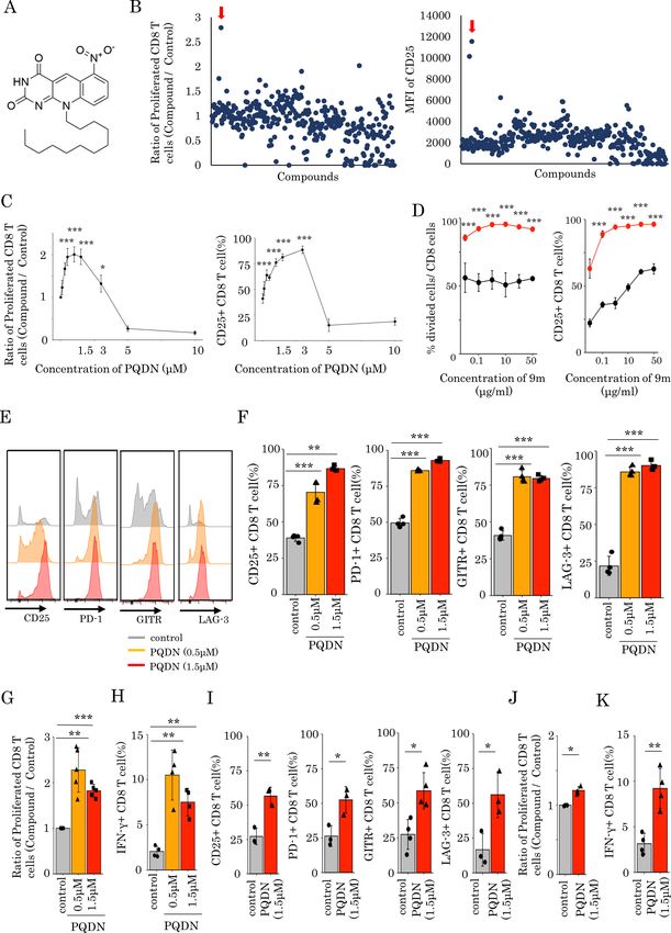
Open access
then evaluated CD25 expression and the prolifera- CH for nitrogen at the N5 position of flavin and is
J Immunother Cancer: first published as 10.1136/jitc-2021-003958 on 3 February 2022. Downloaded from http://jitc.bmj.com/ on March 6, 2022 by guest. Protected by copyright.
tive activity of CD8 T cells. We found that 10-d odecyl similar to that of flavin mononucleotide (FMN). Since
-6-nitro-pyrimido[4,5-b ]quinoline-2 ,4 (3H,10H)-d ione FMN acts as a coenzyme for NADH dehydrogenase in
(figure 1A), named as PQDN, markedly increased the mitochondria, we performed a molecular docking of
CD25 expression and cell proliferation (figure 1B). We PQDN on the FMN-binding site of mouse mitochon-
also observed that PQDN increased CD25 expression drial complex I. The tricyclic ring of PQDN could
and cell proliferation up to 3 µM in a concentration- effectively fit into the hydrophobic pocket in a similar
dependent manner, whereas these were decreased orientation to that of FMN (figure 2A,B). Hydrogen
with further increasing concentrations due to toxicity bond formation between the nitro group and pyrimi-
(figure 1C). Furthermore, we investigated the effect dine moiety of PQDN with side chains such as Tyr184
of PQDN on CD8 T cells when antigen stimulation and Asp98 were also observed. The docking score of
was titrated using 9 m peptide or anti-CD3 antibody the redocked FMN was –9.12 kcal/mol, while that of
and found that PQDN improved T cells activation the PQDN was similar at approximately –8.72 kcal/
when antigen stimulation was weak (figure 1D and mol (online supplemental figure 5A,B). Additionally,
online supplemental figure 1A). Although CD8 T cells we evaluated NAD+/NADH following PQDN treatment
stimulated with the 9 m peptide alone were not fully to determine whether PQDN can activate NADH as
activated, PQDN enhanced their activation. These FMN and found that PQDN promotes the conversion
data show that PQDN improve the T cell activation of NADH to NAD+ (figure 2C). These results suggest
threshold. Additionally, PQDN did not enhance CD8 that PQDN may contribute to the electron transport
T cell activation without TCR stimulation (online chains (ETCs) in mitochondria. Next, we assessed
supplemental file 2). Thus, this hit compound affects T the membrane potential of mitochondria using flow
cells stimulated with a weak antigen dominantly, indi- cytometry with tetramethylrhodamine methyl ester
cating that the effect of this compound is restricted (TMRE) as a fluorescent probe to determine whether
to antigen-specific CD8 T cells. We also investigated PQDN induced mitochondrial activation. The mito-
the effect of PQDN on antigen-s pecific CD8 T cells in chondrial membrane potential is increased 30 min
more detail. The expression of multiple T cell activa- after PQDN treatment in naive T cells (figure 2D), and
tion markers such as CD25, GITR, PD-1 , and LAG-3 similar results were obtained in antigen-stimulated T
on CD8 T cells, as well as IFN-γ -secretion and cell cells; this elevation continued for at least 72 hours
proliferation were increased (figure 1E–H). Next, to after stimulation (figure 2E,F). Similar results were
determine whether the effect of PQDN on CD8 T cells also observed among CD8 tumor-infiltrating lympho-
is mediated by antigen-presenting cells (APCs), sple- cytes (TILs) in a CMS5a murine model (figure 2G).
nocytes were stimulated with anti-CD3 antibody in the As PQDN elevated mitochondrial membrane poten-
presence of PQDN for 72 hours. PQDN enhanced the tial, we investigated whether it affects mitochondrial
function of CD8 T cells by activating marker expres- respiration. OXPHOS profiles of CD8 T cells were
sion, cell proliferation, and IFN-γ secretion (online also determined using extracellular flux analysis for
supplemental figure 2A). We then separated CD8 T the OCR at 7, 24, and 48 hours after PQDN treat-
cells from whole splenocytes and stimulated them ment (figure 2H). Basal respiration was increased
with anti-CD3 antibody for 72 hours. Similar to whole in both T cells with or without PQDN treatment at
splenocytes, PQDN augmented CD8 T cell activation, 24 hours after antigen stimulation, and this increment
suggesting that the effect of PQDN on CD8 T cells was continued in PQDN treated T cells; however, in
is direct and not mediated by APCs (figure 1I–K). peptide-stimulated T cells without PQDN, this incre-
Meanwhile, PQDN also affected CD4 and regulatory ment was not observed even at 48 hours (figure 2I).
T cells (Tregs), suggesting that PQDN can interact Spare respiratory capacity (SRC) was also increased
with common molecules for T cell activation (online in both T cells with or without PQDN at 24 hours, and
supplemental figure 3 A, B). PQDN treatment significantly elevated SRC compared
with the vehicle control (figure 2I). This significant
PQDN enhances mitochondrial function and glycolysis via increase in SRC by PQDN was temporary and was
mitochondrial complex I and III not observed at 48 hours. Mitochondrial respiration
To elucidate the mechanism underlying the effect complexes comprise five complexes (complexes I–V).
of PQDN on T cells, we analyzed the whole gene Of these, proton pumping using ubiquinone and ETC
expression in splenocytes cells treated with antigen mainly occur in mitochondrial complexes I and III,
and PQDN for 24 hours using RNA- S eq. Gene set which are required for mitochondrial activation.23
enrichment analysis revealed that PQDN upregulated Therefore, to verify whether the effect of PQDN on
mitochondria-related genes, especially for mitochon- mitochondrial respiration is mediated by mitochon-
drial membrane potential to transport proteins such drial complexes I and III, we cultured DUC18 lympho-
as TIMM9 and TOMM22 (online supplemental figure cytes in the presence of PQDN for 24 hours with a
4A,B).21 22 Pyrimido[4,5- b ]quinoline-2 ,4 (3H,10H)- mitochondrial complex I inhibitor (rotenone (Rot))
dione, a substructure of PQDN, is a substitution of or complex III inhibitor (antimycin A (AA)); the
Dotsu Y, et al. J Immunother Cancer 2022;10:e003958. doi:10.1136/jitc-2021-003958 3
Open access
J Immunother Cancer: first published as 10.1136/jitc-2021-003958 on 3 February 2022. Downloaded from http://jitc.bmj.com/ on March 6, 2022 by guest. Protected by copyright.
Figure 1 (A) Chemical structure of PQDN. (B) Approximately 310 compounds were screened to identify compounds enhancing
the activation of CD8 T cells despite weak antigen stimulation. Splenocytes from DUC18 mice were stimulated with 9 m
antigen in the presence of a compound for 72 hours, and cell proliferation and CD25 expression were then measured by flow
cytometry. Red arrow indicates PQDN. (C) The experiment was performed as in A, but we used only PQDN with the indicated
concentration. (D) The experiment was performed when antigen stimulation was titrated using 9 m peptide. (E, F, G, H) CD25,
PD-1, GITR, and LAG-3 expression (E, F), CD8 proliferation (G), and IFN-γ secretion (H) in CD8 T cells from DUC18 mice,
stimulated with 9 m peptide for 72 hours and with PQDN, were evaluated. Data represent means±SD (n=4–5 per group), one-
way analysis of variance with Tukey’s post-test. (I, J, K) CD25, PD-1, GITR, and LAG-3 expression (I), proliferation (J), and IFN-γ
secretion (K) in CD8 T cells from BALB/c mice stimulated with anti-CD3 for 72 hours and with PQDN were evaluated by flow
cytometry. Data represent the means±SD. (n=3–4 per group), unpaired Student’s t-test. All experiments were repeated three or
more times with similar results.
4 Dotsu Y, et al. J Immunother Cancer 2022;10:e003958. doi:10.1136/jitc-2021-003958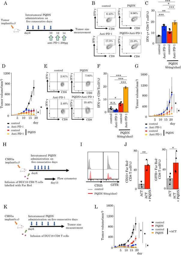
Open access
J Immunother Cancer: first published as 10.1136/jitc-2021-003958 on 3 February 2022. Downloaded from http://jitc.bmj.com/ on March 6, 2022 by guest. Protected by copyright.
Figure 2 (A) Docking model of PQDN to an NADH dehydrogenase FMN-binding site of mouse mitochondrial complex I using
MOE. PQDN is indicated in orange in ball-stick representation. (B) Overlay of the original FMN and PQDN generated using MOE.
FMN is indicated in cyan in line representation. (C) NADH activation induced by PQDN was measured using an NAD+/NADH
assay kit (n=4 per group). (D) Flow cytometric analysis of the mean fluorescence intensity of TMRE in CD8 T cells treated with
PQDN. (n=3 per group). (E, F) Kinetics of the TMRE dependent on stimulation time. (G) Flow cytometric analysis of the TMRE in
CD8 TILs from CMS5a tumor-bearing mice (n=3 per group). (H and I) The oxygen consumption rate (OCR) of CD8 T cells treated
with PQDN. The basal OCR or SRC are shown in figure 2I. (J and K) The OCR of CD8 T cells treated with PQDN for 24 hours
using Rot or AA. The basal OCR or SRC are shown in figure 2K (n=3 per group). (L and M) The extracellular acidification rate
(ECAR) of CD8 T cells treated with PQDN were measured. The basal and stressed ECAR are shown in figure 2M. (N and O) The
ECAR of CD8 T cells treated with PQDN for 24 hours using Rot or AA. Basal and stressed ECAR are shown in figure 2O (n=3
per group). Data are presented as mean±SD. P values were determined using unpaired Student’s t-test or one-way analysis of
variance with Tukey’s post-test. *P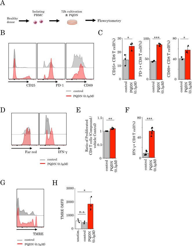
Open access
elevation of basal OCR or SRC by PQDN treatment was initial 24 hours following TCR stimulation. Next, we inves-
J Immunother Cancer: first published as 10.1136/jitc-2021-003958 on 3 February 2022. Downloaded from http://jitc.bmj.com/ on March 6, 2022 by guest. Protected by copyright.
diminished by both Rot and AA (figure 2J,K). Consid- tigated how PQDN induces glycolysis via mitochondrial
ering that OXPHOS as well as glycolysis is necessary respiration. mTOR/AKT signaling pathways can induce
for full activation of T cells after antigen recogni- glucose uptake in antigen-stimulated CD8 T cells,28 where
tion,18 we next evaluated the ECAR. Both basal and glycolysis is regulated by mTOR signaling; this metabolic
stressed ECAR were significantly increased by PQDN shift contributes to CD8 T cell differentiation.29 There-
treatment, and this increase was diminished by both fore, we hypothesized that mTOR/AKT signaling may be
Rot and AA (figure 2L–O). involved in the activation of glycolysis by PQDN; hence,
we investigated whether PQDN accelerates mTOR/AKT
Glycolysis induced by PQDN through mitochondrial respiration signaling in CD8 T cells and found that PQDN induced
is essential for CD8 T cell activation AKT and mTOR phosphorylation, 24 hours following TCR
Thus far, we showed that PQDN augments glycol- stimulation, but not at 4 hour (figure 3I,J). The effect of
ysis following OXPHOS activation via mitochondrial PQDN on the mTOR/AKT signaling was inhibited when
complexes I and III. Next, we investigated whether mito- CD8 T cells were cultured with mitochondrial inhibitors
chondrial activation is necessary for the effects of PQDN (Rot and AA) (figure 3K). These results suggest that
on CD8 T cells. Thus, DUC18 lymphocytes were cultured PQDN accelerates mTOR/AKT signaling through mito-
with PQDN and with mitochondrial complex inhibitors. chondrial activation and subsequently activates glycolytic
Both Rot and AA abolished the effect of PQDN in terms pathway as well as mitochondrial respiration.
of CD25 expression (figure 3A,B). In addition to mito-
chondrial complexes I and III, we also examined whether PQDN tunes inactivated T cell with tumor antigen recognition
this compound affects T cells via reactive oxygen species potent and enhances antitumor immunity
(ROS), mitochondrial complex II, or mitochondrial pyru- Next to evaluate the in vivo effects of PQDN, we used
vate carrier (MPC), which are known to be involved in T the CT26 and CMS5a tumor-bearing mice model. PQDN
cell activation.24–26 However, although PQDN produced has no toxicity effects on these cell lines at concentra-
higher ROS both in vitro and in vivo (online supple- tions lower than 10 uM, which suggests that these cell
mental figure 6A), the antioxidant N-acetylcysteine did lines show high resistance to the direct effect of PQDN,
not inhibit the effect of PQDN (online supplemental at such concentrations, and are suitable for evaluating
figure 6E). The mitochondrial complex II inhibitor the immune system- mediated effect of PQDN (online
(dimethyl malonate, DMM) and MPC inhibitor (UK5099) supplemental figure 8Supplementary Fig. 8 A,B). First,
also did not diminish the effect of PQDN (online supple- we examined the change in immune cells following
mental figure 7A,B). These data suggest that activation PQDN treatment and found that PQDN increased the
of mitochondrial respiration directly through mitochon- tumor-infiltrating CD8 T cells and Tregs and decreases
drial complexes I and III is crucial for CD8 T cell acti- CD4 T cells (online supplemental figure 9). Based on
vation induced by PQDN. Interestingly, this cancelation these data, we focused on CD8 TILs and examined the
of the effect of PQDN was not observed if Rot or AA was effects of PQDN on CD8 TILs from CT26 tumor-bearing
added 24 hours after TCR stimulation (figure 3C,D). mice following stimulation with anti- CD3 antibody
From these data, we hypothesized that T cells treated (online supplemental figure 10A). Similar to splenocytes,
with antigen and PQDN use glycolysis to synthesizes ATP PQDN significantly increased the expression of activation
for their activation after 24 hours later following stimula- markers (online supplemental figure 10B,C). This effect
tion. Therefore, to verify whether PQDN-induced glycol- was observed more notably at 72 hours after stimulation
ysis is required for CD8 T cell activation, we cultured than at 24 hours (online supplemental figure 10D). Next,
DUC18 lymphocytes with PQDN in galactose medium. we evaluated the effect of PQDN on CD8 TILs in the
The effect of PQDN was decreased when CD8 T cells tumor microenvironment (TME) in vivo.
were cultured in glucose-free medium (figure 3E).12 27 To determine how to inject PQDN into tumor-bearing
Notably, though 2-deoxy-d-glucose (2DG), an inhibitor of mice, we examined the optimal dose and times of PQDN
d-glucose metabolism, canceled the effect of PQDN, the injection and decided to inject more than 21 ng PQDN for
effect of PQDN was maintained when cultured with 2DG five consequent days to induce therapeutic effects (online
for only 24 hours since the beginning of stimulation and supplemental figure 11A,B). Similar to ex vivo analysis,
was canceled with 2DG addition at 24 hours after TCR beneficial effects of PQDN on CD8 TILs were observed in
stimulation (figure 3F). Furthermore, we also estimated terms of activated marker expression and IFN-γ secretion in
glucose uptake ability by flow cytometry using a fluores- CT26 tumor-bearing mice (figure 4A–E). Furthermore, we
cent d-glucose analog, 2-[N-(7-nitrobenz-2-oxa-1,3-diazol- investigated whether PQDN augments antitumor efficacy
4-yl) amino]−2-deoxy-D-glucose (2NBDG). We observed in CT26 murine models. We found that PQDN treatment
that PQDN improved glucose uptake in CD8 T cells retarded tumor growth compared with that in the control
24 hours and later after TCR stimulation, consistent with group (figure 4F); however, this effect was not confirmed
the time point of glycolysis acceleration (figure 3G,H). in T cell deficient mice or CD8 T cells depleted mice
These results suggest that glycolysis induced by PQDN is (figure 4G,H). This compound also showed a significant
essential for CD8 T cell activation, especially within the antitumor effect including prolonged survival in CMS5a
6 Dotsu Y, et al. J Immunother Cancer 2022;10:e003958. doi:10.1136/jitc-2021-003958Open access
J Immunother Cancer: first published as 10.1136/jitc-2021-003958 on 3 February 2022. Downloaded from http://jitc.bmj.com/ on March 6, 2022 by guest. Protected by copyright.
Figure 3 (A and B) Flow cytometric analysis of CD25 expression in DUC18 CD8 T cells treated with PQDN using Rot (A) or AA
(B) (n=3 per group) (C and D) Flow cytometric analysis of CD25 and CD69 expression in DUC18 CD8 T cells treated with PQDN
and Rot (C) or AA (D) (n=3 per group). (E) CD25 and GITR expression in DUC18 CD8 T cells treated with PQDN in glucose
or glucose-free medium (n=3 per group). (F) Flow cytometric analysis of GITR and LAG-3 expression in DUC18 CD8 T cells
treated with PQDN with 2DG at 0 and 24 hours after stimulation, and subsequently removed 2DG 24 hours after stimulation
(n=5 per group). (G and H) Glucose uptake ability in CD8 T cells after treatment with antigen and PQDN was evaluated using
2NBDG (n=4 per group). (I and J) Phosphorylation of mTOR and AKT in DUC18 CD8 T cells treated with antigen and PQDN was
measured by flow cytometry at 24 hours (I) or at the indicated time points (J) (n=4 per group). (K) Phosphorylation of mTOR and
AKT in CD8 T cells after treatment with antigen and PQDN for 72 hours using Rot or AA (n=3–4 per group). Data are presented
as mean±SD. P values were determined using unpaired Student’s t-test or one-way analysis of variance with Tukey’s post-test.
*POpen access
J Immunother Cancer: first published as 10.1136/jitc-2021-003958 on 3 February 2022. Downloaded from http://jitc.bmj.com/ on March 6, 2022 by guest. Protected by copyright.
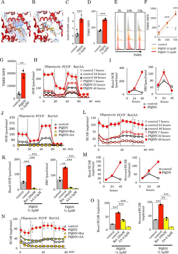
Figure 4 (A) The murine tumor cell line CT26 (B–H) or CMS5a (I–L) was subcutaneously inoculated into BALB/c mice. PQDN
(n=8 mice per group) intratumorally administered on five consecutive days (days 6–10). (B–E) Flow cytometric analysis of CD25,
PD-1, GITR, and LAG-3 expression (B, C, I) or IFN-γ secretion (D, E) in CD8 TILs (n=3–4 per group). (F and J) Tumor-bearing
mice were treated with intratumoral PQDN. Tumor sizes are shown (n=5–6 per group). (G and K) The experiment was performed
in nude mice as described in G or K. (H and L) The experiment was performed in CD8 depleted mice as described in H or L. (M–
P) The murine tumor cell line CT26 was subcutaneously inoculated into BALB/c mice. PQDN (n=5 mice per group) was injected
intratumorally on five consecutive days. Three days after the final administration, PD-1+CD8 TILs were isolated and analyzed
for their TCR repertoire. (N) The circle graph represents the frequency of clones in PD-1+CD8 TILs from CT26 tumors (n=5 per
group). (O and P) The frequency of the top five clones (O) and the number of clones with a frequency of >1% (P) in PD-1+CD8
TILs from CT26 tumors (n=5 per group) are shown. Data are presented as mean±SD. P values were determined using unpaired
Student’s t-test or one-way analysis of variance with Tukey’s post-test. *POpen access
murine fibrosarcoma tumor-bearing models (figure 4I–L of endogenous tumor-specific immune responses were
J Immunother Cancer: first published as 10.1136/jitc-2021-003958 on 3 February 2022. Downloaded from http://jitc.bmj.com/ on March 6, 2022 by guest. Protected by copyright.
and online supplemental figure 12). In addition to the essential to induce the efficacy of this therapy.7–9 It has
therapeutic effect of PQDN, we also analyzed the in vivo been reported that induction of endogenous immune
toxicity. PQDN injection for five consequent days did not responses against tumor through antigen spreading
induce changes in any enzyme levels including aspartate contributes to retardation of tumor growth after adop-
aminotransferase and alanine aminotransferase (online tive cell therapy. Thus, we examined whether intratu-
supplemental figure 13A). Additionaly, weight loss related moral injection of this compound after adoptive T cell
to PQDN treatment was not observed in tumor-bearing therapy activates the function of endogenous T cells at
mice (online supplemental figure 13B). A previous study tumor after infusion of tumor-specific CD8 T cells. CD8
reported that CMS5a is a low immunogenic tumor and T cells derived from DUC18 mice labeled with Far Red
demonstrates high resistance to T cell dependent immu- were intravenously infused into CMS5a tumor- bearing
notherapy, such as immune checkpoint inhibitors and mice 6 days after inoculation. On day 7, the expression
adoptive cell transfer (ACT).5 These data suggest that of surface markers on CD8 TILs was examined, and an
PQDN improves CD8 T cell function and promotes anti- increase in the expression of activated surface markers
tumor immunity in a T cell mediated manner, even in was observed on both endogenous and infused CD8 T
a low immunogenic tumor model. Thus far, we demon- cells in PQDN treated mice compared that in with control
strated that PQDN can augment CD8 T cell activation, mice (figure 5H–J and online supplemental figure
even when TCR signaling is insufficient to induce acti- 15A,B). Furthermore, intratumoral injection of PQDN
vation or proliferation. Furthermore, we confirmed that remarkably increased the therapeutic efficacy of ACT
CD8 T cells from DUC18 mice were more strongly acti- with PQDN in mice bearing CMS5a (figure 5K,L). These
vated in the presence of PQDN compared with the control results suggest that PQDN can improve the function of
medium when cocultured with CMS5a tumor cells, indi- endogenous T cells and enhance immune checkpoint
cating that PQDN can facilitate tumor antigen recogni- inhibitors and adoptive cell therapy, even in low immu-
tion directly in the TME (online supplemental figure nogenic tumors.
14A–C). From these results, we considered that PQDN
may activate even the low-avidity tumor antigen-specific PQDN enhances the activation of human CD8 T cells
CD8 T cells and expand the TCR repertoire diversity of The results from our mouse studies prompted us to inves-
TILs. In order to verify this, we collected PD-1 +CD8 TILs tigate whether PQDN might also augment CD8 T cell
from CT26 tumor-bearing mice, as tumor recognizing T function in human PBMCs. Therefore, we examined the
cells and performed TCR repertoire analysis using TCR cell surface marker expression, cell proliferation, and
beta sequencing to determine the diversity of TCR after IFN-γ secretion in CD8 T cells derived from healthy donor
PQDN treatment (figure 4M). The results indicated that PBMCs stimulated with anti CD3 alone or in combination
PQDN decreased the frequency of the top five clones and with PQDN (figure 6A). We found a significant increase
expanded the number of clones with frequencies >1% in activation markers, cell proliferation, and IFN-γ secre-
(figure 4N–P). tion with PQDN treatment at 0.5 µM (figure 6B–F).
Furthermore, to verify whether PQDN affects the mito-
PQDN has a synergistic potent with T cell dependent chondrial membrane potential in human CD8 T cells, we
immunotherapy even in a low immunogenic tumor assessed the change in TMRE in CD8 T cells and found
As manipulation of mitochondrial quality or quantity or that PQDN elevated TMRE at 2 hours after anti-CD3 stim-
changing mitochondrial activity controls T cell function ulation (figure 6G,H). These results suggest that PQDN is
and improves the efficacy of cancer immunotherapies also a promising compound to augment CD8 T cell func-
such as checkpoint inhibitory therapy or adoptive cell tion through mitochondrial activation in humans.
therapy,26 30–32 we hypothesized that PQDN can enhance
tumors sensitivity to cancer immunotherapy, hence we
investigated whether PQDN can enhance the antitumor DISCUSSION
effects of these therapies in tumor-bearing models. First, In this study, we identified PQDN as a small molecule that
we examined whether PQDN improves the efficacy of PD-1 can augment CD8 T cell activation even with weak TCR
blockade in CT26 murine models (figure 5A). Combi- stimulation, which is insufficient to activate these cells,
nation therapy induces the production of higher IFN-γ indicating that this compound tunes T cell activation
levels (figure 5B,C) and significantly inhibited tumor thresholds following antigen recognition. Mechanisti-
growth compared with monotherapy (anti-PD-1 therapy cally, PQDN enhances mitochondrial reciprocal capacity
or PQDN) in the CMS5a tumor model (figure 5D). and subsequent glycolysis acceleration via the activating
Furthermore, these synergistic effects were also observed of ETC. Furthermore, intratumoral administration of
in CMS5a mouse models showing a resistance for PQDN increases the frequency of PD-1 +CD8 T cells and
immune checkpoint inhibitor therapy (figure 5E–G). expands the TCR repertoire diversity of these cells at the
Next, we determined whether PQDN can also improve tumor site. These results suggest that PQDN can acti-
the efficacy of adoptive cell therapy. Though adoptive cell vate inactive CD8 TILs that recognize the tumor antigen
therapy is effective, it is not sufficient. Because activation but do not respond to a tumor due to weak antigen
Dotsu Y, et al. J Immunother Cancer 2022;10:e003958. doi:10.1136/jitc-2021-003958 9Open access
J Immunother Cancer: first published as 10.1136/jitc-2021-003958 on 3 February 2022. Downloaded from http://jitc.bmj.com/ on March 6, 2022 by guest. Protected by copyright.
Figure 5 (A–G) CT26 (A–D) or CMS5a (E–G) cells were subcutaneously inoculated into BALB/c mice. PQDN was injected
intratumorally into the same mice on five consequent days (6–10). Anti-PD-1 antibody was administrated intravenously on days
10, 12, and 14 into the same mice. (B, C) Flow cytometric analysis of IFN-γ secretion in CD8 TILs from CT26 tumors treated
with the combination of intratumoral PQDN and anti-PD-1 antibody. Data are shown as mean±SD (n=5 per group) and analyzed
using one-way analysis of variance (ANOVA) with Tukey’s post-test. (D) CT26-bearing mice were treated with a combination of
intra tumorous PQDN and anti-PD-1 antibody. Tumor sizes are shown (n=4–5 per group) and analyzed using two-way ANOVA
with Tukey’s post-test. (E, F) Flow cytometric analysis of IFN-γ secretion in CD8 TILs from CMS5a tumors treated with the
combination of intratumoral PQDN and anti-PD-1 antibody. Data are shown as mean±SD (n=3 per group) and analyzed using
one-way ANOVA with Tukey’s post-test. (G) CMS5a-bearing mice were treated with a combination of intratumoral PQDN and
anti-PD-1 antibody. Tumor sizes are shown (n=3–4 per group) and analyzed using two-way ANOVA with Tukey’s post-test.
(H–L) CMS5a cells were subcutaneously inoculated into BALB/c mice. CD8 cells isolated from DUC18 mice were infused on day
6. PQDN was injected intratumorally into the same mice for five consequent days (days 6 to 10). (I, J) CD25, GITR expression in
endogenous CD8 T cells (Far Red – T cells) from CMS5a tumors. Data are shown as mean±SD (n=3–4 per group) and analyzed
using unpaired Student’s t-test. (L) Tumor sizes are shown (n=3 per group) and analyzed using two-way ANOVA with Tukey’s
post-test. TILs, tumor-infiltrating lymphocytes.
10 Dotsu Y, et al. J Immunother Cancer 2022;10:e003958. doi:10.1136/jitc-2021-003958Open access
J Immunother Cancer: first published as 10.1136/jitc-2021-003958 on 3 February 2022. Downloaded from http://jitc.bmj.com/ on March 6, 2022 by guest. Protected by copyright.
Figure 6 (A–F) PBMCs isolated from healthy donors were stimulated with anti-CD3 antibody in the presence or absence of
PQDN for 72 hours. CD25, PD-1, and CD69 expression (B, C), proliferation (D, E) and IFN-γ secretion (D, F) in CD8 T cells were
measured by flow cytometry. Data represent means±SD (n=3–5 per group), unpaired Student’s t-test. (G, H) Mean fluorescence
intensity of TMRE in CD8 T cells from healthy donor PBMCs stimulated with anti-CD3 for 2 hours. Data represent means±SD
(n=3 per group), one-way analysis of variance with Tukey’s post-test.
Dotsu Y, et al. J Immunother Cancer 2022;10:e003958. doi:10.1136/jitc-2021-003958 11Open access
stimulation. Previous studies have reported that diverse the activation threshold. Considering this, modification
J Immunother Cancer: first published as 10.1136/jitc-2021-003958 on 3 February 2022. Downloaded from http://jitc.bmj.com/ on March 6, 2022 by guest. Protected by copyright.
qualities of TCR are associated with the therapeutic effi- of the activation threshold in PQDN-treated cells might
cacy of cancer immunotherapy, including checkpoint be induced by accelerated glycolysis, which leads to T cell
blockade therapy or cancer vaccines.33–35 Therefore, an activation despite weak antigen stimulation.
increase in functional TCR diversity by PQDN may be Although PQDN activates mitochondrial respiration,
useful for improving cancer immunotherapy. Further- mode of action of PQDN is unique. Several compounds
more, these findings suggest that tumor-specific T cells that activate mitochondria have been reported including
with low antigen recognition ability exist at the tumor site dichloroacetate (DCA)44 or triptolinamide (TLAM).45
and that these T cells might be a target for developing DCA, an inhibitor of PDK, enhances the phosphorylation
novel cancer immunotherapies. of PDH and increases the flux of pyruvate into mitochon-
Modulating the metabolism of T cells at the tumor dria, resulting in OXPHOS activation.44 Furthermore,
site improves these functions and augments antitumor TLAM increases OXPHOS via the AMPK/fatty acid
immune responses.26 30–32 Attenuation of mitochondrial oxidation pathway by inhibiting phosphofructokinase-1.45
activity induces TIL dysfunction.26 31 32 36 37 Reduction of However, these compounds simultaneously reduce glycol-
PPAR- gamma coactivator 1a (PGC1a) in TILs reduces ysis, because PDK and PFK are important regulators of
mitochondrial function and mass and leads to metabolic glycolysis,46 47 meaning that these compounds enhance
exhaustion, resulting in T cell dysfunction. However, mitochondrial function as a replacement for glycolysis.
enforced expression of PGC1a in TILs or treatment with Unlike these compounds, PQDN enhanced mitochon-
bezafibrate, a pan-PPAR agonist, bolsters mitochondrial drial function as well as glycolysis. The reason for this
biogenesis and improves antitumor T cell function.32 36 might be that OXPHOS was directly activated by PQDN
ROS generation is also a crucial mechanism for enhancing and not by inhibiting the molecules involved in glycol-
T cell activity following mitochondrial activation.38 ROS ysis. Antigen-stimulated T cells require a large amount
are generated at mitochondrial complexes I and III of energy for activation and acquire energy by activating
through the ETC39 and activate CD8 T cells by enhancing both OXPHOS and glycolysis.18 Thus, PQDN is an ideal
NFAT signaling and cytokine secretion.24 25 Trifluoro- compound that augments T cell activation through mito-
methoxy carbonylcyanide phenylhydrazone (FCCP), an chondrial activation. Meanwhile, PQDN may also activate
ROS inducer, increases the functional potency of T cells Tregs at tumor site. Since mitochondrial capacity is essen-
by activating mitochondria.26 In this study, we observed tial for the Tregs survival and function,48 ETC activation
that PQDN enhanced mitochondrial activation and ROS by PQDN may contribute to Tregs increment. However,
generation in antigen-stimulated T cells. However, ROS although PQDN increased Tregs at tumor site, it can lead
generation by PQDN was not related to CD8 T cell acti- to tumor growth retardation, possibly due to the PQDN
vation (online supplemental figure 6A–E). Although induction of antigen specific-CD8 T cells to overcome
we investigated whether bezafibrate or FCCP enhance suppression by Tregs.
the activation of T cells receiving weak stimulation, we In this study, we indicated that PQDN may bind to
did not observe an effect like PQDN was not observed NADH dehydrogenase in the mitochondrial respiratory
(online supplemental figure 16A,B). This difference complex I like FMN (figure 2A,B). PQDN was previ-
might be caused by differences in the mode of mitochon- ously reported as a cell growth inhibitory compound.49
drial activation by these compounds; for instance, bezafi- Shinkai et al50 reported that pyrimido[4,5-b]quinoline-2,4
brate or FCCP increases mitochondrial activity indirectly (3H,10H)-dione structure, known as 5-deazaflavin, can
in a PPAR-mediated or ROS-mediated manner, whereas promote electron bridge from NADH to FMN in an in
PQDN affects the mitochondria directly via ETC. Manip- vitro benzylamine oxidation reaction model. However, to
ulating mitochondrial function directly might be critical date, there have been no reports of the biological effect of
for tuning the T cell activation threshold in low antigen- PQDN on mitochondrial function. Conversely, when FMN
stimulated T cells. Guo et al reported that IL-10 directly binds to NADH dehydrogenase as a prosthetic group,
enhances the expansion and effector function of TILs by FMN receives hydrogen from NADH along with two elec-
promoting OXPHOS.31 This reactivation of OXPHOS in trons and transfers these electrons to iron-sulfur clusters.
TILs is dependent on MPC, which plays a role in cytosolic This electron transport induces leakage of protons into
pyruvate incorporation into the mitochondrial matrix.40 the mitochondrial intermembrane space (IMS); ATP is
However, the effect of PQDN was not mediated by MPC. then synthesized from ADP when the protons transfer
Altogether, although PQDN augments TIL activation by from IMS to the matrix through complex V. Altogether,
enhancing mitochondrial function, the mode of action based on the structure of PQDN, this compound is
of PQDN differs from that of various mitochondrial acti- considered to promote ETC in mitochondria, resulting
vators reported previously. in increased mitochondrial activity. However, FMN could
CD28 signaling or IL-2 signaling modulates the activa- not augment the activation of CD8 T cells after weak TCR
tion threshold and induces the biological response of T stimulation (online supplemental figure 17). The reason
cells.41 42 Additionally, these signaling pathways induce of this difference might be attributed to the differences
glycolysis in antigen-stimulated T cells.43 Therefore, these in chemical structures between PQDN and FMN. FMN is
reports suggest that glycolysis plays a crucial role in tuning water soluble while PQDN is lipophilic. The ClogP value
12 Dotsu Y, et al. J Immunother Cancer 2022;10:e003958. doi:10.1136/jitc-2021-003958Open access
of FMN is remarkably lower than that of PQDN (online Open access This is an open access article distributed in accordance with the
J Immunother Cancer: first published as 10.1136/jitc-2021-003958 on 3 February 2022. Downloaded from http://jitc.bmj.com/ on March 6, 2022 by guest. Protected by copyright.
supplemental figure 18). Thus, PQDN might enhance the Creative Commons Attribution Non Commercial (CC BY-NC 4.0) license, which
permits others to distribute, remix, adapt, build upon this work non-commercially,
ETC at mitochondria after penetrating cell membranes, and license their derivative works on different terms, provided the original work is
which FMN might not be able to. It is unclear how acti- properly cited, appropriate credit is given, any changes made indicated, and the use
vating mitochondrial function by PQDN accelerates is non-commercial. See http://creativecommons.org/licenses/by-nc/4.0/.
glycolytic activity. Further studies will provide insight into ORCID iD
the fundamental mechanisms of T cell activation and Yosuke Dotsu http://orcid.org/0000-0002-4493-7052
metabolic reprogramming.
In conclusion, we discovered a novel compound,
PQDN, that increases mitochondrial respiration capacity REFERENCES
and enhances glycolysis following mitochondrial activa- 1 Brahmer JR, Drake CG, Wollner I, et al. Phase I study of single-agent
tion. To our knowledge, no other compound that can anti-programmed death-1 (MDX-1106) in refractory solid tumors:
safety, clinical activity, pharmacodynamics, and immunologic
simultaneously activate mitochondria and glycolysis has correlates. J Clin Oncol 2010;28:3167–75.
been reported; therefore, this compound is valuable for 2 Robbins PF, Kassim SH, Tran TLN, et al. A pilot trial using
lymphocytes genetically engineered with an NY-ESO-1-reactive T-cell
cancer immunotherapy. Furthermore, this compound receptor: long-term follow-up and correlates with response. Clin
augments antitumor immunity by expanding functional Cancer Res 2015;21:1019–27.
3 Maude SL, Laetsch TW, Buechner J, et al. Tisagenlecleucel in
TCR diversity. Our findings indicate that PQDN can create children and young adults with B-cell lymphoblastic leukemia. N Engl
an immune-responsive environment even in a low immu- J Med 2018;378:439–48.
nogenic tumor by modulating the activation threshold. 4 Stevanović S, Pasetto A, Helman SR, et al. Landscape of
immunogenic tumor antigens in successful immunotherapy of virally
However, further research is necessary to understand its induced epithelial cancer. Science 2017;356:200–5.
antitumor effects in patients with cancer. Taken together, 5 Muraoka D, Seo N, Hayashi T, et al. Antigen delivery targeted
to tumor-associated macrophages overcomes tumor immune
our results indicate that PQDN activates CD8 T cells in a resistance. J Clin Invest 2019;129:1278–94.
previously unknown manner and is highly potent in over- 6 Schmidt J, Smith AR, Magnin M, et al. Prediction of neo-epitope
immunogenicity reveals TCR recognition determinants and provides
coming immunotherapy resistance in tumors. insight into immunoediting. Cell Rep Med 2021;2:100194.
7 Robbins PF, Li YF, El-Gamil M, et al. Single and dual amino acid
Author affiliations substitutions in TCR CDRs can enhance antigen-specific T cell
1
Department of Oncology, Nagasaki University Graduate School of Biomedical functions. J Immunol 2008;180:6116–31.
8 Parkhurst MR, Joo J, Riley JP, et al. Characterization of genetically
Sciences, Nagasaki, Japan
2 modified T-cell receptors that recognize the CEA:691-699 peptide
Department of Respiratory Medicine, Nagasaki University Graduate School of in the context of HLA-A2.1 on human colorectal cancer cells. Clin
Biomedical Sciences, Nagasaki, Japan Cancer Res 2009;15:169–80.
3
Division of Translational Oncoimmunology, Aichi Cancer Research Institute, Naogya, 9 Kaczanowska S, Joseph AM, Guo J, et al. A synthetic CD8α:MyD88
Japan Coreceptor enhances CD8+ T-cell responses to weakly immunogenic
4 and lowly expressed tumor antigens. Cancer Res 2017;77:7049–58.
Center for Drug Discovery, Graduate School of Pharmaceutical Sciences, University
10 Phan AT, Doedens AL, Palazon A, et al. Constitutive glycolytic
of Shizuoka, Shizuoka, Japan metabolism supports CD8+ T cell effector memory differentiation
5
Department of Immunology, Juntendo University School of Medicine, Tokyo, Japan during viral infection. Immunity 2016;45:1024–37.
11 Ron-Harel N, Santos D, Ghergurovich JM, et al. Mitochondrial
biogenesis and proteome remodeling promote one-carbon
Correction notice This article has been corrected since it was first published. The
metabolism for T cell activation. Cell Metab 2016;24:104–17.
author name Daisuke Muraoka was incorrectly spelt as Daisuk Muraoka. 12 Chang C-H, Curtis JD, Maggi LB, et al. Posttranscriptional control of
Acknowledgements We would like to thank S Yamaguchi and H Masumoto for T cell effector function by aerobic glycolysis. Cell 2013;153:1239–51.
providing technical assistance. 13 Wang R, Dillon CP, Shi LZ, et al. The transcription factor Myc controls
metabolic reprogramming upon T lymphocyte activation. Immunity
Contributors DM, HYam, HM and HI supervised the study. YD, DM, KY and HI 2011;35:871–82.
conceived and designed the experiments. YD and DM performed the experiments. 14 Klein-Hessling S, Muhammad K, Klein M, et al. NFATc1 controls the
YD and DM performed data analysis. YS, NO, AA, and HYag contributed the reagents cytotoxicity of CD8+ T cells. Nat Commun 2017;8:511.
and materials. YD and DM wrote the manuscript. All authors reviewed and approved 15 Toriyama K, Kuwahara M, Kondoh H, et al. T cell-specific deletion
of Pgam1 reveals a critical role for glycolysis in T cell responses.
the final manuscript. DM is a guarantor for this study and publication.
Commun Biol 2020;3:394.
Funding This study was partially supported by the Japan Society for the Promotion 16 Menk AV, Scharping NE, Moreci RS, et al. Early TCR signaling
of Science KAKENHI (Grant Number 17K07222 and 21H03005). induces rapid aerobic glycolysis enabling distinct acute T cell
effector functions. Cell Rep 2018;22:1509–21.
Competing interests None declared. 17 Xu K, Yin N, Peng M, et al. Glycolysis fuels phosphoinositide
3-kinase signaling to bolster T cell immunity. Science
Patient consent for publication Not applicable. 2021;371:405–10.
Ethics approval This study does not involve human participants. 18 Levine LS, Hiam-Galvez KJ, Marquez DM, et al. Single-cell analysis
by mass cytometry reveals metabolic states of early-activated
Provenance and peer review Not commissioned; externally peer reviewed. CD8+ T cells during the primary immune response. Immunity
Data availability statement Data are available on reasonable request. 2021;54:e825:829–44.
19 Ikeda H, Ohta N, Furukawa K, et al. Mutated mitogen-activated
Supplemental material This content has been supplied by the author(s). It has protein kinase: a tumor rejection antigen of mouse sarcoma. Proc
not been vetted by BMJ Publishing Group Limited (BMJ) and may not have been Natl Acad Sci U S A 1997;94:6375–9.
peer-reviewed. Any opinions or recommendations discussed are solely those 20 Hanson HL, Donermeyer DL, Ikeda H, et al. Eradication of
of the author(s) and are not endorsed by BMJ. BMJ disclaims all liability and established tumors by CD8+ T cell adoptive immunotherapy.
Immunity 2000;13:265–76.
responsibility arising from any reliance placed on the content. Where the content
21 Qiu J, Wenz L-S, Zerbes RM, et al. Coupling of mitochondrial import
includes any translated material, BMJ does not warrant the accuracy and reliability and export translocases by receptor-mediated supercomplex
of the translations (including but not limited to local regulations, clinical guidelines, formation. Cell 2013;154:596–608.
terminology, drug names and drug dosages), and is not responsible for any error 22 Sotgia F, Whitaker-Menezes D, Martinez-Outschoorn UE,
and/or omissions arising from translation and adaptation or otherwise. et al. Mitochondria “fuel” breast cancer metabolism: fifteen
Dotsu Y, et al. J Immunother Cancer 2022;10:e003958. doi:10.1136/jitc-2021-003958 13Open access
J Immunother Cancer: first published as 10.1136/jitc-2021-003958 on 3 February 2022. Downloaded from http://jitc.bmj.com/ on March 6, 2022 by guest. Protected by copyright.
markers of mitochondrial biogenesis label epithelial cancer 36 Scharping NE, Menk AV, Moreci RS, et al. The tumor
cells, but are excluded from adjacent stromal cells. Cell Cycle microenvironment represses T cell mitochondrial biogenesis to drive
2012;11:4390–401. intratumoral T cell metabolic insufficiency and dysfunction. Immunity
23 Cogliati S, Lorenzi I, Rigoni G, et al. Regulation of mitochondrial 2016;45:374–88.
electron transport chain assembly. J Mol Biol 2018;430:4849–73. 37 Yu Y-R, Imrichova H, Wang H, et al. Disturbed mitochondrial
24 Weinberg SE, Sena LA, Chandel NS. Mitochondria in the regulation dynamics in CD8+ TILs reinforce T cell exhaustion. Nat Immunol
of innate and adaptive immunity. Immunity 2015;42:406–17. 2020;21:1540–51.
25 Weinberg F, Hamanaka R, Wheaton WW, et al. Mitochondrial 38 Sena LA, Li S, Jairaman A, et al. Mitochondria are required for
metabolism and ROS generation are essential for KRAS-mediated antigen-specific T cell activation through reactive oxygen species
tumorigenicity. Proc Natl Acad Sci U S A 2010;107:8788–93. signaling. Immunity 2013;38:225–36.
26 Chamoto K, Chowdhury PS, Kumar A, et al. Mitochondrial activation 39 Turrens JF. Mitochondrial formation of reactive oxygen species.
chemicals synergize with surface receptor PD-1 blockade for J Physiol 2003;552:335–44.
T cell-dependent antitumor activity. Proc Natl Acad Sci U S A 40 Herzig S, Raemy E, Montessuit S, et al. Identification and
2017;114:E761–70. functional expression of the mitochondrial pyruvate carrier. Science
27 Cham CM, Driessens G, O'Keefe JP, et al. Glucose deprivation 2012;337:93–6.
inhibits multiple key gene expression events and effector functions in 41 Acuto O, Michel F. CD28-mediated co-stimulation: a quantitative
CD8+ T cells. Eur J Immunol 2008;38:2438–50. support for TCR signalling. Nat Rev Immunol 2003;3:939–51.
28 Wofford JA, Wieman HL, Jacobs SR, et al. IL-7 promotes GLUT1 42 Au-Yeung BB, Smith GA, Mueller JL, et al. IL-2 modulates the TCR
trafficking and glucose uptake via STAT5-mediated activation of Akt signaling threshold for CD8 but not CD4 T cell proliferation on a
to support T-cell survival. Blood 2008;111:2101–11. single-cell level. J Immunol 2017;198:2445–56.
29 Buck MD, O'Sullivan D, Pearce EL. T cell metabolism drives 43 Frauwirth KA, Riley JL, Harris MH, et al. The CD28 signaling pathway
immunity. J Exp Med 2015;212:1345–60. regulates glucose metabolism. Immunity 2002;16:769–77.
30 Eikawa S, Nishida M, Mizukami S, et al. Immune-mediated antitumor 44 Shen Y-C, Ou D-L, Hsu C, et al. Activating oxidative phosphorylation
effect by type 2 diabetes drug, metformin. Proc Natl Acad Sci U S A by a pyruvate dehydrogenase kinase inhibitor overcomes sorafenib
2015;112:1809–14. resistance of hepatocellular carcinoma. Br J Cancer 2013;108:72–81.
31 Guo Y, Xie Y-Q, Gao M, et al. Metabolic reprogramming of terminally 45 Kobayashi H, Hatakeyama H, Nishimura H, et al. Chemical reversal
exhausted CD8+ T cells by IL-10 enhances anti-tumor immunity. Nat of abnormalities in cells carrying mitochondrial DNA mutations. Nat
Immunol 2021;22:746–56. Chem Biol 2021;17:335–43.
32 Chowdhury PS, Chamoto K, Kumar A, et al. PPAR-induced fatty 46 Mor I, Cheung EC, Vousden KH. Control of glycolysis through
acid oxidation in T cells increases the number of tumor-reactive regulation of PFK1: old friends and recent additions. Cold Spring
CD8+ T cells and facilitates anti-PD-1 therapy. Cancer Immunol Res Harb Symp Quant Biol 2011;76:211–6.
2018;6:1375–87. 47 Takubo K, Nagamatsu G, Kobayashi CI, et al. Regulation of
33 Han J, Duan J, Bai H, et al. TCR repertoire diversity of peripheral glycolysis by PDK functions as a metabolic checkpoint for cell
PD-1+CD8+ T cells predicts clinical outcomes after immunotherapy cycle quiescence in hematopoietic stem cells. Cell Stem Cell
in patients with non-small cell lung cancer. Cancer Immunol Res 2013;12:49–61.
2020;8:146–54. 48 Weinberg SE, Singer BD, Steinert EM, et al. Mitochondrial complex
34 Poran A, Scherer J, Bushway ME. Combined TCR repertoire profiles III is essential for suppressive function of regulatory T cells. Nature
and blood cell phenotypes predict melanoma patient response to 2019;565:495–9.
personalized neoantigen therapy plus anti-PD-1. Cell Rep Med 49 Kawamoto T, Ikeuchi Y, Hiraki J, et al. Synthesis and evaluation of
2020;1:100141. nitro 5-deazaflavins as novel bioreductive antitumor agents. Bioorg
35 Hopkins AC, Yarchoan M, Durham JN, et al. T cell receptor repertoire Med Chem Lett 1995;5:2109–14.
features associated with survival in immunotherapy-treated 50 Shinkai S, Kuroda H, Manabe O, et al. Flavin plus 5-deazaflavin as a
pancreatic ductal adenocarcinoma. JCI Insight 2018;3 doi:10.1172/ turnover oxidation system: model for the electron bridge from NAD+
jci.insight.122092 to flavin. J Chem Soc Chem Commun 1981;8:391–2.
14 Dotsu Y, et al. J Immunother Cancer 2022;10:e003958. doi:10.1136/jitc-2021-003958You can also read