Chemical and structural studies provide a mechanistic basis for recognition of the MYC G- quadruplex
←
→
Page content transcription
If your browser does not render page correctly, please read the page content below
ARTICLE
                  DOI: 10.1038/s41467-018-06315-w                OPEN

                  Chemical and structural studies provide a
                  mechanistic basis for recognition of the MYC G-
                  quadruplex
                  David R. Calabrese1, Xiang Chen2, Elena C. Leon3, Snehal M. Gaikwad3, Zaw Phyo3, William M. Hewitt1,
                  Stephanie Alden1, Thomas A. Hilimire1, Fahu He2, Aleksandra M. Michalowski3, John K. Simmons3,
                  Lindsey B. Saunders1, Shuling Zhang3, Daniel Connors3, Kylie J. Walters2,
1234567890():,;

                  Beverly A. Mock 3 & John S. Schneekloth Jr. 1

                  G-quadruplexes (G4s) are noncanonical DNA structures that frequently occur in the pro-
                  moter regions of oncogenes, such as MYC, and regulate gene expression. Although G4s are
                  attractive therapeutic targets, ligands capable of discriminating between different
                  G4 structures are rare. Here, we describe DC-34, a small molecule that potently down-
                  regulates MYC transcription in cancer cells by a G4-dependent mechanism. Inhibition by DC-
                  34 is significantly greater for MYC than other G4-driven genes. We use chemical, biophysical,
                  biological, and structural studies to demonstrate a molecular rationale for the recognition of
                  the MYC G4. We solve the structure of the MYC G4 in complex with DC-34 by NMR
                  spectroscopy and illustrate specific contacts responsible for affinity and selectivity. Mod-
                  ification of DC-34 reveals features required for G4 affinity, biological activity, and validates
                  the derived NMR structure. This work advances the design of quadruplex-interacting small
                  molecules to control gene expression in therapeutic areas such as cancer.

                  1 Chemical Biology Laboratory, National Cancer Institute, Frederick, MD 21702, USA. 2 Structural Biophysics Laboratory, National Cancer Institute, Frederick,

                  MD 21702, USA. 3 Laboratory of Cancer Biology and Genetics, National Cancer Institute, Bethesda, MD 20892, USA. These authors contributed equally:
                  David. R. Calabrese, Xiang Chen, Elena C. Leon. Correspondence and requests for materials should be addressed to K.J.W. (email: kylie.walters@nih.gov)
                  or to B.A.M. (email: mockb@mail.nih.gov) or to J.S.S.Jr. (email: schneeklothjs@mail.nih.gov)

                  NATURE COMMUNICATIONS | (2018)9:4229 | DOI: 10.1038/s41467-018-06315-w | www.nature.com/naturecommunications                                               1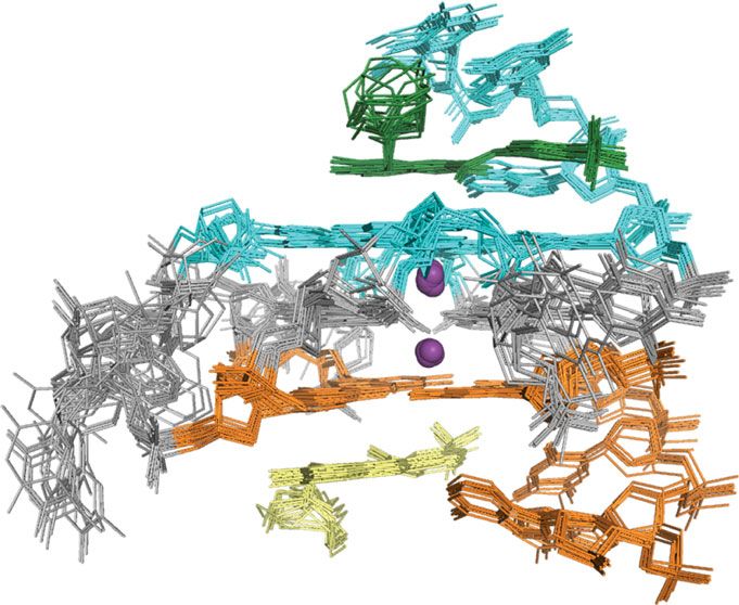
ARTICLE                                                                          NATURE COMMUNICATIONS | DOI: 10.1038/s41467-018-06315-w

I
    n addition to the double helical structure of DNA, the genome          clinical trials28. Another example, Quarfloxin (CX-3552, Cylene
    is known to fold into a variety of noncanonical three-                 Pharmaceuticals, Tetragene), is reported to act through the
    dimensional structures that offer unique opportunities for             inhibition of rRNA biogenesis by disrupting interaction between
small molecule binding1,2. Molecules that recognize specific DNA            nucleolin and ribosomal G4 DNA and advanced to phase II
folds that are scarce in the genome could be used to alter gene            trials29. Despite considerable efforts, a G4-modulating drug is not
expression or regulatory pathways controlled by these elements,            yet available, and molecules that discriminate between different
thus offering potential as therapeutics. To develop chemical               G4 structures have proved challenging to develop29.
probes that bind to folded DNA in the genomic context, small                  In this study, we report the discovery of a drug-like compound
molecules with high affinity and selectivity are needed3,4. One             with dramatic effects on MYC expression in multiple myeloma
barrier to generating such reagents is the paucity of atomic level         cells, demonstrate that it acts by a G4-dependent mechanism of
structural information available for nucleic acid-small molecule           action, and solve a complete structure by NMR of the compound
complexes, as the resulting mechanistic detail could guide ligand          in complex with the MYC G4. We synthesize a focused library of
design efforts. Furthermore, many molecules reported to bind to            analogs30, which are evaluated for their affinity, and their ability
nucleic acids have multiple mechanisms of action, fall far outside         to silence MYC and limit cell growth in a MYC-driven multiple
“drug-like” chemical space, and/or are characterized by high               myeloma cell line. The most potent analog (DC-34) inhibits MYC
molecular weights, multiple cationic charges, or intercalating             at the transcriptional level only when a G4 is present in the
scaffolds5,6. While it is routine to characterize protein-binding          promoter. Importantly, DC-34 does not transcriptionally down-
small molecules using X-ray crystallography or NMR spectro-                regulate several other G4-dependent genes to the same extent. To
scopy, there are comparatively few structures of “drug-like” small         establish a structural basis for this selectivity, we synthesized an
molecules in complex with nucleic acids. Understanding the                 isotopically labeled DC-34 for use in NMR studies. This probe,
chemical and structural basis for nucleic acid-small molecule              along with an unlabeled version, is used to solve the NMR
interactions will greatly improve our ability to rationally design         structure of DC-34 in complex with the G4. DC-34 adopts a
selective, high affinity small molecules and further explore nucleic        three-dimensional conformation that enables specific contacts
acid-binding compounds as mechanistically novel therapeutics.              with the G4 that govern selectivity and biological activity. Insights
   Developing small molecules that bind to and alter the function          gained from this structure and the corresponding chemical
of regulatory nucleic acid sequences is particularly attractive            derivatives provide a basis for the recognition of the MYC G4 and
when they govern the expression of so-called “undruggable”                 have implications for the development of selective nucleic acid-
proteins, such as MYC7,8. The MYC gene encodes the tran-                   binding compounds with biological activity.
scription factor MYC (also known as c-Myc), which is responsible
for affecting the expression of a large number of genes in the
human genome9–13 and associated with proliferation, differ-                Results
entiation, apoptosis, and oncogenesis. Importantly, MYC is                 Structure of DC-34 influences binding and cellular activity. As
upregulated in 70% of all cancers10 and linked to ~100,000 deaths          part of an effort to understand the factors that govern molecular
per year14. However, it has proven difficult to develop efficacious          recognition and selectivity for G4 structures we designed an
small molecule inhibitors of the MYC protein due to a lack of              efficient, flexible, and scalable synthetic route that enabled the
small molecule binding pockets and a short protein half-life of            generation of a focused library of 25 compounds (Fig. 1a, b, and
20–30 min7,8. An attractive alternative route is the prevention of         Supplementary Table 1)31,32. Most compounds were evaluated for
MYC transcription via small molecule-mediated stabilization of             affinity and effects on cell viability, and selected compounds also
the G-quadruplex (G4) present within the MYC promoter                      for effects on MYC protein levels in L363 cells (a MYC-driven
region15–17. G4s are non-B DNA structures that occur in G-rich             multiple myeloma cell line33). Affinities were generated by mea-
sequences and are characterized by stacks of Hoogsteen-bonded              suring KD values determined by the compound-induced change
guanine tetrads stabilized by central potassium ions and flanked            in fluorescence of an Alexa Fluor® 647 tag conjugated to the 5′
by loop regions1,18. G4s have been identified in genome-wide                end of Pu27. Selected compounds are illustrated in Fig. 1b
structural probing studies using a G4-specific antibody19, as well          (Supplementary Table 1).
as by a chemical probing approach employing ss-DNA seq20,21,                  From this effort, clear trends were established to generate a
which together, have identified about 10,000 G4-forming                     structure–activity relationship and determine requirements for
sequences in cells. About 90% of MYC expression is regulated               binding. Substitution of an electron withdrawing trifluoromethyl
by a G4-forming 27 nucleotide sequence found in the CT element             group in place of the methyl group on the aryl amide yielded
(sometimes referred to as the nuclease hypersensitive element III          increased affinity, decreased cell viability, and decreased MYC
or NHEIII region) of the MYC gene (Pu27)13,14,22,23. Small                 expression (DC-34, Fig. 1b). With DC-34, the most potent
molecules that bind and stabilize the MYC G4 have been shown               compound, MYC protein expression was reduced to 0.4% at a 10
to decrease MYC expression and present a potential method for              μM dose compared to 50% for the methyl-substituted compound
targeting cancers where MYC contributes to the oncogenic phe-              and an increase in potency was observed in the cell viability assay.
notype3,4. However, many of these ligands that silence MYC                 Conversely, replacement of the methyl group with an electron
expression in cells are not selective, and therefore their activity        donating methoxy substituent or substituted pyridine resulted in
cannot always be attributed to a MYC-dependent mechanism of                worse activity than the methyl-bearing compound for all assays,
action24.                                                                  likely indicating a weaker interaction for the aromatic ring with
   As with many nucleic acids, there are few structures of small           the electron rich guanine tetrads. A fluoro substituent in place of
molecule ligands bound to G4s. As a method to provide insights             the methyl group yielded a compound with minimal activity and
for rational design for small molecule ligands, Yang and co-               weaker binding affinity (Supplementary Table 1).
workers solved an NMR structure of the MYC G4 (Pu22 G14T/                     The position of the trifluoromethyl group on the benzene ring
G23T)25 and later in complex with a ligand26. Others have also             was also evaluated next. The para-trifluoromethyl group emerged
reported small molecule ligands for G4s, and they are a target of          as the most potent, while the meta-analogs and ortho-analogs had
considerable interest3,4,16,27. In one example, CX-5461 was found          weaker activity. Substitution of the aromatic ring in the ortho-
to target BRCA1/2-deficient tumors and is reported to stabilize             position is likely to force the amide substituent out of plane
G4 structures and dsDNA, and is being investigated in phase I              with the arene, suggesting that these substituents are required to

2                        NATURE COMMUNICATIONS | (2018)9:4229 | DOI: 10.1038/s41467-018-06315-w | www.nature.com/naturecommunications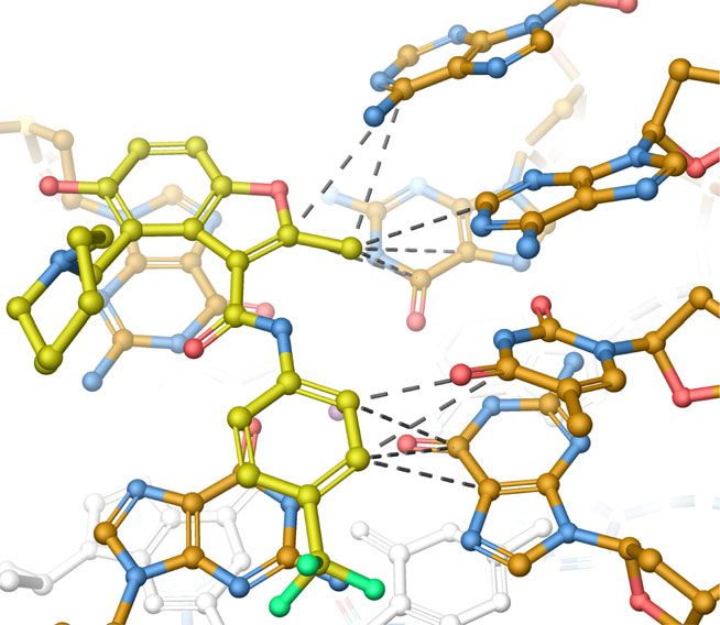
NATURE COMMUNICATIONS | DOI: 10.1038/s41467-018-06315-w ARTICLE
a O
R R R
H2N O N O
R O NH N NH
O O H
DMAP O O In(OTf)3 HO HO
Formaldehyde
H3C O CH3 Toulene H3C N Acetonitrile CH3 CH3
EtOH, 70 °C O
reflux H O
b Aromatic ring electronics Position of ring substituent
OCH3 CH3 CF3 CF3
DC-34 DC-34 F3C
F3C
N O N O N O N O N O N O
NH NH NH NH NH NH
HO HO HO HO HO HO
CH3 CH3 CH3 CH3 CH3 CH3
O O O O O O
% Myc: 81% 50% 0.44% 0.44% 12% N.D.
IC50: 7.2 μM 5.8 μM 3.5 μM 3.5 μM 3.3 μM >50 μM
KD: >100 μM 23.2 ± 7.6 9.4 ± 1.1 μM 9.4 ± 1.1 μM 23.2 ± 2.5 μM >100 μM
Length of amine linker Other structural alterations
DC-34 CF3 CF3 CF3 CF3 CF3 CF3
N N
N
N O O O N O N O HO O
NH NH NH N NH NH
HO CH3 HO HO
HO HO HO
CH3 CH3 CH3 CH3 CH3 CH3
O O O O O O
% Myc: 0.44% N.D. N.D. N.D. N.D. N.D.
IC50: 3.5 μM 38.2 μM >50 μM 23.8 μM >50 μM 26.5 μM
KD: 9.4 ± 1.1 μM 18.8 ± 3.2 μM N.D. >50 μM >80 μM 57.6 ± 7.4 μM
Fig. 1 Structure–activity relationship of DC-34. a Synthesis scheme of MYC G4-binding scaffold. b Table of selected analogs, with the parent compound on
the left. %Myc indicates to the percent of MYC protein expressed at a 10 µM dose in L363 MM cells, IC50 is the half maximum concentration for
cytotoxicity in L363 MM cells at 48 h, and KD determined by a fluorescence intensity assay. N. D. indicates that value was not determined
be co-planar for binding. Similarly, N-methylation of the amide of Pu27 and Pu22 increased by 7.5 ± 2.0 and 7.4 ± 0.5 °C,
also resulted in loss of activity and binding affinity by likely respectively (Fig. 2a for Pu22 and Supplementary Fig. 1 for Pu27).
influencing the orientation of the amide bond, further high- Increasing the length of loops 1 and 2 of Pu22 resulted in lower
lighting a role for this group in the interaction with the MYC ΔTm values with DC-34, while changing the length of the third
G434. loop had no effect (Supplementary Table 2). An A25T mutant G4
Since the para-trifluoromethylbenzene emerged as the most had a similar decrease in stability as removing both 5′ and 3′ tails,
effective substituent, we then investigated the amine ring of the 4- highlighting the importance of this residue for DC-34 binding
position on the benzofuran. Alteration of the azepane ring to (Supplementary Table 2). DC-34 exhibited a smaller ΔTm of 4.2 ±
other aliphatic amines did not result in dramatic increases in 0.4 °C for the BCL-2 G435, and minimal change was observed
potency, while bicyclic heterocycles were uniformly inferior with the G4 oligos from VEGF36, KRAS37, MYB38, and HIF1α39
(Supplementary Table 1). Increasing the number of carbons genes (Fig. 2b). Similarly, no change in Tm was observed for two
between the benzofuran core and amine decreased activity, different antiparallel G4 structures including a G4 derived from
indicating that the benzylic amine substituent exhibited the best telomeric DNA40 or dsDNA (Supplementary Table 2).
performance in all assays. The relative position of the cationic Surface plasmon resonance (SPR) experiments were used to
amine is thus clearly important for binding, though the nature of further quantitate the binding of DC-34 to the MYC G4. DC-34
the aliphatic ring is somewhat less consequential. Variation of the had a KD of 1.4 ± 1.2 μM with a 5′-biotin labeled Pu27 (Fig. 2d).
methyl group on the 2-position of the benzofuran core with This was in reasonable agreement with the KD of 9.4 ± 1.1 μM
branched aliphatic groups resulted in considerably weaker affinity from the fluorescence intensity assay (FIA) with a 5′-Alexa Fluor ®
(Supplementary Table 1). Taken together, DC-34 displayed the labeled Pu27 (Fig. 2c). By FIA, DC-34 displayed a tighter binding
best combination of affinity, ability to silence MYC, and limit cell affinity for MYC than several other G4 oligos, including DNA and
growth. Therefore, this compound was the focus of further study. RNA G4 oligos from the VEGF41, KRAS37, BCL235, telomeric
DNA40, and NRAS42 genes (Fig. 2b and Supplementary Fig. 2).
Thus, unlike other G4-binding compounds such as CX-4561, DC-
Biophysical analysis of DC-34 reflects MYC G4 preference. To 34 does not bind to B-DNA and has weaker binding to a variety of
evaluate the interaction between DC-34 and the MYC G4 in other quadruplex sequences3.
greater detail, we performed in-depth biophysical analyses. To
test whether DC-34 confers stability to the MYC G4, we per-
formed a circular dichroism (CD)-based thermal melt assay, in DC-34 requires the G4 to downregulate MYC in cancer cells.
which molar ellipticity was measured as a function of increasing We next performed time course experiments of cells treated with
temperature3. In the presence of DC-34, the melting temperature DC-34 to assess tumor cell viability and MYC protein levels
NATURE COMMUNICATIONS | (2018)9:4229 | DOI: 10.1038/s41467-018-06315-w | www.nature.com/naturecommunications 3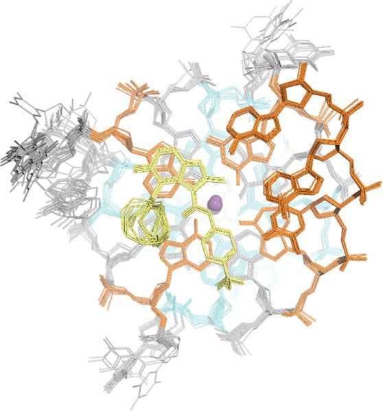
ARTICLE                                                                                                                                 NATURE COMMUNICATIONS | DOI: 10.1038/s41467-018-06315-w

                       a                                                                                              b                                             ΔTm (°C)            KD FIA (μM)
                                                                                                                                                Oligo

                       Normalized ellipticity/mdeg
                                                                                                MYC G4                                       MYC (pu27)             7.5 ±2.0                9.4 ±1.1
                                                     1.0
                                                                                                MYC G4 + DC-34                               MYC (pu22)             7.4 ±0.5                 N. D.
                                                                                                                                                KRAS                0.7 ±0.4               44.2 ±3.8
                                                                                                                                                MYB                 1.5 ±0.3                 N. D.
                                                     0.5
                                                                                                                                                BCL2                4.2 ±0.4               17.3 ±4.4
                                                                                                                                            Telomeric DNA           0.3 ±0.3               40.3 ±10.0
                                                                                                                                                VEGF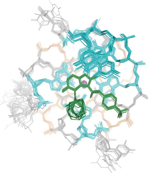
NATURE COMMUNICATIONS | DOI: 10.1038/s41467-018-06315-w ARTICLE
a c d DMSO
120
MYC expression (%unt)
100 100 MYC
Cell viability (%)
80
75
β-actin
60
50
40 Time (min) UNT 15 30 45 60 75
IC50 = 1.9 μM
25 20 DC-34
0
0 0.01 0.1 1 10 MYC
0.1 1 10 100
[DC-34] μM
[DC-34] μM β-actin
MYC Time (min) UNT 15 30 45 60 75
b 5 μM treatment
UNT 4 24 48 72 h 0 0.1 1 1.5 3 4 5 μM 1.0
CX
MYC
0.8 CX + DC-34
% MYC protein
GAPDH 0.6
0.4
e [DC-34] μM f GFP CMV-MYC
0.2
0 0.1 1 2 3 5 [DC-34] (μM) 0 1 5 0 1 5
MYC MYC 0.0
0 20 40 60 80
L363 β-actin
GAPDH Time (min)
MYC
CA46
GAPDH
Fig. 3 DC-34 silencs MYC expression in cancer cells. a Inhibition of L363 MM cell proliferation at 24 (IC50 = 3.4 µM) (red), 48 (IC50 = 3.4 µM) (green)
and 72 h (IC50 = 3.1 µM) (blue). b Inhibition of MYC protein translation with 5 μM of DC-34 is sustained over time in L363 cells. c The half maximal
inhibitory concentration (IC50) of DC-34 with respect to MYC protein inhibition as determined by Peggy protein expression. d A Western blot of MYC
protein during a representative cycloheximide-chase degradation experiment with L363 multiple myeloma cells. Cells were treated with 10 µg/mL of
cycloheximide (CX) in the absence or presence of DC-34 (5 µM) and MYC protein expression was assessed at indicated time points. β-actin was used as
loading control. e MYC protein levels are inhibited as a function of the dose of DC-34 in L363 cells; only the highest dose of DC-34 affected MYC in the
more resistant CA46 Burkitt’s lymphoma cells. f Western blot analysis of 293T cells transiently transfected with either GFP or CMV-MYC plasmid (the
CMV promoter lacks a MYC G4) (right) and dosed with different concentrations of DC-34. All western blots were exposed for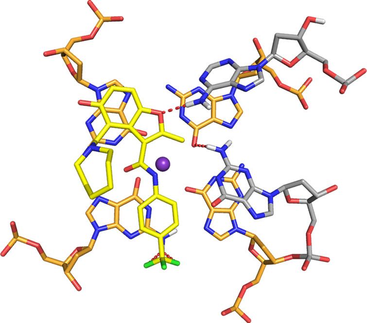
ARTICLE                                                                          NATURE COMMUNICATIONS | DOI: 10.1038/s41467-018-06315-w

DC-34 binds independently to the MYC G4 3′ and 5′ ends. We                 with flanking residues and 5′ and 3′ G-tetrads (Fig. 6c–e).
used NMR experiments to probe the structural origin of the                 Intermolecular NOEs were not observed to the central G-tetrad
interaction between DC-34 and the MYC Pu22 G14T/G23T                       or loop residues. G7, G11, G16, and G20 H1 of the 5′ G-tetrad
mutant25,26. We performed WaterLOGSY46,47 experiments on                   interact with DC-34 methyl and methanediyl groups (Fig. 6c, top
DC-34 in the presence and absence of the MYC G4 with N-                    and middle panels respectively); G7 H8 interacts with the DC-34
methyl-L-valine used as an internal, non-binding control mole-             methyl group (Fig. 6e); and G11 and G16 H8 form NOEs with the
cule (Fig. 5a). In the absence of DNA, all peaks phased negatively         DC-34 methanediyl group (Fig. 6d). The G7 and A6 ribose H1′
in the WaterLOGSY experiment, confirming that DC-34 does not                protons are overlapped but show NOE interactions with the DC-
aggregate in buffer. Upon the addition of DNA, only the peaks for          34 para-trifluoromethylbenzene protons (Fig. 6f). The DC-34
DC-34 became positively phased and chemical shift perturbations            methyl group also shows NOEs to A6 H2, H8, and H1′, as well as
were observed, confirming a direct interaction between DC-34                G5 H8 (Fig. 6e). These interactions define a distinct orientation
and the G4.                                                                for DC-34 at the 5′ end.
   We titrated DC-34 into a sample of the Pu22 G14T/G23T G4                   At the 3′ end, intermolecular NOE interactions are detected
to demonstrate chemical shift perturbation of the imino protons.           between G9, G13, G18, and G22 H1 and DC-34 methyl,
Fast exchange chemical shift perturbations were observed,                  methanediyl, and/or para-trifluoromethylbenzene protons
allowing all 12 guanine imino protons from the MYC G4 tetrad               (Fig. 6c). G9 H8 and H1′, A24 and A25 H2, and T23 and A24
planes25 to be tracked upon addition of DC-34. The observation             H1′ interact with the DC-34 methyl group (Fig. 6e). In contrast to
of a new set of 12 well-resolved imino proton peaks (Fig. 5b)              the 5′ end, NOEs were observed from DC-34 trifluoromethyl-
indicate the formation of a well-defined DC-34/MYC G4                       benzene to G13, G18, and G22 H1 (Fig. 6c, bottom panel) as well
complex. Perturbations saturated above six molar equivalents of            as G18 and G22 H8 (Supplementary Fig. 5E). However, only G11
DC-34, indicating that the effect is governed by affinity as well as        H8 of the 5′ end G-tetrad base region exhibited NOEs to the DC-
stoichiometry. The largest perturbations were observed for G9              34 para-trifluoromethylbenzene group (Supplementary Fig. 5E),
and G18 (3′ face) and G11 and G16 (5′ face) (Fig. 5b). Minimal             although NOEs were also observed to the H1′ of overlapped A6
chemical shift perturbations were observed for the imino protons           and G7 (Fig. 6f). Collectively, these NOEs place the DC-34 para-
from G8 and G21, in the central guanine tetrad. Plots of the               trifluoromethylbenzene group closer to the flanking and first
chemical shift changes observed for G9, G18, G11, and G16 imino            residue of the 5′ G-tetrad, and more centered over the G-tetrad
protons at varying molar ratio of DC-34 to MYC G4 yielded an               base region at the 3′ end.
overall KD value of 16.5 ± 1.1 μM, by fitting the data to a non-               To confirm the intermolecular NOE interactions, we
cooperative binding mode with the software package Bindfit48                synthesized tetra-13C-labeled DC-34, including a 13C-labeled
(Fig. 5d). The Pu22 G14T/G23T sequence used in NMR studies                 methyl group (Fig. 6b). This probe was synthesized using a
differs slightly from the wild-type Pu27 used in affinity                   route similar to Fig. 1a but with 13C-labeled reagents. Twofold
measurements by fluorescence (Fig. 2c), a potential source of               molar excess of tetra-13C-labeled DC-34 was then mixed with
the small difference in observed affinities. Altogether, these              unlabeled Pu22 DNA. A 1H, 13C half-filtered NOESY experi-
findings suggest that DC-34 likely stacks independently on the 3′           ment was acquired to record interactions between the DC-34
and 5′ faces of the MYC G4, with similar affinity for each site.            methyl group and MYC G4 (Fig. 6g). Intermolecular NOE
                                                                           interactions between the DC-34 methyl group and MYC G4 5′
                                                                           face residues (G5, A6, G7) or 3′ face residues (G9, T23, A24,
Distinct binding of DC-34 at the 5′ and 3′ ends of MYC G4.                 A25) were observed (Fig. 6g), as well as intramolecular NOE
We assigned 92% of DC-34-bound MYC G4 protons; most of the                 interactions involving 12C bound DC-34 protons. The observed
unassigned protons are guanine and adenine NH2 groups that are             intermolecular NOE interactions recorded using the 13C-
not present in the spectra. For clarity, we provide a representative       labeled sample were consistent with those observed with
G-quadruplex structure for MYC G4 with nucleic acid numbers                unlabeled DC-34/MYC G4 complex (Fig. 6c–f). No NOE
indicated (Fig. 6a) and the DC-34 chemical structure with our              interaction was observed between DC-34 and residues of the
numbering scheme and 13C-labeling included (Fig. 6b). The base             central G-tetrad or 1:2:1 loop region. Thus, the NOESY
imino protons (H1) of the MYC G-tetrads were assigned by                   experiments demonstrate the presence of two DC-34 molecules
tracking in the 1D titration spectra (Fig. 5b), validated by inter-        bound to the MYC G4, one at the 5′ end and the other at the 3′
residue NOE interactions (Fig. 6c, bottom panel), and used to              end. Unique NOE interactions at each of these ends further
assign the G-tetrad base aromatic protons (H8) (Fig. 6c, bottom            demonstrate distinct binding modes.
panel). The ribose protons (H1′, H2′, H2”, H3′, H4′, H5′, and
H5”) were assigned by NOE interactions (Fig. 6d and Supple-
mentary Fig. 5A) and TOCSY spectra (Supplementary Fig. 5B).                Structure of the DC-34/MYC G4 complex. We solved the
Our NMR data indicated a single G-quadruplex conformation for              structure of DC-34 complexed with the MYC G4 using NOE-
DC-34-bound MYC G4, with each proton exhibiting a single                   derived distance constraints, hydrogen bonds, electrostatic bonds,
chemical shift value and NOEs characteristic of the three G-tetrad         and dihedral angle constraints, as described in Experimental
stacked structure (Fig. 6c).                                               Procedures and summarized in Table 1. The 15 lowest energy
   The regions flanking the G-quadruplex were similarly assigned            structures converged with an overall root-mean-square-deviation
by NOE interactions with neighboring residues (Fig. 6c, bottom             (r.m.s.d.) for all atoms including hydrogen of 0.89 ± 0.26 Å
panel, and Fig. 6d) and intra-residue NOEs (Fig. 6d). Involvement          (Table 1) and higher convergence was found for the three G-
of these bases in DC-34 binding was suggested by comparing                 tetrad structures alone (r.m.s.d. = 0.55 ± 0.12 Å), or the adjacent
their chemical shift values in the free and complexed state, as            5′- (T4-G5-A6) and 3′- (T23-A24-A25) flanking regions
demonstrated for G22, T4, and T23 (Supplementary Fig. 5C).                 (Table 1). The superposition for the three G-tetrads is displayed
Loop residues T10, T14, A15, and T19 shifted only slightly                 for three different orientations (Fig. 7a, b) to highlight the MYC
following DC-34 addition (Supplementary Fig. 5D, demonstrated              G4 parallel-stranded G-quadruplex with two bound potassium
for A15), suggesting that they are not at the binding surface.             ions and an additional stacked plane contributed by DC-34 over
   The protons of DC-34 in the complexed state were readily                each external G-tetrad of the MYC G4. The DC-34 benzofuran
assigned and intermolecular NOEs confirmed direct interaction               and para-trifluoromethylbenzene rings form pi–pi stacking

6                        NATURE COMMUNICATIONS | (2018)9:4229 | DOI: 10.1038/s41467-018-06315-w | www.nature.com/naturecommunications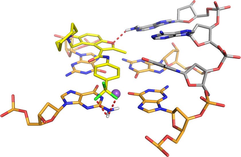
NATURE COMMUNICATIONS | DOI: 10.1038/s41467-018-06315-w ARTICLE
a
!
!
7.9 7.7 7.5 7.3 7.1 6.9 1.8 1.6 1.4 1.2 1.0
b G21,G20 G17,G8 MYC G4:DC-34
G16 G7 G11 G12 G13 G22 G18 G9
1:6
G17,G8
G16 G7 G11 G12 G21 G20 G13 G22 G18 G9
1:5
G17,G8
G16 G7 G11 G12 G21 G20 G13 G22 G18 G9
1:4
G17,G8
G16 G7 G11 G12 G21 G20 G13 G22 G18 G9
1:3
G17,G8
G7 G11 G12 G21 G20 G13 G22 G18
G16 G9
1:2
G17,G8
G16 G7 G11 G12 G21 G20 G13 G22 G18
G9
1:1.5
G17
G16 G7 G11 G12 G21 G20 G8 G13 G22 G18 G9
G17 1:1
G20 G8 G22
G16 G7 G11 G12 G21 G13 G18
G9
G17 G22 1:0.5
G16 G7 G11 G12 G18
G21 G20 G8 G13 G9
1:0
11.75 11.65 11.55 11.45 11.35 11.25 11.15 11.05 10.95 10.85 10.75 10.65 10.55 10.45
1
H (p.p.m.)
c 0.2 G9
d
0.2
0.18 G16 0.18 G9
CSP of imino proton (p.p.m.)
0.16 0.16 G16
0.14 0.14
CSP (p.p.m.)
0.12 0.12 G18
G11 G18
G11
0.1 0.1
0.08
0.08
0.06
0.06
0.04
0.04
0.02
0.02 KD: 16.5 ± 1.1 μM
0
0 0 1 2 3 4 5 6
7 11 16 20 8 12 17 21 9 13 18 22 Molar ratio ([DC-34]/[MYC G4])
G-tetrad imino proton of MYC G4
Fig. 5 Shifting of imino protons during a titration experiment indicates DC-34 binding to each end of MYC G4. a WaterLOGSY NMR spectra of DC-34 and
N-methyl-L-valine (non-binding control, peaks indicated with !) in the absence (top) and presence (bottom) of MYC G4. b Expanded 1D 1H NMR spectra
illustrating the imino region during titration of DC-34 into MYC G4. Molar ratios of MYC G4:DC-34 are as indicated at 1:0, 1:0.5, 1:1, 1:1.5, 1:2, 1:3, 1:4, 1:5,
and 1:6. The G-tetrad imino protons are labeled in each spectrum. c Plot of chemical shift perturbation (CSP) comparing MYC G4 G-tetrad imino protons
alone and with sixfold molar excess DC-34. The red dashed line indicates one standard deviation above the average value. d CSP values for G9 (red), G11
(purple), G16 (blue), and G18 (orange) H1 plotted against molar ratio of DC-34 to MYC G4. An overall KD value of 16.5 ± 1.1 µM was obtained by fitting the
data to a non-cooperative binding mode with the software package Bindfit v0.5
NATURE COMMUNICATIONS | (2018)9:4229 | DOI: 10.1038/s41467-018-06315-w | www.nature.com/naturecommunications 7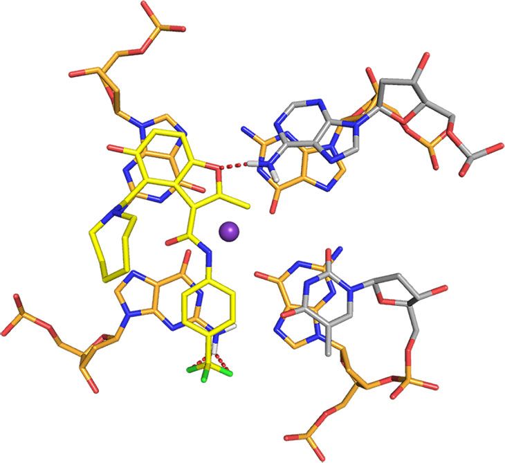
ARTICLE NATURE COMMUNICATIONS | DOI: 10.1038/s41467-018-06315-w
a T4
c 5’-end G21G17 G8 3’-end
5’
H1: G16 G7G11 G12 G20 G13G22 G18 G9
G5 2.3
DC-34 18 2.5
4.2
DC-34 5
4.3
DC-34
A6 13,15
12,16 7.5
G16 G20 G8A6H2 G18 G8
A6H2 G22 G13
H8/H2 G20 G17 G17 G9
G11 G7
G17 G11 G7 G7 G12 G21 G21 G12 8.0
G21 G16 G11
A15 G16 A6
G12 G8
8.5
G18
T14
G22 G20
G13 G9 9.0
G17
T23 H21/H22 G8
1
H (p.p.m.)
9.5
MYC G4 G12 G21
A24
3’ 10.0
A25
b
10.5
F G9
F G18 11.0
G22
DC-34 F G13
G8
H1 G17
G21 G20
21 12 G12 11.5
22 20 G11 G7
16 Para-trifluoro
13 G16
19 -methylbenzene
23 15 11.8 11.6 11.4 11.2 11.0 10.8
N O 1
H (p.p.m.)
Azepane 24 5 NH
ring 13
C-labeled
HO g DC-34 18 13CH3
2.396 2.392 (ppm)
CH3 13 1.0
18 C
2 15.39 ppm
O 21,22
1 20,23
benzofuran
2.0
d f H1’
H8 G7A6
A6 G16 G7 A24 G11 7.3
*
H2’/ A6
DC-34 5 H2”
G5H4’ 4.05 DC-34
13,15 7.4 19,24 3.0
G7H5’/H5” 12,16
4.10 7.5
A6H4’
A24H4’ 4.15 7.6
G12H5’
1
4.0
1
H (p.p.m.)
H (p.p.m.)
or H5” 5
7.7
4.20
1
G8H8
H (p.p.m.)
G16H5’/H5” 7.8
4.25 A6H2
G11H5’/H5” 7.9 5.0
4.30
8.0
G8H5’ 4.35 G7H8
or H5” A6H8 8.1 G5 A24
H1’ T23 6.0
4.40 A6 H1’
8.1 8.05 8.0 6.10 6.05 G9
1
1
H (p.p.m.) H (p.p.m.) 2
e 2.36
1 7.0
1
13,15
H (p.p.m.)
12,16 A24 H2
DC-34 2.40 H8 G5 A25
18 H2 A6 G9 H8
G7
H8 A6 8.0
2.44
H8 H8 H2 H1’ H8 H8 H1’ H2 H2 H1’ H1’ 12,16 13,15
G5 A6 G7 G9 A25 A24 T23 A24 DC-34 2.5 2.4 2.3
1
H (p.p.m.)
interactions with the G-tetrads, from which the azepane ring is MYC G4 complex, A6 is displaced to form pi–pi stacking
directed away (Fig. 7a, middle panel). The overall fold for the G- interactions with G20 while the DC-34 benzofuran and para-
tetrad region is similar to the unbound state25, although differ- trifluoromethylbenzene rings stacks over G16 and G7, which are
ences exist in the flanking regions to accommodate DC-34. diagonal in the G-tetrad (Fig. 8a). This configuration enables the
When unbound, A6 stacks over G11 with T4 and G5 directed DC-34 benzofuran ring to interact with A6. DC-34 at the 3′ end
away from the 5′ G-tetrad25 (Supplementary Fig. 6A). In the stacks over G13 and G18, which are G-tetrad neighbors, while
8 NATURE COMMUNICATIONS | (2018)9:4229 | DOI: 10.1038/s41467-018-06315-w | www.nature.com/naturecommunications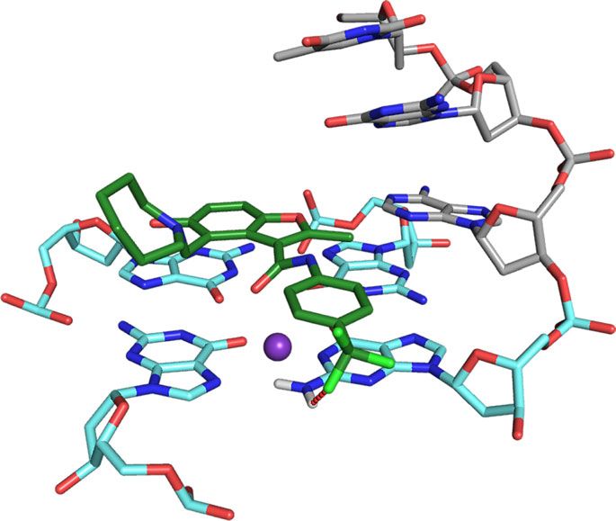
NATURE COMMUNICATIONS | DOI: 10.1038/s41467-018-06315-w                                                                                 ARTICLE

Fig. 6 Intermolecular NOE interactions indicate distinct binding modes for DC-34 at the 5′ and 3′ end of MYC G4. a Representative G-quadruplex structure
for the MYC G4 with nucleic acid number indicated. The lowest energy structure displayed in Fig. 7 was used to make this figure. b Annotation of DC-34
indicating numbering scheme and 13C-labeling. c–f Expanded regions of a 2D NOESY spectrum acquired on MYC G4 with twofold molar excess unlabeled
DC-34 at pH 6.4 in buffer A (25 mM Tris-d11 and 50 mM KCl) with 90% H2O/10% DMSO-d6. H1–H1, H21/H22–H1, and H8/H2–H1 NOE interactions
within MYC G4 are labeled (c, bottom panel), as are intermolecular NOE interactions between MYC G4 H1 and DC-34 methyl (c, top panel) or methanediyl
(c, middle panel) groups. g Selected region from a 1H, 13C half-filtered NOESY experiment acquired with MYC G4 and twofold molar excess DC-34 with
selective 13C-labeling as indicated by red arrows in b. Intermolecular NOE interactions between the DC-34 methyl group and MYC G4 protons are
displayed and labeled as are intramolecular NOE interactions involving 12C bound DC-34 protons. A breakthrough intramolecular signal from the DC-34
methyl group is indicated by a black asterisk. Labeling of NOEs in c–g is color coded with DC-34 in red (intermolecular) or pink (intramolecular) and MYC
G4 in black, light blue (for the 5′ G-tetrad and flanking residues), or orange (for the 3′ G-tetrad and flanking residues)

 Table 1 Structural and NMR statistics for the DC-34/MYC                          Discussion
 G4 complex                                                                       Here, we demonstrate a molecular basis for specific recognition of
                                                                                  the MYC G4 structure with a drug-like small molecule. Insights
                                                                                  are provided by chemical approaches, e.g., alteration of the che-
                                                                  DC-34/MYC G4
                                                                                  mical structure of the ligand, as well as biochemical and bio-
 NMR distance and dihedral constraints                                            physical experiments. Analysis of a focused library of analogs has
 Distance constraints
  Intramolecular NOEs                                             907
                                                                                  revealed a direct structure–activity relationship. Through this
  Intra-residue                                                   610             work we identified trifluoromethyl-substituted compound DC-34
  Inter-residue                                                   297             as the analog with the best affinity and ability to decrease MYC
   Sequential (|i – j| = 1)                                       253             expression. The KD measurements from SPR demonstrated a
   Medium range (|i – j| ≤ 4)                                     22              roughly sevenfold tighter binding interaction than FIA. This
   Long range (|i – j| ≥ 5)                                       22              apparent difference in affinity may be due to the use of different
  Hydrogen bonds                                                  27              labels on the oligonucleotide or the different biophysical techni-
  Intermolecular NOEs                                             45              ques used to measure affinity. Furthermore, FIA experiments use
 Dihedral angle restraints                                                        a two-site binding model while SPR experiments only measure
  χ (O4′-C1′-N9-C4)                                               12
                                                                                  the higher affinity binding site. However, consistent with the low
 G-tetrad planarity (5 atoms sub-groups)                          24
 Coordination bond (O6-K+)                                        16
                                                                                  micromolar affinity, exposure of cells to low micromolar con-
 Structure statistics                                                             centrations of DC-34 caused decreases in MYC protein levels,
 Violations (mean and s.d.)                                                       indicating that it is likely saturated at working concentrations.
  Distance constraints (Å)                                        0               This effect was confirmed to occur at the transcriptional level. In
  Dihedral angle constraints (°)                                  0               293T cells transfected with a plasmid expressing MYC from the
  Max. dihedral angle violation (°)                               0               CMV promoter (which lacks the MYC G4), DC-34 had no effect
  Max. distance constraint violation (Å)                          0               on MYC levels. In CA46 cells, which harbor a chromosomal
 Deviations from idealized geometry                                               translocation resulting in one MYC allele being driven by the IgH
  Bond lengths (Å)                                                0.005 ± 0.000   promoter (lacking the MYC G4)43, DC-34 had minimal effects on
  Bond angles (°)                                                 0.705 ± 0.005
                                                                                  MYC protein levels at IC50 doses required to limit myeloma
  Impropers (°)                                                   0.461 ± 0.031
 Average pairwise r.m.s.d (Å)a                                                    growth. In biophysical experiments, DC-34 preferentially stabi-
  All atoms                                                       0.89 ± 0.26     lized the MYC G4 over six other known G4s and had no effect on
  G-tetrads                                                       0.55 ± 0.12     the Tm of dsDNA. At doses of 5 µM and 7.5 µM, DC-34 exhibited
  5′-end (T4-G5-A6)                                               0.29 ± 0.10     a pronounced ability to decrease MYC mRNA, while having
  3′-end (T23-A24-A25)                                            0.29 ± 0.08     minor effects on other G4-driven genes.
 aPairwise
                                                                                     To aid in solving the structure defined at the atomic level, we
             r.m.s.d. was calculated among 15 refined structures
                                                                                  generated a 13C-labeled DC-34 sample for use in a 13C half-
                                                                                  filtered NOESY experiment. Intermolecular NOEs observed with
T23 packs against G22 (Fig. 8b); this configuration requires                       both this experiment and conventional NOESY experiments
displacement of A25, which in the free state, forms pi–pi stacking                acquired with unlabeled DC-34 yielded a structure with distinct
interactions with G1325 (Supplementary Fig. 6B). At each end,                     binding of DC-34 to each end of the MYC G4 with pi–pi stacking
distances and angles between DC-34 fluorines and NH2 groups                        interactions between the benzofuran and methylbenzene rings of
from the guanines involved in G-tetrads are within the range of                   DC-34 and the terminal G-tetrads, as has been observed for other
bonding interactions49–52. At the 5′ end, G7 is 3.3 Å from a                      molecules22. Hydrogen bonding no doubt contributes to the
fluorine on DC-34 with an angle of 140° (Fig. 8a, c). At the 3′ end,               specificity observed in the biophysical and biological assays. The
DC-34 is a bifurcated donor with G18, at distances of 2.8 Å and                   oxygen in the benzofuran core forms a hydrogen bond to NH2
3.0 Å and angles of 101° and 98°, respectively (Fig. 8b, d).                      group of A25 and fluorines at the para position of the benzene
Additionally, on the 3′ face, the benzylic amine carbon of DC-34                  ring forms hydrogen bonds with G7 and G18 NH2 groups; these
is 3.5 Å away from G13, indicating a cation–pi interaction52,53.                  interactions are not observed with other reported ligands that
The 5′ flanking residues T4 and G5 stack against each other                        generally stack between flanking residues on the tail26,54. Changes
(Fig. 8c), placing T4 far from DC-34. By contrast, A25 does not                   to either of these functional groups decreased the affinity and
stack over A24 (Fig. 7a, middle panel) and instead adopts a                       activity of the corresponding analog. The carbon of the benzylic
configuration that enables a hydrogen bond between its NH2                         amine forms a cation–pi interaction with G13 on the 3′ face.
group and the DC-34 benzofuran oxygen (Fig. 8b). A25, together                    Increasing the number of carbons between the benzofuran core
with A24, also forms hydrophobic interactions with the                            and the amine ring alters the location of the positive charge
benzofuran group of DC-34 (Fig. 8e), as supported by NOEs                         relative to the tetrads, decreasing activity. Additionally, in order
involving the methyl group (Fig. 6e–g).                                           to accommodate ligand binding, both the 5′ and 3′ tails move

NATURE COMMUNICATIONS | (2018)9:4229 | DOI: 10.1038/s41467-018-06315-w | www.nature.com/naturecommunications                                           9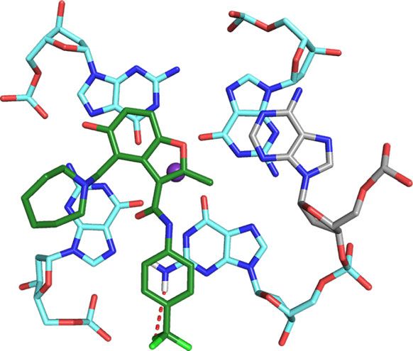
ARTICLE NATURE COMMUNICATIONS | DOI: 10.1038/s41467-018-06315-w
a
T4 5’
G5
DC-34 A25
A6 G13 G9
T4 A6
G5 A24
G16
K+
K+ G7
K+ T23
90° 90°
G18 G22
G11 DC-34 T23 DC-34
A24
A25
DC-34
3’
b
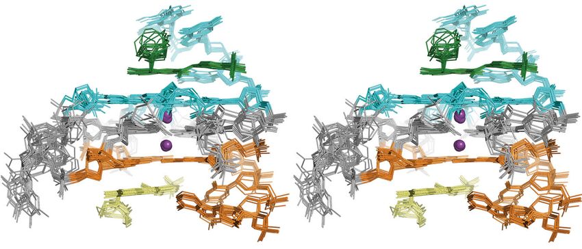
Fig. 7 Structure of the DC-34/MYC G4 complex indicates an additional stacked layer at each end and rearrangement of the flanking residues. a The 15
lowest energy structures of the DC-34/MYC G4 complex are displayed with a top (left panel), side (middle panel), and bottom (right panel) view relative
to the cylindrical axis of the DNA. Residues of MYC G4 from the 5′ G-tetrad and flanking residues are highlighted in light blue, whereas those from the 3′
end are indicated in orange. The two DC-34 molecules are colored green and yellow, and the two potassium ions displayed as purple spheres. b Stereo
view of the 15 lowest energy structures of the DC-34/MYC G4 complex
away from the tetrads to generate hydrophobic-binding pockets. 57,58 whereas DC-34 and the quindoline derivative stack over only
To stabilize the MYC G4 1:2:1 isomer we used a previously two guanines at each terminal G-tetrad plane with flanking seg-
reported Pu22 DNA sequence with G14 and G23 substituted with ments reconfigured to cap the ligand26 (Fig. 8 and Supplementary
thymine24,25. Whereas T14 showed no interaction with DC-34, Fig. 7E). At the 3′ end, these two ligands stack over G13 and G18
T23 of the 3′ flanking region interacts with the tri- and form a hydrogen bond to a 3′ flanking residue; namely, DC-
fluoromethylbenzene group. We therefore substituted T23 with 34 benzofuran oxygen and quindoline derivative N1 form
the wild-type guanine in the MYC G4/DC-34 structure and hydrogen bonds to A25 NH2 (Fig. 8b) and T23 O4 (Supple-
performed energy minimization. The bulkier G23 purine base was mentary Fig. 7E), respectively. A greater difference is observed at
readily accommodated in the DC-34 binding pocket with the the 5′ end where DC-34 stacks over G7 and G16 while the
NH2 group positioned to form a potential hydrogen bond with O6 quindoline derivate overlaps with G11 and G16 (Fig. 8a and
from G9 (Fig. 8f); this possibility may further explain the stronger Supplementary Fig. 7E). In addition to the pi–pi interactions,
affinity measured for the Pu22 wild-type sequence. In addition, positively charged atoms present in the various other G4-binding
the wild-type MYC G4 is known to exist in multiple equilibrating ligands were observed to contribute electrostatic interactions with
conformations. However, we see comparable affinity for both negatively charged G4 phosphates55–57. In the quindoline deri-
wild-type (Pu22, Pu27) and mutant (Pu22 G14T/G23T) quad- vative, the protonated aminoalkylamino side chain is within 3 Å
ruplexes, suggesting that the major conformation in solution is of the G16 phosphate group (red circle in Supplementary Fig. 7E)
recognized by DC-34. whereas the DC-34 azepane ring is too short for such an inter-
Several G4/ligand structures are available3,4 and reveal a action. By contrast, bonding interactions are formed between the
common stacking of ligand on one end of the G4 to form a 1:1 DC-34 fluorines and G7 or G18, and a cation–pi interaction
complex; as exemplified by the telomeric G4/MM41 and G4/ forms between the DC-34 benzylic amine carbon and G13
L2H2-6M(2)OTD complexes55,56 (Supplementary Fig. 7A and (Fig. 8).
7B) and MYC Pu24 G4 complexes with TMPyP4 and Phen Although G4-binding ligands stack on a common tetrad
DC357,58 (Supplementary Fig. 7C and 7D). By contrast, DC-34 structure, variations in groove dimensions, the inter-G-tract
molecules bound to the MYC G4 5′ and 3′ external tetrads loops, and flanking segments allow for specificity4,26,55,60. In the
forming a 2:1 complex, similar to a previously characterized MYC MYC G4, each end has flanking regions that contribute favorably
G4 ligated quindoline derivative complex26,59 (Supplementary to DC-34 binding as mentioned above, with mutations in these
Fig. 7E). All G4/ligand complexes were stabilized by pi–pi regions reducing affinity (Supplementary Table 2). Moreover,
interactions. TMPyP4 and Phen DC3 overlap extensively with all neither DC-34-binding site in the MYC G4 is obscured by resi-
four guanines of the 5′ G-tetrad (Supplementary Fig. 7C and 7D) dues from the loop regions, whereas other G4s that we examined
10 NATURE COMMUNICATIONS | (2018)9:4229 | DOI: 10.1038/s41467-018-06315-w | www.nature.com/naturecommunicationsNATURE COMMUNICATIONS | DOI: 10.1038/s41467-018-06315-w ARTICLE
a b
5’ 3’
G16 A25
G20 G13
A6 2.2Å
Benzofuran
Benzofuran
G9
DC-34 K+ DC-34 K+
T23
G7
G11
G18
3.3Å 2.8Å 3.4Å G22
Para-trifluoro Para -trifluoro
-methylbenzene -methylbenzene
c d
5’ 3’
T4 A25
DC-34
Azepane G5
ring DC-34 A24
A6 Azepane
ring
G13
G9 T23
G20
G16 K+
K+
G18 2.8Å 3.4Å
G7 G22
G11 3.3Å
e f
A25
A25
G13
DC-34
2.3Å
A24
G9
G9 T23 2.6Å
DC-34 K+ G23
G13
K+
G18
G22 G18
3.0Å 3.2Å G22
Fig. 8 Specific contacts formed at each end of the MYC G4 provide a rationale for DC-34 as the preferred compound from the benzofuran-containing
molecules screened. a–d Expanded views of the DC-34/MYC G4 complex to display interactions at the 5′ (a, c) and 3′ (b, d, e) ends of the MYC G4. The
color scheme follows Fig. 7 but with flanking residues gray and oxygen, nitrogen and fluorine in red, navy, and light green. Hydrogen bonds between MYC
G4 and DC-34 are indicated by dotted red lines. In e hydrophobic contacts to flanking residues are indicated by gray-dashed lines. f Expanded view of an
energy minimized model structure of DC-34/MYC G4 with wild-type guanine substituted for T23
have features that make the DC-34 binding sites less accessible. clashes and unfavorable electrostatic interactions observed
We generated a model of a KRAS G4/DC-34 structure using the between DC-34 and KRAS G4 in the model supports the lack of
KRAS G4 structure (PDB 5I2V)61 with DC-34 placed in the binding and effect on gene expression measured experimentally.
analogous region compared to its MYC-binding sites. At the 5′ Similarly, in the BCL2 G4 (PDB 2F8U), two lateral loops are
end, the KRAS G4 contains a four-base loop (A14-A15-T16-A17) located at both the 5′ and 3′ faces, a well-defined three nucleotide
while the MYC G4 has a one base loop (T19) (Supplementary loop at the 3′-end and a seven-nucleotide loop that caps the 5′ G-
Fig. 8A and 8B). This loop narrows the cavity in the KRAS G4, tetrad to form potential reversed Watson–Crick hydrogen bonds
inducing steric clashes with DC-34 (Supplementary Fig. 8B). The (Supplementary Fig. 9)62. These lateral loops are likely to be more
KRAS 3′ end is less crowded; however, the additional loop restricted than those of MYC G4, which we observed to be
nucleotide T8 inserted between G7 and G9 of stacked G-tetrads reconfigured upon DC-34 binding. The MYC G4 contains short
causes the G9 phosphate group to be ~4 Å from the DC-34 loops (1-nucleotide or 2-nucleotide) and flexible flanking regions,
hydroxyl group (circled in Supplementary Fig. 8C). The steric which collectively make it more accessible for DC-34 binding.
NATURE COMMUNICATIONS | (2018)9:4229 | DOI: 10.1038/s41467-018-06315-w | www.nature.com/naturecommunications 11ARTICLE NATURE COMMUNICATIONS | DOI: 10.1038/s41467-018-06315-w
A variety of compounds have been reported to bind to the storage. Cell lines were obtained from Michael Kuehl (NCI) and tested by CNV
MYC G4; however, these compounds generally do not have drug- fingerprinting to verify their authenticity64.
like properties and are not selective for the MYC G4 over other
G4 structures24,27 or other protein targets63, despite often exhi- Cell viability (mts) experiments. Cell viability experiments were performed using
biting good anticancer effects. Our work demonstrates that drug- CellTiter 96 AQueous One Solution Cell Proliferation Assay System (Promega).
Cells were plated in quadruplicate on clear, flat-bottomed 96-well tissue cultured
like compounds can discriminate between different quadruplexes treated plates (Corning Costar) and incubated for each designated time in a 37 °C
by making discrete interactions or altering the conformation of incubator with 5% CO2. Concentrated drug stocks were diluted down in Eppendorf
the tail, loop, and tetrad portions of the quadruplex. These con- tubes to each specific dose point before being added to the plate. After incubation,
formational changes and bonding interactions provide a plausible MTS reagent was added directly to the wells and incubated again at 37 °C with 5%
CO2 for 90 min. The absorbance of the MTS formazan was immediately read at
basis for G4 recognition and selectivity in controlling gene 500 nm on an Omega 640 spectrophotometer. A blank measurement was taken
expression in cells. Here, we provide a structural basis for one from the absorbance of the wells with media only and subtracted accordingly.
such recognition event by solving an NMR structure of DC-34 Percentage cell viability was normalized to the absorbance of untreated wells after
bound to the MYC G4. Additionally, we demonstrate through blanking and averaged from the four quadruplicate wells.
chemical and functional studies that affinity for the target cor-
relates directly with cellular activity and ability to regulate gene Protein assays and western blotting. Cell pellets were first lysed in RIPA buffer
expression. Thus, further structure-based investigation of G4- (RIPA, sodium orthovanadate, PMSF, protease inhibitor, and phosphatase inhi-
bitors A and B), vortexed to homogenize, and sonicated in an ice water bath for 5
binding compounds is likely to be fruitful in developing high min on high, with intervals of 30 s on, 1 min off. Homogenized pellets were then
affinity and selective probes of gene expression, as well as incubated on ice for 90 min before being spun down and transferred to new
potential therapeutics. Eppendorf tubes. Protein was next quantitated by a standard BCA protocol. 15 µg
of protein was loaded into each well of 4–12% Bis-Tris Gels (Novex), electro-
phoresed at 135 V for 90 min to achieve optimal band separation, and transferred
Methods with the iBlot2 system (Life Technologies). Equal protein loading and transfer was
Thermal melt assays. The thermal stability of the MYC oligonucleotide (TGA confirmed by Ponceau staining (Thermo Scientific) after the initial protein transfer.
GGG TGG GGA GGG TGG GGA A) or other appropriate oligonucleotide with Western blots were blocked in 5% dry milk in 1XTBST (10X TBS, DI H2O, Tween
and without compound was determined using an Aviv Biomedical Model 420 20) for 1 h, washed three times in 1XTBST for 10 min each, and incubated on a
Circular Dichroism (CD) Spectrometer equipped with a ThermoCube temperature rocker in a 4 °C cold room overnight with primary monoclonal antibodies in 5%
regulator. To anneal the oligonucleotide, the sample was heated to 95 °C for three BSA at the designated dilution by the manufacturer. Blots were washed three times
minutes and allowed to cool to RT over 1–2 h. The oligonucleotide was then with 1XTBST before incubation with the appropriate species and dilution of
diluted to 10 µM in 10 mM Tris buffer (pH 6.3, containing 30 mM KCl) and four polyclonal secondary antibodies in 5% dry milk on a rocker at room temperature
equiv of compound were added to yield 40 µM compound in 1% DMSO. Spectra for 1 h. Blots were again washed three times with 1XTBST before imaging with
were recorded from 224 to 312 nm at 25 °C with a step size of 2 nm, followed by SuperSignal West Dura Chemiluminescent Substrate (Thermo Scientific) on a
heating from 25 to 97 °C at 1 °C/min in a 0.1 cm quartz cuvette. To calculate the Tm GBOX F3 Imager (Syngene). Primary antibodies were purchased and used as
of each sample, ellipticity was plotted as a function of temperature and fit in follows: c-myc (abcam ab32072, rabbit, 1:10,000), Rb1 (Cell Signaling 9309, mouse,
GraphPad Prism 7 software using a nonlinear sigmoidal dose-response model with 1:2000), Bcl2 (Cell Signaling 2870 S, rabbit, 1:1000), GAPDH (abcam ab128915,
a variable slope. Each condition was performed in triplicate, with ΔTm values rabbit, 1:25,000), and Vinculin (Cell Signaling 13901 P, rabbit, 1:1000). Secondary
calculated using Tm(+compound) – Tm(apo) and then averaged to yield the final value. antibodies were purchased and used as follows: HRP-linked Anti-rabbit IgG (Cell
Signaling 7074, 1:2500), and HRP-linked Anti-mouse IgG (Cell Signaling 7076,
Fluorescence intensity titration. Alexa Fluor 647-labeled MYC (pu27): (TGG 1:2500). For a higher throughput quantitation of MYC protein from treated cells, a
GGA GGG TGG GGA GGG TGG GGA AGG) or other appropriate oligonu- size-based automated capillary immunoassay system (Peggy Sue Simple Western,
cleotide was heated at 95 °C for three minutes, allowed to cool to RT, and diluted to ProteinSimple, Santa Clara, CA) was used by the Center for Cancer Research
50 nM in 25 mM Tris buffer (pH 6.4, containing 50 mM KCl). Compound was Collaborative Protein Technology Resource Group and operated according to
added as a solution both in buffer containing 2–3% DMSO, and the sample was manufacturer’s protocols. Uncropped western blots are shown in Supplementary
allowed to equilibrate for 10 min. Fluorescence intensity spectra were recorded at Figs. 82–97.
RT using a Photon Technology International, Inc. QuantaMaster 600TM Spectro- For more precise quantification of the MYC protein in determining the half
fluorometer equipped with Felix GX 4.2.2 software. Fluorescence intensity was maximal inhibitory concentration (IC50) due to drug treatment, the size-based
recorded at an excitation wavelength of 645 nm, with the resulting emission automated capillary immunoassay system (Simple Western, ProteinSimple, Santa
spectrum recorded from 650 to 800 nm, and the fluorescence intensity at the Clara, CA) was performed by the Center for Cancer Research Collaborative Protein
emission maximum was used in all calculations. KD values were fit using a 2:1 Technology Resource group according to the manufacturer’s protocol. A more
binding model. comprehensive description of the experimental procedures have been described
previously65.
Surface plasmon resonance. SPR was conducted using a Biacore 3000 (Biacore,
Inc) instrument. Streptavidin (Rockland) was immobilized to 20000 RU in both Quantitative PCR. RNA was first isolated from cells using manufacturer’s pro-
flow cells using EDC/NHS coupling to a CM5 chip (GE). The surface was then tocols from the Qiagen RNeasy Mini Kit with the addition of QIAshredder col-
blocked with ethanolamine. Next, 1 μM MYC (pu27) 5′-biotin-TEG BiotinTEG umns. RNA concentration was evaluated using a NanoDrop ND-1000
TGG GGA GGG TGG GGA GGG TGG GGA AGG (obtained as an HPLC-purified Spectrophotometer. cDNA was next reverse transcribed using a master mix of
sample from Integrated DNA Technologies, Inc.) was refolded in 10 mM TRIS, 30 MultiScribeTM reverse transcriptase (1 U/µL, Applied Biosystems), 1X RT buffer
mM KCl, 3% DMSO, pH 6.4 by heating at 98 °C in a heater block for 2 min then (Applied Biosystems), dNTP mix (2 mM, Applied Biosystems), MgCl2 (5.5 mM,
cooled to room temperature, and immobilized on one flow cell of the SPR chip to a Applied Biosystems), RNAse inhibitor (0.4 U/µL, Applied Biosystems), and 1X RT
density of 450 RU. Compounds were injected at a flow rate of 30 mL/min in 10 mM random primers (Applied Biosystems). Master mix was added to 1 µg of RNA and
TRIS, 30 mM KCl, 3% DMSO, 0.01% Tween-20, pH 6.4 for 1 min. Each injection thermal cycled at 25 °C for 10 min, 48 °C for 60 min, 95 °C for 5 min, and finally
was repeated twice for consistency. Each trace was fit individually to a Langmuir held at 4 °C until use. cDNA was diluted 1:10 with ultrapure, RNAse free water
model (1:1 binding, reporting on the highest affinity binding site) for DC-34. before use in qPCR. Samples for qPCR were prepared with 1:4 between diluted
cDNA and primer master mix (1X SYBR Green PCR Mix: Applied Biosystems, 0.2
µM forward primer, 0.2 µM reverse primer, ultrapure H2O). qPCR was performed
Cell culture methods. Human multiple myeloma cell lines L363, KMS12PE, JIM1, on an Applied Biosystems 7500 Fast Real-Time PCR System per manufacturer’s
AMO1, KMM1, KMS27, ARD, OPM1, KHM11, KARPAS417, and H929 were protocol. All primers were validated to ensure the absence of primer-dimers, the
cultured in Advanced RPMI 1640 (6% heat-inactivated fetal bovine serum (FBS): presence of a single peak dissociation curve, and an acceptable standard curve from
Gibco by Life Technologies, 2 mM L-glutamine: Gibco by Life Technologies, 100 U/ serial dilutions of the same cDNA.
mL Penicillin and 100 μg/mL Streptomycin: Gibco by Life Technologies, 100 µg/mL
Normocin: InvivoGen) and incubated at 37 °C with 5% CO2. Human Burkitt′s
lymphoma cell line CA46 was cultured in RPMI 1640 (10% heat-inactivated fetal Cycloheximide-chase degradation assay. L363 multiple myeloma cells were
bovine serum (FBS): Gibco by Life Technologies, 2 mM L-glutamine: Gibco by Life grown at 37 °C in Gibco RPMI Advanced Media (6% FBS) and plated at ~1.0 × 10⁶
Technologies, 100 U/mL Penicillin and 100 μg/mL Streptomycin: Gibco by Life cells per ml density. Cycloheximide was added to a final concentration of 10 µg/mL
Technologies, 100 µg/mL Normocin: InvivoGen) and incubated at 37 °C with 5% and DC-34 was added to a final concentration of 5 µM. An equivalent amount of
CO2. Media was changed every 2 days. For cells plated and harvested for protein or DMSO was added for control samples. Cell samples were obtained every 15 min at
RNA, pellets were washed twice with cold PBS. After aspirating off the PBS, pellets indicated time points by centrifugation and washed once with PBS prior to flash
were flash frozen in dry ice and transferred to a −80 °C freezer for short term freezing. Whole-cell protein extracts were prepared using the RIPA lysis buffer
12 NATURE COMMUNICATIONS | (2018)9:4229 | DOI: 10.1038/s41467-018-06315-w | www.nature.com/naturecommunicationsNATURE COMMUNICATIONS | DOI: 10.1038/s41467-018-06315-w ARTICLE
(ThermoFisher #89900) for 1 h with intermittent vortexing. Proteins were sepa- intramolecular and 45 intermolecular NOE-derived distance restraints, 27 hydro-
rated by SDS-PAGE and analyzed by immunoblotting with c-myc (ab32072) and gen bond restraints, 16 coordination bond restraints, 12 dihedral angles restraints,
β-actin (cell signaling #4967) primary antibodies. A secondary goat anti-rabbit and 24 planarity restraints (Table 1) were combined to calculate the structure of the
antibody (IgG (H + L)-HRP, Invitrogen G21234) was used for detection by the MYC G4/DC-34 complex by using simulated annealing in XPLOR-NIH 2.45 with
ChemidocTM Touch Imaging System. Three replicates of the assay were performed the RNA-ff1 force field68. During the calculations, thymine methyl protons were
and signal intensity was normalized to β-actin and averaged, with error bars replaced by pseudo-atoms.
representing standard deviation. The overall structure calculation was performed through two sequential stages,
as described in the Xplor-NIH distribution package68, whereby the structure of
MYC G4 was established followed by that of the MYC G4/DC-34 complex. For the
Water ligand observed gradient spectroscopy (waterLOGSY). A reference 1D-
first stage, 20 linear starting structures of MYC G4 with two K+ ions were subjected
1H and 1D WaterLOGSY spectrum of 100 µM N-methyl-L-valine (Chem-Impex-
to simulated annealing with all restraints mentioned above and in Table 1 except
International) and 100 µM compound was collected, followed by a separate
sample containing 5 µM MYC, 100 µM N-methyl-L-valine, and 100 µM com- for the intermolecular NOE-derived distance restraints. The lowest energy MYC
G4 structure with best geometry was then used as the starting structure for the
pound. MYC oligonucleotide (TGA GGG TGG GGA GGG TGG GGA A) was
second stage with two DC-34 molecules added. The topology and parameter files of
buffer exchanged into 10 mM Tris-d11 buffer (pH 6.4, containing 50 mM KCl)
DC-34 were generated by the GlycoBioChem PRODRG2 Server69. A second
using centrifugal filtration (3 kDa MWCO, EMD Millipore). A sample of DC-34
iteration of simulated annealing was performed with all restraints included
and N-methyl-L-valine, each at 100 µM, was prepared in 25 mM Tris-d11 buffer
(Table 1) to generate 100 structures. For each stage of the structure calculations,
(pH 6.4, containing 50 mM KCl and 10% DMSO-d6), and 1D reference proton
simulated annealing was performed at high temperature (3000 K), followed by
and WaterLOGSY spectra without oligonucleotide were recorded. These spectra
cooling with 1 K decrements to 25 K, and the resulting structures subjected to
were recorded at 20 °C on a Bruker AVANCE III 500 MHz spectrometer
500 steps of Powell minimization.
equipped with TCI cryogenically cooled probe. The “zgesgp” excitation sculpting
The 15 lowest energy structures without distance or dihedral angle violations
water suppression pulse sequence from Bruker was used for data acquisition
greater than 0.5 Å or 5°, respectively were selected for presentation and statistical
with 128 scans. All data were processed and visualized with MestReNova soft-
ware (Version 8.1.2–11880). analyses. Visualization was performed with PyMOL (PyMOL Molecular Graphics
System, http://www.pymol.org), UCSF Chimera70 and Shrödinger Maestro (www.
schrodinger.com).
NMR spectroscopic experiments of MYC G4/DC-34. NMR samples of MYC G4,
DC-34, or the MYC G4/DC-34 mixtures (at equimolar or twofold molar excess
Model generation. The lowest energy MYC G4/DC-34 structure was modified by
DC-34) were at pH 6.4 in buffer A (25 mM Tris-d11 and 50 mM KCl) with 90%
replacing T23 with the wild-type G23 in UCSF Chimera v1.11.2 to generate a
H2O/10% D2O, 90% H2O/10% DMSO-d6, or 90% D2O/10% DMSO-d6 as indi-
starting model structure for MYC G4 T23G/DC-34. This structure was subse-
cated. DC-34 was first dissolved in DMSO-d6 to a 10 mM stock concentration, and
quently energy minimized by Shrödinger Maestro. Only G22-A25 and the nearby
a shortened MYC oligonucleotide (TGA GGG TGG GTA GGG TGG GTA A) was
DC-34 molecule were free to move during minimization. To generate a model
buffer exchanged (3 kDa MWCO spin column) into buffer A. With the exception
KRAS G4/DC-34 structure, the G-tetrad region of the lowest energy MYC G4/DC-
of the WaterLOGSY experiments, all NMR data were acquired at 25 °C on Bruker
34 structure was superimposed onto the corresponding region of an available KRAS
Avance 700, 800, or 850 MHz spectrometers equipped with cryogenically cooled
probes. 1D 1H and 2D homonuclear 1H–1H experiments including NOESY (80, G4 structure (PDB 5I2V) in PyMOL.
150, 300, and 400 ms mixing time), TOCSY (80 ms mixing time) and COSY were
collected for MYC G4 with DC-34 at twofold molar excess and using relaxation RNA isolation for nanostring nCounter® gene expression. RNA was isolated
delays of 2 s. A 2D NOESY (300 ms mixing time) experiment was also collected for from cells using Trizol reagent (Sigma) and further purified using a Qiagen RNeasy
MYC G4 with equimolar DC-34. Intermolecular NOE interactions between MYC Mini Kit. Isolated RNA was eluted in a 30 μL volume and its purity assessed using a
G4 and the DC-34 methyl group were confirmed by using a 13C-half-filtered NanoDrop ND-1000 spectrophotometer (OD 260/280 nm 1.7–2.5).
NOESY spectrum (300 ms mixing time) recorded on unlabeled MYC G4 mixed
with twofold molar excess DC-34 with selective 13C-labeling (Fig. 6g). 1D 1H and
13C spectra, 2D homonuclear 1H–1H (NOESY, TOCSY, COSY), and heteronuclear NanoString nCounter® gene expression quantification. RNA (100 ng) was
1H–13C (HSQC, HMBC) spectra were collected for DC-34. For the 1D 1H titration analyzed by NanoString nCounter XT CodeSet Gene Expression Assays which
delivers direct, multiplexed measurements of gene expression through digital
experiment, DC-34 was first diluted in buffer A to 1 mM with a final DMSO-d6
readouts of the abundance of mRNA transcripts. The nCounter XT CodeSet Gene
concentration of 10%, and titrated into 0.1 mM MYC G4 in buffer A with varying Expression Assay system uses gene-specific probe pairs that hybridize directly to
molar ratios of DC-34 (MYC G4:DC-34 at 1:0, 1:0.5, 1:1, 1:1.5, 1:2, 1:3, 1:4, 1:5,
the mRNA sample in solution eliminating any enzymatic reactions that might
1:6). NMR spectra were processed and visualized by MestReNova for 1D experi-
introduce bias in the results. A Reporter Probe carries the fluorescent signal; a
ments (mestrelab.com) and NMRPipe66 and XEASY67 for 2D and 3D experiments.
Capture Probe allows the complex to be immobilized for data collection. The
nCounter XT assay simultaneously measures the expression levels of 730 target
KD fitting of the NMR titration data. Chemical shift perturbation (CSP) of MYC genes plus 40 endogenous control (house-keeping) genes in a single hybridization
G4 imino protons was calculated by using Eq. ( 1), in which ΔδH represents the reaction using an nCounter CodeSet. Each assay run includes a reference sample
change in proton value (in parts per million). consisting of in vitro transcribed RNAs of six targets that are used for normal-
ization purposes. The raw expression data were normalized using nSolver Analysis
CSP ¼ ΔδH ð1Þ software version 3.0. Platform specific variability was accounted for with the
geometric mean of the four positive controls (ERCC_00117.1, ERCC_00112.1,
ERCC_00002.1, and ERCC_00002.1), followed by assay-specific normalization
A KD value was determined by using Bindfit v0.5 software (http:// with the geometric mean of six house-keeping genes (AGK, EDC3, FCF1, MRPS5,
supramolecular.org). Raw data, including MYC G4 and DC-34 concentrations and PRPF38A, and USP39) chosen by the geNorm algorithm. Genes with low
CSP values of MYC G4 G9, G11, G16, and G18 imino protons, were inputted into expression in all samples (normalized expressionYou can also read