Cement bridging phenomenon in percutaneous vertebroplasty for adjacent vertebral compression fracture
←
→
Page content transcription
If your browser does not render page correctly, please read the page content below
www.nature.com/scientificreports
OPEN Cement bridging phenomenon
in percutaneous vertebroplasty
for adjacent vertebral compression
fracture
Yun‑Da Li1,2,3,4, Tsung‑Ting Tsai1,2,3, Chi‑Chien Niu1,2,3 & Po‑Liang Lai1,2,3*
In some cases of vertebroplasty for adjacent fractures, we observed a cement bridging phenomenon,
in which the injected cement flowed from the newly fractured vertebra to the previously cement-
augmented vertebra through the space between the abutting anterior longitudinal ligament and the
vertebral column. The purpose of this retrospective study was to investigate this phenomenon. From
January 2012 to December 2014, patients who sustained new-onset adjacent vertebral compression
fracture and who were again treated with vertebroplasty were enrolled. We divided the patients into
two groups, the bridging group and the nonbridging group, to analyze the difference between them.
Results showed that the cement bridging phenomenon occurred in 18 (22.8%) of the 79 patients.
Significant differences between the bridging and nonbridging groups were identified in the following
3 imaging features: severe loss of the anterior vertebral body height at the new-onset adjacent
vertebra on plain film (odds ratio [OR] = 4.46, p = 0.014), fluid accumulation (OR = 36.27, p < 0.001)
and hypointense signaling (OR = 15.67, p < 0.001) around the space anterior to the abutting vertebral
bodies and the corresponding intervertebral disc on MRI. After a 2-year follow-up, both the mean
value of the focal kyphotic angle and anterior body height ratio were significantly better in the cement
bridging group than in the nonbridging group. The cement bridging phenomenon, which has never
been reported in the literature, is not rare in clinical practice. This phenomenon was associated with
better maintenance of focal kyphotic angle and anterior body height ratio during the 2-year follow-up.
Abbreviations
CBP Cement bridging phenomenon
OR Odds ratio
PMMA Polymethylmethacrylate
VCF Vertebral compression fracture
BMI Body mass index
BMD Bone mineral density
Percutaneous vertebroplasty was first introduced in 1984 in France by Galibert for the treatment of a painful
hemangioma at the C2 vertebral b ody1. Since then, this procedure has been used extensively worldwide for the
treatment of osteoporotic vertebral compression fracture (VCF). The advantages of percutaneous vertebroplasty
are that it is minimally invasive and efficient not only for providing pain relief and spinal stabilization but also
for reducing the side effects of medications and the risks of long-term limited m obility2–4.
However, some complications remain, such as cement leakage, pulmonary embolism and new-onset vertebral
compression fracture5–7. The incidence of subsequent new-onset VCF after percutaneous vertebroplasty has
been reported as 6.5–52% in the literature, usually occurring in adjacent vertebral bodies7–11. Secondary cement
augmentation is an effective procedure for treating adjacent V CF12.
1
Department of Orthopedic Surgery, Spine Section, Chang Gung Memorial Hospital, No. 5, Fusing St., Guishan
Dist., Taoyuan 33305, Taiwan. 2Bone and Joint Research Center, Chang Gung Memorial Hospital, Taoyuan,
Taiwan. 3College of Medicine, Chang Gung University, Taoyuan, Taiwan. 4Department of Orthopedic Surgery,
New Taipei Municipal TuCheng Hospital, Chang Gung Memorial Hospital, Taoyuan, Taiwan. *email: polianglai@
gmail.com
Scientific Reports | (2021) 11:10184 | https://doi.org/10.1038/s41598-021-89412-z 1
Vol.:(0123456789)
www.nature.com/scientificreports/

                                            Figure 1.  (A–F) Serial intraoperative fluoroscopy images that illustrated the cement bridging phenomenon, in
                                            which the injected cement filling the newly fractured vertebra flowed through the anterior margin of the spinal
                                            column and then reached the previously cement-augmented vertebra.

                                               During the procedure of percutaneous vertebroplasty for new-onset adjacent vertebral compression fracture,
                                            we observed a cement bridging phenomenon (CBP), in which the injected cement flowed from the newly frac-
                                            tured vertebra to the previously cement-augmented vertebra through the space between the abutting anterior
                                            longitudinal ligament and the vertebral column (Fig. 1). This phenomenon has never been described in the lit-
                                            erature. Therefore, the purpose of our study was to elucidate this phenomenon, and further analyze the imaging
                                            predictors and clinical significance of the cement bridging phenomenon.

                                            Materials and methods
                                            Data collection. Between January 2012 and December 2014, all the patients in our institute were included
                                            in this study if they: (1) sustained new-onset adjacent vertebral compression fractures; and (2) were treated again
                                            with vertebroplasty. The local institutional review board approved this retrospective study (Institutional Review
                                            Board of Chang Gung Medical Foundation Reference Number: 201900794B0) and waived the requirement for
                                            written informed patient consent. All methods were conducted in accordance with the relevant guidelines and
                                            regulations.
                                                The cement bridging phenomenon was defined as the presence of injected cement filling the newly fractured
                                            vertebra, flowing through the anterior margin of the spinal column, and then reaching the previously cement-
                                            augmented vertebra. The CBP was observed by orthogonal intraoperative fluoroscopy and confirmed by post-
                                            operative plain films. Each image was independent interpretated by two authors (YD Li and PL Lai) to reach
                                            agreement on the presence or absence of the CBP in one patient.
                                                Preoperative patient characteristics, including age, sex, body mass index (BMI), T-score of bone mineral
                                            density (BMD), findings from preoperative radiographic studies and MRI data, were recorded. The preopera-
                                            tive MRI analysis focused on both fluid accumulation or hypointense signaling around the space anterior to the
                                            abutting vertebral bodies and the corresponding intervertebral disc. The details of vertebroplasty were inves-
                                            tigated, including the volume of injected cement and patterns of cement distribution. Postoperative data were
                                            reviewed, including the incidence rates of further adjacent fractures, serial changes in the anterior body height
                                            ratio of the newly fractured vertebra and the focal kyphotic angle (Fig. 2), and clinical outcomes (based on the
                                            frequency of analgesics prescription in the outpatient clinics) within 2 years. We divided the patients into two
                                            groups, the bridging group (with the cement bridging phenomenon) and the nonbridging group, to analyze the
                                            difference between them.

                                            Operative procedures. At the beginning of the procedure, the patient was placed in the prone position
                                            on the operating table. The surgical field and surgical drapes were carefully disinfected. After proper admin-
                                            istration of local anesthesia, a vertebroplasty needle was inserted into the vertebral body through the pedicle

          Scientific Reports |   (2021) 11:10184 |               https://doi.org/10.1038/s41598-021-89412-z                                                   2

Vol:.(1234567890)
www.nature.com/scientificreports/
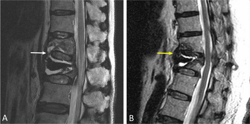
Figure 2. Measurement of the anterior body height and focal kyphotic angle. a, anterior body height of the
VCF; b, posterior vertebral height of the adjacent cranial vertebra; c, posterior vertebral height of the adjacent
caudal vertebra; p, estimate of the posterior body height of the VCF (average of b and c). The anterior body
height ratio is a/p; U, the line parallel to the upper endplate of the cranial vertebra (red line); L, the line parallel
to the lower endplate of the caudal vertebra (blue line). The focal kyphotic angle is the intersecting angle of U
and L.
under fluoroscopic guidance. After the needle was gently hammered into the anterior third of the vertebral body,
preparation for the polymethylmethacrylate (PMMA) bone cement injection was initiated. The injection tim-
ing was chosen between the liquid phase and the paste phase of the bone cement. The injection was performed
until symmetrical filling of the central and anterior parts of the vertebral body was obtained. Special attention
was given to the flow of the bone cement while cement was distributed across the anterior margin of the spinal
column.
While the cement bridging phenomenon developed, the cement was continuously and cautiously injected.
Injection typically ceased when cement reached the posterior quarter of the vertebral bodies (either the newly
fractured vertebra or previously cement-augmented vertebra), leaked into veins, or leaked laterally out of osse-
ous structures.
Postoperative care. It was suggested that all patients wear an orthosis when ambulating for 3 months.
In general, antiosteoporosis medications and calcium and vitamin D supplementation were prescribed for the
patients after surgery for osteoporotic VCFs. However, because of the potential adverse interaction with coex-
isting morbidities, antiosteoporosis medication was used on a case-by-case basis. These patients were usually
discharged on the day of surgery or the next day and followed-up at 1 and 3 months postoperatively and then
annually at outpatient clinics.
Statistical analysis. Associations between the cement bridging phenomenon and potential predictors were
analyzed by the chi-square test or Fisher’s exact test for categorical variables and Student’s t test for continuous
variables. Logistic regression analysis was used for the calculation of odds ratios and 95% confidence intervals. A
p value < 0.05 was considered significant. Statistical analysis was performed using SPSS 25.0 (IBM, Armonk, New
York). Power analysis was performed using the following parameters: test family—t tests, difference between two
independent means (two groups), two-tailed, α = 0.05. (G*Power 3.1.9.4, University of Kiel, Germany).
Results
Patient characteristics and imaging features. A total of 79 patients (94 vertebrae) sustained new-
onset adjacent VCF and then underwent further vertebroplasty. In 18 of 79 (22.8%) patients, or 20 of 94 (21.3%)
adjacent vertebrae, the cement bridging phenomenon was observed. The mean age of patients who developed
adjacent VCFs was 78.0 ± 6.3 years (range 66–95 years), and female patients made up the majority (87%). The
most commonly involved vertebral level was T12 (36.2%), followed by L1 (21.3%) and L2 (16.0%).
The demographic data and the imaging features in the bridging and nonbridging groups are listed in Table 1.
Significant differences were found with regard to three preoperative imaging features (p < 0.05), which included
severe loss (more than 50%) of the anterior vertebral body height at the new-onset adjacent vertebra on a
Scientific Reports | (2021) 11:10184 | https://doi.org/10.1038/s41598-021-89412-z 3
Vol.:(0123456789)www.nature.com/scientificreports/
Bridging group (n = 20) Nonbridging group (n = 74) OR (95% CI) p value
Age (years) 79.1 ± 7.3 77.8 ± 6.0 0.97 (0.89–1.05) 0.414
Sex (female) 20 (100%) 63 (85%) N/A 0.965
BMI 21.1 21.6 0.93 (0.77–1.13) 0.478
T-score of BMD − 3.12 − 3.02 0.91 (0.47–1.78) 0.789
Cement volume (ml) 7.3 6.2 0.83 (0.68–1.02) 0.072
Severe anterior body height loss on X-ray* 16 35 4.46 (1.36–14.61) 0.014
Fluid accumulation on MRI** 17 10 36.27 (8.97–146.58) < 0.001
Hypointense signal on MRI*** 18 27 15.67 (3.37–72.76) < 0.001
Cement volume increase**** 14 0 N/A < 0.001
Table 1. Patient characteristics and imaging features of the bridging and nonbridging groups. *Severe loss
(more than 50%) of anterior vertebral body height at the new-onset adjacent vertebra on X-ray. **Fluid
accumulations around the space anterior to the abutting vertebral bodies and the corresponding intervertebral
disc on MRI. ***Hypointense signal around the space anterior to the abutting vertebral bodies and the
corresponding intervertebral disc on MRI. ****Cement volume increase in the previously augmented vertebra.
BMI body mass index, BMD bone mineral density, OR odds ratio, CI confidence interval, N/A not applicable.
Figure 3. Preoperative MRI predictors of the cement bridging phenomenon include (A) fluid accumulation
(white arrow) and (B) hypointense signaling (yellow arrow), indicating a redundant anterior longitudinal
ligament, around the space anterior to the abutting vertebral bodies and the corresponding intervertebral disc.
lateral-view plain film (odds ratio [OR] = 4.46, p = 0.014), fluid accumulation (OR = 36.27, p < 0.001) or hypoin-
tense signaling (OR = 15.67, p < 0.001) around the space anterior to the abutting vertebral bodies and the cor-
responding intervertebral disc on MRI (Fig. 3). Furthermore, 14 vertebrae (70%) that were previously cement-
augmented vertebrae demonstrated increased cement volume due to the cement bridging phenomenon. No
statistical significance in patient age, sex ratio, BMI, T-score of BMD or injected cement volume was found
between the bridging and nonbridging groups (p > 0.05).
There was no major complication that was directly attributable to vertebral augmentation in the bridging and
nonbridging groups, such as epidural hematoma, pulmonary emboli, or cement leakage into the spinal canal.
Follow‑up and clinical correlation. A total of 18 patients (20 vertebrae) in the bridging group and 56
patients (68 vertebrae) in the nonbridging group who had followed up for at least 2 years were enrolled for fur-
ther analysis of clinical correlations.
No statistical significance in the incidence rate of adjacent fractures was found between the bridging and
nonbridging groups during 2-year follow-up. The incidence rates of adjacent fractures in the bridging group
were 15.0%, 20.0%, and 20.0% at 3 months, 1 year, and 2 years postoperatively, respectively. The incidence rates
of adjacent fractures in the nonbridging group were 14.7%, 17.7%, and 19.1% at 3 months, 1 year, and 2 years
postoperatively, respectively.
Scientific Reports | (2021) 11:10184 | https://doi.org/10.1038/s41598-021-89412-z 4
Vol:.(1234567890)www.nature.com/scientificreports/
Bridging group (n = 20) Nonbridging group (n = 68) p value
Postop 3 months 3 10 0.974
Adjacent fracture Postop 1 year 4 12 0.810
Postop 2 years 4 13 0.930
Preop 21.2∘ 21.9∘ 0.774
Postop 1 day 15.6∘ 18.9∘ 0.173
Mean focal kyphotic angle (degrees) Postop 3 months 17.1∘ 26.4∘ 0.012
Postop 1 year 18.0∘ 27.3∘ 0.017
Postop 2 years 19.9∘ 29.4∘ 0.007
Preop 0.688 0.683 0.850
Postop 1 day 0.745 0.718 0.324
Mean anterior body height ratio Postop 3 months 0.730 0.678 0.119
Postop 1 year 0.709 0.636 0.026
Postop 2 years 0.692 0.610 0.005
Table 2. Two-year follow-up of the incidence rate of adjacent fractures and radiographic parameters in the
bridging and nonbridging groups. Preop preoperative, postop postoperative.
Compared with the nonbridging group, the cement bridging group demonstrated a significantly better mean
value of the focal kyphotic angle (p < 0.05) at 3 months, 1 year, and 2 years postoperatively. The mean anterior
body height ratio of the newly fractured vertebra was also significantly better (p < 0.05) in the cement bridging
group than in the nonbridging group 1 year and 2 years postoperatively. The detailed values are listed in Table 2.
Postoperatively, patients were scheduled to return for follow-up at 1 and 3 months after the surgery and then
annually at outpatient clinics. However, some patients might visit the clinic more frequently than the scheduled
follow-up if pain sustained. The number of patients who visited the outpatient clinics as scheduled follow-up
and more frequently than the scheduled follow-up for the bridging group (n = 18) were 12 and 6; for the non-
bridging group (n = 56) were 39 and 17, respectively. No statistical significance in the frequency of analgesics
prescription in the outpatient clinics was found between the bridging and nonbridging groups during 2-year
follow-up (p = 0.812).
Power analysis showed that under the setting of α = 0.05 and current sample size, the power of our study
about the three preoperative imaging features and two postoperative radiographic parameters at 2-year follow-
up were all more than 0.9.
Discussion
The cement bridging phenomenon (CBP) is sometimes observed during the procedure of cement augmentation
but has, to the best of the authors’ knowledge, not been published before. Due to the unknown mechanism and
cause of formation, the cement bridging phenomenon usually presents unpredictably. When the phenomenon
occurred, we were unclear about the clinical manifestation of this phenomenon. To solve this question, we tried
to describe this phenomenon clearly, investigate the predictors of occurrence, and correlate the results with
clinical application.
The severe loss (more than 50%) in the anterior vertebral body height at the new-onset adjacent vertebra
on plain film, fluid accumulation and hypointense signaling around the space anterior to the abutting vertebral
bodies and the corresponding intervertebral disc on MRI were the three predictors of the CBP identified in this
study. These three characteristics were presumed to be related to structural disruption anterior to the vertebral
bodies and the corresponding intervertebral disc, indicating disruption of the anterior cortex and detachment
of the anterior longitudinal ligament. The severe loss of anterior vertebral body height at the new-onset adjacent
vertebra and hypointense signaling changes around the space anterior to the abutting vertebral bodies and the
corresponding intervertebral disc on MRI suggest redundancy of the anterior longitudinal ligament. Therefore,
an anterior intervertebral space existed, which may be related to the phenomenon of bone cement flowing from
this space. Fluid accumulation around the space anterior to the abutting vertebral bodies and corresponding
intervertebral disc results in the possibility of bone cement flowing from this space as well.
The cement bridging phenomenon is essentially an event of cement leakage because cement leakage has been
defined as the presence of any extravertebral cement. Yeom et al.13 proposed three patterns of cement leakage: (1)
leakage via the basivertebral vein (B-type), (2) leakage via the segmental vein (S-type), and (3) leakage through a
cortical defect (C-type). The cement bridging phenomenon develops leakage through a cortical defect, belong-
ing to the C-type. Nieuwenhuijse et al.5 reported that the incidence of cortical leakage increased with advancing
severity grade, which is consistent with the results of our research, that is, the severe loss of anterior vertebral
body height was highly associated with the CBP.
Some studies14–17 have shown that cement leakage into a disk is associated with adjacent VCFs. This made us
wonder whether the CBP would also have a similar effect. However, there was no statistical significance in the
incidence rates of secondary adjacent fracture between the bridging and nonbridging groups during the 2-year
follow-up in our study. In fact, this issue is still inconclusive because several s tudies4,18,19 have demonstrated
leakage unrelated to new fractures either at adjacent or non-adjacent levels. A meta-analysis conducted by Yi
Zhan et al.20 mentioned that cement extravasation can also lead to serious complications including neurologic
Scientific Reports | (2021) 11:10184 | https://doi.org/10.1038/s41598-021-89412-z 5
Vol.:(0123456789)www.nature.com/scientificreports/
Figure 4. (A) A 95-year-old woman sustained a new-onset adjacent L1 compression fracture 3 weeks after L2
vertebroplasty, (B) the cement bridging phenomenon was observed during L1 vertebroplasty. Increased cement
volume of the previously cement-augmented L2 vertebrae was noted compared with the preoperative image. (C)
Two years postoperatively, the focal kyphotic angle and bony alignment were maintained.
deficit and fetal sequalae such as pulmonary embolism, but it is generally of no clinical symptoms. Hassan
et al.21 reported that there was no significantly difference in patient satisfaction between those who had cement
extravasation and those who did not, in both groups.
In spite of no major complication with regarding to the cement bridging phenomenon in our study, there
was one case who developed lateral cement extravasation into paraspinal soft tissue during cement bridging
phenomenon. This patient felt temporary paraspinal burning sensation during the cement curing process, and
this symptom subsided completely after the procedure. In our opinion, leakage of bone cement to the lateral
side is usually far more prone to produce symptoms than leakage to the front area. Faded color of the injected
cement during the injection in the lateral view was usually a sign of lateral extravasation.
Fourteen vertebrae (70%) that were previously cement-augmented demonstrated increased cement volume
in the bridging group, which means that injected cement filled the newly fractured vertebra and the previously
cement-augmented vertebra simultaneously via the cement bridging phenomenon. This finding was presumed
to be related to the concurrent refracture of the previously cement-augmented vertebra and represented the real
existence of the cement bridging phenomenon. Repeated percutaneous vertebroplasty for refracture of cemented
vertebrae is a feasible t reatment22, but an alternative method is to fill these two neighboring vertebrae through
one portal via the cement bridging phenomenon. To achieve this condition, we recommended to insert needles
in these two neighboring vertebrae concurrently before cement injection. Then, we suggested to inject cement
through the needle in the newly fractured vertebra first. Once CBP and cement filling in these refractured
vertebrae developed, cement can be continuously injected with caution. If satisfactory filling pattern in the
refractured vertebra was already achieved by this way, no further injection through the needle in the refractured
vertebra was needed.
The cement bridging phenomenon may offer anterior support of the vertebral column. Both the mean value of
the focal kyphotic angle and mean anterior body height ratio were significantly better (p < 0.05) postoperatively
in the cement bridging group than in the nonbridging group during the 2-year follow-up (Fig. 4). We think that
it is difficult to promote and is unnecessary to pursue the cement bridging phenomenon. However, we should
not worry this phenomenon when it occurs.
There are several limitations in this retrospective study, including the relatively small number of new-onset
VCFs in the bridging group and the different end points of the operator during cement injection, which may
result in different cement distribution patterns by different operators even under the same conditions. We did
not analyze the influence on cement bridging between vertebroplasty and kyphoplasty, because most surgeons
performed vertebroplasty in our institute. In addition, we did not objectively measure the bone cement viscosity,
although the injection timing was consistently chosen between the liquid phase and paste phase. We think this
method can minimize the possible impact of this factor.
Conclusion
The cement bridging phenomenon (CBP), which has never been reported in the literature, is not rare in clinical
practice. The severe loss of anterior vertebral body height at the new-onset adjacent vertebra, fluid accumulation
and hypointense signaling around the space anterior to the abutting vertebral bodies and the corresponding
intervertebral disc on MRI were identified as predictors of the CBP. The cement extravasation could lead to
complications, but may offer positive effect with occurrence of CBP through cautious control of cement injec-
tion. Cement bridging phenomenon was associated with better maintenance of focal kyphotic angle and anterior
body height ratio during the 2-year follow-up.
Scientific Reports | (2021) 11:10184 | https://doi.org/10.1038/s41598-021-89412-z 6
Vol:.(1234567890)www.nature.com/scientificreports/
Received: 12 December 2020; Accepted: 23 April 2021
References
1. Galibert, P., Deramond, H., Rosat, P. & Le Gars, D. Preliminary note on the treatment of vertebral angioma by percutaneous acrylic
vertebroplasty. Neurochirurgie 33, 166–168 (1987).
2. Yang, E. Z. et al. Percutaneous vertebroplasty versus conservative treatment in aged patients with acute osteoporotic vertebral
compression fractures: a prospective randomized controlled clinical study. Spine (Phila Pa 1976) 41, 653–660 (2016).
3. Zhang, H., Xu, C., Zhang, T., Gao, Z. & Zhang, T. Does percutaneous vertebroplasty or balloon kyphoplasty for osteoporotic
vertebral compression fractures increase the incidence of new vertebral fractures? A meta-analysis. Pain Physician 20, E13–E28
(2017).
4. Klazen, C. A. et al. Vertebroplasty versus conservative treatment in acute osteoporotic vertebral compression fractures (Vertos II):
an open-label randomised trial. Lancet 376, 1085–1092 (2010).
5. Nieuwenhuijse, M. J., Van Erkel, A. R. & Dijkstra, P. D. Cement leakage in percutaneous vertebroplasty for osteoporotic vertebral
compression fractures: identification of risk factors. Spine J. 11, 839–848 (2011).
6. Shridhar, P. et al. A review of PMMA bone cement and intra-cardiac embolism. Materials (Basel) 9, 821 (2016).
7. Ren, H. L., Jiang, J. M., Chen, J. T. & Wang, J. X. Risk factors of new symptomatic vertebral compression fractures in osteoporotic
patients undergone percutaneous vertebroplasty. Eur. Spine J. 24, 750–758 (2015).
8. Seo, D. H. et al. Risk factors of new adjacent compression fracture after percutaneous vertebroplasty: effectiveness of bisphosphonate
in osteoporotic or osteopenic elderly patients. Korean J. Neurotrauma. 10, 86–91 (2014).
9. Lee, D. G., Park, C. K., Park, C. J., Lee, D. C. & Hwang, J. H. Analysis of risk factors causing new symptomatic vertebral compres-
sion fractures after percutaneous vertebroplasty for painful osteoporotic vertebral compression fractures: a 4-year follow-up. J.
Spinal Disord. Tech. 28, E578-583 (2015).
10. Yang, S., Liu, Y., Yang, H. & Zou, J. Risk factors and correlation of secondary adjacent vertebral compression fracture in percutane-
ous kyphoplasty. Int. J. Surg. 36, 138–142 (2016).
11. Ko, B. S., Cho, K. J. & Park, J. W. Early adjacent vertebral fractures after balloon kyphoplasty for osteoporotic vertebral compression
fractures. Asian Spine J. 13, 210–215 (2019).
12. Song, D. et al. Secondary balloon kyphoplasty for new vertebral compression fracture after initial single-level balloon kyphoplasty
for osteoporotic vertebral compression fracture. Eur. Spine J. 26, 1842–1851 (2017).
13. Yeom, J. S. et al. Leakage of cement in percutaneous transpedicular vertebroplasty for painful osteoporotic compression fractures.
J. Bone Joint Surg. Br. 85, 83–89 (2003).
14. Komemushi, A. et al. Percutaneous vertebroplasty for osteoporotic compression fracture: multivariate study of predictors of new
vertebral body fracture. Cardiovasc. Intervent Radiol. 29, 580–585 (2006).
15. Lin, E. P., Ekholm, S., Hiwatashi, A. & Westesson, P. L. Vertebroplasty: cement leakage into the disc increases the risk of new
fracture of adjacent vertebral body. AJNR Am. J. Neuroradiol. 25, 175–180 (2004).
16. Chen, W. J. et al. Impact of cement leakage into disks on the development of adjacent vertebral compression fractures. J. Spinal
Disord. Tech. 23, 35–39 (2010).
17. Rho, Y. J., Choe, W. J. & Chun, Y. I. Risk factors predicting the new symptomatic vertebral compression fractures after percutaneous
vertebroplasty or kyphoplasty. Eur. Spine J. 21, 905–911 (2012).
18. Lin, W. C. et al. Refractures in cemented vertebrae after percutaneous vertebroplasty: a retrospective analysis. Eur. Spine J. 17,
592–599 (2008).
19. Lee, K. A. et al. Analysis of adjacent fracture after percutaneous vertebroplasty: does intradiscal cement leakage really increase the
risk of adjacent vertebral fracture?. Skeletal Radiol. 40, 1537–1542 (2011).
20. Zhan, U., Jiang, J. Z., Liao, H. F., Tan, H. T. & Yang, K. Q. Risk factors for cement leakage after vertebroplasty or kyphoplasty: a
meta-analysis of published evidence. World Neurosurg. 101, 633–642 (2017).
21. Hassan, S. et al. Clinical outcome and subsequent sequelae of cement extravasation after percutaneous kyphoplasty and vertebro-
plasty: a comparative review. Acta Radiol. 59, 861–868 (2018).
22. Chen, L. H. et al. Repeated percutaneous vertebroplasty for refracture of cemented vertebrae. Arch. Orthop. Trauma. Surg. 131,
927–933 (2011).
Acknowledgements
The authors thank the Center for Big Data Analytics and Statistics, Chang Gung Memorial Hospital at Linkou,
for statistical consultation.
Author contributions
Y.D.L.: data acquisition, statistical evaluation, figure and table preparation, manuscript writing. T.T.T.: data evalu-
ation, statistical evaluation. C.C.N.: supervision, study design. P.L.L.: supervision, study design, data interpreta-
tion, manuscript editing. All authors reviewed and approved the final manuscript.
Funding
This study was supported by the grants (CORPG3J0381, CMRPG3J1431, and CMRPG3J1432) from the Linkou
Chang Gung Memorial Hospital. The funding body did not have any role in the design of the study or in the
data analysis.
Competing interests
The authors declare no competing interests.
Additional information
Correspondence and requests for materials should be addressed to P.-L.L.
Reprints and permissions information is available at www.nature.com/reprints.
Publisher’s note Springer Nature remains neutral with regard to jurisdictional claims in published maps and
institutional affiliations.
Scientific Reports | (2021) 11:10184 | https://doi.org/10.1038/s41598-021-89412-z 7
Vol.:(0123456789)www.nature.com/scientificreports/
Open Access This article is licensed under a Creative Commons Attribution 4.0 International
License, which permits use, sharing, adaptation, distribution and reproduction in any medium or
format, as long as you give appropriate credit to the original author(s) and the source, provide a link to the
Creative Commons licence, and indicate if changes were made. The images or other third party material in this
article are included in the article’s Creative Commons licence, unless indicated otherwise in a credit line to the
material. If material is not included in the article’s Creative Commons licence and your intended use is not
permitted by statutory regulation or exceeds the permitted use, you will need to obtain permission directly from
the copyright holder. To view a copy of this licence, visit http://creativecommons.org/licenses/by/4.0/.
© The Author(s) 2021
Scientific Reports | (2021) 11:10184 | https://doi.org/10.1038/s41598-021-89412-z 8
Vol:.(1234567890)You can also read