CE Credit - Case Study Animal Eponyms in Eye Care - CRO Journal
←
→
Page content transcription
If your browser does not render page correctly, please read the page content below

CE Credit - Case Study

Animal Eponyms in Eye Care

Alely Hostia, OD; Jeffrey Kyle Wells, OD; Alexandra Vlad, OD; Ruth Hyatt, OD; Darcy L. Eberle, OD; Son Thai Ho, MD

Synopsis                                                                          Introduction
There are many eponyms derived from the animal world that                         The characterization of ophthalmological conditions through
help describe what is observed during an ophthalmological                         pertinent findings and descriptions is continually expanding.
examination. These zoologically-based eponyms tell a                              Mnemonics, triads, acronyms, acrostics, chunking, and
story between the animal kingdom and the human eye.                               eponyms are commonly utilized in medicine to enhance the
Medical nomenclature could be described as suffering from                         understanding of a plethora of medical conditions. Eponyms can
eponymophilia. Eponymous descriptions are ubiquitous in the                       aid in identifying and understanding ophthalmological findings.
field of medicine as seen in text in these fields: dermatology,1                  Unfortunately, although some zoological eponyms may have
neurology,2 and trichology.3 Eponyms, however practical,                          a derogative connotation, causing some authors to advocate
are profoundly affected by local geography and culture,4                          their omission,3 the eponyms reviewed here do not fall under
which can contribute to confusion and misunderstanding,                           that subgroup. Instead, they allow conditions to become more
particularly amongst practitioners from different cultures and                    memorable, and in many cases, help newer clinicians make the
locations. Deriving meaning from eponyms often necessitates                       connection between certain diseases and their manifestations.
ethnocentric or linguistically exclusive knowledge.5 While the                    Some eponyms (e.g. cat scratch disease) may nudge novel
debate surrounding whether to continue the use of eponyms                         clinicians down the correct systemic work-up path.
in medicine roars on,4 to the authors’ knowledge, a review of
animal eponyms used in ophthalmology has been limited, as                         Animal Eponyms
such, this manuscript will review animal eponyms encountered
in ophthalmology and optometry with the aim to clarify meaning
and provide a useful reference for practicing and trainee eye
                                                                                  1  Bag of worms: the appearance of numerous arteriovenous
                                                                                     anastomoses seen as enlarged, tortuous blood vessels, with
                                                                                  the artery and vein appearing similar. Treatment is generally not
physicians. As this work was undertaken to review commonly                        required; however ipsilateral lesions involving the brain can be
encountered conditions, no detailed pathophysiology will be                       found in Wyburn-Mason syndrome.6
described as it is beyond the scope of this article. Of note, not
all eponyms reviewed are based on physical appearances (e.g.
chicken eyes).

Keywords: animal eponyms, disease, eponyms, eye,
ophthalmology, optometry, sign, syndrome

Alely Hostia, OD - Daytona Beach VA Multispecialty Clinic, FL                     Figure 1. Retinal racemose angioma in Wyburn Mason syndrome. This
Jeffrey Kyle Wells, OD - Daytona Beach VA Multispecialty Clinic, FL               image was originally published in the Retina Image Bank® website. Henry
Alexandra Vlad, OD - Daytona Beach VA Multispecialty Clinic, FL                   Kaplan. Wyburn Mason Syndrome. Retina Image Bank. 2013; 6211.
Ruth Hyatt, OD - Daytona Beach VA Multispecialty Clinic, FL                       ©the American Society of Retina Specialists
Darcy L. Eberle, OD - Daytona Beach VA Multispecialty Clinic, FL
Son Thai Ho, MD - Orlando VA Medical Center, Department of Ophthalmology
Correspondence to: Ruth Hyatt, OD
Daytona Beach VA Multispecialty Clinic
551 National Health Care Drive, Daytona Beach, FL 32114
E-mail: Ruth.Hyatt@va.gov
The authors have no financial or proprietary interest in any material or method
mentioned in this article. This article has been peer reviewed.

428              Clinical & Refractive Optometry 32.4, 2021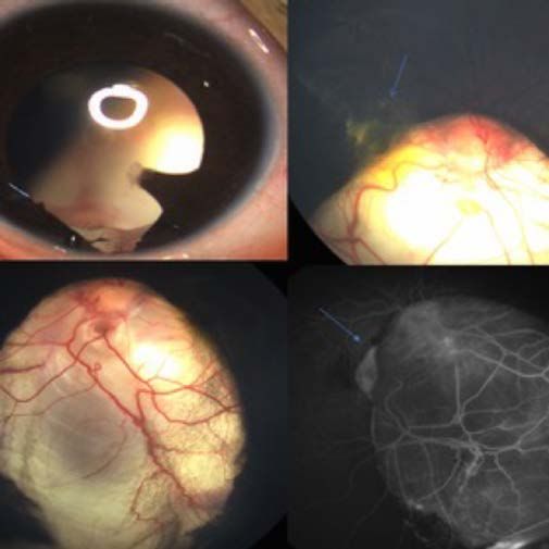
2     Bear tracks: also known as grouped pigmentation of the
      retina, bear tracks are flat, well-demarcated, hyperpigmented
lesions in the retinal pigment epithelium (RPE) and are a
subgroup of congenital hypertrophy of the RPE (CHRPE).7
CHRPE can be associated with familial adenomatous polyposis
if bilateral, occurs in multiple quadrants, has a pisciform shape,
and irregular borders.7

                                                                                                               Figure 7. Target.

                                                                          Figure 6. Bull’s eye maculopathy.
                                                                          ©EyeRounds.org

                                                                          b. Bull’s eye rash: also known as erythema migrans, a gradually
                                                                          expanding circular skin lesion that develops at the site of a
                                                                          tick bite containing Borrelia burgdorferi and signifies localized
Figure 2. Bear tracks.              Figure 3. Bear tracks.                infection.13-15
©EyeRounds.org

3    Birdshot chorioretinopathy: a rare autoimmune posterior
     uveitis with poorly understood pathogenesis8 characterized by
multiple lightly colored oval choroid lesions (the long axis is radial
to the optic disc) with a juxtopapillary predilection.9 Presentation is
most commonly bilateral and symmetric.8 Although uncommon,
a mild anterior uveitis can be present.8 Cystoid macular edema
is common and is the predominant cause of vision loss.8 The
name was coined because the pattern of these lesions mimics
the pattern of birdshot from shotgun scatter.10

                                                                          Figure 8. Erythema migrans in Lyme disease. This image was originally
                                                                          published in the Retina Image Bank® website. Henry Kaplan. Uveitis.
                                                                          Retina Image Bank. 2013; 4904.
                                                                          ©the American Society of Retina Specialists

Figure 4. Birdshot chorioretinopathy.
                                                                          5   Butterfly rash: a raised or flat malar rash that is non-
                                                                              pruritic and occurs over the cheeks and bridge of the nose,
                                                                          with nasolabial folds spared.16,17 A feature of systemic lupus
©2020 American Academy of Ophthalmology 		                                erythematosus,16,17 an autoimmune disease of unknown etiology
                                                                          that affects multiple organ systems.17 Lupus is Latin for wolf.18
                                                                          Translated19 as “a systemic disease in which a wolf turns red”. It
                                                                          may also be associated with hydroxychloroquine use.20
                              Figure 5. Paper shooting target depicting
                              birdshot from shotgun scatter.

4    Bull’s eye:
     a. Bull’s eye maculopathy: the distinct pattern of retinopathy
in patients with chloroquine11 or hydroxycholoroquine12 toxicity          Figure 9. Butterfly rash. DermNet    Figure 10. Butterfly
                                                                          (https://creative commons.
that may appear funduscopically as macular granularity, thinning          org/licenses/by-nc-nd/3.0/nz/
of parafoveal retinal pigmented epithelium and photoreceptors             legalcode)
on optical coherence tomography, or a ring defect on visual
field testing.12 The mechanism of toxicity is not understood.12
Retinopathy in patients of Asian ancestry may be more peripheral
than that of Caucasians.12

                                                                                                      Animal Eponyms in Eye Care            429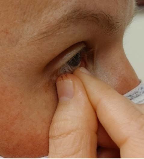
6    Butterfly-shaped pattern dystrophy: a bilateral, autosomal
     dominant condition21 in which pigmented lipofuscin22
accumulates in the macular retinal pigment epithelium in a
                                                                              9   Cat scratch disease: a systemic illness contracted through
                                                                                  a scratch or bite that transmits Bartonella henselae; the
                                                                              classic ocular manifestation is unilateral optic disc edema
pattern resembling the wings of a butterfly.23 The condition                  and exudates that form the pattern of a star at the macula
is due to genetic mutations at the same locus as mutations                    (termed neuroretinitis), and visual acuity can recover after
found in pattern dystrophies, retinitis pigmentosa, and fundus                several weeks.28 Mild inflammation of the anterior chamber
flavimaculatus, resulting in photoreceptor membrane integrity.22              and vitreous is common, as is retinal thickening and exudative
                                                                              retinal detachment.28

                                                                                                                                 Figure 15. Starburst.

                                       Figure 12. Butterfly
                                                                              Figure 14. Neuroretinitis with macular star.
Figure 11. Butterfly-shaped pattern                                           ©2020 American Academy of Ophthalmology
dystrophy.

                                                                              10
©Online Journal of Ophthalmology
                                                                                      Cat’s paw retractor: an instrument used in ophthalmic
                                                                                      procedures that involve retracting the skin, subcutaneous

7    Camel’s sign: also known as camel’s second hump sign,24                  tissues, and ligaments.29
     the appearance of a second peak on the densitometry graph
of a Scheimpflug image25 due to higher reflection of Descemet’s
membrane indicative of corneal guttata.26

8    Cat eye syndrome: coloboma and anal atresia resulting
     from an extra acrocentric chromosome, whose expression
and transmission varies widely.27 Ocular features of cat eye
                                                                              Figure 16. Cat’s paw retractor.

syndrome have sizeable variation- and coloboma may be found
in the iris or choroid, and may be bilateral or unilateral.27
                                                                                                                      Figure 17. Cat’s paw.

                                                                              11      Cattle track sign: also known as cattle trucking or box-
                                                                                      carring, a beaded appearance of retinal vessels resulting
                                                                              from interrupted blood flow resulting in “sludge phenomenon”,
                                                                              whereby there is a “stagnation of the arterial blood stream
                                                                              without diminution in the size of the arterial column”.30,31 Of
                                                                              note, in one large retrospective study, nearly all patients with
                                                                              total central retinal artery occlusion (CRAO) had a cattle track
                                                                              sign, which may be an important distinction as patients having
                                                                              incomplete CRAO showed a better prognosis.30

                                                                                                                      Figure 19. Cattle car.
Figure 13. Iris and optic nerve coloboma in cat eye syndrome. Blue arrows
depict choroidal neovascularization. This image was originally published in   Figure 18. Cattle track sign in central retinal artery occlusion. This image
the Retina Image Bank® website. Sophia El Hamichi. Giselle De Oliveira. Cat   was originally published in the Retina Image Bank® website. Hyung-Woo
Eye Syndrome. Retina Image Bank. 2020; 49706. ©the American Society of        Kwak. Central Retinal Artery Occlusion. Retina Image Bank. 2012; 1683.
Retina Specialists                                                            ©the American Societyof Retina Specialists

430             Clinical & Refractive Optometry 32.4, 2021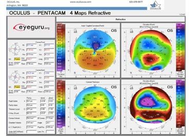
12    Chicken eyes: Vitamin A deficiency impedes rhodopsin
      production, resulting in poor rod function which
manifests as nyctalopia. Chickens lack rod photoreceptors,
                                                                          17      Crow’s feet: also known as periorbital rhytids, wrinkles
                                                                                  that form at the lateral canthus due to hyperkinesia of
                                                                          the orbicularis oculi,39 appearing similar to the talons of a crow.
making them night blind.32

13     Cotton wool spots: fluffy, white lesions caused by
       accumulated axoplasmic debris within the ganglion cell
axon bundle due to arteriolar occlusion and ischemia.33

                                                                          Figure 24. Crow’s feet.              Figure 25. Crow’s feet.

Figure 20. Cotton wool spots.       Figure 21. Cotton wool ball.
©2020 American Academy of
Ophthalmology                                                             18      Cutaneous horns of the eyelid: a keratinous mass
                                                                                  protruding from the skin, resembling animal horns
                                                                          (unlike animal horns, there is no bony core).40 Most often occur
                                                                          on sun-exposed skin of elderly men, and lesions are usually

14      Crab claws: also known as kissing birds or butterfly
        pattern,34 the appearance of the corneal topographical
pattern in which there is steepening of the inferior peripheral
                                                                          asymptomatic.40

cornea with flattening along the vertical meridian,35 suggestive of
pellucid marginal degeneration (PMD) or keratoconus.36 In PMD,
peripheral corneal thinning in the 4:00 to 8:00 position occurs
bilaterally, with epithelium remaining intact.34

                                                                          Figure 26. Cutaneous horn.
                                                                          ©EyeRounds.org, The University of Iowa

                                                                          19      Elschnig’s pearls: the accumulation of equatorial
                                                                                  epithelial cells onto the posterior capsule after cataract
                                                                          surgery are clinically visible as “pearls” in posterior capsular
                                                                          opacification.41
Figure 22. Crab claws or kissing dove pattern     Figure 23. Crab claw.
on axial curve.

15      Crocodile shagreen: symmetrical, gray-white polygonal
        opacities with indistinct edges and clear zones in
between (resembling the skin of a crocodile) in the central
posterior corneal stroma.37 Electron microscopy shows vacuoles
throughout the corneal stroma.37

16      Crocodile tear syndrome: also known as gusto-
        lacrimation, following traumatic facial paralysis or Bell’s
palsy, aberrant gustatory nerve fiber regeneration to the lacrimal
gland (rather than the salivary gland) causes non-emotional
tearing whilst eating.38                                                  Figure 27. Elschnig’s pearls. This image was originally published in the
                                                                          Retina Image Bank® website. Rene Parada. Elschnig’s Pearls. Retina Image
                                                                          Bank. 2015; 25591. ©the American Society of Retina Specialists

                                                                                                     Animal Eponyms in Eye Care            431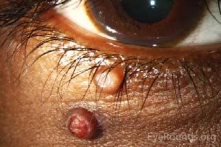
20       Fish eye disease: severe corneal opacification resulting
         from low high density lipoproteins cholesterol.42                  23    Lobster claw intraocular lens: also known as iris-claw
                                                                                  lens46, an intraocular lens used when there is not
                                                                            adequate capsular support47 for a traditional intraocular lens.

                                                                            Figure 33. Claw intraocular lens.   Figure 34. Lobster claw.
Figure 28. Corneal clouding.         Figure 29. Cloudy fish eye.
                                                                            ©EyeRounds.org, The University
©EyeRounds.org, The University
                                                                            of Iowa
of Iowa

21    Halsted mosquito forceps and Hartman mosquito
      forceps: hemostats to clamp tissues or vessels43 during
                                                                            24     Leopard spots: also known as giraffe spots, a
                                                                                   chorioretinopathy of characteristic hypo- and
                                                                            hyperpigmented spots.48,49
ophthalmic surgeries.44

                                                                            Figure 35. Leopard spots. This image was originally published in the
                                                                            Retina Image Bank® website. Mohammad Hossein Jabbarpoor Bonyadi.
                                                                            Miscellaneous. Retina Image Bank. 2019; 27167.
Figure 30. Mosquito forceps                                                 ©the American Society of Retina Specialists

22        Horseshoe retinal tear: a U-shaped flap tear which
          occurs most frequently in the superior temporal
peripheral quadrant of the retina, probably due to a combination
                                                                                                                   Figure 36. Spotted pattern of a
                                                                                                                   leopard’s coat.
of gravity acting on the vitreous as well as thinner peripheral
retinal tissue.45

                                                                            25     Maddox wing: a dissociated test to control
                                                                                   accommodation in pre-presbyopic patients that utilizes
                                                                            a septum to measure heterophoria at near.50

                                                                                                                   Figure 37. Maddox wing. Image
Figure 31. Horseshoe retinal tear.   Figure 32. The heel of a horseshoe
                                                                                                                   courtesy of Neil Handley. The
©Online Journal of Ophthalmology     oriented with the base of the
                                                                                                                   College of Optometrists, London
                                     horseshoe retinal tear; the toe of a
                                                                                                                   UK. 2021.
                                     horseshoe oriented with the apex
                                     of the horseshoe retinal tear.

432            Clinical & Refractive Optometry 32.4, 2021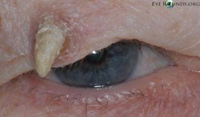
26       Molluscum contagiosum: round, centrally umbilicated
         waxy popular lesions usually occurring in clusters found
on the skin and mucous membranes caused by a poxvirus.51
                                                                      29     Muscae volitantes: collagen fibril accumulation results
                                                                             in light scattering and perceived vitreous floaters which
                                                                      move when the eyes or head is moved,55 derived from the Latin
Commonly seen in children, and suspicion of immunodeficiency          phrase meaning “flying flies”.56
is raised if found in adults.51

                                                                      Figure 42. Vitreous floater. This image was originally published in the
Figure 38. Molluscum contagiosum. ©EyeRounds.org                      Retina Image Bank® website. Gary R. Cook. Pre-Macular Floater. Retina
                                                                      Image Bank. 2019; 29867. ©the American Society of Retina Specialists.

27       Moth-eaten iris: atrophy of the iris stroma and/or pigment
         epithelium, most notably in Fuch’s heterochromic
uveitis,52 appearing as if eaten by the larvae of moths.53
                                                                      30       Mutton-fat keratic precipitates: leukocytes that appear
                                                                               on the corneal endothelium as ‘greasy’ fat globules,57,58
                                                                      similar to the fat of sheep. This can be an indicator of systemic
                                                                      disease such as tuberculosis, syphilis, or sarcoidosis.57

                                   Figure 40. Moth-eaten garments.

Figure 39. Fuchs’ heterochromic
cyclitis.©EyeRounds.org                                               Figure 43. Mutton-fat keratic precipitates. ©2020 American Academy of
                                                                      Ophthalmology.

28       Mucus fishing syndrome: chronic cycles initiated by
         ocular irritation leads to conjunctival mucus production
which prompts the patient to mechanically remove (“fish”)             31     Octopus perimeter: a perimeter designed to be “as fast
                                                                             as an eight-armed technician”, which was the original
mucus, which in turn causes more irritation and a continuous          automated perimeter, marking the first time a device of its
cycle.54                                                              kind could store results and offer comparison to a normative
                                                                      database.59

                                                                      32     Owl-eye facial rash: also known as “raccoon-
                                                                             like” appearance,60 a periorbital cutaneous rash
                                                                      characteristically observed in neonatal lupus erythematosus.61

                                                                      Figure 44. Owl-eye facial           Figure 45. Owl.
                                                                      rash in neonatal lupus
Figure 41. “Fishing” for mucous.                                      erythematosus. DermNet (https://
                                                                      creativecommons. org/licenses/by-
                                                                      nc-nd/3.0/nz/legalcode)

                                                                                                Animal Eponyms in Eye Care            433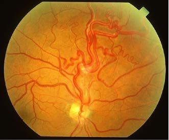
33      Panda sign in nevus of Ota: bluish hyperpigmentation
        that persists after treating periorbital nevus of Ota lesion
with laser surgery.62 The exact mechanism is unknown but could
                                                                          36      Polar bear tracks: also known as congenital grouped
                                                                                  albinotic spots of the retinal pigment epithelium, sharply
                                                                          demarcated chalky lesions occurring in patterns similar to
be due to operator over-caution using lower laser intensity or            footprints.66
avoidance of areas in close proximity to the eye; or perhaps
macrophage clearing of laser irradiated periorbital tissue is less
effective than other locations.62

                                                                                                               Figure 52. Polar bear tracks.

                                                                          Figure 51. Polar bear tracks. ©EyeRounds.org, The University of Iowa

Figure 46. Naevus of Ota. DermNet   Figure 47. Panda bear.
(https://creativecommons.
org/licenses/by-nc-nd/3.0/nz/
legalcode)                                                                37      Pterygium: a triangular-shaped corneal over-growth
                                                                                  originating from the interpalpebral bulbar conjunctiva,
                                                                          often with epithelial secretory goblet cells not typically found in
                                                                          corneal epithelial tissue.67 The term originates from the Greek

34      Periorbital spider angioma: also known as spider nevus
        or spider telangiectasia, a vascular lesion consisting of
a central arteriole body, radiating capillaries like a spider’s legs,
                                                                          word pterygion, which is in turn derived from the Greek roots
                                                                          pterón (feather, wing) and ptéryx, ptérigos (wing, fin, bird).68

and surrounding erythema resembling a spider web.63

                                                                          Figure 53. Pterygium.                Figure 54. Extended wing of a bird.
                                                                          ©2020 American Academy of
                                                                          Ophthalmology
Figure 48. Periorbital spider       Figure 49: Spider web.
angioma. DermNet (https://
creativecommons.org/licenses/by-
nc-nd/3.0/nz/legalcode)                                                   38        Racoon eyes: also known as raccoon sign or panda
                                                                                    sign,69 a periorbital hematoma that can be caused by
                                                                          basal skull fracture,70 particularly if hemorrhaging is absent
                                                                          beyond the tarsal plate.69,71 In the absence of trauma, it may be

35     Pisciform flecks: yellow fish-shaped64 lesions which
       over time atrophy and leave barely perceptible
hyperpigmentation in the retinal pigment epithelium
                                                                          associated with neuroblastoma metastasis in and around the
                                                                          orbit,72,73 or primary amyloidosis from amyloid fibril infiltration
                                                                          into periorbital blood vessels.74 Anecdotal case reports also
characteristic of fundus flavimaculatus.65                                attribute etiology to Kaposi’s sarcoma,75 multiple myeloma,76
                                                                          and other vascular, infectious, immune-mediated, metabolic,
                                                                          genetic, and malignant etiologies.69

                                                                          Figure 55. Periorbital hematoma      Figure 56. Characteristic “black
Figure 50. Pisciform flecks in Stargardt’s disease. This image was        in a 2-year-old with bilateral       mask” of a raccoon.
originally published in the Retina Image Bank® website. David Callanan.   metastasis of neuroblastoma.
Fundus Flavimaculata/Stargardt’s. Retina Image Bank. 2013; 12774.         ©2020 American Academy of
©the American Society of Retina Specialists                               Ophthalmology

434            Clinical & Refractive Optometry 32.4, 2021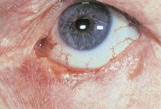
39     Retinal operculum: a free-floating flap of detached retinal
       tissue within the vitreous body.77 “Operculum” is derived
from the Latin term operire, meaning “to cover”,78 and is also the
                                                                          42      Salmon patch:
                                                                                  a. of Hutchinson:82 stromal vascularization and
                                                                          inflammation that appears in syphilitic interstitial keratitis.83 Most
technical term for the gill coverings of many bony fish.79                often congenital, although may occur with acquired infection.83

Figure 57. Optical coherence tomography of full thickness macular hole    Figure 60. Salmon patch. ©EyeRounds.org, The University of Iowa
with operculum. This image was originally published in the Retina Image
Bank® website. Samarth Mishra. Aditya Birla. Stage 3 Macular Hole With
Operculum. Retina Image Bank. 2018; 28613.                                b. a painless pink-colored mass on the bulbar conjunctiva
©the American Society of Retina Specialists                               or adnexa, it can be associated with lymphoma.84 Feeder
                                                                          vessels, rapid invasive growth, and ulceration raise suspicion
                                                                          of lymphoma.84

40       Rhinophyma: Greek for “nose growth”; sebaceous gland
         hyperplasia in stage IV rosacea.80

                                                                          Figure 61. This image was             Figure 62. Salmon filet.
                                                                          originally published in the Retina
                                                                          Image Bank® website. Nichole
                                                                          Lewis. Salmon Patch Lymphoma.
                                                                          Retina Image Bank. 2017; 27223.
Figure 58. Rhinophyma. DermNet
                                                                          ©the American Society of Retina
(https://creativecommons.org/licenses/by-nc-nd/3.0/nz/legalcode)
                                                                          Specialists

41     Rodent ulcer: refers to the excavated appearance with
       elevated pearly margins81 characteristic of basal cell
carcinoma; in Latin, rodere means “to gnaw”.18
                                                                          43     Sea fan neovascularization: similar in appearance to
                                                                                 the aquatic invertebrate Gorgonia flabellum,85 retinal
                                                                          neovascularization that proliferates anteriorly from perfused to
                                                                          non-perfused tissue, associated with sickle cell disease.

                                                                          Figure 63. Sea fan                    Figure 64. Sea fan coral.
Figure 59. Basal cell carcinoma of the lower eyelid.                      neovascularization of the retina.
©2020 American Academy of Ophthalmology                                   This image was originally published
                                                                          in the Retina Image Bank®
                                                                          website. Maurice Rabb. Macro And
                                                                          Microvascular Sea Fan Changes.
                                                                          Retina Image Bank. 2013; 12289.
                                                                          ©the American Society of Retina
                                                                          Specialists

                                                                                                      Animal Eponyms in Eye Care            435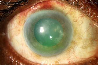
44       Serpiginous choroiditis: retinal pigment epithelium
         atrophy relapses and remits leaving behind subretinal
areas of swelling that spread equatorially in a creeping, snake
                                                                              46      Soft shell technique: a cataract surgery technique using
                                                                                      two ophthalmic viscosurgical devices (OVD) within the
                                                                              anterior chamber to create different physical environments
like fashion.86 The etymology of the word serpiginous is derived              improving the success of cataract surgery particularly in difficult
from the Latin verb sepere meaning to creep or creeping in a                  cases.91
similar fashion to a ringworm or snake, hence the root is used
in the 13th century English word ‘serpent’.87,88
                                                                              47      Steinert double-ended claw chopper: a surgical
                                                                                      instrument whose curvature allows easier engagement
                                                                              with the cataract’s nucleus.92

                                                                              48      Tigroid fundus: a striped appearance93 seen when
                                                                                      prominent choroidal pigmentation is interrupted by
                                                                              choroidal vessels, which appear lighter in comparison.94 Also
                                                                              referred to as tessellated fundus, this physiologic variant is a
                                                                              protective factor in diabetic retinopathy.94
Figure 65. Serpiginous choroiditis.
©2020 American Academy of Ophthalmology

                                  Figure 66. Lateral undulation of a snake.

                                                                                                                   Figure 72. Tiger stripes.

45     Snail tracks:
       a. Posterior polymorphous corneal dystrophy: an
asymptomatic dystrophy affecting Descemet’s membrane and
                                                                              Figure 71. Tigroid fundus with a Drance hemorrhage. This image was
                                                                              originally published in the Retina Image Bank® website. Gregg Kokame.
endothelium that results in horizontal band-like lesions.89                   Miscellaneous. Retina Image Bank. 2012; 1354. ©the American Society
                                                                              of Retina Specialists

                                                                              49      Tri-soft shell technique: a technique used in cataract
                                                                                      surgery that specifically uses a dispersive OVD with
                                                                              a viscoadaptive OVD to increase space creation and stability
                                                                              within the anterior chamber.91

Figure 67. Snail tracks.               Figure 68. A snail track               50      Vermiform movement: refers to the worm-like movement
                                                                                      of the iris caused by sectoral constriction of the iris
                                                                              sphincter, most commonly with Adie’s tonic pupil.95 The term
©EyeRounds.org, The University
of Iowa                                                                       ‘vermiform’ is derived from the Latin word vermis, meaning
                                                                              ‘worm’.96

b. Can be an early stage of retinal lattice degeneration, whereby
thinning inner retinal tissue appears opaque or furrowed.90                   51       Vitelliform macular dystrophy: originates from the Latin
                                                                                       term vitulus, originally meaning ‘calf’ and later meaning
                                                                              ‘yolk of an egg’.97 Foveal dome-shaped deposits of lipofuscin and
                                                                              degraded photoreceptor segments that initially appear as an egg
                                                                              cooked “sunny-side up”, and later progressing to a “scrambled
                                                                              egg” appearance. Variants of this condition include Best disease
                                                                              and Adult-onset vitelliform macular dystrophy.98

                                       Figure 70. A snail track

Figure 69. Snail track degeneration of the retina.
©Online Journal of Ophthalmology
                                                                                                                   Figure 74. “Sunny-side up” fried egg.

                                                                              Figure 73. “Egg yolk” stage of Best disease.
436             Clinical & Refractive Optometry 32.4, 2021                    ©2020 American Academy of Ophthalmology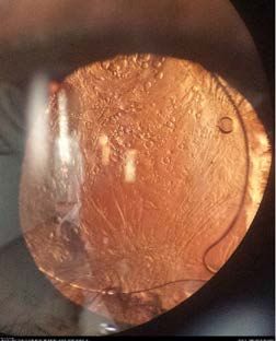
Conclusion                                                          13. Bressler M, Happel P, Blazey W, et al. Northeastern Climate
Eponyms can enhance our understanding of medical conditions;            is Bullseye for Lyme Disease. Family Doctor: A Journal of the
however, repetitive eponym use can introduce a degree of                New York State Academy of Family Physicians [Internet]. 2020
confusion for different conditions. Eponymous syndromes that            [cited 2020 May 15];40:9-14. Available from: https://www.
refer to the physician who originally reported the condition can        researchgate.net/publication/340595139_Northeastern_
also add a level of complexity: one paper reviewed seventeen            Climate_is_Bullseye_for_Lyme_Disease
eponyms attributed to Sir Jonathan Hutchinson alone.82 Some         14. Moore A, Nelson C, Molins C, Mead P, Schriefer M. Current
of his more notable ophthalmic eponyms include: Hutchinson’s            Guidelines, Common Clinical Pitfalls, and Future Directions
disease, Hutchinson’s facies, Hutchinson’s patch, Hutchinson’s          for Laboratory Diagnosis of Lyme Disease, United States.
pupil, Hutchinson’s triad,82 and Hutchinson’s sign.99                   Emerg Infect Dis. 2016 Jul;22(7):1169–77. doi: 10.3201/
                                                                        eid2207.151694. PMID: 27314832; PMCID: PMC4918152.
Whether the eponymous association is obvious or more                15. Wormser G, Dattwyler RJ, Shapiro ED, et al. The Clinical
obscure, there is a diverse array of creatures unknowingly              Assessment, Treatment, and Prevention of Lyme Disease,
responsible for the names of countless conditions, findings,            Human Granulocytic Anaplasmosis, and Babesiosis: Clinical
and instruments in ophthalmology. Without these animals, there          Practice Guidelines by the Infectious Diseases Society of
would be more eponyms based on scientists’ names, providing             America. Clin Infect Dis [Internet]. 2006 Nov 1 [cited 2020
little to no context about the function, appearance, or etiology        May 15];43(9):1089-1134. Available from: https://academic.
of new discoveries. Despite the dimensions of each creature,            oup.com/cid/article/43/9/1089/422463
the animal kingdom plays a sizeable role in expanding the ever-     16. Kumar RR, Jha S, Dhooria A, et al. Butterfly rash: hallmark of
growing glossary of eye-related terms, and it will continue to do       lupus. Q J Med. 2019. P. 877. DOI 10.1093/qjmed/hcz091
so until the cows come home.                                        17. Uva L, Miguel D, Pinheiro, et al. Cutaneous manifestations of
                                                                        systemic lupus erythematosus. Autoimmune Dis. 2012. DOI:
References                                                              10.1155/2012/834291.
1. Jindal N, Jindal P, Kumar J, et al. Animals Eponyms in           18. Fox GH. Dermatologic etymology. Arch Derm Syphilol.
    Dermatology. Indian J Dermatol. 2014 Nov-Dec;59(6):63.              1921;3:404-12.
2. Beh S, Frohman T, Frohman E. The menagerie of neurology.         19. Pisetsky D. The butterfly rash of lupus: an example of
    Animal signs and the refinement of clinical acumen. Neurol          aposematism?. Arthritis Res Ther. 2013;15(106).
    Clin Pract. 2014 Jun;4(3):e1-e9.                                20. Pelechas E, Drosos A. Hydroxychloroquine-induced dark
3. Trueb RM. Value of Eponyms in Dermato-Trichological                  butterfly rash in a rheumatoid arthritis patient. Rheumatology.
    Nomenclature. Skin Appendage Disord. 2018;4(71):71-7.               2018;57:849.
4. Woywodt A, Matteson E. Should eponyms be abandoned?              21. Saksens NT, Krebs MP, Schoenmaker-Koller FE, et al. Mutations
    Yes. BMJ 2007;335:424.                                              in CTNNA1 cause butterfly-shaped pigment dystrophy and
5. O’Flynn C. Epo-Nots: issues with non-traditional eponyms.            perturbed retinal pigment epithelium integrity. Nat Genet
    Journal of Communication in Healthcare 2020;13(3)201-4.             [Internet]. 2016 Feb [ cited 2020 May 14];48(2)144-51.
6. Kanski JJ. Clinical Ophthalmology. 5th ed. Menon J.                  Available from: https://www.ncbi.nlm.nih.gov/pmc/articles/
    Philadelphia: Butterworth-Heinemann;2003. 344 p.                    PMC4787620/pdf/nihms741151.pdf
7. Deibert B, Ferris L, Sanchez N, et al. The link between          22. Zhang K, Garibaldi DC, Li Y, Green WR, Zack DJ. Butterfly-shaped
    colon cancer and congenital hypertrophy of the retinal              pattern dystrophy: a genetic, clinical, and histopathological
    pigment epithelium (CHRPE). Am J Ophthalmol Case Rep.               report. Arch Ophthalmol [Internet]. 2002 Apr [cited 2020 May
    2019;15:100524.                                                     19];120(4):485-90. Available from: https://jamanetwork.com/
8. Minos E, Barry RJ, Southworth S, et al. Birdshot                     journals/jamaophthalmology/fullarticle/270267 Full free text
    chorioretinopathy: current knowledge and new concepts               download. doi:10.1001/archopht.120.4.485
    in pathophysiology, diagnosis, monitoring, and treatment.       23. Deutman AF, van Blommestein JD, Henkes HE, Waardenburg
    Orphanet J Rare Dis. 2016;11(61).                                   PJ, Driest ES. Butterfly-shaped pigment dystrophy of the
9. Garcia EM, Vicente LR, Chavarri Garcia JJ, del Rio Mayor JL.         fovea. Arch Ophthalmol [Internet]. 1970 May [cited 2020 May
    Birdshot chorioretinopathy: importance of early diagnosis and       15];83:558-69. Available from: https://jamanetwork.com/
    therapeutic intervention. Pan Am J Ophthalmol. 2020;2:3.            journals/ jamaophthalmology/fullarticle/629863
10. Ryan SJ, Maumenee AE. Birdshot chorioretinopathy. Am J          24. Kwon R, Price M, Price F, et al. Pentacam Characterization
    Ophthalmol. 1980;89(1):31-45.                                       of Corneas with Fuchs Dystrophy Treated with Descemet
11. Kearns TP, Hollenhorst RW. Chloroquine Retinopathy                  Membrane Endothelial Keratoplasty. J Refract Surg
    Evaluation by Fluorescein Fundus Angiography. Arch                  [Internet]. 2010 Dec 26 [cited 2020 May 15];12:972-9.
    Ophthalmol. 1966 Sep;76(3):378-384.                                 Available from: https://www.healio.com/ophthalmology/
12. Kowalski T, Baker C, Mack HG. Hydroxycholoquine retinal             journals/jrs/2010-12-26-12/%7Bab0e6445-1a5b-4637-a9c3-
    toxicity in two patients with dermatological conditions.            c2d306dbe20d%7D/pentacam-characterization-of-corneas-
    Australasian Journal of Dermatology. 2018 May                       with-fuchs-dystrophy-treated-with-descemet-membrane-
    02;59(4):266-8.                                                     endothelial-keratoplasty#divReadThis

                                                                                             Animal Eponyms in Eye Care           437
25. Ambrósio Jr. R, Correia FF. Analyzing Tomographic Thickness 38. Mccoy F J, Goodman R C. The Crocodile Tear Syndrome.
for Detecting Corneal Ectatic Diseases [Internet]. Switzerland: Plastic and Reconstructive Surgery. 1979;63(1):58-62.
Springer International Publishing; 2017 [cited 2020 May 39. Spiegel JH. Treatment of periorbital rhytids with botunlinum.
22]. 8p. Available from: https://www.researchgate.net/ Arch Facial Plast Surg [Internet]. 2005 May 1 [cited 2020 May
publication/311960596_Analyzing_Tomographic_Thickness_ 21];7(3)198-202. Available from: https://www.liebertpub.
for_Detecting_Corneal_Ectatic_Diseases com/abs/doi/10.1001/archfaci.7.3.198 doi: 10.1001/
26. Ramos I, Belin MW, Valbon BF, et al. Keratoconus associated archfaci.7.3.198
with Corneal Guttata. Int J Keratoconus Ectatic Corneal Dis 40. Mencía-Gutiérrez E, Gutiérrez-Díaz E, Redondo-Marcos I, et al.
[Internet]. 2012 Sep-Dec [cited 2020 May 22]; 1(3):75. Available Cutaneous Horns of the Eyelid: A Clinicopathological Study
from: https://www.researchgate.net/publication/273713043_ of 48 Cases. J Cutan Pathol [Internet]. 2004 Sep [cited 2020
Keratoconus_associated_with_Corneal_Guttata May 22];31(8):539-43. Available from: https://onlinelibrary.
27. Cory C, Jamison D. The Cat Eye Syndrome. Arch Ophthalmol. wiley.com/resolve/openurl?genre=article&sid=nlm:pubme
1974;92(3):259–62. d&issn=0303-6987&date=2004&volume=31&issue=8&spa
28. Curi AL, Kahloun R. Cat-scratch disease. In: Chee S-P, Khairallah ge=539
M, editors. Emerging infectious uveitis. Switzerland: Springer 41. Yanoff M, Duker J S, Wiggs J L, et al. Ophthalmology. 3rd rev.
International Publishing; 2017. p. 57-8. DOI 10.1007/978-3- ed. United Kingdom: Mosby Elsevier; 2009. 497 p.
319-23416-8¬6. 42. Carlson LA. Fish Eye Disease: a New Familial Condition with
29. Basak, SK. Essentials of Ophthalmology [Internet]. New Massive Corneal Opacities and Dyslipoproteinaemia Clinical
Delhi: Jaypee Brothers Medical Publishers. 2019 [cited and Laboratory Studies in Two Afflicted Families. Eur J Clin
2020 May 13]. 544 p. Available from: https://www.google. Invest. 1982;12(1):41-53.
com/books/edition/Essentials_of_Ophthalmology/ 43. Jung, D. Suture Types, Needle Types, and Instruments. In:
ZXKSDwAAQBAJ?hl=en&gbpv=1&dq=essentials+of+ Sutton J, Beckwith A, Johnson B, Knod J, Walther A, Watson
Essentials+of+Ophthalmology+Basak,+Samar+K&printsec C, et. al, editors. The Mont Reid Surgical Handbook 7th ed.
=frontcover#spf=1589556656348 Philadelphia: Elsevier; 2018.
30. Schmidt D, Schulte-Monting J, Schumacher M. Prognosis of 44. Sihota R, Tandon R. Parson’s Disease of the Eye. 22nd ed. New
central retinal artery occlusion: local intraarterial fibrinolysis Delhi: Elsevier; 2015. p. 585-601.
versus conservative treatment. Am J Neuroradiol. 2002 45. Combs JL, Welch RB. Retinal breaks without detachment:
Sep;23(8):1301-7. natural history, management, and long-term follow-up. Trans
31. Hayreh S, Podhajsky P, Zimmerman M. Retinal artery Am Ophthalmol. 1982;80:64-97.
occlusion: associated systemic and ophthalmic abnormalities. 46. Güell JL, Montes DA. Toric Iris Claw Lens Implantation for
Ophthalmol. 2009 Oct;116(10):1928–1936. Keratoconic Eyes. European Ophthalmic Review [Internet].
32. Sommer A. Vitamin A deficiency and its consequences. A field 2012 [cited 29 May 2020];6(2);115-8. Available from:
guide to detection and control, 3rd ed. Geneva: World Health https://www.touchophthalmology.com/toric-iris-claw-lens-
Organization; 1995. 9-11 p. implantation-for-keratoconic-eyes/
33. Schmidt D. The mystery of cotton-wool spots - a review of 47. Worst J. Iris claw lens. J Am Intraocul Implant Soc. 1980;6:166-
recent and historical descriptions. Eur J Med Res [Internet]. 7.
2008 [cited 2020 May 27]; Jun 24;13(6):231-66. Available from: 48. Jabbarpoor M, Ownagh V, Rahimy E, Soheilian M. Giraffe or
https://pubmed.ncbi.nlm.nih.gov/18558551/ leopard spot chorioretinopathy as an outstanding finding: case
34. Gruenauer-Kloevekorn C, Fischer U, Kloevekorn-Norgall K, et report and literature review. Int Ophthalmol. 2019 39: 1405-12.
al. Pellucid marginal corneal degeneration: evaluation of the 49. Ayachit G, Ayachit A, Joshi S, Vasudevan S. A rare case
corneal surface and contact lens fitting. Br J Ophthalmol. 2006 of unilateral diffuse melanocytic proliferation. Indian J
Mar;90(3): 318-323. Ophthalmol. 2018;66(4):588‐90.
35. Maguire LJ, Klyce SD, McDonald MB, Kaufman HE. Corneal 50. Evans B. Binocular vision assessment. In: Rosenfield M,
topography of pellucid marginal degeneration. Ophthalmology Logan N, Edwards K, editors. Optometry science, techniques,
[Internet]. 1987 May [cited 2020 May 14];94(5):519-24]. and clinical management 2nd ed. Edinburgh: Butterworth-
Available from: https://www.sciencedirect.com/science/ Heinemann; 2009. 247 p.
article/ abs/pii/S0161642087334165 doi: 10.1016/S0161- 51. Schornack MM, Siemsen DW, Bradley EA, et al. Ocular
6420(87)33416-5 manifestations of molluscum contagiosum. Clin Exp Optom.
36. Lee BW, Jurkunas UV, Harissi-Dagher M, Poothullil AM, Tobaigy 2006;89(6):390-393.
FM, Azar DT. Ectatic disorders associated with a claw-shaped 52. Singh SR, Gupta PC, Ram J. Fuchs uveitis. JAMA Ophthalmol
pattern on corneal topography. Am. J. Ophthmol. [Internet]. [Internet]. 2015 Jun 11 [cited 2020 May 15];133(6):e15676.
2007 Jul 1 [cited 2020 Jun 19];144(1):154-6 e3. Available Available from: https://jamanetwork.com/journals/
from: https://www.ajo.com/article/S0002-9394(07)00198- jamaophthalmology/fullarticle/2319762 doi:10.1001/
5/fulltext. jamaophthalmol.2015.76
37. Belliveau M J, Brownstein S, Agapitos P, et al. Ultrastructural 53. Moth-eaten. (n.d.) in Dictionary.com. Available from https://
Features of Posterior Crocodile Shagreen of the Cornea. Surv www.dictionary.com/browse/moth-eaten
of Ophthalmol. 2009;54(5):569-75.
438 Clinical & Refractive Optometry 32.4, 202154. Slagle WS, Slagle AM, Brough GH. Mucus fishing syndrome: 67. Golu T, Mogoanta L, Streba CT, Pirici DN, Malaescu D,
case report and new treatment option. Optometry [Internet]. Mateescu GO, Mutiu G. Pterygium: histological and
2001 Oct [cited 2020 May 13];72(10):634-640. Available immunohistochemical aspects. Rom J Morphol Embryol
from: https://www.researchgate.net/publication/11641928_ [Internet]. 2011 [cited 2020 May 20];52(1):153-8. Available
Mucus_fishing_syndrome_Case_report_and_new_treatment_ from: https://www.researchgate.net/publication/50597546_
option Pterygium_Histological_and_immunohistochemical_aspects
55. Milston R, Madigan MC, Sebag J. Vitreous floaters: etiology, 68. Murube, J. Pterygium: descriptive nomenclature of the past.
diagnostics, and management. Surv Ophthalmol [Internet]. Ocul Surf [Internet]. 2008 Jul 1 [cited 2020 May 20];6(3):104-
2016 Mar 1 [cited 2020 May 21];61(2):211-27. Available 7. Available from: https://www.clinicalkey.com/service/
from: https://www.clinicalkey.com/service/content/pdf/ content/pdf/watermarked/1-s2.0-S1542012412702781.
watermarked/1-s2.0-S003962571530014X.pdf?locale=en_ pdf?locale=en_US&searchIndex=
US&searchIndex= 69. Das JM, Munakomi S. Raccoon sign. StatPearls [Internet].
56. Muscae volitantes. (n.d.) in Merriam-Webseter.com dictionary. Treasure Island (FL): StatPearls Publishing; 2020 Jan. Available
Available from https://www.merriam-webster.com/dictionary/ from: https://www.ncbi.nlm.nih.gov/books/NBK542227/
muscae%20volitantes 70. Herbella FA, Mudo M, Delmonti C, et al. ‘Raccoon eyes’
57. Pierce RG, Wong M, Skalet AB. More to mutton than meets (periorbital haematoma) as a sign of skull base fracture. Injury.
the eye. J Gen Intern Med [Internet]. 2010 Jun 8 [cited 2020 2001;32:745-7.
May 14];25:989[1p.]. Available from: https://link.springer.com/ 71. McPheeters RA, White S, Winder A. Raccoon eyes. West J
content/pdf/10.1007/s11606-010-1406-x.pdf Emerg Med. 2010;11(1):97.
58. Millodot M. Dictionary of Optometry and Visual Science 72. Gumus K. A child with raccoon eyes masquerading as trauma.
[Internet]. 8th ed. Oxford (UK). Elsevier. c2017 Oct 19. Keratic Int Ophthalmol. 2007;27: 379-81.
precipitates [cited 2020 May 18] 165 p. Available from: https:// 73. Timmermann R. Raccoon eyes and neuroblastoma. NEJM.
www.elsevier.com/books/dictionary-of-optometry-and-vision- 2003;349(4):e4.
science/ unknown/978-0-7020-7223-9 Google preview. 74. van Woerkom JM, van Toorn DW. A domestic fight or
59. Leffler CT. The History of Glaucoma [Internet]. The Netherlands: something else? Nephrol Dial Transplant. 2000;15:1253-4.
Wayenborgh Publications. 2020 [cited 29 May 2020]. 409 p. 75. Schwartz RA, Spicer MS, Thomas I, et al. Ecchymotic Kaposi’s
Available from: https://www.google.com/books/edition/The_ sarcoma. Cutis. 1995;56(2):104-6.
History_of_Glaucoma/NHXlDwAAQBAJ?hl=en&gbpv=1&dq= 76. Loo H, Forman WB, Levine MR, et al. Periorbital ecchymoses
the+history+of+glaucoma+octopus+perimeter as the initial sign in multiple myeloma. Ann Ophthalmol.
+history&pg=PA409&printsec=frontcover 1982;14(11):1066-8.
60. Vanoni F, Lava S, Fossali EF. Neonatal systemic lupus 77. Millodot M. Dictionary of Optometry and Visual Science
erythematosus syndrome: a comprehensive review. Clinic [Internet]. 7th ed. Oxford (UK). Elsevier. 2008 Sep 22.
Rev Allerg Immunol. 2017;53:469-476. Operculum [cited 2020 May 18] 253 p. Available from: https://
61. Weston WL, Morelli JG, Lee LA. The clinical spectrum of anti- www.elsevier.com/books/dictionary-of-optometry-and-visual-
Ro-positive cutaneous neonatal lupus erythematosus. J Am science/unknown/ 978-0-7020-2958-5 Google preview
Acad Dermatol. 1998;40:675-81. 78. Operculum. (n.d.) in Dictionary.com. Retrieved from https://
62. Chan HH, Lam L, Wong D. Nevus of Ota: a new classification www.dictionary.com/browse/operculum.
based on the response to laser treatment. Lasers Surg Med. 79. Wildlife Journal Junior [Internet]. Durham: New Hampshire
2001;28:267-272. PBS; c2020. Osteichthyes – bony fish; [cited 2020 May 14];
63. Samant H, Kothadia JP. Spider Angioma [Internet]. StatPearls. [about 1 screen]. Available from: https://nhpbs.org/wild/
Treasure Island (FL): StatPearls Publishing; 2020 Jan [cited osteichthyes.asp
2020 May 22]. [1 p.]. Available from: https://www.ncbi.nlm.nih. 80. Laun J, Gopman J, Elston JB, Harrington MA. Rhinophyma.
gov/books/NBK507818/ Eplasty [Internet]. 2015 May [cited 2020 May 14];
64. Pisciform. (n.d.) in CollinsDictionary.com dictionary. Available 15(ic25):[5p.]. Available from: https://www.researchgate.net/
from https://www.collinsdictionary.com/us/dictionary/ publication/277087144_Rhinophyma
english/pisciform. 81. Weidmayer S, McGinty-Tauren M. A close look at common lid
65. Klein R, Lewis RA, Meyers SM, et al. Subretinal lesions. Review of Optometry. 2019.
neovascularization associated with fundus flavimaculatus. 82. van Ruth S, Toonstra J. Eponyms of Sir Jonathan Hutchinson.
Arch Ophthalmol. 1978;96:2054-7. Int J Dermatol. 2008 Jun;47:754–8.
66. Kim DY, Hwang JC, Moore AT, et al. Fundus autofluorescence 83. Margo C, Hamed L. Ocular syphilis. Surv Ophthalmol.
and optical coherence tomography of congenital grouped 1992;37:203-20.
albinotic spots. Retina. 2010;30(8):1217-22. 84. Pallavi R, Popescu-Martinez A. More than Meets the Eye: the
'Pink Salmon Patch'. BMJ Case Rep. 2014 Aug 28; 2014.
85. De Melo MB. An eye on sickle cell retinopathy. Rev Bras
Hematol Hemoter [Internet]. 2014 Aug 12 [cited 2020 May
14];36(5):319-21. Available from: https://www.ncbi.nlm.nih.
gov/pmc/articles/PMC4318455/pdf/main.pdf
Animal Eponyms in Eye Care 43986. Spalton D, Hitchings RA, Hunter PA, et al. Atlas of Clinical 94. Pokharel S, Sherpa D, Shakya K, et al. Diabetic retinopathy in
Ophthalmology. 3rd rev. ed. London: Mosby Elsevier; 2005. tessellated fundus. J Nepal Health Res Counc. 2014;12(26):49-
326 p. 53.
87. Serpigo. (n.d.) in Merriam-Webster.com dictionary. Retrieved 95. Thompson HS. Segmental palsy of the iris sphincter in Adie’s
from https://www.merriam-webster.com/dictionary/serpigo. Syndrome. Arch Ophthalmol [Internet]. 1978 Sep [cited 2020
88. Sinous. (n.d.) in Merriam-Webster.com dictionary. Retrieved May 13];96(9):1615-20. Available from: https://jamanetwork.
from https://www.merriam-webster.com/dictionary/sinous. com/journals/jamaophthalmology/fullarticle/632728 Full free
89. Mendoza-Adam G, Hernandez-Camarena JC, Valdez-Garcia text download. doi:10.1001/archopht.1978.03910060249012
JE. Posterior polymorphous dystrophy, case report and 96. Vermi (n.d.). Collins English Dictionary [Internet]. c2020 [cited
literature review. Arch Soc Esp Oftalmol. 2015;90(9):439-41. 2020 May 20]. Available from: https://www.collinsdictionary.
90. Zinn KM. Clinical Atlas of Peripheral Retinal Disorders. New com/dictionary/english/vermi
York: Sprnger-Verlag;1988. p. 54. 97. Vitellus (n.d.). Collins English Dictionary [Internet]. c2020 [cited
91. Arshinoff, SA, Norman R. Tri-Soft Shell Technique. J 2020 May 20]. Available from: https: //www.collinsdictionary.
Cataract Refract Surg [Internet]. 2013 Aug [cited 2020 May com/dictionary/english/vitellus
13];39(8):1196–1203. Available from: https://journals.lww. 98. Creel, DJ. Best disease. Handbook of Clinical Neurology
com/jcrs/Fulltext/2013/08000/Tri_soft_shell_technique.12. [Internet]. 2019 [cited 2020 May 15];160:495-9. Available
aspx from: https://www.sciencedirect.com/topics/medicine-and-
92. Steinert, R. Cataract Surgery [Internet]. [place unknown]: dentistry/vitelliform-macular-dystrophy
Elsevier. 2010 [cited 2020 May 13]. 210 p. Available 99. Gautam M, Sheth P. Hutchinson’s signs in dermatology. Indian
from: https://books.google.com/books?id=NbM_ J Paediatr Dermatol. 2018;19:371-4.
MAd0dLIC&printsec=frontcover&source=gbs_ge_
summary_r&cad=0#v=onepage&q&f=false
93. Tigroid. (n.d.) in Merriam-Webseter.com dictionary. Retrieved
from https://www.merriam-webster.com/medical/tigroid.
Call for Papers
Clinical & Refractive Optometry
welcomes original articles based
on clinical research, case reports,
and review papers related to the
practice of optometry.
Please submit manuscripts to:
Clinical & Refractive Optometry
www.crojournal.com/author
440 Clinical & Refractive Optometry 32.4, 2021INSTRUCTIONS FOR 1 HOUR OF COPE CE CREDIT
This course is valid for 1 hour of COPE CE Credit, if the post-course test shown below is taken and
submitted online for grading no later than August 11, 2024, and a score of 70% or more is achieved.
CLICK HERE TO TAKE AND SUBMIT THIS POST-COURSE TEST ONLINE
COPE ACCREDITED POST-COURSE TEST
COURSE QUESTIONS ONLINE MAY BE PRESENTED IN A DIFFENT ORDER THAN SHOWN BELOW.
Please Note: In order for you to be able to take this (or any other) CRO Online post-course test, you must first have
registered on the CRO website at www.crojournal.com to open a Personal Dashboard Account. This will allow you to
enroll, complete and submit this or any of our other CRO Online CE Credit Courses for grading. The registration process
is quick and simple and only needs to be done once.
Following your taking and submitting a completed post course test with a score of 70% or more, a personalized COPE
CE Credit Certificate will automatically be made available for you to download from your Personal Dashboard Account.
The passing score may vary by State regulatory authority.
For answers to any questions about either the registration or post course test processes, or to report a specific problem,
please contact CRO Support at support@crojournal.com.
POST-COURSE TEST
Animal Eponyms in Eye Care
1. Why are zoological based eponyms useful in ophthalmic care?
Allow conditions to become more memorable
Help clinicians down the correct systemic workup path
Help clinicians become familiar with diseases and their manifestations
All of the above
2. What eponym is commonly used to describe an autoimmune posterior uveitis characterized by multiple lightly
colored oval chorioretinal lesions with a juxtopapillary predilection?
Birdshot Chorioretinopathy
Bear Tracks
Bag of worms
Butterfly rash
3. What eponym is commonly used to describe fluffy, white lesions caused by accumulated axoplasmic debris
within the ganglion cell axon bundle due ischemia?
Chicken eyes
Crab Claws
Cattle Track sign
Cotton Wool Spots
4. What definition is used to describe the eponym ‘Mutton-fat keratic precipitates’?
Round, centrally umbilicated lesions found on the skin and mucous membranes caused by a poxvirus
Ocular irritation leads to conjunctival mucus production which prompts the patient to mechanically remove “fish” mucus
Leukocytes that appear on the posterior cornea as ‘greasy’ large fat globules, similar to the fat of sheep.
Pigmentation that persists after treating periorbital nevus of Ota lesion with laser surgery
Animal Eponyms in Eye Care 4415. What eponym is commonly used to describe a triangular-shaped growth originating from the interpalpebral
bulbar conjunctiva?
Polar bear tracks
Pterygium
Pisciform
Periorbital spider angioma
COPE ACCREDITED POST-COURSE TEST
6. What eponym is commonly used to describe a periorbital hematoma that can be caused by basal skull fracture?
Raccoon eyes; also known as raccoon sign or panda sign
Salmon patch
Tri-soft shell technique
Steinert double-ended claw chopper
7. What definition is used to describe the eponym ‘rodent ulcer’?
benign swelling and reddening of the nose in stage IV rosacea
A free-floating flap of detached retinal tissue
Refers to basal cell carcinoma
Retinal neovascularization that proliferates anteriorly from perfused to non-perfused tissue, associated with sickle
cell disease
8. What definition is used to describe the eponym ‘serpiginous choroiditis’?
The accumulation of epithelial cells that are clinically visible as “pearls” in posterior capsular opacification
A striped appearance seen when prominent choroidal pigmentation is interrupted by choroidal vessels, which
appear lighter in comparison
Also known as giraffe spots, a chorioretinopathy of characteristic hypo- and hyperpigmented spots viewed by
funduscopy and with angiography
Retinal pigment epithelium atrophy relapses and remits leaving behind subretinal areas of swelling that spread
equatorially in a creeping, snake like fashion
9. What eponym is commonly used to describe foveal dome-shaped deposits of lipofuscin and degraded
photoreceptor segments that initially appear as an egg cooked “sunny-side up”, and later progressing to a
“scrambled egg” appearance?
Vermiform movement
Vitelliform macular dystrophy
Snail tracks
Periorbital spider angioma
10. Why do animal eponyms provide better understanding to conditions as opposed to using physicians who
originally report the condition?
They provide context about the function, appearance or etiology of the condition
Non-repetitive use in ophthalmic context
Cannot be considered derogatory
None of the above
442 Clinical & Refractive Optometry 32.4, 2021You can also read