Catecholaminergic Vasopressors Reduce Toll-Like Receptor Agonist-Induced Microvascular Endothelial Cell Permeability But Not Cytokine Production
←
→
Page content transcription
If your browser does not render page correctly, please read the page content below
ONLINE LABORATORY INVESTIGATION

Catecholaminergic Vasopressors Reduce
Toll-Like Receptor Agonist-Induced
Microvascular Endothelial Cell Permeability
But Not Cytokine Production
                                                                                 Jérémie Joffre, MD, PhD1
OBJECTIVES: Catecholaminergic vasopressors are the cornerstone of
                                                                                 Elliot Lloyd, BA1
circulatory shock management. Nevertheless, catecholamines have prob-
                                                                                 Erika Wong, MD1,2
lematic side effects, arousing a growing interest in noncatecholaminergic
agents such as vasopressin or angiotensin-II. However, their respective          Che Chung-Yeh, PhD1
effects on sepsis-associated microvascular endothelial dysfunction such          Nina Nguyen, BA1
as permeability or inflammation remain elusive. We investigated the role         Fenguyn Xu, PhD1
of catecholamines and other vasopressors on Toll-like receptor agonists-         Matthieu Legrand, MD, PhD1
induced microvascular endothelial permeability and inflammation.
                                                                                 Judith Hellman, MD1
SETTING: University research laboratory/cell research.
SUBJECTS: Human pulmonary microvascular endothelial cells from mul-
tiple donors.
INTERVENTION: Confluent monolayers of human pulmonary micro-
vascular endothelial cells were treated with Toll-like receptor agonists
(lipopolysaccharide, Poly[I:C], or tripalmitoyl-S-glyceryl cysteine) in
the presence or absence of epinephrine, norepinephrine, vasopressin,
and angiotensin-II. Permeability was inferred from transendothelial re-
sistance, measured using electrical cell impedance sensing, where
decreased transendothelial resistance is consistent with increased per-
meability. Cell-cell junction molecule expression was assessed via immu-
nofluorescence microscopy and flow cytometry. We quantified cytokines
in supernatants of Toll-like receptor agonist-treated human pulmonary
microvascular endothelial cells.
MEASUREMENTS AND MAIN RESULTS: Epinephrine and norep-
inephrine both ameliorate lipopolysaccharide, polyinosinic:polycytidylic
acid, or tripalmitoyl-S-glyceryl cysteine–induced reductions in transendo-       Copyright © 2021 The Author(s).
thelial resistance, a surrogate for endothelial permeability. In contrast, the   Published by Wolters Kluwer Health,
noncatecholaminergic agents, vasopressin, and angiotensin-II did not affect      Inc. on behalf of the Society of Critical
Toll-like receptor agonists-induced reductions in transendothelial resist-       Care Medicine and Wolters Kluwer
ance. β1- and β2-adrenergic receptor antagonists reduced the effects of          Health, Inc. This is an open-access ar-
                                                                                 ticle distributed under the terms of the
the catecholamines on transendothelial resistance, whereas α-adrenergic
                                                                                 Creative Commons Attribution-Non
receptor antagonists did not. We observed that epinephrine and norep-            Commercial-No Derivatives License
inephrine induced actin cytoskeletal rearrangement and normalized the            4.0 (CCBY-NC-ND), where it is per-
membrane expression of proteins involved with adherens-junctions (vas-           missible to download and share the
cular endothelial-cadherin) and tight-junctions (zona occludens-1). Despite      work provided it is properly cited. The
having a substantial effect on endothelial permeability, epinephrine and         work cannot be changed in any way or
norepinephrine did not affect human pulmonary microvascular endothe-             used commercially without permission
lial cell survival or production of interleukin-8, interleukin-6, or monocyte    from the journal.
chemoattractant protein-1 (CCL-2) induced by Toll-like receptor agonists,        DOI: 10.1097/CCM.0000000000004854

Critical Care Medicine                                                           www.ccmjournal.org        e315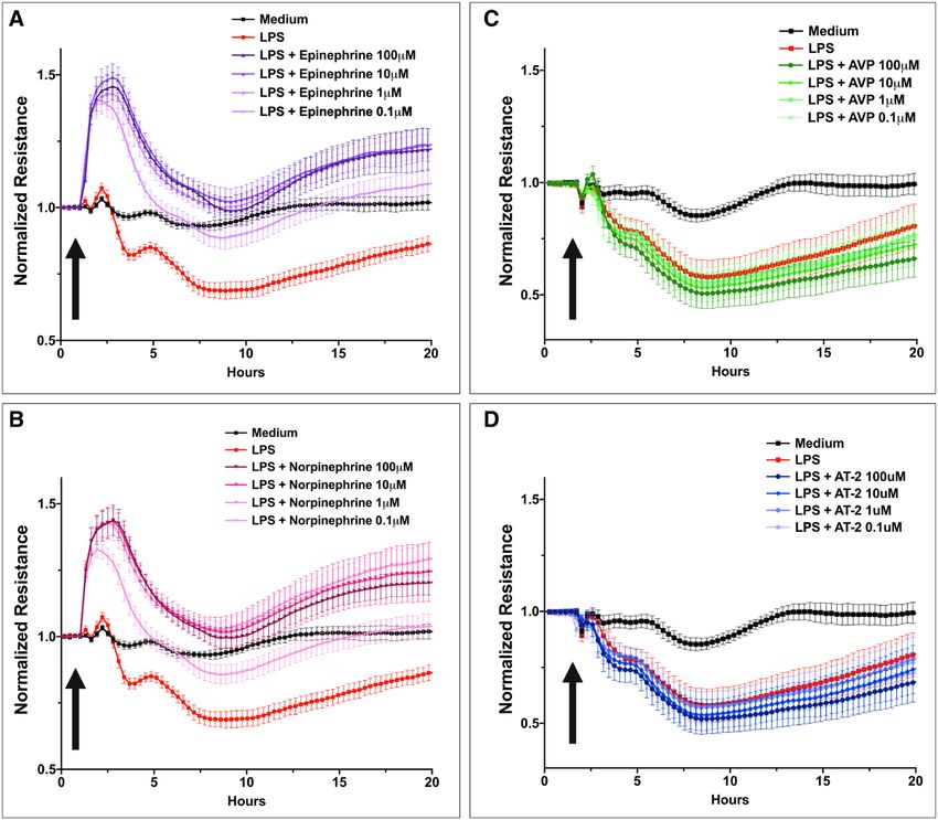
Joffre et al

suggesting that these functions are regulated sepa-             classes of vasopressors on the function of microvascular
rately from permeability.                                       endothelial cells are poorly defined. Here, we explored
CONCLUSIONS: Our findings demonstrate that                      the microvascular endothelial cell effects of catechol-
treatment with epinephrine or norepinephrine                    aminergic (epinephrine and norepinephrine) and non-
strongly reduces endothelial permeability induced               catecholaminergic vasopressors such as vasopressin and
by agonists of multiple Toll-like receptors (Toll-like          angiotensin-II. We tested the effects of catecholaminer-
receptor-2, Toll-like receptor-3, Toll-like receptor-4)         gic and noncatecholaminergic vasopressors in vitro on
in vitro. Our studies suggest that both β1- and β2-             permeability, cytokine and chemokine production, and
adrenergic receptors mediate the stabilizing effects            survival of primary human pulmonary microvascular
of epinephrine and norepinephrine on the endothe-               endothelial cells (HMVEC) activated by microbial and
lial barrier.                                                   endogenous inflammatory agonists.
KEY WORDS: catecholamines; endothelial cells;                   METHODS
inflammation; permeability; sepsis; vasopressors
                                                                Cell Culture and Stimuli

D
          espite prompt care and improvements in sepsis         Primary HMVEC from male and female cadavers were
          management (1, 2), many patients develop a            purchased (Lonza, Basel, Switzerland) and used at pas-
          microcirculatory dysfunction and multiple             sage 3–6. HMVEC were plated and grown to 90–100%
organ failure (MOF). Thus, septic shock still carries           confluence before treatment (at 37°C, 5% Co2), using
unacceptably high mortality rates. Microvascular alter-         the appropriate medium (endothelial cell growth me-
ations play an essential role in developing organ               dium [EGM]–2) (Lonza). Cells were incubated with
failure in critically ill patients, especially in sepsis (3).   lipopolysaccharide (LPS) (1 μg/mL; Sigma-Aldrich,
Persistent microcirculatory alterations, such as mot-           St. Louis, MO), tripalmitoyl-S-glyceryl cysteine
tling (4, 5), alterations in tissue saturation, or sublin-      (Pam3Cys, 10 μg/mL; Abcam, Cambridge, United
gual orthogonal polarization spectral, are associated           Kingdom), polyinosinic:polycytidylic acid (10 μg/mL;
with MOF and death, even though global hemody-                  Abcam), recombinant human interleukin (IL)–1β (10
namic and oxygenation variables are corrected (6–8).            ng/mL; R&D Systems, Minneapolis, MN), recombi-
The first-line recommended vasopressor in septic shock          nant human tumor necrosis factor (TNF)–α (10 ng/
is the catecholaminergic amine norepinephrine (9).              mL; R&D Systems), or cytomix (TNF-α, interferon-γ,
In clinical trials, catecholaminergic and noncatechol-          and IL-1-β; R&D Systems). Simultaneously or after 8
aminergic agents can restore macrocirculatory variables,        hours, cells were stimulated with epinephrine (Sigma-
but thus far, no class of vasopressor has consistently          Aldrich), norepinephrine (Hospira, Lake Forest, IL),
exhibited superiority over the others in improving              vasopressin (Anaspect, Fremont, CA), or angiotensin-2
sepsis outcomes (10–15). The current expert recom-              (Sigma-Aldrich) from 0.1 to 100 μM, encompass-
mendation to treat septic shock by adding either vaso-          ing clinically relevant concentration. The following
pressin or epinephrine to norepinephrine to raise mean          reagents have been used to decipher the involvement
arterial pressure to target is considered a “weak recom-        of different adrenergic receptors (ARs): dobutamine,
mendation supported by a low quality of evidence” (16).         atenolol, propranolol (Cerilliant, Round Rock, TX),
Much remains to be understood about the physiologic             ICI-118,551 (Tocris, Bristol, United Kingdom), phen-
and cellular effects of different vasopressors, including       tolamine methanesulfonate salt (PMS; Sigma-Aldrich),
their impact on the endothelial functions and organ             and phenylephrine (Hikma, Eatontown, NJ). UCSF’s
                                                                Institutional Review Board waived the need for ap-
failure. Sepsis-induced microvascular endothelial dys-
                                                                proval to work with deidentified human cells purchased
function is a key feature of sepsis-related organ failure
                                                                from a vendor.
(17). Furthermore, endothelial permeability, which is
regulated by adherens (cadherin-cadherin) and tight
                                                                Electric Cell-Substrate Impedance Sensing
junction (claudin-occludin) proteins, leads to capillary
leak that can worsen tissue hypoperfusion by increas-           HMVEC barrier integrity was inferred from transen-
ing interstitial pressure. Notably, the effects of different    dothelial resistance (TER) measured using electrical

e316     www.ccmjournal.org                                                                March 2021 • Volume 49 • Number 3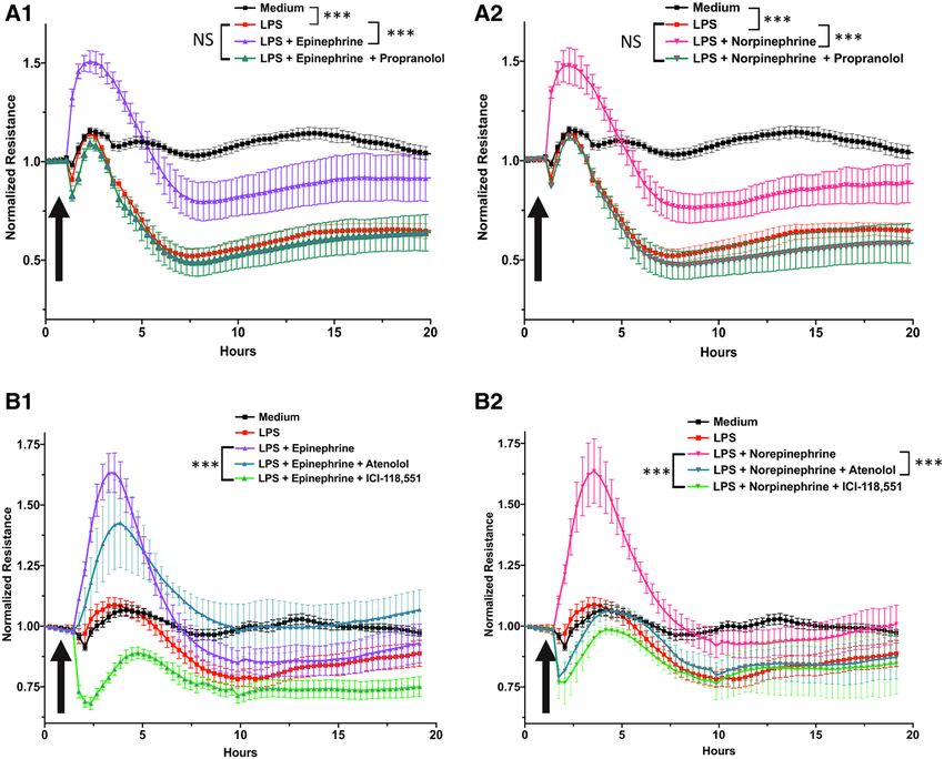
Online Laboratory Investigation

cell impedance sensing (ECIS) ZΘ TEER technology            (Invitrogen), F-actin (rhodamine phalloidin; Invitrogen),
(Applied Biophysics Inc., Troy, NY). ECIS measures a cell   or anti–vascular endothelial (VE)-cadherin (Santa Cruz
monolayer’s electrical impedance in real time and at mul-   Biotechnologies) followed by AlexaFluor488-tagged
tiple electrical frequencies (18, 19). A decrease in TER    secondary antibody. Nuclei were counterstained with
reflects an increase in permeability. A 96W20idf plate      4′6-diamidino-2-phenylindole. Slides were visualized
(Applied Biophysics Inc.) was stabilized with cysteine      with a Cytation 5 Biotek (Fischer scientific, Waltham,
(10 mM) for 10 minutes. Cells were then seeded in 300       MA) and recorded at 20X objective on Gen5 Image+
μL EGM-2 at a density of 60,000/cm2. Cells were consid-     Software (Biotek,Winooski, VT).
ered confluent upon reaching stable baseline resistances
at 4,000 Hz, an optimal frequency specific for endothe-     Immunoassays
lial cells to be modeled using ECIS Software V.1.2.163.0.
                                                            Endothelial cell culture supernatants were collected at
PC (Applied Biophysics Inc.) (20). Endothelial mono-
                                                            different timepoints and stored at –80°C until analysis.
layers were then stimulated with inflammatory ago-
                                                            Cytokines were measured using Duoset ELISA Kit
nists and simultaneously or sequentially with the drugs
                                                            (R&D Systems) for IL-8, IL-6, and CCL-2, according
described above, using four to eight replicates per con-
                                                            to the appropriate dilution and following recommen-
dition. Supplemental Figure 1 (Supplemental Digital
                                                            dations of the manufacturer.
Content 1, http://links.lww.com/CCM/G58; legend,
Supplemental Digital Content 12, http://links.lww.com/
                                                            Statistics
CCM/G69) describes how we measured the area under
the curve as an integrative marker of permeability and      Results are reported as means (± sd), data were ana-
the maximal conductance peak.                               lyzed using one-way ordinary Kruskal-Wallis with
                                                            multiple Dunn’s post hoc test or a formal combined
Flow Cytometry                                              analysis of variance when multiple donors were ana-
After detachment with the trypsin-free reagent Accutase     lyzed on the same graph. p values of less than 0.05 were
(Innovative Cell Technologies INC., San Diego, CA),         considered statistically significant. Statistics and graph-
single cells in suspension were labeled with  -       ical representations were performed using GraphPad
CD102 (Clone CBR-IC1/2) (Biolegend, San Diego,              Prism 8.0 (Graph Pad Software, San Diego, CA). Each
CA),  -CD31 (Clone WM59) (ebiosci-               experiment has been replicated at least twice on differ-
ences, San Diego, CA), and  -CD144 (Clone 16B-          ent donors from both genders.
1) (Invitrogen, Carlsbad, CA). Single-cell suspensions
stained with fluorophore-conjugated antibodies were         RESULTS
acquired the same day using an LSRII Fortessa (BD           Catecholaminergic Vasopressors, But Not
Biosciences, San Jose, CA) flow cytometer and analyzed      Vasopressin or Angiotensin-2, Reduce Toll-Like
with FlowJo software (Miltenyi, Bergisch Gladbach,          Receptor Agonist-Induced Permeability on
Germany).                                                   HMVEC
                                                            To assess for microvascular endothelial barrier dys-
Immunofluorescence Microscopy
                                                            function, we grew HMVEC to confluency on ECIS
HMVEC were grown to confluence on LabTek II chamber         plates and stimulated them with LPS (Toll-like re-
slides (Nunc, thermofischer scientific, Waltham, MA) that   ceptor [TLR]–4 agonist) in the presence or absence
had been precoated with collagen and then treated for 6     of increasing concentrations of each vasopressor.
hours with LPS in the presence and absence of catechol-     We observed that epinephrine and norepinephrine
aminergic agents, and antagonists and agonists of α- and    at concentrations from 0.1 to 100 μM significantly
β-AR. Cells were then fixed with 4% paraformaldehyde        reduced LPS-induced permeability without an appar-
for 15 minutes and permeabilized with 0.5% Triton X-100     ent concentration-dependent effect (Fig. 1, A and B)
in phosphate-buffered saline (PBS). After blocking with     (Supplemental Fig. 2, A and B, Supplemental
1% bovine serum albumin in PBS, cells were incubated        Digital Content 2, http://links.lww.com/CCM/
with either anti–zona occludens (ZO)-1-AlexaFluor594        G59; legend, Supplemental Digital Content 12,

Critical Care Medicine                                                                 www.ccmjournal.org       e317
Joffre et al

Figure 1. Effect of vasopressors on lipopolysaccharide (LPS)–induced permeability of human pulmonary microvascular endothelial cell
(HMVEC) effects of increasing concentrations of epinephrine (A), norepinephrine (B), vasopressin (AVP) (C), an angiotensin (AT)–2
(D) on HMVEC LPS-induced permeability. HMVEC were simultaneously coincubated with indicated concentrations of vasopressors
and LPS (1 μg/mL). Arrows indicate the time of addition of treatments. *p < 0.05, **p < 0.01, ***p < 0.001, any condition versus LPS.

http://links.lww.com/CCM/G69). Conversely, neither                   12, http://links.lww.com/CCM/G69). Overall, we esti-
vasopressin nor angiotensin-2 affected LPS-induced                   mate that 10 μM of catecholamines reduce LPS-induced
permeability at any concentrations (Fig. 1, C and D)                 permeability about 73.2% (± 24.9%; p = 0.01) for ep-
(Supplemental Fig. 2, C and D, Supplemental Digital                  inephrine and 70.8% (± 25.9%; p = 0.02) for norepi-
Content 2, http://links.lww.com/CCM/G59; legend,                     nephrine. Similar results were observed in HMVEC
Supplemental Digital Content 12, http://links.lww.                   treated with TLR-3 agonist (Poly[I:C]) (Supplemental
com/CCM/G69). Despite substantial variability be-                    Fig. 4, Supplemental Digital Content 4, http://links.
tween the human donors regarding the intensity of                    lww.com/CCM/G61; legend, Supplemental Digital
response to both LPS and vasopressors, all donors                    Content 12, http://links.lww.com/CCM/G69) and
studied showed the same pattern (Supplemental Fig.                   TLR-2 agonist (Pam3Cys) (Supplemental Fig. 5,
3, Supplemental Digital Content 3, http://links.lww.                 Supplemental Digital Content 5, http://links.lww.com/
com/CCM/G60; legend, Supplemental Digital Content                    CCM/G62; legend, Supplemental Digital Content 12,

e318     www.ccmjournal.org                                                                          March 2021 • Volume 49 • Number 3
Online Laboratory Investigation
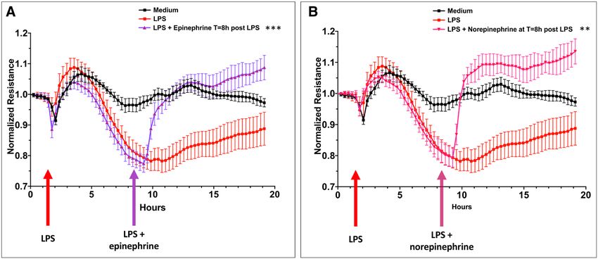
Figure 2. Posttreatment with catecholaminergic vasopressors reverses lipopolysaccharide (LPS)–induced permeability of human
pulmonary microvascular endothelial cell. Epinephrine (10 μM) (A) and norepinephrine (10 μM) (B) were added to wells 8 hr after the
addition of LPS (1 μg/mL). Arrows indicate the respective time of addition of treatments. **p < 0.01, ***p < 0.001, area under the curve
group LPS versus LPS + catecholamine at 8 hr.
http://links.lww.com/CCM/G69). Epinephrine and legend, Supplemental Digital Content 12, http://links.
norepinephrine also significantly reduced permea- lww.com/CCM/G69).
bility induced by non-TLR ligands endogenous media-
β-ARs, But Not α-Adrenergic, Are Responsible
tors: IL-1β, TNF-α, and Cytomix (Supplemental Fig. 5,
for the Reduced LPS-Induced Permeability of
Supplemental Digital Content 5, http://links.lww.com/
Catecholamines
CCM/G62; legend, Supplemental Digital Content 12,
http://links.lww.com/CCM/G69). Neither vaso- Given their affinity for α-AR, we initially hypothesized
pressin nor angiotensin-2 had significant effect on that catecholamines reduce endothelial permeability
permeability induced by these inflammatory agonists induced by inflammatory agonists via α-AR signal-
(Supplemental Fig. 5, Supplemental Digital Content 5, ing. However, the competitive blockade of α1- and
http://links.lww.com/CCM/G62; legend, Supplemental α2-receptors using PMS (10 μM) did not reverse the
Digital Content 12, http://links.lww.com/CCM/G69). effects of epinephrine or norepinephrine (Fig. 3, A
The effects of catecholamines on permeability induced and B). PMS alone had no impact on endothelial bar-
by IL-1β, TNF-α, and cytomix are smaller compared rier maintenance at baseline or in LPS-stimulated
with their effects on TLR agonists-induced permea- cells (Supplemental Fig. 7A, Supplemental Digital
bility (LPS, Poly[I:C], and Pam3Cys). We speculate that Content 7, http://links.lww.com/CCM/G64; legend,
these differences may be due to apoptosis of HMVEC Supplemental Digital Content 12, http://links.lww.
treated with IL-1β, TNF-α, and cytomix, but not by the com/CCM/G69). In addition, the pure α-AR agonist
TLR agonists. Notably, the addition of catecholamines phenylephrine did not modify LPS-induced perme-
8 hours post LPS challenge, when permeability was ability (Fig. 3C). Conversely, the simultaneous addi-
maximal (resistance nadir), restored the endothelial tion of the nonselective β-AR antagonist, propranolol
monolayer to its baseline resistance, in approximately (10 or 25 μM), to LPS + epinephrine/norepineph-
1 hour (Fig. 2). Combining a catecholaminergic and a rine fully abrogated the effect of these catecholamines
noncatecholaminergic agent does not further modify (Fig. 4, A1 and A2). We also observed that pro-
the permeability effect than treatment with catechol- pranolol on its own transiently reduces TER and
amines alone, whether they were added simultaneously that propranolol prohibits full recovery of HMVEC
or sequentially (Supplemental Fig. 6, Supplemental from LPS-induced permeability (Supplemental
Digital Content 6, http://links.lww.com/CCM/G63; Fig. 7B, Supplemental Digital Content 7,
Critical Care Medicine www.ccmjournal.org e319Joffre et al
http://links.lww.com/CCM/G64;
legend, Supplemental Digital
Content 12, http://links.lww.
com/CCM/G69). We also
observed that the selective β1-
antagonist atenolol (10 or 25 μM)
and selective β2-antagonist ICI-
118,551 (10 or 25 μM) both abro-
gated norepinephrine’s effect,
whereas only ICI-118,551 coun-
teracted the effect of epinephrine
(Fig. 4, B1 and B2). Neither aten-
olol nor ICI-118,551 on their own
affected TER (Supplemental Fig.
7, C and D, Supplemental Digital
Content 7, http://links.lww.com/
CCM/G64; legend, Supplemental
Digital Content 12, http://links.
lww.com/CCM/G69). Finally, se-
lective β2-agonist albuterol and
β1-agonist dobutamine signifi-
cantly reduced the LPS-induced
permeability (Supplemental Fig.
7, E and F, Supplemental Digital
Content 7, http://links.lww.com/
CCM/G64; legend, Supplemental
Digital Content 12, http://links.
lww.com/CCM/G69).
Catecholaminergic
Vasopressors Restore
Adherens- and Tight
Junction Membrane Protein
Organization Via Actin-
Filament Rearrangement
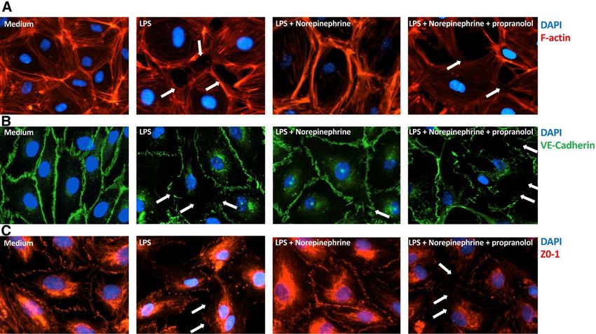
Immunofluorescence of HMVEC,
6 hours post stimulation, re-
vealed that LPS directly induces
gaps in cell-cell junctions and
reduces membrane VE-cadherin
and ZO-1 expression. We
observed that treatment with cat-
Figure 3. Catecholamines normalize human pulmonary microvascular endothelial cell (HMVEC) echolaminergic agents (10 μM)
permeability independently of the α-adrenergic receptor. Synchronous HMVEC stimulation with reduces LPS-induced forma-
phentolamine methanesulfonate salt (PMS) (10 μM) (an α1 and α2 receptors blocker) does not
tion of cell-cell junction gaps
reverse the protective effect of epinephrine (10 μM) (A1 and A2) nor norepinephrine (10 μM)
(B1 and B2). Pure α-agonist phenylephrine does not modify lipopolysaccharide (LPS)–induced and restores the expression
permeability (1 μg/ml) (C1 and C2). Arrows indicate the time of addition of treatments. **p < of VE-cadherin and ZO-1.
0.01, ***p < 0.001, any condition versus LPS. AUC = area under the curve. Catecholaminergic agents also
e320 www.ccmjournal.org March 2021 • Volume 49 • Number 3Online Laboratory Investigation Figure 4. Catecholamines reduce lipopolysaccharide (LPS)–induced human pulmonary microvascular endothelial cell (HMVEC) permeability via β1- and β2-adrenergic receptors. Synchronous HMVEC stimulation with propranolol (25 μM) (a β1 and β2 receptors blocker) fully abrogates the antipermeability effects of epinephrine (10 μM) (A1) and norepinephrine (10 μM) (A2). β2-antagonist ICI- 118,551 (25 μM) fully abrogates the reduced permeability induced by epinephrine (10 μM) and norepinephrine (10 μM), β1-antagonist atenolol (25 μM) reverses the effects of norepinephrine (but not epinephrine) in reducing LPS-induced permeability (1 μg/mL) (B1 and B2). Arrows indicate the time of addition of treatments. **p < 0.01, ***p < 0.001. NS = not significant. induced substantial changes in the F-actin submembrane legend, Supplemental Digital Content 12, http://links. cytoskeleton. Indeed, treatment with epinephrine or lww.com/CCM/G69). norepinephrine induced actin-filament contraction with a reinforcement under the membrane and parallel rear- Treatment of HMVEC With Catecholaminergic rangement in “railroad tracks.” Cotreatment with pro- Vasopressors Does Not Modify LPS or pranolol fully reversed the cytoskeletal changes induced Pam3Cys-Induced Inflammatory Mediators by catecholamines and augmented HMVEC monolayer Secretion or Cell Survival disruption. Representative images are shown in Figure 5. The microvascular endothelium is a critical component Catecholaminergic vasopressor-induced restoration of of the immune response in sepsis and acute sterile inflam- VE-Cadherin membranous expression was confirmed mation. We, therefore, assessed the effects of vasopressors by flow cytometry (Supplemental Fig. 8, Supplemental on HMVEC production of IL-6, IL-8, and CCL-2, which Digital Content 8, http://links.lww.com/CCM/G65; are involved in the recruitment and transendothelial Critical Care Medicine www.ccmjournal.org e321
Joffre et al

Figure 5. Catecholaminergic vasopressors restore adherens- and tight-junctions of lipopolysaccharide (LPS)–treated human pulmonary
microvascular endothelial cell (HMVEC) via actin-filament rearrangement. Representative pictures of F-actin (A), VE-cadherin (B),
and ZO-1 (5C) immunofluorescent staining on HMVEC, after 6 hr of stimulation with LPS (1 μg/mL). Arrows indicate gaps in cell-cell
junctions. DAPI = 4′6-diamidino-2-phenylindole.

migration of monocytes and neutrophils. We observed                catecholamines, which are widely used in sepsis and
no significant effects of epinephrine and norepinephrine           other forms of shock for their vasoconstrictive and
on LPS-induced IL-6, IL-8, or CCL-2 at any time point.             inotropic effects, can directly reduce endothelial cell
In contrast, angiotensin-2 significantly augmented LPS-            permeability induced by bacterial and viral TLR ago-
induced IL-6 and CCL-2 at 24 hours (Supplemental                   nists. Catecholamines also limit permeability induced
Fig. 9, Supplemental Digital Content 9, http://links.lww.          by proinflammatory cytokines. Our results raise the
com/CCM/G66; legend, Supplemental Digital Content                  possibility that catecholamines may affect the course of
12, http://links.lww.com/CCM/G69). Similar results                 shock not only through their effects on vascular tone
were observed with Pam3Cys (Supplemental Fig. 10,                  and cardiac contractility but also by reducing micro-
Supplemental Digital Content 10, http://links.lww.com/             vascular endothelial permeability.
CCM/G67; legend, Supplemental Digital Content 12,                     During sepsis, microbial products directly stim-
http://links.lww.com/CCM/G69). None of the vasopres-               ulate endothelial cells through pattern-recognition
sors affected HMVEC survival as assessed by crystal vi-            receptors (PRRs) (21, 22) and downstream inflam-
olet assays (Supplemental Fig. 11, Supplemental Digital            matory pathways mediated by nuclear factor kappa
Content 11, http://links.lww.com/CCM/G68; legend,                  B and the mitogen-activated protein kinases (23).
Supplemental Digital Content 12, http://links.lww.com/             Engagement of endothelial cell PRRs promotes a pro-
CCM/G69).                                                          inflammatory phenotype with increased production
                                                                   of cytokines, chemokines and procoagulant factors,
                                                                   and up-regulated expression of proadhesive molecules
DISCUSSION                                                         and antifibrinolytic factors. Additionally, damaged
Permeability and endothelial dysfunction are hall-                 glycocalyx and endothelial cell apoptosis lead to an
marks of sepsis and are being explored as tar-                     increase in permeability to proteins and fluids, caus-
gets for sepsis therapies. Our study indicates that                ing interstitial edema (17, 24). Our data indicate that
e322     www.ccmjournal.org                                                                       March 2021 • Volume 49 • Number 3
Online Laboratory Investigation catecholaminergic vasopressors significantly decrease edema is believed to promote organ failure in many TLR-agonist–induced permeability, whereas noncat- conditions, including septic shock, acute respiratory echolaminergic vasopressors do not. Even when LPS- distress syndrome (ARDS), ischemia-reperfusion, and induced endothelial permeability is at its peak, the noninfectious shock (e.g., pancreatitis, trauma) (32). addition of catecholamines restores TER within min- Restoration of endothelial functions is considered a po- utes of introduction. The effect of catecholamines on tential target of resuscitation strategies (33). Concerns permeability is preserved in a vast range of concentra- with reducing permeability center around the concept tions (from 0.1 to 100 μM). Furthermore, no additive that vascular permeability may serve a beneficial role or synergistic effect was observed when combining dif- in recruiting leukocytes to sites of infection and injury ferent classes of vasopressors. Our data using selective by promoting leukocyte-transendothelial migrations, pharmacologic antagonists implicate β1 and β2-AR in which are necessary to fight infection and initiate re- the endothelial stabilizing effects of catecholamines. pair of injured tissues. Several studies, however, have We observed that antagonists for both β1 and β2 challenged the notion that vascular leak is required reversed the antipermeability effect of norepinephrine, for leukocyte tissue migration (34, 35) and have docu- whereas only β2-antagonist reversed the antiperme- mented that fluid leakage, leukocyte recruitment, and ability effects of epinephrine. This dichotomy may be transendothelial migration are regulated separately due to the 10- to 100-fold stronger affinity of epineph- (36–38). rine for the β2 receptor than the β1 receptor, leading Early fluid loading is a cornerstone of hemodynamic to negligible effects of selective blockade of β1 on the septic and nonseptic vasoplegic shock management. stabilizing effects of epinephrine. However, fluid resuscitation protocols are intensely Catecholamines rapidly restore the endothelial bar- debated, regarding the importance of achieving he- rier within minutes. This suggests that protein-protein modynamic stability versus the risk of overloading. interactions may be responsible for the restoration, Observational studies suggest that insufficient fluid rather than transcriptional regulation of genes in- volume, in early phase of septic shock resuscitation, volved in permeability. β-ARs have been reported to is deleterious (39–41). Conversely, numerous stud- play a role in forming focal adhesions and dynamic ies also suggest that high-volume resuscitation and a remodeling of the actin cytoskeleton. Activation via positive fluid balance might be harmful (42–44), sup- the Gs subunit of β2-AR has been shown to enhance porting a more restrictive approach to fluid resusci- intracellular cyclic adenosine monophosphate (cAMP) tation (45–48). Reports in humans with septic shock generation transiently. Intracellular cAMP activates suggest that early treatment with norepinephrine may protein kinase A, which stabilizes the actin cytoskel- be beneficial (49–51). Notably, in the CENSER study, eton and counteracts the cell retractile force (25–27). patients receiving early norepinephrine had a lower β-AR signaling is also mediated via the β-arrestin 2 occurrence of pulmonary edema (14.4 % vs 27.7 %; and p115RhoGEF complexation, activating the mem- p = 0,004) despite receiving the equivalent volumes of brane RhoA-kinase (28, 29). Inhibition of Rho-kinase fluids (51). Our results raise the possibility that early has been demonstrated to increase LPS-induced endo- catecholamine administration may limit the capillary thelial permeability (30). Thus, β-AR blockade induces leak syndrome induced by sepsis and therefore limit intracellular gaps via actin myosin-driven endothelial interstitial edema. retraction, and conversely, we observed in our study Despite having substantial effects on TLR- that its activation reduces LPS-induced permeability agonist–induced permeability, we observed that through cytoskeletal rearrangement and contributes the catecholaminergic vasopressors did not affect to the maintenance of endothelial barrier properties TLR-agonist–induced production of cytokines and under baseline conditions (31). chemokines by HMVEC. Our data suggest that TLR- Although this is an in vitro study of primary human dependent permeability and inflammation are inde- endothelial cells, we speculate that our results may pendently regulated in microvascular endothelial cells have relevance to the choice and timing of initiation (52). This suggests that it may be possible to develop of vasopressors in humans with shock. Endothelial endothelial based therapies that prevent or treat vas- barrier dysfunction leading to vascular leak and tissue cular leakage, without impairing the endothelium’s Critical Care Medicine www.ccmjournal.org e323
Joffre et al

immunoinflammatory response. Although Staedtke             in a critically ill patient’s pulmonary microcircula-
et al (53) reported that epinephrine increases IL-6 se-    tion. Finally, although we observed a similar effect
cretion by peritoneal macrophages and T cells, but not     of LPS and catecholamines in every donor tested, we
endothelial cells, it has been recently published that     noticed substantial interindividual variability in the
norepinephrine infusion correlates with a “anti-inflam-    intensity of the permeability response to LPS and
matory cytokine profile” in septic mice and healthy        catecholamines.
human volunteers following LPS challenge (54).
Also, the effects of catecholamines on inflammation        CONCLUSIONS
mediators might depend on the cell type and the in-
                                                           Our findings demonstrate that treatment of cultured
flammatory conditions. In our study, we found that
                                                           primary human lung microvascular endothelial cells
high concentrations of noncatecholaminergic vaso-
                                                           with epinephrine or norepinephrine strongly reduces
pressors (angiotensin-2 and to a lesser extent vas-
                                                           endothelial permeability induced by agonists of mul-
opressin) increased LPS- and Pam3Cys-induced
                                                           tiple TLRs. Furthermore, posttreatment with catechol-
production of IL-6 and CCL-2 at 24 hours. In vitro, the
                                                           amines many hours into treatment with LPS restores
absence of vasopressors’ clearance makes it difficult to
                                                           endothelial permeability to baseline. Both β1- and
fully mimic the potential in vivo effects. However, ex-
                                                           β2-AR, but not α-AR, mediate the stabilizing effects
perimental studies reported that continuous infusion
                                                           of epinephrine and norepinephrine on the endothelial
of angiotensin-2 in mice dose-dependently increases
                                                           barrier. Our results suggest that epinephrine and nor-
intra-aortic IL-6 production and recruitment of mac-
                                                           epinephrine may act to restore vascular barrier integ-
rophages suggesting a vascular induction of chemo-
                                                           rity during sepsis and shock, in addition to their known
kines (55, 56) and supporting our findings.
                                                           effects on vasomotor tone and cardiac function.
   β-blockers have been proposed as potential ther-
apeutic adjuvants for septic shock because they
have been reported to decrease cardiac oxygen con-          1 Department of Anesthesia and Perioperative Care, University
sumption, limit hyperglycemia, reduce the catabolic           of California, San Francisco, CA.
response to sepsis, and alleviate Type-1 T-helper in-       2 Division of Pediatric Critical Care, UCSF Benioff Children’s
flammation (57–59).                                           Hospitals, San Francisco, CA.
   Our study raises the possibility that β-blockers        Drs. Joffre and Lloyd participated in the study concept and de-
                                                           sign, acquisitions of data, statistical analysis, drafting, and re-
might exacerbate vascular leakage in sepsis. Our
                                                           vision of the article. Drs. Wong, Chung-Yeh, Nguyen, and Xu
study has several limitations. First, we worked only       participated in cell culture, stimulation, and immunoassays. Drs.
with lung derived microvascular cells. We focused          Legrand and Hellman provided input on the design and critical
on lung-derived-EC because of their susceptibility         comments and edits to the article. The study was performed in
to capillary leakage driving acute lung injury and         Dr. Hellman’s laboratory.
ARDS in critical illness. However, given the heter-        Supplemental digital content is available for this article. Direct
ogeneity of microvascular beds we can't extrapolate        URL citations appear in the printed text and are provided in the
these findings to other organs (60, 61). Second, we        HTML and PDF versions of this article on the journal’s website
                                                           (http://journals.lww.com/ccmjournal).
explored only barrier and inflammatory functions,
                                                           Supported, in part, by awards from the University of California, San
but the response of the microvascular endothelium
                                                           Francisco (UCSF) Department of Anesthesia and Perioperative
to inflammation is multifaceted. We did not study the      Care and the San Francisco Foundation (to Dr. Hellman). The
effects of vasopressors on other essential functions,      UCSF Flow Cytometry Facility acknowledges Diabete research
such as of glycocalyx maintenance, coagulation, fi-        center Center Grant National Institutes of Health P30 DK063720.
brinolysis, and leukocyte adhesion (17). Assessing         Dr. Joffre is supported by a fellowship from SRLF (French Society
endothelial barrier function in vivo is extremely chal-    of Intensive Care) and Amicale des Anciens Internes en medi-
lenging. Our in vitro system has the advantage that        cine des Hopitaux de Paris and Assistance Publique—Hopitaux
it allows real-time assessment of the effect of vaso-      de Paris. The remaining authors have disclosed that they do not
                                                           have any potential conflicts of interest.
pressors on endothelial barrier function regardless of
                                                           For information regarding this article, E-mail: Jeremie.joffre@ucsf.
changes in vasomotor tone. But, it cannot pretend to
                                                           edu
fully reproduce the integrative effects of vasopressors

e324     www.ccmjournal.org                                                                 March 2021 • Volume 49 • Number 3
Online Laboratory Investigation
REFERENCES 18. Tiruppathi C, Malik AB, Del Vecchio PJ, et al: Electrical method
for detection of endothelial cell shape change in real time:
1. Kaukonen KM, Bailey M, Suzuki S, et al: Mortality related to Assessment of endothelial barrier function. Proc Natl Acad Sci
severe sepsis and septic shock among critically ill patients USA 1992; 89:7919–7923
in Australia and New Zealand, 2000-2012. JAMA 2014; 19. Meng J, Roy S: Study of epithelium barrier functions by real-
311:1308–1316 time TER measurement. Bio Protoc 2016; 6:e1815
2. Dellinger RP, Hussain S: From Barcelona to New York: 15 20. Szulcek R, Bogaard HJ, van Nieuw Amerongen GP: Electric
years of transition of sepsis performance improvement. J cell-substrate impedance sensing for the quantification of en-
Thorac Dis 2017; 9:3453–3455 dothelial proliferation, barrier function, and motility. J Vis Exp
3. Sakr Y, Dubois MJ, De Backer D, et al: Persistent microcircula- 2014; 28:51300
tory alterations are associated with organ failure and death in 21. Salvador B, Arranz A, Francisco S, et al: Modulation of en-
patients with septic shock. Crit Care Med 2004; 32:1825–1831 dothelial function by toll like receptors. Pharmacol Res 2016;
4. Ait-Oufella H, Lemoinne S, Boelle PY, et al: Mottling score predicts 108:46–56
survival in septic shock. Intensive Care Med 2011; 37:801–807 22. Khakpour S, Wilhelmsen K, Hellman J: Vascular endothelial
5. Preda G, Bourcier S, Joffre J, et al: Mottling score is associ- cell toll-like receptor pathways in sepsis. Innate Immun 2015;
ated with 28-day mortality in critically ill patients with sepsis. 21:827–846
Minerva Anestesiol 2017; 83:664–666 23. Wang W, Deng M, Liu X, et al: TLR4 activation induces nontol-
6. Ait-Oufella H, Joffre J, Boelle PY, et al: Knee area tissue erant inflammatory response in endothelial cells. Inflammation
oxygen saturation is predictive of 14-day mortality in septic 2011; 34:509–518
shock. Intensive Care Med 2012; 38:976–983 24. Colbert JF, Schmidt EP: Endothelial and microcirculatory
7. Rovas A, Seidel LM, Vink H, et al: Association of sublingual function and dysfunction in sepsis. Clin Chest Med 2016;
microcirculation parameters and endothelial glycocalyx dimen- 37:263–275
sions in resuscitated sepsis. Crit Care 2019; 23:260 25. Rangarajan S, Enserink JM, Kuiperij HB, et al: Cyclic AMP
8. Johansson PI, Stensballe J, Ostrowski SR: Shock induced induces integrin-mediated cell adhesion through Epac and
endotheliopathy (SHINE) in acute critical illness - a unifying Rap1 upon stimulation of the beta 2-adrenergic receptor. J
pathophysiologic mechanism. Crit Care 2017; 21:25 Cell Biol 2003; 160:487–493
9. Rhodes A, Evans LE, Alhazzani W, et al: Surviving sepsis 26. Qiao J, Huang F, Lum H: PKA inhibits RhoA activation: A pro-
campaign: international guidelines for management of tection mechanism against endothelial barrier dysfunction. Am
sepsis and septic shock: 2016. Intensive Care Med 2017; J Physiol Lung Cell Mol Physiol 2003; 284:L972–L980
43:304–377 27. Moore TM, Chetham PM, Kelly JJ, et al: Signal transduc-
10. Khanna A, Ostermann M, Bellomo R: Angiotensin II for the tion and regulation of lung endothelial cell permeability.
treatment of vasodilatory shock. N Engl J Med 2017; 377:2604 Interaction between calcium and cAMP. Am J Physiol 1998;
11. Sakr Y, Reinhart K, Vincent JL, et al: Does dopamine admin- 275:L203–L222
istration in shock influence outcome? Results of the sepsis 28. Holinstat M, Mehta D, Kozasa T, et al: Protein kinase cal-
occurrence in acutely ill patients (SOAP) Study. Crit Care Med pha-induced p115RhoGEF phosphorylation signals en-
2006; 34:589–597 dothelial cytoskeletal rearrangement. J Biol Chem 2003;
12. Myburgh JA, Higgins A, Jovanovska A, et al; CAT Study inves- 278:28793–28798
tigators: A comparison of epinephrine and norepinephrine in 29. Ma X, Zhao Y, Daaka Y, et al: Acute activation of β2-adrenergic
critically ill patients. Intensive Care Med 2008; 34:2226–2234 receptor regulates focal adhesions through βArrestin2- and
13. Russell JA, Walley KR, Singer J, et al; VASST Investigators: p115RhoGEF protein-mediated activation of RhoA. J Biol
Vasopressin versus norepinephrine infusion in patients with Chem 2012; 287:18925–18936
septic shock. N Engl J Med 2008; 358:877–887 30. Xiaolu D, Jing P, Fang H, et al: Role of p115RhoGEF in lipo-
14. Marks JA, Pascual JL: Selepressin in septic shock: Sharpening polysaccharide-induced mouse brain microvascular endothe-
the VASST effects of vasopressin?*. Crit Care Med 2014; lial barrier dysfunction. Brain Res 2011; 1387:1–7
42:1747–1748 31. Spindler V, Waschke J: Beta-adrenergic stimulation contrib-
15. Laterre PF, Berry SM, Blemings A, et al: Effect of selepressin utes to maintenance of endothelial barrier functions under
vs placebo on ventilator- and vasopressor-free days in patients baseline conditions. Microcirculation 2011; 18:118–127
with septic shock: The SEPSIS-ACT Randomized Clinical Trial. 32. Lee WL, Slutsky AS: Sepsis and endothelial permeability. N
JAMA 2019; 322:1476–1485 Engl J Med 2010; 363:689–691
16. Demiselle J, Fage N, Radermacher P, et al: Vasopressin and 33. Hernández G, Ospina-Tascón GA, Damiani LP, et al; The
its analogues in shock states: A review. Ann Intensive Care ANDROMEDA SHOCK Investigators and the Latin America
2020; 10:9 Intensive Care Network (LIVEN): Effect of a resuscitation
17. Joffre J, Hellman J, Ince C, et al: Endothelial responses in strategy targeting peripheral perfusion status vs serum lactate
sepsis. Am J Respir Crit Care Med 2020; 202:361–370 levels on 28-day mortality among patients with septic shock:
Critical Care Medicine www.ccmjournal.org e325Joffre et al
The ANDROMEDA-SHOCK randomized clinical trial. JAMA 48. Boyd JH, Forbes J, Nakada TA, et al: Fluid resuscitation in
2019; 321:654–664 septic shock: A positive fluid balance and elevated central
34. Martin TR, Pistorese BP, Chi EY, et al: Effects of leukotriene venous pressure are associated with increased mortality. Crit
B4 in the human lung. Recruitment of neutrophils into the al- Care Med 2011; 39:259–265
veolar spaces without a change in protein permeability. J Clin 49. Bai X, Yu W, Ji W, et al: Early versus delayed administration of
Invest 1989; 84:1609–1619 norepinephrine in patients with septic shock. Crit Care 2014;
35. Wessel F, Winderlich M, Holm M, et al: Leukocyte extravasa- 18:532
tion and vascular permeability are each controlled in vivo by 50. Ospina-Tascón GA, Hernandez G, Alvarez I, et al: Effects of
different tyrosine residues of VE-cadherin. Nat Immunol 2014; very early start of norepinephrine in patients with septic shock:
15:223–230 A propensity score-based analysis. Crit Care 2020; 24:52
36. Sugiyama MG, Armstrong SM, Wang C, et al: The Tie2-agonist 51. Permpikul C, Tongyoo S, Viarasilpa T, et al: Early use of norep-
vasculotide rescues mice from influenza virus infection. Sci inephrine in septic shock resuscitation (CENSER). A random-
Rep 2015; 5:11030 ized trial. Am J Respir Crit Care Med 2019; 199:1097–1105
37. Gutbier B, Jiang X, Dietert K, et al: Vasculotide reduces pulmo- 52. Filewod NC, Lee WL: Inflammation without vascular leakage.
nary hyperpermeability in experimental pneumococcal pneu- Science fiction no longer? Am J Respir Crit Care Med 2019;
monia. Crit Care 2017; 21:274 200:1472–1476
38. London NR, Zhu W, Bozza FA, et al: Targeting Robo4- 53. Staedtke V, Bai RY, Kim K, et al: Disruption of a self-amplify-
dependent slit signaling to survive the cytokine storm in sepsis ing catecholamine loop reduces cytokine release syndrome.
and influenza. Sci Transl Med 2010; 2:23ra19 Nature 2018; 564:273–277
39. Kuttab HI, Lykins JD, Hughes MD, et al: Evaluation and pre- 54. Stolk RF, van der Pasch E, Naumann F, et al: Norepinephrine
dictors of fluid resuscitation in patients with severe sepsis and dysregulates the immune response and compromises host
septic shock. Crit Care Med 2019; 47:1582–1590 defense during sepsis. Am J Respir Crit Care Med 2020;
40. Leisman DE, Doerfler ME, Schneider SM, et al: Predictors, 202:830–842
prevalence, and outcomes of early crystalloid responsiveness 55. Gomolak JR, Didion SP: Angiotensin II-induced endothelial
among initially hypotensive patients with sepsis and septic dysfunction is temporally linked with increases in interleukin-6
shock. Crit Care Med 2018; 46:189–198 and vascular macrophage accumulation. Front Physiol 2014;
41. Lane DJ, Wunsch H, Saskin R, et al: Association between early 5:396
intravenous fluids provided by paramedics and subsequent 56. Han Y, Runge MS, Brasier AR: Angiotensin II induces inter-
in-hospital mortality among patients with sepsis. JAMA Netw leukin-6 transcription in vascular smooth muscle cells through
Open 2018; 1:e185845 pleiotropic activation of nuclear factor-kappa B transcription
42. Sakr Y, Rubatto Birri PN, Kotfis K, et al; Intensive Care Over factors. Circ Res 1999; 84:695–703
Nations Investigators: Higher fluid balance increases the risk 57. Oberbeck R, Kobbe P: Beta-adrenergic antagonists:
of death from sepsis: Results from a large international audit. Indications and potential immunomodulatory side effects in
Crit Care Med 2017; 45:386–394 the critically ill. Curr Med Chem 2009; 16:1082–1090
43. Tigabu BM, Davari M, Kebriaeezadeh A, et al: Fluid volume, 58. Schmitz D, Wilsenack K, Lendemanns S, et al: Beta-Adrenergic
fluid balance and patient outcome in severe sepsis and septic blockade during systemic inflammation: Impact on cellular
shock: A systematic review. J Crit Care 2018; 48:153–159 immune functions and survival in a murine model of sepsis.
44. Marik PE, Byrne L, van Haren F: Fluid resuscitation in sepsis: Resuscitation 2007; 72:286–294
The great 30 mL per kg hoax. J Thorac Dis 2020; 12:S37–S47 59. Elenkov IJ, Wilder RL, Chrousos GP, et al: The sympathetic
45. Byrne L, Van Haren F: Fluid resuscitation in human sepsis: nerve–an integrative interface between two supersystems:
Time to rewrite history? Ann Intensive Care 2017; 7:4 The brain and the immune system. Pharmacol Rev 2000;
46. Maitland K, Kiguli S, Opoka RO, et al; FEAST Trial Group: 52:595–638
Mortality after fluid bolus in African children with severe infec- 60. Aird WC: Phenotypic heterogeneity of the endothelium:
tion. N Engl J Med 2011; 364:2483–2495 I. Structure, function, and mechanisms. Circ Res 2007;
47. Andrews B, Semler MW, Muchemwa L, et al: Effect of an early 100:158–173
resuscitation protocol on in-hospital mortality among adults 61. Klijn E, Den Uil CA, Bakker J, et al: The heterogeneity of
with sepsis and hypotension: A Randomized Clinical Trial. the microcirculation in critical illness. Clin Chest Med 2008;
JAMA 2017; 318:1233–1240 29:643,viii–654, viii
e326 www.ccmjournal.org March 2021 • Volume 49 • Number 3You can also read


