Case Report Successful Treatment of a Coxofemoral Luxation in a Shetland Pony by Closed Reduction and Prolonged Immobilization Using a Full-Body ...
←
→
Page content transcription
If your browser does not render page correctly, please read the page content below
Hindawi
Case Reports in Veterinary Medicine
Volume 2020, Article ID 2424653, 5 pages
https://doi.org/10.1155/2020/2424653

Case Report
Successful Treatment of a Coxofemoral Luxation in a Shetland
Pony by Closed Reduction and Prolonged Immobilization Using a
Full-Body Animal Rescue Sling

          Miriam Sprick and Christoph Koch
          Department of Clinical Veterinary Medicine, Swiss Institute for Equine Medicine (ISME), Vetsuisse Faculty, University of Bern
          and Agroscope, Bern, Switzerland

          Correspondence should be addressed to Christoph Koch; christoph.koch@vetsuisse.unibe.ch

          Received 31 July 2019; Accepted 17 December 2019; Published 6 January 2020

          Academic Editor: Maria Teresa Mandara

          Copyright © 2020 Miriam Sprick and Christoph Koch. This is an open access article distributed under the Creative Commons
          Attribution License, which permits unrestricted use, distribution, and reproduction in any medium, provided the original work
          is properly cited.

          A 12-year-old, 170 kg, Shetland pony mare was presented with an acute severe right pelvic limb lameness and concurrent upward
          fixation of the right patella. The affected limb was rotated externally and adducted with a prominent greater trochanter and the right
          calcaneal tuber being more proximal than its left counterpart. Radiographic examination revealed complete dislocation of the right
          femoral head from the acetabular cavity in a dorsal and caudal direction. A closed reduction of the coxofemoral luxation was
          performed successfully under general anaesthesia. A full-body animal rescue and transportation sling (ARTS) was applied for
          the recovery. The reduction was followed by a right-sided medial patellar desmotomy. The pony was supported in the ARTS for
          a total of eight weeks combined with crossties for the first six weeks. Subsequently, the mare was discharged with instructions to
          slowly increase walking exercise over a period of two months before returning to her intended use. A follow-up after 22 months
          attested the successful treatment of a coxofemoral luxation by closed reduction and prolonged immobilization resulting in a
          regularly exercised pony without any residual lameness.

1. Introduction                                                          combined with a toggle pin or prosthetic capsule technique
                                                                         [9]. Total hip arthroplasty is not an established treatment
Coxofemoral luxation is an uncommon injury in equids                     option in equids since it has only been described in a single
described mostly in young horses, miniature horses, and                  case report, and a long-term follow-up could not be obtained
ponies [1–3]. The remarkably low prevalence in mature                    because the pony succumbed to pulmonary fat embolism
horses is attributed to a deep acetabulum, strong ligamentous            syndrome and small intestinal infarction following surgery
support of the joint, including the equine exclusive accessory           [10]. Femoral head ostectomy can be performed as a last
ligament, and heavy musculature that firmly stabilizes the                resort treatment but usually does not result in acceptable
coxofemoral joint [2, 4–6]. In adult horses, there is a higher           comfort except in very small equids [11–13].
chance of a fracture of the ileum than a luxation of the coxo-               Closed reduction is reported to have limited success in
femoral joint [7]. The most common cause of a coxofemoral                equids [1, 2, 4, 8, 14].
luxation is trauma from a fall or a kick, but there are also                 To the authors’ best knowledge, only a small number of
reports of luxation secondary to limb immobilization in a full           reports on successful closed reduction of the coxofemoral
limb cast or to upward fixation of the patella [2, 4, 8].                 luxation in equids can be found in the veterinary literature.
    Surgical options for case management include open                    These include one Quarter horse filly with an unknown
reduction followed by lateral stabilization, at least in small           long-term outcome and two Shetland ponies with a good
equids [6]. Since failure of this fixation has to be expected             long-term outcome, followed for up to 2 and 3 years, respec-
in equids with a bodyweight exceeding 150 kg, this can be                tively [2, 4, 14]. However, both Shetland ponies were reported
2 Case Reports in Veterinary Medicine
to have a noticeable residual lameness following the coxofe-
moral luxation [2, 3]. The aim of this report was to describe
the successful management and long-term outcome of a
170 kg Shetland pony mare following correction of coxofe-
moral luxation by closed reduction.
2. Case History
A 12-year-old, 170 kg, Shetland pony mare was presented to
the emergency service of the ISME Equine Clinic Berne for
evaluation of an acute severe right pelvic limb lameness.
The mare was used for pleasure riding by children. There
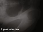
was a history of laminitis, and the owner reported episodes Figure 1: Ventrodorsal radiographic projection of the right
of intermittent upward fixation of the right patella. The pony acetabulum with complete dislocation of the right femoral head in
was found lame on pasture on the day of admission. a dorsal and caudal direction.
3. Clinical Findings
Upon presentation, the mare was in a good general condi-
tion and showed a severe right pelvic limb lameness (Grade
4 of 5 according to the scale of the American Association of
Equine Practitioners) with concurrent upward fixation of the
right patella. External rotation and adduction of the right
pelvic limb were observed with the right greater trochanter
more prominent and the right calcaneal tuber being more
proximal than its left counterpart. Physical examination of
the mare was normal, and no wounds could be detected. Based
on these findings, the differential diagnoses were a coxofe-
moral luxation or pelvic fracture involving the right acetabu- Figure 2: Ventrodorsal radiographic projection of the right
lum, and radiographs of the right coxofemoral joint were acetabulum after closed reduction of the coxofemoral luxation.
obtained. The mare was sedated with romifidine (0.04 mg/kg The right femoral head is seated in its correct position within the
intravenously (IV)) and L-methadone (0.05 mg/kg IV). Gen- acetabulum.
eral anaesthesia was induced with ketamine (2.5 mg/kg IV)
and diazepam (0.05 mg/kg IV), and anaesthesia was main- A full-body animal rescue and transportation sling
tained with isoflurane in oxygen and a continuous rate infusion (ARTS) was applied for the recovery [15]. Additionally, an
of romifidine (0.04 mg/kg/h IV). Radiographic examination Ehmer sling was placed on the right pelvic limb for the recov-
of the pelvis was subsequently performed with the pony in ery period and for the first 12 hours following closed reduc-
dorsal recumbency. tion. The following day, the Ehmer sling was removed, due
to a lack of compliance for the sling by the pony, but the pony
4. Diagnosis was maintained in the full-body ARTS. Furthermore, the
pony was crosstied, and a couloir of approximately 5 feet
A laterolateral and ventrodorsal radiographic projection width was made with large bales of wood shavings to keep
centred over the right acetabulum revealed complete disloca- the pony from moving from side to side with its rear end
tion of the right femoral head from the acetabular cavity in a (Figure 3). Within hours after removing the Ehmer sling,
dorsal and caudal direction (Figure 1). the pony showed an upward fixation of the right patella.
Therefore, a right-sided medial patellar ligament desmotomy
5. Treatment was performed under local anaesthesia and sedation.
Closed reduction of the coxofemoral luxation was per- 6. Further Case Management
formed while the mare was still anaesthetised. To achieve
this, the pony was left in dorsal recumbency and a rope Nonsteroidal anti-inflammatory drugs, flunixin meglumine
was placed around the pastern of the right pelvic limb to (1.1 mg/kg IV, once daily) for four consecutive days, and
apply distal traction on the extremity using a hoist. After meloxicam (0.6 mg/kg orally, once daily) were administered
several attempts, traction combined with external rotation for an additional five days. Analgesia was continued with
and adduction by the surgeon allowed a reduction of the firocoxib (0.1 mg/kg orally, once daily) for three additional
femoral head back into the acetabular cavity. Reduction weeks. The pony was supported in the ARTS net and
was completed by internal rotation of the limb. Laterolat- crosstied for six weeks. After these six weeks, the bales
eral and ventrodorsal radiographs confirmed the complete of wood shavings and crossties were removed, and the mare
reduction (Figure 2). was allowed to move around freely in the box. However,Case Reports in Veterinary Medicine 3
prior to submission of this case report, 22 months following
closed reduction of the coxofemoral joint luxation, and the
mare was still used regularly as a riding pony for children
without any observable residual lameness.
8. Discussion
In select cases, coxofemoral luxation can be successfully
treated by closed reduction and adequate immobilization in
the weeks following the closed reduction. A full-body animal
rescue and transportation sling and crosstying were well-
tolerated means of constraint to achieve adequate postopera-
tive immobilization in the described case.
Both the accessory ligament and the smaller ligament
capitis ossis femoris need to rupture for a luxation to occur
[7]. Interestingly, coxofemoral luxation may also be associ-
ated with upward fixation of the patella, especially in ponies
and miniature horses [2, 4, 16], and speculated to occur sec-
ondary to violent quadriceps femoris contractions caused by
forceful attempts to flex the limb locked in extension [4].
Alternatively, upward fixation of the patella can occur sec-
ondary to coxofemoral luxation when rotation of the limb
impedes the function of the rectus and biceps femoris muscles
in the so-called patellar release mechanism [16]. Based on
this information, it can be argued that medial patellar des-
motomy is an effective, adjunctive treatment for coxofemoral
luxation and should be routinely performed immediately
Figure 3: A crosstied Shetland pony mare in a full-body animal after open or closed reduction of a coxofemoral luxation to
rescue and transportation sling (ARTS) after closed reduction of a reduce the risk of reluxation, especially in animals that have
right coxofemoral luxation. a history of previous upward fixation of the patella. In the
present case, the upper fixation of the patella was released fol-
lowing closed reduction of the coxofemoral luxation but
the mare was kept in the ARTS to prevent her from lying reoccurred immediately after removing the Ehmer sling the
down. Because intermittent left-sided upward fixation of following day.
the patella had been observed, a left-sided medial patellar lig- A complete diagnostic radiographic evaluation of the
ament desmotomy was performed under local anaesthesia coxofemoral joint in dorsal recumbency under general anaes-
and sedation. thesia is recommended to confirm luxation [7, 17]. However,
Starting seven weeks after closed reduction, the pony was dynamic ultrasonography or standing lateral oblique radio-
walked out of the box stall and allowed to hand-graze daily. graphs of the pelvis, alone or in combination, can provide
After a total of eight weeks, the ARTS was removed and the diagnostic images when assessing a suspected coxofemoral
pony was allowed to lie down and move freely in the box luxation in small equids [18–20]. In the present case, gen-
stall. In week nine, after observing that the pony had laid eral anaesthesia for radiographic assessment of the highly
down and gotten back up on its feet without complications, suspected luxation was preferred so that immediate closed
the pony was discharged from the hospital with instructions reduction could be performed. Nonetheless, standing ultra-
for continued box rest and progressively increasing daily sonography was performed successfully and repeatedly in
hand walking for up to 30 minutes per day for the following the present case after closed reduction, to assess the posi-
two months. tion of the femoral head and to rule out a reluxation,
whenever the pony was reluctant to bear full weight on
7. Outcome the affected limb.
Closed reduction of coxofemoral luxation is rarely suc-
The owner and referring veterinarian provided weekly cessful and is commonly associated with a high incidence of
updates and videos of the pony. No complications were reluxation (up to 80%) [1, 2, 4, 8]. However, three isolated
reported in the two months following hospital discharge, cases of successful closed reduction have been reported in
and the pony was returned to regular pasture turnout four the veterinary literature [2, 4, 14]. The likely causes of the
months after the closed reduction. By six months after hospi- high rate of reluxation are accumulation of blood clots and
tal discharge, the pony was used again for pleasure riding by debris within the acetabular socket, damage to the capsular
young children without any signs of residual lameness, as muscles, and trapping of the fibrocartilage rim within the
assessed on video footage and on site by the referring veteri- acetabulum preventing proper seating of the femoral head
narian. The last follow-up information was obtained just [6, 21, 22]. Based on experiences in cattle, closed reduction4 Case Reports in Veterinary Medicine
is more likely to be successful if performed within 36 hours References
after luxation. Once this time window has passed, closed
reduction will likely be difficult to achieve, because of muscle [1] J. R. Field, R. McLaughlin, and M. Davies, “Surgical repair of
contraction and the formation of organised blood clots coxofemoral luxation in a miniature horse,” The Canadian
within the acetabulum [21, 22]. Therefore, a short time frame Veterinary Journal, vol. 33, no. 6, pp. 404-405, 1992.
between initial injury and closed reduction, as in the present [2] J. A. Malark, A. J. Nixon, M. A. Haughland, and M. P. Brown,
case, seems to be an important factor for success. “Equine coxofemoral luxations: 17 cases (1975-1990),” The
Cornell Veterinarian, vol. 82, no. 1, pp. 79–90, 1992.
A controlled and uneventful recovery from general
anaesthesia and adequate immobilization during the postre- [3] D. Platt, I. M. Wright, and J. E. Houlton, “Treatment of
duction period are critical factors for a successful outcome. chronic coxofemoral luxation in a Shetland pony by excision
arthroplasty of the femoral head: a case report,” The British
Reluxation after closed reduction is described to occur in
Veterinary Journal, vol. 146, no. 4, pp. 374–379, 1990.
80% of the cases during recovery from general anaesthesia
[4] P. D. Clegg and R. J. Butson, “Treatment of a coxofemoral lux-
or in the ensuing 3 days [2]. This was also the case in the first
ation secondary to upward fixation of the patella in a Shetland
attempt of closed reduction in a Shetland pony published in pony,” The Veterinary Record, vol. 138, no. 6, pp. 134–137,
the English veterinary literature [4]. In this original report, 1996.
Clegg and coworkers applied an Ehmer sling during recovery
[5] J. Frewein, K. H. Wille, and H. Wilkens, “Gelenkslehre -
and the first 4 days postoperatively. Likewise, an Ehmer sling Hüftgelenk,” in Lehrbuch der Anatomie der Haustiere, R.
was used in the case presented here, but the use of the sling Nickel, A. Schummer, and E. Seiferle, Eds., pp. 260-261, Paul
was restricted to the first 12 hours following general anaes- Parey, Berlin and Hamburg, Germany, 6th edition, 1992.
thesia. The Ehmer sling subsequently was removed because [6] J. M. Kuemmerle and A. E. Fürst, “Treatment of a coxofe-
the pony was constantly trying to free the affected limb from moral luxation in a pony using a prosthetic capsule tech-
the restrictive bandage. Besides the lack of compliance, as nique,” Veterinary Surgery, vol. 40, no. 5, pp. 631–635, 2011.
experienced in this case, another potential disadvantage asso- [7] K. E. Sullins and G. M. Baxter, “The femur and coxofemoral
ciated with placing an Ehmer sling is the increased risk of joint,” in Adams and Stashak's Lameness in Horses, G. M. Bax-
support limb laminitis. ter, Ed., pp. 814–832, Wiley-Blackwell, UK, 6th edition, 2011.
We speculate that preventing the pony from lying down [8] G. W. Trotter, J. A. Auer, W. Arden, and A. Parks, “Coxofe-
during the 2 months following reduction and avoiding sud- moral luxation in two foals wearing hindlimb casts,” Journal
den forces to act on the capsule of the affected coxofemoral of the American Veterinary Medical Association, vol. 189,
joint were not only essential in preventing reluxation but also no. 5, pp. 560-561, 1986.
allowed for a functional healing of the traumatized joint cap- [9] J. M. Garcia-Lopez, R. J. Boudrieau, and P. J. Provost, “Surgical
sule. The combined use of the ARTS and crossties on the repair of coxofemoral luxation in a horse,” Journal of the
pony in a well-confined space proved to be an effective American Veterinary Medical Association, vol. 219, no. 9,
method of immobilization, and the pony tolerated it well. pp. 1254–1258, 2001.
The fortunate combination of timely corrective intervention [10] N. Huggons, R. Andrea, B. Grant, and C. Duncan, “Total hip
and effective postreduction management allowed this pony arthroplasty in the horse: overview, technical considerations
to return to its prior function without any residual lameness. and case report,” Equine Veterinary Education, vol. 22,
no. 11, pp. 547–553, 2010.
[11] I. B. François, A. L. Thomas, and O. M. Lepage, “Treatment of
9. Conclusion coxofemoral luxation in a mature Welsh pony by femoral head
ostectomy: long-term outcome,” Equine Veterinary Education,
Closed reduction under general anaesthesia followed by vol. 29, no. 10, pp. 528–533, 2017.
medial patellar desmotomy and two months of immobiliza- [12] D. W. Richardson and K. F. Ortved, “Femur and pelvis,” in
tion using a commercially available ARTS combined with Equine Surgery, J. Auer, J. Stick, J. Kuemmerle, and T. Prange,
crossties can be a valid treatment approach for the treatment Eds., pp. 1777–1789, Elsevier, St. Louis, MO, USA, 5th edition,
of craniodorsal coxofemoral luxation in mature equids 2019.
weighing less than 200 kg. Provided that the collateral dam- [13] F. Toth, H. S. Adair, T. E. C. Holder, and J. Schumacher,
age to the coxofemoral articulation is limited and a prompt “Femoral head ostectomy to treat a donkey for coxofemoral
and properly executed closed reduction is performed, a full luxation,” Equine Veterinary Education, vol. 19, no. 9, pp. 478–
return to previous use is possible. 481, 2007.
[14] B. Nyack, M. J. Willard, J. Scott, and C. L. Padmore, “Non-sur-
gical repair of coxofemoral luxation in a quarter horse filly,”
Conflicts of Interest Equine Practice, vol. 4, pp. 11–14, 1982.
No conflicts of interest have been declared. [15] A. E. Fürst, R. Keller, M. Kummer et al., “Evaluation of a new
full-body animal rescue and transportation sling in horses: 181
horses (1998–2006),” Journal of Veterinary Emergency and
Acknowledgments Critical Care, vol. 18, no. 6, pp. 619–625, 2008.
[16] D. Bennett, J. R. Campbell, and J. R. Rawlinson, “Coxofemoral
We thank Dr. Dorian Bindler for the case referral and Dr. luxation complicated by upward fixation of the patella in the
Shannon Axiak Flammer for the thorough revision of the pony,” Equine Veterinary Journal, vol. 9, no. 4, pp. 192–194,
manuscript. 1977.Case Reports in Veterinary Medicine 5
[17] J. M. García-López, “Coxofemoral luxations in the horse: sur-
gical options and challenges,” Equine Veterinary Education,
vol. 22, no. 11, pp. 554–556, 2010.
[18] F. N. Amitrano, S. D. Gutierrez-Nibeyro, and S. K. Joslyn,
“Radiographic diagnosis of craniodorsal coxofemoral luxation
in standing equids,” Equine Veterinary Education, vol. 26,
no. 5, pp. 255–258, 2014.
[19] S. Brenner and M. B. Whitcomb, “Ultrasonographic diagnosis
of coxofemoral subluxation in horses,” Veterinary Radiology &
Ultrasound, vol. 50, no. 4, pp. 423–428, 2009.
[20] F. Geburek, A. K. Rötting, and P. M. Stadler, “Comparison of
the diagnostic value of ultrasonography and standing radiog-
raphy for pelvic–femoral disorders in horses,” Veterinary Sur-
gery, vol. 38, no. 3, pp. 310–317, 2009.
[21] P. D. Clegg and E. J. Comerford, “Coxofemoral luxation-how
does our knowledge of treatment in other species help us in
the horse?,” Equine Veterinary Education, vol. 19, no. 9,
pp. 482-483, 2007.
[22] N. G. Ducharme and S. S. Trostle, “Coxofemoral luxation/su-
bluxation,” in Farm Animal Surgery, S. L. Fubini and N. G.
Ducharme, Eds., pp. 346-347, Saunders, St. Louis, MO, USA,
1st edition, 2004.The Scientific
Zoology
International Journal of
Case Reports in Veterinary Medicine
Hindawi
Veterinary Medicine
Hindawi
Scientifica
Hindawi
International
Hindawi
World Journal
Hindawi Publishing Corporation
www.hindawi.com Volume 2018 www.hindawi.com Volume 2018 www.hindawi.com Volume 2018 www.hindawi.com Volume 2018 http://www.hindawi.com
www.hindawi.com Volume 2018
2013
Journal of
Veterinary Medicine
International Journal of
Hindawi
Microbiology
Hindawi
www.hindawi.com Volume 2018 www.hindawi.com Volume 2018
International Journal of International Journal of
Agronomy Ecology
Submit your manuscripts at
www.hindawi.com
Hindawi Hindawi
www.hindawi.com Volume 2018 www.hindawi.com Volume 2018
Applied &
International Journal of International Journal of Environmental Biochemistry BioMed
Genomics
Hindawi
Cell Biology
Hindawi
Soil Science
Hindawi Volume 2018
Research International
Hindawi
Research International
Hindawi
www.hindawi.com Volume 2018 www.hindawi.com Volume 2018 www.hindawi.com www.hindawi.com Volume 2018 www.hindawi.com Volume 2018
Advances in
Journal of
Parasitology Research
Hindawi
www.hindawi.com Volume 2018
Biotechnology
Research International
Hindawi
www.hindawi.com Volume 2018
Virolog y
Hindawi
www.hindawi.com Volume 2018
Psyche
Hindawi
www.hindawi.com Volume 2018
Archaea
Hindawi
www.hindawi.com Volume 2018You can also read


