CASE REPORT Primary infectious aortic aneurysm: a case series and review of the literature
←
→
Page content transcription
If your browser does not render page correctly, please read the page content below
ISSN 1677-7301 (Online)
CASE REPORT
Primary infectious aortic aneurysm: a case series and review of
the literature
Aneurisma infeccioso primário da aorta: série de casos e revisão da literatura
Fernanda Beatriz Araújo de Albuquerque1 , Matheus Oliveira Feijó1 , Jacob Hindrik Antunes Smit2 ,
Ricardo Bernardo da Silva3 , Adenauer Marinho de Oliveira Góes Junior1,2
Abstract
Infectious aneurysms, formerly known as mycotic aneurysms, are rare, most often involve the aorta in young patients,
and have a greater tendency to rupture than aneurysms of other etiologies. The most characteristic shape is saccular and
the most common etiologic agents are Staphylococcus sp. and Salmonella sp. There is scant and imprecise information
in the literature about correct nomenclature, diagnosis, and treatment. The authors present three cases in which
diagnostic and therapeutic procedures were documented. In addition to reporting this case series, the authors also
present a review of the subject, outlining pertinent diagnostic and therapeutic strategies.
Keywords: infectious aneurysm; aortic aneurysm; abdominal aortic aneurysm; ruptured aortic aneurysm.
Resumo
Aneurismas infecciosos, anteriormente chamados de aneurismas micóticos, são raros; acometem com maior frequência
a aorta de pacientes jovens e apresentam maior tendência à rotura do que aneurismas de outras etiologias. O formato
sacular é o mais característico, e os agentes etiológicos mais comuns são Staphylococcus sp e Salmonella sp. A literatura
fornece informações limitadas e imprecisas sobre a correta nomenclatura, diagnóstico e tratamento da doença. Os
autores reuniram três casos cujos procedimentos diagnósticos e terapêuticos foram documentados. Além de relatar
essa série de casos, realiza-se uma revisão sobre o tema, a fim de estabelecer estratégias diagnósticas e terapêuticas
pertinentes.
Palavras-chave: aneurisma infectado; aneurisma aórtico; aneurisma da aorta abdominal; aneurisma aórtico roto.
How to cite: Albuquerque FBA, Feijó MO, Smit JHA, Silva RB, Góes Junior AMO. Primary infectious aortic aneurysm:
a case series and review of the literature. J Vasc Bras. 2022;21:e20210206. https://doi.org/10.1590/1677-5449.202102062
1
Universidade Federal do Pará – UFPA, Belém, PA, Brasil
2
Centro Universitário do Estado do Pará – CESUPA, Belém, PA, Brasil
3
Santa Casa de Londrina, Londrina, PR, Brasil
Financial support: None.
Conflicts of interest: No conflicts of interest declared concerning the publication of this article.
Submitted: December 08, 2021. Accepted: May 31, 2022.
The study was carried out at Centro Universitário do Estado do Pará (CESUPA), Belém, PA, Brazil.
Copyright© 2022 The authors. This is an Open Access article distributed under the terms of the Creative Commons Attribution License, which
permits unrestricted use, distribution, and reproduction in any medium, provided the original work is properly cited.
Albuquerque et al. J Vasc Bras. 2022;21:e20210206. https://doi.org/10.1590/1677-5449.202102062 1/8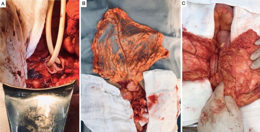
Primary infected aortic aneurysms
INTRODUCTION patient was using subcutaneous adalimumab injections.
He denied fever, weight loss, and using illicit drugs.
Infectious aneurysms, formerly known as mycotic Physical examination identified a painful pulsating
aneurysms, are rare, accounting for 1-3% of all mass with an audible murmur in the mesogastrium.
aneurysms.1,2 They most frequently affect the aorta2,3 in Hemoglobin and leukocytes were within normal
young men,4 the most characteristic shape is saccular,5 limits, but erythrocyte sedimentation rate (ESR) and
and they have a greater tendency to rupture than non- C-reactive protein (CRP) were elevated (22 and 12,
infectious aneurysms.6,7 The most common etiologic respectively). Angiotomography showed a fusiform
agents are Staphylococcus sp. and Salmonella sp.6-8 aneurysm of the infrarenal aorta, with a maximum
Infectious aneurysms may be associated with injection diameter of 3.6 cm and lobed outlines, surrounded
of illicit drugs,4,8 immunosuppression, or sepsis.1,4 by hypodense hematoma, with no signs of active
Diagnosis is challenging because of the low prevalence bleeding, but with interrupted parietal calcifications
and the nonspecific signs and symptoms.6-8 Fever, and penetrating ulcerations, in addition to an inaccurate
pulsating mass, and abdominal pains are described definition of the posterior aortic outline, suggestive of
classically, as are elevated inflammatory markers and
an infrarenal abdominal aortic aneurysm with signs of
positive blood cultures, but are not always present.3,9
contained rupture (Figure 1). The patient was admitted.
Treatment for this type of aneurysm must be initiated
Blood cultures were negative and an echocardiogram
rapidly, with antibiotic therapy (ideally guided by
showed no signs of endocarditis. Intravenous
cultures) and surgery.1,6-9 Few surgeons accumulate
ciprofloxacin and clindamycin were administered for
experience with their treatment and the literature
14 days. During laparotomy on the 15th day, there
contains imprecise information on nomenclature,
was no fetid odor or liquid collections, but there was
diagnostic criteria, and therapeutic strategies. This
significant fibrosis, thickening of the artery wall, and
article presents a series of three cases and highlights
adherence to adjacent tissues. Proximal control was
aspects of their clinical presentation, diagnosis, and
achieved by infrarenal clamping. Dissection of the
treatment, in addition to reviewing the literature on
distal neck was not feasible because of fibrosis. Distal
the subject to help to standardize the management of
control was obtained by intraluminal inflation of Foley
this uncommon, but extremely serious, disease. This
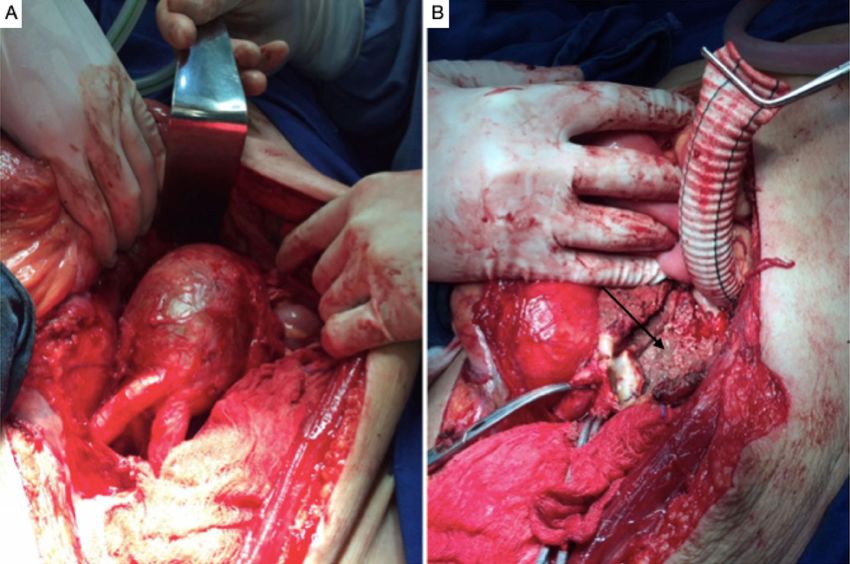
catheters in the common iliac arteries (Figure 2A).
case series has been analyzed and approved by the
After longitudinal arteriotomy and removal of
Ethics Committee at the originating institution, under
thrombi, contained rupture of the posterior aneurysm
decision number 48950921.5.0000.5169.
wall was confirmed. The aorta was resected to the
maximum extent possible, including its posterior
CASE DESCRIPTIONS wall, and the stumps of the lumbar arteries were
sutured. An aorto-aortic Dacron graft was used for
Case 1 reconstruction, wrapped in a vascularized pedicle of
A 44-year-old man was referred for follow-up the greater omentum (Figures 2B, 2C, and 3). Cultures
after deep venous thrombosis (DVT). He presented of the thrombus and aorta wall were negative. The
with pain, erythema, and edema involving both legs. intravenous antibiotic therapy was maintained up to
He had been treated for erysipelas with penicillin discharge on the 14th postoperative (PO) day. Oral
G benzathine and 4 months later suffered a similar ciprofloxacin and clindamycin were prescribed for a
episode, with more severe symptoms, restricted to further 6 weeks and the adalimumab was withdrawn.
the left lower limb, including edema and clubbing. The patient has been in outpatients follow-up for 6
He was prescribed another course of penicillin and months.
a Doppler ultrasonography examination confirmed
DVT of the common, superficial, and deep femoral Case 2
veins, for which he was prescribed rivaroxaban for A 62-year-old man was admitted for recurrent
6 months. This treatment had been concluded by the abdominal pains after outpatient consultations. He
time the patient was seen by the vascular surgeon. reported severe gastroenteritis, treated with antibiotics
However, he still complained of abdominal pains. 4 months previously. He had diffuse abdominal pain,
During the second episode of erysipelas (8 months more intense in the mesogastrium; angiotomography
before the reference consultation), he reported an confirmed a juxtarenal aortic aneurysm with a diameter
episode of acute and intense lumbar and abdominal of 5.3 cm and signs of periaortic inflammation. He
pain, which improved after a few days, persisting at a had elevated CRP (34 mg/L) and leukocytosis at
lower intensity. This pain was attributed to ankylosing 17,000/µL. Blood cultures were negative. After 7
spondylitis (diagnosed 13 years previously), for which the days on intravenous ciprofloxacin and clindamycin,
Albuquerque et al. J Vasc Bras. 2022;21:e20210206. https://doi.org/10.1590/1677-5449.202102062 2/8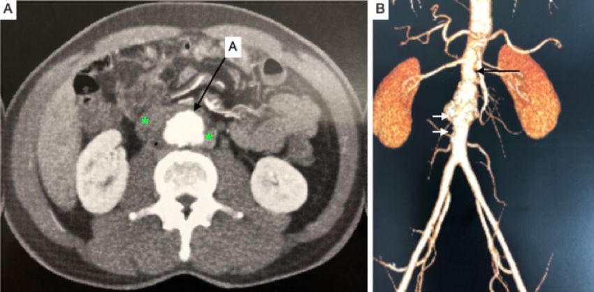
Primary infected aortic aneurysms

Figure 1. Computed tomography with intravenous contrast. (A) Axial slice; A: aorta; the asterisks mark an image compatible with
periaortic collection/mass. (B) Reconstruction with the maximum intensity projection (MIP) technique. Observe the irregular
outlines not just of the aneurysm, but also of the aortic segments proximal (black arrow) and distal (white arrows) of the aneurysm.

Figure 2. Intraoperative images. (A) Temporary hemostasis with endoluminal inflation of Foley catheters in the iliac arteries; (B)
Vascularized pedicle of the great omentum; (C) Pedicle of the great omentum in position to be wrapped around the tubular
Dacron graft.

the patient was operated. The following interoperative                   therapy was changed to piperacillin with tazobactam
findings were observed: intestinal loops with signs                      and vancomycin. The patient died on the ninth PO day.
of inflammation and a fetid odor after the aneurysm
sac was opened. Proximal supraceliac clamping was                        Case 3
performed prior to reconstruction with an 18 mm                             A 59-year-old man with a prior history of admissions
Dacron aorto-aortic graft (Figure 4). During the                         for psychiatric conditions was admitted because of
postoperative period, the patient developed renal                        abdominal pains. On physical examination, he reported
dysfunction and nosocomial pneumonia; an aorta wall                      pain in response to deep palpation of the mesogastrium,
culture revealed Escherichia coli, and the antibiotic                    with a pulsating mass. Angiotomography confirmed

Albuquerque et al. J Vasc Bras. 2022;21:e20210206. https://doi.org/10.1590/1677-5449.202102062                                    3/8
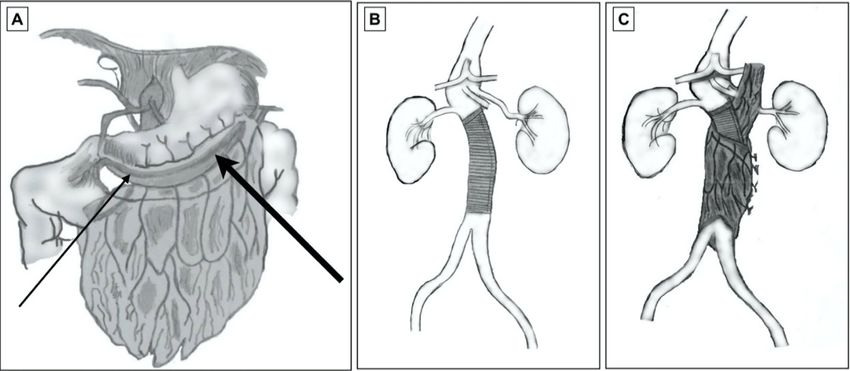
Primary infected aortic aneurysms

Figure 3. Diagrams illustrating the tubular Dacron graft. (A) Omentum, right gastroepiploic artery (thin arrow) and left gastroepiploic
artery (thick arrow); (B) Abdominal aorta after implantation of the Dacron graft. C) Omentum wrapped around the abdominal
aortic graft.

Figure 4. Intraoperative images. (A) Large aneurysm of the infrarenal aorta. (B) Tubular Dacron graft. The arrow indicates where
the posterior wall of the aorta was resected. The proximal anastomosis is already concluded.

a 5.7 cm juxtarenal saccular aneurysm and periaortic                     teicoplanin and piperacillin with tazobactam. After 7
collection (Figure 5). His ESR was normal, CRP was                       days, by when blood cultures were already negative,
elevated (27 mg/L), and he had leukocytosis (21,000/µL).                 the patient underwent surgical treatment. Supraceliac
Blood cultures were positive for coagulase-negative                      clamping was needed to achieve proximal control
Staphylococcus and antibiotic therapy was started with                   of the aorta. When the aneurysm sac was opened,

Albuquerque et al. J Vasc Bras. 2022;21:e20210206. https://doi.org/10.1590/1677-5449.202102062                                     4/8
Primary infected aortic aneurysms
DISCUSSION
The term mycotic aneurysm was coined by William
Osler in 1885,10 because of the mushroom-like
appearance of the aneurysmal lesions.10,11 However,
the term induces the erroneous idea that etiology is
fungal.5,11 The name infectious aortitis could denote
several different conditions, such as aortoenteric
fistulas and infections after surgical manipulation,
including infections of aortic grafts.5,8,12-14 The best
term is primary infectious aneurysm, which denotes a
dilation secondary to infection of the aorta wall.2,5,11,14,15
The condition is rare,1,5-7,9,16 but highly lethal.6-7,9,16
In the West, infectious aneurysms account for
no more than 3% of all aneurysms1,2,5,6,8,9,11,14,16-18
Figure 5. Computed tomography with intravenous contrast, and tend to occur in men8,14 who are younger than
axial slice. (A) Aorta; the asterisks mark an image compatible those who develop degenerative aneurysms.4,7,11
with periaortic collection/mass.
These aneurysms can grow rapidly and there is a
high risk of rupture,2,4,8,9,12,16,17 with mortality rates
as high as 60%.2 The following etiologic agents
have been reported: gram-positive bacteria such as
Staphylococcus sp.,2,4,6-8,11,14,19 Enterococcus sp.,11
Streptococcus sp.2,8,11,18,19 and Clostridium sp.,11 and
gram-negative bacteria such as Salmonella sp.,2-4,6-8,11,14
Pasteurella sp.,7 Brucella sp.,20 Coxiella burnetti,11 and
Pseudomonas aeruginosa,2,19 in addition to fungi.5,11,18
The most frequently identified agents are members of
the Staphylococcus and Salmonella genera.6,12,18 The
source of infection is not identified in 1/3 of cases
and the etiologic agent is not established in 20-40%
of cases.11,14,15 Infectious aneurysms can be caused by
contiguity21 or, frequently, by bacteremia.1,2,5,11,14,17,19
After attaching to the artery wall, the microorganism
provokes acute inflammation with neutrophilic
infiltration, leading to activation of enzymes and
weakening of the artery wall,8 resulting in suppuration
and arterial dilatation.1,2,5,9,11,17,20,22 Aortic involvement
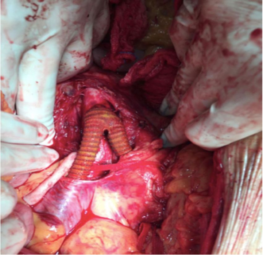
Figure 6. Intraoperative images. Aortoiliac graft with bifurcated
is more common because of the more pronounced vasa
silver-impregnated Dacron prosthesis.
vasorum of larger caliber arteries, which facilitates
bacterial colonization.8,11,14,19
Early diagnosis is key to therapeutic success.6,8,9,12,16
an intense odor was noted and a contained rupture
Classically, there is fever,1,3,4,6-8,14,18 abdominal/lumbar
of the posterior wall was observed. Reconstruction pains, and a pulsating mass1,4,6-8,14,18 in the presence
was performed with an 18x9 mm bifurcated Dacron of an infectious condition (osteomyelitis, urinary
aortoiliac graft wrapped in a vascularized pedicle of infections, tuberculosis, gastroenteritis, and soft tissue
the greater omentum (Figure 6). Antibiotic therapy infections)1,4,6,11,14 and/or immunosuppression caused by
was maintained for 30 days postoperatively. One diseases or medications (cancer, renal failure requiring
year after discharge, the patient underwent surgery dialysis, HIV, diabetes, corticosteroids). However,
for bilateral degenerative aneurysms of the common asymptomatic cases can also occur.4,14 All three of the
femoral artery (infectious etiology was ruled out). cases reported above had abdominal pains; cases 1 and
The patient is in outpatients follow-up 2 years after 2 had a history of an infectious condition, and there was
treatment of the infected aortic aneurysm. also use of immunosuppressant medication in case 1.
One feature that the three cases described in this Laboratory tests generally show Leukocytosis1,6,8,11,12,14
series all have in common is that angiotomography and inflammatory markers such as elevated ESR and
did not show periaortic gas. CRP,6,12 in addition to positive blood cultures.1,3,4,6,7,16
Albuquerque et al. J Vasc Bras. 2022;21:e20210206. https://doi.org/10.1590/1677-5449.202102062 5/8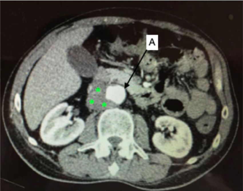
Primary infected aortic aneurysms

However, blood cultures can be negative even during                      treatment of infectious aneurysms of the thoracic
the acute phase in up to 50% of cases,14,23 particularly                 aorta was described in 1998.25 Since then, several
in patients who are being given antibiotics, which is                    other reports have been published.1,3,6,8,12,15,21,23 This
common.8,14,15 Two of the cases in the present series                    strategy is less invasive because it avoids dissections
had positive blood cultures. Angiotomography will                        in an anatomy compromised by infection and the
often show parietal irregularities, saccular dilatations,                aorta is not clamped, which in theory benefits patients
changes suggestive of inflammation, perivascular liquid                  with a high surgical risk.1,7,11 However, implantation
collections/masses, periaortic gas buildup, signs of                     of endoprostheses in infected tissues increases the
free or contained rupture, and rapid progression over                    incidence of complications, including endoprosthesis
a series of examinations.1,4,5,8,9,11,12,14,17,24 Hepatic and            infection,1,6,9,23 and also of malpositioning and consequent
splenic abscesses should be sought on tomography.9,21                    endoleaks with the potential for aneurysm rupture.4,8
Echocardiography is a convenient method for investigating                Other undesirable outcomes include sepsis, fistulas,8
endocarditis.4 Positron emission tomography/computed                     and expansion of the aneurysm.16 It is undeniable that
tomography scanning (PET-CT SCAN) is a tool with                         endovascular treatment does not enable the removal
high diagnostic accuracy and very good sensitivity,                      of infected tissues9,12,16,23 and that there is a high risk
but its specificity is affected by false-positive results                of merely delaying open surgery, adding the need for
in cases of inflammatory aneurysms and arterites.24                      explantation of the endoprosthesis to an operation
   There is no consensus on how to define the primary                    that is already highly complex.
infectious etiology of these aneurysms.5,15,17 It is                        Conventional surgery is associated with morbidity
suggested that diagnosis is based on the combination                     and mortality of up to 44%12 and remains the gold
of clinical status, laboratory tests, and tomographic                    standard11,12,16,22 because, although recovery is
findings1,6,12,17,21 (Table 1). It is essential to be clear              slower, reintervention rates are lower.3,6 In addition
that this diagnosis can be made in the absence of                        to resection of the aneurysm,9,12,13 surgery should
fever and positive blood cultures.1,3,8,9,15,16,23 After                 involve extensive retroperitoneal debridement,
drawing blood for cultures, antibiotic therapy for                       circumferential aortic resection,9,13,16,22,23 and in situ
Staphylococcus sp. (vancomycin) and antibiotic therapy                   or extra-anatomic revascularization with prosthetic
should be initiated for Salmonella sp. (quinolones or                    grafts or allografts.1,9,11-13,16,22 Use of an extra-anatomic
third-generation cephalosporins),1,4,11,12,15,16,23 to be                bypass avoids grafting in an infected field; however,
changed or supplemented with antifungals, depending                      rupture of the stump of the aorta, lower limb amputation,
on the results. There is no consensus on the duration                    or reinfection can occur.8,13 In turn, with in situ
of treatment.6-8,11,19 The majority of authors suggest                   revascularization, using a graft impregnated with silver
preoperative intravenous antibiotic therapy for 1 to 6                   or antibiotics,13 anastomosis in the infected bed involves
weeks.4,6,12 Clinical treatment alone, with antibiotics,                 the risk of dehiscence and pseudoaneurysm formation.1
                                                                         There are insufficient data to compare complications
is associated with mortality exceeding 80%.13,14,23
                                                                         associated with in situ and extra-anatomic grafts, but
   Surgical treatment can be with endovascular procedures
                                                                         they are more common with extra-anatomic grafts.13,22
or conventional surgery.1,3,6,7,9,14,15,21,23 Endovascular
                                                                         Studies report mortality of 5-49% for in situ grafts vs.
                                                                         24-50% for extra-anatomic grafts, while infectious
Table 1. Criteria suggestive of infectious aneurysm etiology.            complications occur in approximately 20% of cases
Clinical presentation Abdominal/lumbar pains                             with both strategies.11
                      Fever                                                 Dissection in the midst of thickened/adherent tissues
                      Sepsis/shock                                       increases the risk of bleeding and iatrogenic injuries,
Laboratory            Elevated C-reactive protein                        primarily involving the vena cava and ureters. Use
                      Elevated leukocytes                                of double J stents in advance can help to identify the
                      Positive blood/aortic tissue culture               ureters within the thickened retroperitoneal space,21,22
Computed              Saccular/multilobed outlines                       as was done in case 1. If dissection of the distal
tomography            Periaortic gas                                     neck is not possible, clamping can be substituted
                      Amorphous periaortic mass/collection               by endovascular occlusion of the iliac arteries with
                      Rapid expansion (days)                             Foley catheters,1,23 as was done in cases 1 and 3. The
                      Rupture                                            prosthesis can be isolated from adjacent tissues by
                      Location in an atypical aortic segment             wrapping it with a vascularized pedicle of the greater
                      (for example, paravisceral)                        omentum2,11,22,23 as in cases 1 and 3; vascularized
                      Multiple aneurysms                                 omentum also improves the delivery of antibiotics
Source: compiled by the authors, 2021.                                   to the prosthesis.2,11,22 There are also descriptions

Albuquerque et al. J Vasc Bras. 2022;21:e20210206. https://doi.org/10.1590/1677-5449.202102062                                       6/8
Primary infected aortic aneurysms
of cryopreserved cadaveric aorta (not available in 2016;63(6):1638-46. http://dx.doi.org/10.1016/j.jvs.2016.01.031.
Brazil) in reconstruction using grafts made from both PMid:26951998.
femoral veins.11,13,22 Technical details that improve the 5. Sörelius K, di Summa PG. On the diagnosis of mycotic aortic
aneurysms. Clin Med Insights Cardiol. 2018;12:1179546818759678.
results of surgery include the use of double J stents to
http://dx.doi.org/10.1177/1179546818759678. PMid:29497343.
identify the ureters, preparation of the colon to reduce
6. Guo Y, Bai Y, Yang C, Wang P, Gu L. Mycotic aneurysm due
the need to displace loops out of the cavity, central to Salmonella species: clinical experiences and review of the
venous access, invasive blood pressure monitoring, literature. Braz J Med Biol Res. 2018;51(9):e6864. http://dx.doi.
use of an 8F angiographic introducer in the internal org/10.1590/1414-431x20186864. PMid:29947649.
jugular to enable rapid infusion of blood products, 7. Kano Y, Takamatsu A, Honda H. Mycotic aneurysm due to
fluid balance positive by at least 1,000 mL before Pasteurella multocida. QJM. 2020;113(9):667-9. PMid:32016425.
the conclusion of the procedure, and heating with a 8. Deipolyi AR, Czaplicki CD, Oklu R. Inflammatory and infectious
aortic diseases. Cardiovasc Diagn Ther. 2018;8(Suppl 1):S61-70.
thermal blanket. There is no consensus on the duration
http://dx.doi.org/10.21037/cdt.2017.09.03. PMid:29850419.
of postoperative antibiotic therapy.8 Some authors
9. Zeng Z, Li Z, Zhao Y, et al. Endovascular repair combined with
recommend 6 weeks,6,15,18,22 while other suggest 3 to 6 staged drainage for the treatment of infectious aortic aneurysm:
months,4,6,7,11,12,15 or even lifelong antibiotic therapy.6,12,22 a case report. BMC Cardiovasc Disord. 2020;20(1):406. http://
Rare diseases and complex treatment demand dx.doi.org/10.1186/s12872-020-01694-9. PMid:32894058.
sharing of information; discussion with colleagues who 10. Osler W. The gulstonian lectures, on malignant endocarditis. Br Med
are experts in aortic surgery and sharing experiences J. 1885;1(1262):467-70. http://dx.doi.org/10.1136/bmj.1.1262.467.
PMid:20751186.
in groups that practice collective intelligence26 are
important to increase the likelihood of success. 11. Wanhainen A, Verzini F, Van Herzeele I, et al. Editor’s Choice -
European Society for Vascular Surgery (ESVS) 2019 Clinical Practice
Infectious etiology should always be considered Guidelines on the Management of Abdominal Aorto-iliac Artery
when faced with fever and abdominal/lumbar pains Aneurysms. Eur J Vasc Endovasc Surg. 2019;57(1):8-93. http://
with a pulsating mass, particularly in the presence dx.doi.org/10.1016/j.ejvs.2018.09.020. PMid:30528142.
of a confirmed infection or immunosuppression 12. Zhu C, Zhao J, Huang B, Yuan D, Yang Y, Wang T. Long‐term
caused by diseases/medications and if leukocytes and outcome of endovascular aortic repair for mycotic abdominal
aortic aneurysm. ANZ J Surg. 2020;90(7-8):1376-80. http://dx.doi.
inflammatory markers are elevated. Blood cultures
org/10.1111/ans.16122. PMid:32648327.
are often negative. Suggestive angiotomographic
13. Berchiolli R, Mocellin DM, Marconi M, et al. Ruptured mycotic
images include saccular dilatations, perivascular aneurysm after intravesical instillation for bladder tumor. Ann
collections, and contained ruptures. Waiting for Vasc Surg. 2019;59:310.e7-310.e11. http://dx.doi.org/10.1016/j.
“classic” presentations and positive blood cultures avsg.2018.12.100. PMid:30802589.
before initiating the correct treatment can compromise 14. Wilson WR, Bower TC, Creager MA, et al. Vascular Graft
the patient’s prognosis. Infections, Mycotic Aneurysms, and Endovascular Infections:
a scientific statement from the American Heart Association.
For postoperative control, it is recommended
Circulation. 2016;134(20):e412-e460. http://dx.doi.org/10.1161/
that computed tomography angiographies should be CIR.0000000000000457. PMid:27737955.
conducted at 1 and 6 months and annually thereafter, to 15. Dang Q, Van Eps RG, Wever JJ, et al. Nationwide study of the
check for complications and a need for reintervention.12 treatment of mycotic abdominal aortic aneurysms comparing
Limitations of this series include the small number open and endovascular repair in The Netherlands. J Vasc Surg.
of cases and the lack of documentation of imaging 2020;72(2):531-40. http://dx.doi.org/10.1016/j.jvs.2019.09.060.
PMid:32061482.
exams conducted for postoperative follow-up of the
16. Sörelius K, Wanhainen A, Furebring M, et al. Nationwide study of
patients.
the treatment of mycotic abdominal aortic aneurysms comparing
open and endovascular repair. Circulation. 2016;134(23):1822-
REFERENCES 32. http://dx.doi.org/10.1161/CIRCULATIONAHA.116.024021.
PMid:27799273.
1. Hurtado DFG, Neira JCH, Atala SH, Lawrence PT. Manejo de un
17. Wang TKM, Griffin B, Cremer P, et al. Diagnostic utility of CT and
aneurisma infeccioso. Rev Cir. 2019;71(5):446-9.
MRI for Mycotic Aneurysms: a meta-analysis. AJR Am J Roentgenol.
2. Dsouza R, Kota AA, Jain S, Agarwal S. Mycotic abdominal aortic 2020;215(5):1257-66. http://dx.doi.org/10.2214/AJR.19.22722.
aneurysm complicated by infective spondylitis due to Pseudomonas PMid:32930605.
aeruginosa. BMJ Case Rep. 2020;13(2):1-3. http://dx.doi.org/10.1136/ 18. Watanabe N, Koyama S, Tabira M, et al. Infected aortic aneurysm
bcr-2019-233461. caused by Streptococcus pyogenes: a case report. J Infect Chemother.
3. Nagrodzki J, Sharrocks KE, Wong VK, Carmichael AJ. A mycotic 2021;27(4):647-9. http://dx.doi.org/10.1016/j.jiac.2020.11.008.
aneurysm related to Salmonella Rissen infection: a case report. PMid:33277175.
BMC Infect Dis. 2020;20(1):97. http://dx.doi.org/10.1186/s12879- 19. Matsuo T, Mori N, Mizuno A, et al. Infected aortic aneurysm
020-4819-0. PMid:32005105. caused by Helicobacter cinaedi: case series and systematic review
4. Kordzadeh A, Watson J, Panayiotopolous YP. Mycotic aneurysm of the literature. BMC Infect Dis. 2020;20(1):854. http://dx.doi.
of the superior and inferior mesenteric artery. J Vasc Surg. org/10.1186/s12879-020-05582-7. PMid:33203370.
Albuquerque et al. J Vasc Bras. 2022;21:e20210206. https://doi.org/10.1590/1677-5449.202102062 7/8Primary infected aortic aneurysms
20. Alhaizaey A, Alassiri M, Alghamdi M, Alsharani M. Mycotic Correspondence
aortic aneurysm due to brucellosis. J Vasc Surg Cases Innov Adenauer Marinho de Oliveira Góes Junior
Tech. 2016;2(2):50-2. http://dx.doi.org/10.1016/j.jvsc.2016.03.009. R. Domingos Marreiros, 307, apartamento 802
PMid:31193364. CEP 66055-210 - Belém (PA), Brasil
Tel.: +55 (91) 98127-9656
21. Patel AP, Cantos A, Butani D. Mycotic Aneurysm of the Hepatic E-mail: adenauerjunior@gmail.com
Artery: a case report and its management. J Clin Imaging Sci.
2020;10:41. http://dx.doi.org/10.25259/JCIS_89_2020. PMid:32754376.
Author information
22. Tshomba Y, Sica S, Minelli F, et al. Management of mycotic aorto- FBAA and MOF - Medical students, Universidade Federal do Pará
iliac aneurysms: a 30-year monocentric experience. Eur Rev Med (UFPA).
Pharmacol Sci. 2020;24(6):3274-81. PMid:32271445. JHAS - MD, Medicina, Faculdade de Medicina, Centro Universitário
23. Kazuno K, Kinoshita H, Hori M, et al. Endovascular treatment do Estado do Pará (CESUPA).
for mycotic aneurysm using pyoktanin- applied devices. Cvir RBS - Full member, SBACV; Assistant professor, Pontifícia
Universidade Católica do Paraná (PUCPR); Chief of Serviço de
Endovascular. 2020;3(1):1-8. http://dx.doi.org/10.1186/s42155-
Cirurgia Vascular, Santa Casa de Londrina.
020-00151-0. PMid:32886250.
AMOGJ - Full member, SBACV; Adjunct professor, Faculdade
24. Husmann L, Huellner MW, Ledergerber B, et al. Diagnostic de Medicina, Universidade Federal do Pará (UFPA) and Centro
accuracy of PET/CT and contrast enhanced CT in patients with Universitário do Estado do Pará (CESUPA).
suspected infected aortic aneurysms. Eur J Vasc Endovasc Surg.
2020;59(6):972-81. http://dx.doi.org/10.1016/j.ejvs.2020.01.032. Author contributions
PMid:32340877. Conception and design: FBAA, RBS, MOF and AMOGJ
25. Semba CP, Sakai T, Slonim SM, et al. Mycotic aneurysms of the Analysis and interpretation: FBAA, MOF and AMOGJ
thoracic aorta: repair with use of endovascular stent-grafts. J Vasc Data collection: AMOGJ and RBS
Writing the article: FBAA, MOF and JHAS
Interv Radiol. 1998;9(1 Pt 1):33-40. http://dx.doi.org/10.1016/
Critical revision of the article: AMOGJ, RBS and JHAS
S1051-0443(98)70479-8. PMid:9468393.
Final approval of the article*: AMOGJ and RBS
26. Erzinger FL, de Araujo WJB, Ordinola AAM, et al. Vascular Forum: Statistical analysis: N/A.
collective intelligence in the resolution of vascular clinical cases. Overall responsibility: AMOGJ
J Vasc Bras. 2018;17(3):193-200. http://dx.doi.org/10.1590/1677-
5449.005018. PMid:30643504. *All authors have read and approved of the final version of the article
submitted to J Vasc Bras.
Albuquerque et al. J Vasc Bras. 2022;21:e20210206. https://doi.org/10.1590/1677-5449.202102062 8/8You can also read


