Case Report Comminuted Trapezium Fracture: Case Presentation and Review of Surgical Fixation Techniques
←
→
Page content transcription
If your browser does not render page correctly, please read the page content below
Hindawi
Case Reports in Orthopedics
Volume 2021, Article ID 5532713, 4 pages
https://doi.org/10.1155/2021/5532713

Case Report
Comminuted Trapezium Fracture: Case Presentation and
Review of Surgical Fixation Techniques

                                           1                                             2
          Amanda F. Spielman                   and Sriram Sankaranarayanan
          1
              Miller School of Medicine, University of Miami, 1600, NW 10th Avenue, Miami, Florida, USA 33136
          2
              Department of Orthopedic Surgery, NYU Langone Health, Woodhull Hospital Center, New York City, New York, USA 11206

          Correspondence should be addressed to Sriram Sankaranarayanan; sriram1812@gmail.com

          Received 9 February 2021; Revised 30 April 2021; Accepted 18 May 2021; Published 28 May 2021

          Academic Editor: Hiroshi Takahashi

          Copyright © 2021 Amanda F. Spielman and Sriram Sankaranarayanan. This is an open access article distributed under the Creative
          Commons Attribution License, which permits unrestricted use, distribution, and reproduction in any medium, provided the
          original work is properly cited.

          We report a case of a 28-year-old male who sustained a comminuted trapezium fracture with carpometacarpal subluxation of the
          right hand. Treatment with internal fixation with a headless compression screw resulted in excellent outcomes.

1. Introduction                                                       1.1. Statement of Institutional Review Board. As no patient
                                                                      information was divulged in the presentation of the case,
Trapezium fractures are rare injuries occurring in less than 4        no IRB approval was required per our institutional policy.
percent of all hand fractures [1–5]. The most common types
of trapezium fractures include body and ridge avulsion frac-
tures. Body fractures can be a vertical split or a comminuted         2. Case Presentation
fracture.
     The trapezium is palpable dorsally at the base of the            A 28-year-old male with no significant medical history pre-
thumb or distal portion of the snuffbox as well as at the base         sented to our emergency room five days after sustaining a
of the thenar eminence. The most common presentation of               right-hand injury from a fall on an outstretched hand while
fracture is pain and tenderness at the base of the first meta-         playing basketball. On examination, there was swelling and
carpal bone and snuffbox [6]. Patients may present with pain           tenderness on palpation at the base of the right thumb. Right
with wrist flexion with resistance due to the position of the          hand radiographs (Figure 1) and CT with 3D reconstruction
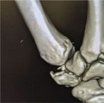
flexor carpal radialis [7]. The trapezium is an important              (Figure 2) showed a comminuted trapezium fracture.
component aiding in the versatile range of motion of the                  The patient was taken to the operating room seven days
thumb.                                                                after his initial injury. A dorsal radial approach was used to
     Early detection of trapezium fractures can help prevent          expose the trapezium protecting the radial sensory nerves,
permanent impairment such as chronic pain, joint stiffness,            radial arteries and EPL and EPB tendons. After exposing
and reduced grip strength [4, 5]. There are limited reports           the trapezium, we reduced the fracture with a dental pick
of trapezium fractures and no consensus on the management             and provisional Kirschner wires. C arm was utilized through-
of these fractures. While nondisplaced fractures may be               out the procedure to confirm fracture and joint reduction as
treated with immobilization, displaced fractures often                well as to appropriately size the screw. A 2 mm headless com-
require surgical intervention [8].                                    pression screw (HCS, Synthes Inc, West Chester, PA, USA)
     We present a case of a 28-year-old male who sustained a          was utilized to definitively fix the fracture (Figure 3).
comminuted trapezium fracture with carpometacarpal                        Intraoperatively, there was excellent stability of the
subluxation of the right hand.                                        fracture and restoration of the articular surface.
2 Case Reports in Orthopedics
Figure 3: Fixation with 2 mm headless screw demonstrating
excellent fixation of fracture and congruity of the carpometacarpal
articulation.
ture, and a CT scan can be obtained in case of a suspected tra-
Figure 1: Plan radiograph at presentation demonstrating trapezium pezium fracture. A CT scan delineates the fracture pattern
fracture with associated carpometacarpal subluxation. and aids in surgical planning [3]. Magnetic resonance imag-
ing (MRI) is an option to identify occult trapezium fractures
and bone bruising; however, it is used infrequently [10].
The most common types of trapezium fracture include a
vertical split pattern or a comminuted pattern. Often there
may be an overlap between these two types. The Walker Clas-
sification describes five types of trapezium fractures based on
pattern and joint involvement: I—horizontal, IIa—radial tuber-
osity through CMC joint, IIb—radial tuberosity though sca-
photrapezial joint, III—ulnar tuberosity, IV—vertical, and
V—comminuted fracture [11]. Trapezium fractures may be
seen in association with Bennet’s fracture of the metacarpal [7].
While there is a paucity of literature on the appropriate
management of trapezium fractures, a few case series have
Figure 2: CT with 3D reconstruction detailing trapezium fracture. reported different treatment options for these injuries includ-
ing nonoperative management. Authors have advocated dif-
ferent surgical techniques for management of comminuted
The patient’s joint was immobilized with a thumb spica trapezium fractures [12–14]. The different techniques
splint for two weeks. Unrestricted range of motion exercises include the use of K wires, cross pinning of the first metacar-
as well as occupational therapy were initiated two weeks fol- pal to the second metacarpal, headless compression screws,
lowing surgery. Three months after surgery, repeated X-rays or plates for fracture fixation. Bone grafts have been used
showed a healed trapezium fracture (Figure 4). During this for greater defects. These case reports have demonstrated
time, his DASH score was 10 and his SANE score [9] was 95. good to excellent outcomes with almost all patients regaining
The patient demonstrated full range of motion of the pain-free and useful range of motion [7, 11, 15–17]. While
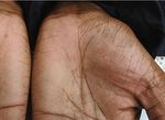
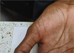
thumb (Figure 5) and returned to all previous activities. there are a few reports on the use of K wire for fixation of
these fractures, there is no clear consensus exists on whether
3. Discussion open or closed pinning is preferred.
Our case describes a patient with a comminuted trape-
Trapezium fractures are rare among hand fractures. A high zium fracture with carpometacarpal subluxation. We
degree of suspicion for the injuries aids in their diagnosis. hypothesize that the comminuted trapezium fracture was
AP wrist radiographs can identify these fractures. A pronated caused by an axial loading of the trapezium by the first meta-
view can allow for better visualization of the trapezio meta- carpal during the fall. A 3D CT analysis allowed for visualiza-
carpal articulation and identify displacements. However, tion of the complex fracture and aided in surgical planning.
there are reports of missed trapezium fractures in the litera- As there was significant displacement of the fracture alongCase Reports in Orthopedics 3
Figure 4: At 3 months follow-up, healing of the fracture demonstrated.
Figure 5: At 3 months follow-up, full range of motion of thumb demonstrated.
with a carpometacarpal subluxation, a decision was made to used to evaluate a patient’s rating of current function of the
perform an open reduction and internal fixation of the frac- affected hand in relation to normal. The assessment is a sin-
ture. We elected to use a headless compression screw over gle patient-reported value from 0–100 (100 being normal
K wires as we believed that we would be able to begin early function) [9]. The SANE score has been validated as a quick
range of motion in our patient with the use of headless and useful tool to obtain patient outcomes in hand surgery
compression screw. [9]. The patient had a near normal return of his hand
At follow-up, the patient demonstrated excellent out- function as seen by his self-reported SANE score of 95 as well
comes and use of his thumb. His DASH score was 10, and as his DASH score.
his SANE score was 95. The Single Assessment Numeric The novelty of this case report lies in that trapezium frac-
Evaluation (SANE) is a patient reported single outcome score tures are rare injuries of the hand. There are no clear4 Case Reports in Orthopedics
guidelines on the best management strategy for these frac- [11] J. L. Walker, T. L. Greene, and P. A. Lunseth, “Fractures of the
tures. We believed that an adequate restoration of the articu- body of the trapezium,” Journal of Orthopaedic Trauma, vol. 2,
lar congruity of the trapezium was important for the long- no. 1, pp. 22–28, 1988.
term hand function in a young patient, such as the one pre- [12] R. J. Foster, “Treatment of Bennett, Rolando, and vertical
sented in our case report. Hence, we elected to perform an intraarticular trapezial fractures,” Clinical Orthopaedics and
open reduction and internal fixation of the trapezium frac- Related Research, vol. 214, pp. 121–129, 1987.
ture with a headless compression screw. The case report [13] J. Pointu, “Fracture of the trapezuim: mechanism, pathology
demonstrates the excellent outcome that can be obtained and indications for treatment,” Journal of orthopaedic surgery,
with this approach. vol. 3, pp. 380–391, 1988.
[14] E. J. McClain and J. H. Boyes, “Missed fractures of the greater
multangular,” JBJS, vol. 48, no. 8, pp. 1525–1528, 1966.
4. Conclusion [15] R. H. Gelberman, R. M. Vance, and G. S. Zakaib, “Fractures at
the base of the thumb: treatment with oblique traction,” The
Trapezium fractures are a rare injury of the hand with no Journal of Bone and Joint Surgery. American Volume, vol. 61,
clear consensus in the literature with regard to their manage- no. 2, pp. 260–262, 1979.
ment. We report a case of a patient with a comminuted trape- [16] L. Alonso and J. Couceiro, “Comminuted fracture of the body
zium fracture who underwent an open reduction and internal of the trapezium and thumb carpometacarpal dislocation: a
fixation with a headless compression screw with excellent particular pattern,” The Surgery Journal, vol. 4, no. 1,
outcomes. pp. e34–e36, 2018.
[17] S. Kohyama, T. Tanaka, A. Ikumi, Y. Totoki, K. Okuno, and
N. Ochiai, “Trapezium fracture associated with thumb carpo-
Conflicts of Interest metacarpal joint dislocation: a report of three cases and litera-
The authors declare that they have no conflicts of interest. ture review,” Case Reports in Orthopedics, vol. 2018, Article ID
2408708, 9 pages, 2018.
References
[1] E. B. H. van Onselen, R. B. Karim, J. J. Hage, and M. J. P. F.
Ritt, “Prevalence and distribution of hand fractures,” The Jour-
nal of Hand Surgery, vol. 28, no. 5, pp. 491–495, 2003.
[2] M. Garcia-Elias, A. Henríquez-Lluch, P. Rossignani,
P. Fernandez de Retana, and J. de Elízaga, “Bennett’s fracture
combined with fracture of the Trapezium,” Journal of Hand
Surgery (British), vol. 18, no. 4, pp. 523–526, 1993.
[3] L. Cordrey and M. Ferror-Torrells, “Management of fractures
of the greater multangular. Report of five cases,” The Journal of
Bone and Joint Surgery. American Volume, vol. 42, no. 7,
pp. 1111–1260, 1960.
[4] A. Palmer, “Trapezial ridge fractures,” The Journal of Hand
Surgery, vol. 6, no. 6, pp. 561–564, 1981.
[5] J. Marchessault, M. Conti, and M. Baratz, “Carpal fractures in
athletes excluding the scaphoid,” Hand Clinics, vol. 25, no. 3,
pp. 371–388, 2009.
[6] A. Arabzadeh and F. Vosoughi, “Isolated comminuted trape-
zium fracture: a case report and literature review,” Interna-
tional Journal of Surgery Case Reports, vol. 78, pp. 363–368,
2021.
[7] F. McGuigan and R. Culp, “Surgical treatment of intraarticular
fractures of the trapezium,” The Journal of Hand Surgery,
vol. 27, no. 4, pp. 697–703, 2002.
[8] S. E. Clarke and J. R. Raphael, “Combined dislocation of the
trapezium and the trapeszoid: a case report with review of
the literature,” The Hand, vol. 5, no. 1, pp. 111–115, 2010.
[9] J. D. Gire, J. C. B. Koltsov, N. A. Segovia, D. E. Kenney, J. Yao,
and A. L. Ladd, “Single assessment numeric evaluation
(SANE) in hand surgery: does a one-question outcome instru-
ment compare favorably?,” The Journal of Hand Surgery,
vol. 45, no. 7, pp. 589–596, 2020.
[10] R. Vassa, A. Garg, and I. M. Omar, “Magnetic resonance imag-
ing of the wrist and hand,” Polish Journal of Radiology, vol. 85,
no. 1, pp. 461–e488, 2020.You can also read


