Case Report Carney Complex: A Rare Case of Multicentric Cardiac Myxoma Associated with Endocrinopathy
←
→
Page content transcription
If your browser does not render page correctly, please read the page content below
Hindawi
Case Reports in Cardiology
Volume 2018, Article ID 2959041, 7 pages
https://doi.org/10.1155/2018/2959041

Case Report
Carney Complex: A Rare Case of Multicentric Cardiac Myxoma
Associated with Endocrinopathy

          Yehia Saleh ,1,2 Basma Hammad,2,3 Abdallah Almaghraby,2 Ola Abdelkarim,2
          Mohamed Seleem,2 Mahmoud Abdelnaby,3 Hoda Shehata,2 Mahmoud Hammad,2
          Bassem Ramadan,2 Mohamed Elshafei,2 Eman Elsharkawy,2
          and Mohamed Ayman Abdel-hay2
          1
            Michigan State University, East Lansing, MI, USA
          2
            Faculty of Medicine, Alexandria University, Alexandria, Egypt
          3
            Massachusetts General Hospital and Harvard Medical School, Boston, MA, USA

          Correspondence should be addressed to Yehia Saleh; yehia.saleh@hc.msu.edu

          Received 3 March 2018; Revised 22 May 2018; Accepted 30 May 2018; Published 2 July 2018

          Academic Editor: Kuan-Rau Chiou

          Copyright © 2018 Yehia Saleh et al. This is an open access article distributed under the Creative Commons Attribution
          License, which permits unrestricted use, distribution, and reproduction in any medium, provided the original work is
          properly cited.

          Carney complex is a rare autosomal dominant disorder characterized by multiple tumors, including cardiac and extracardiac
          myxomas, skin lesions, and various endocrine disorders. We are reporting a 21-year-old female patient with past surgical history
          significant for excision of a cutaneous myxoma who presented with multicentric cardiac myxomas involving the four cardiac
          chambers. She also presented with endocrinal disorders in the form of an enlarged right lobe of the thyroid, hyperthyroid state,
          and an incidentally noted adrenal cyst; hence, she was diagnosed with carney complex syndrome.

1. Introduction                                                        echocardiography (TTE) showed a large echogenic mobile
                                                                       mass with central constriction attached to the interventricu-
Cardiac myxoma is the most common benign cardiac tumor.                lar septum (IVS), occupying the entire right atrium and right
It usually presents as a sporadic isolated condition in the left       ventricle (RV) and obstructing the flow of the tricuspid valve.
atrium of middle-aged women [1]. A condition of multiple               There were two other masses of the same echogenicity: one
cardiac myxomas was reported in the familial forms. Carney             was occupying the left ventricle (LV) and the other was in
complex (CNC) is one of those familial forms characterized             the left atrium attached to the interatrial septum at the site
by multiple cardiac myxomas, skin lesions, and various                 of fossa ovalis (Figure 1, Videos 1–5). The left ventricular
endocrine disorders [2].                                               dimensions and function were normal. Cardiac magnetic
                                                                       resonance showed similar findings with no septal invasion
2. Case Presentation                                                   and tissue characterization suggestive of multiple myxomas
                                                                       (Figure 2). Computed tomography of the chest, abdomen,
A 21-year-old female patient presented with progressive                and pelvis revealed the same findings (Figure 3) in addi-
exertional dyspnea and irregular palpitations for 3 months.            tion to an enlarged thyroid nodule and a left adrenal cyst
She had past surgical history significant for excision of a             that measures 65 × 57 mm (Figure 4). Ultrasonography of
cutaneous myxoma in her left arm. Physical examination                 the thyroid gland revealed a markedly enlarged right lobe
revealed a high jugular venous pressure and a diastolic                of the thyroid with normal vascularity. Serum aldosterone,
murmur. An electrocardiogram showed atrial fibrillation.                dexamethasone suppression test, dehydroepiandrosterone
Laboratory investigations were within normal limits except             sulfate, and 24-hour urine metanephrines were within
for a low TSH and elevated free T3 and T4. Transthoracic               normal limits.
2 Case Reports in Cardiology
Lenght 7.76 cm
Lenght 3.83 cm
Lenght 1.53 cm
Lenght 1.35 cm
Myxoma
occupying the LV
Myxoma occupying the
RV extending into th RA
G
P R
1.7 3.4
Figure 1: Transthoracic echocardiogram modified apical 4-chamber view showing a large mass in the right ventricle and atrium measuring
7.7 × 3.8 cm and another mass in the left ventricle attached to interventricular septum measuring 1.5 × 1.3 cm.
Myxoma occupying the
RV extending into the RA
Myxoma
occupying the LV
Figure 2: Cardiac magnetic resonance showing a myxoma occupying the right ventricle and atrium and another myxoma in the left ventricle
attached to the interventricular septum.
The patient underwent surgery where all three masses cellular proliferations with sparse collagen fibers consistent
were excised. However, the tricuspid valve was inseparable with multiple myxomas (Figures 6(a) and 6(b)). The patient
from the RV mass; hence, it was replaced with a tissue pros- was followed up on 6-month intervals. After 2 years of
thesis. The masses were grossly reddish grey in color, fleshy, follow-up, the adrenal cyst was stable in size; however,
and gelatinous in consistency (Figure 5). The histopatholog- TTE showed a mass in the left ventricular outflow tract
ical examination of the excised masses revealed myxomatous (Figures 7 and 8, Videos 6–10). The newly developed massCase Reports in Cardiology 3
Myxoma occupying the
RV extending into the RA
R L
RA
LV 1 cm
P
Figure 3: Computed tomography showing a large myxoma occupying the right ventricle and atrium, another myxoma occupying the left
atrium, and a third myxoma occupying the left ventricle.
mm
A P
0.50 cm
kVP: 120
mA: 350
msec: 400
mAs: 140
Krn: FC11
Thk: 1 mm
Aquilion
Figure 4: Computed tomography showing a large adrenal cyst measuring 65 × 57 mm.
was surgically excised, and histopathology revealed myxo- most common primary cardiac neoplasm [1]. Cardiac
matous tissue. myxoma usually presents as a sporadic mass; it can
occur in any chamber of the heart but mainly occurs
3. Discussion in the left atrium, and it affects women more than
men with a mean age of 56. If myxoma presents as
Primary intracardiac tumors are extremely rare in com- multiple masses, which rarely occurs, it is usually a
parison to secondary tumors. However, myxoma is the familial form [2].4 Case Reports in Cardiology
RV myxoma
LA myxoma
LV myxoma
Figure 5: Gross specimen of the myxomas.
(a) (b)
Figure 6: (a, b) Histopathology of mass showing myxomatous cellular proliferations with sparse collagen fibers consistent with myxoma.
Carney complex (CNC) is one of the familial forms 15 mm in diameter and most commonly affects the head,
inherited in an autosomal dominant fashion. CNC is most neck, and trunk. When nodules are confirmed to be
frequently associated with mutations in the protein kinase myxomas histologically, the diagnosis of CNC is highly
A type I-alpha regulatory subunit gene (PRKAR1A). How- suggested [5, 6]. Hence, we would suggest screening any
ever, twenty-five percent of the cases occur sporadically patient who present with cutaneous myxomas for other
secondary to de novo mutation [3]. CNC is characterized features of CNC. Our patient did not develop any skin
by multiple tumors, including cardiac and extracardiac pigmentation. However, she had history of a surgically
myxomas, skin lesions, and various endocrine disorders [2]. removed cutaneous myxoma.
Cardiac myxomas associated with CNC are characterized Endocrine abnormalities due to tumors of the adrenal
by the following: occurring at a younger age, multicentric, and pituitary glands or testicular tumors are the most fre-
and a higher tendency of recurrence after resection in quent systemic manifestations of CNC. Cushing’s syndrome
comparison to solitary ones [2, 4]. Our patient presented can occur in up to 45% of patients diagnosed with CNC [7].
with three myxomas initially and had a recurrence after Our patient had no symptoms that would suggest Cushing’s
2 years in a different location. syndrome, and the adrenal gland laboratory work-up was
CNC-associated skin lesions can present as a spotty skin within normal limits. However, an adrenal cyst was detected
pigmentation which occurs in more than 80% of patients [5] on computed tomography and was followed up for 2 years
or a cutaneous myxoma which is a benign dermal tumor without any change in size.
present in less than 50% of patients. Cutaneous myxoma is Thyroid disease is also common; almost 75% of patients
a well-demarcated subcutaneous nodule that can reach with CNC will develop thyroid nodules typically early in life,Case Reports in Cardiology 5
Myxoma occupying
the LVOT
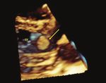
Figure 7: Transthoracic echocardiogram parasternal long-axis view 3-dimensional reconstruction showing a myxoma in the left ventricular
outflow tract.
x
Figure 8: Transthoracic echocardiogram modified parasternal long-axis view showing a myxoma in the left ventricular outflow tract.
and 25% will develop adenomatous disease. Less than 10% Although the clinical diagnostic criteria are reliable,
will develop thyroid cancer. Hence, fine-needle aspiration is nowadays genetic testing for mutations in the PRKAR1A
indicated for patients with thyroid nodules. Regardless of gene is more commonly used for diagnostic certainty. In
the previous pathologies, most CNC patients are euthyroid addition, genetic screening of potentially affected family
[8]. Our patient presented with a nodule and was diagnosed members of patients with CNC could be useful especially in
with hyperthyroidism which is uncommon. younger family members. However, clinical surveillance is
In order to diagnose CNC, Stratakis et al. defined the still advisable for family members even when a PRKAR1A
diagnostic criteria (Table 1). Diagnosis is established if pathogenic variant is not identified.
the patient has two or more major criteria, or, alternatively, In cases of cardiac myxomas, surgical resection is the
one major criterion in addition to either an inactivating treatment of choice with a 6-month follow-up via TTE
mutation of the PRKAR1A gene or a first-degree relative afterwards due to the high incidence of recurrence. In
has CNC [9, 10]. addition, a multidisciplinary approach is recommended in6 Case Reports in Cardiology
Table 1: Diagnostic criteria for carney complex. Supplementary Materials
(1) Spotty skin pigmentation with a typical distribution Supplementary 1. Video 1: an echocardiogram in parasternal
(lips, conjunctiva and inner or outer canthi, vaginal and short-axis view showing a dilated right ventricle containing a
penile mucosa) large highly mobile myxoma.
(2) Myxoma (cutaneous and mucosal)a
Supplementary 2. Video 2: an echocardiogram in an
(3) Cardiac myxomaa atypical view showing a large myxoma in the right ventri-
(4) Breast myxomatosisa or fat-suppressed magnetic resonance cle, a myxoma in the left ventricle, and another myxoma
imaging findings suggestive of this diagnosis in the left atrium.
(5) PPNADa or paradoxical positive response of urinary
glucocorticosteroids to dexamethasone administration during Supplementary 3. Video 3: an echocardiogram in subcostal
Liddle’s test view showing a large myxoma in the right ventricle, dilated
(6) Acromegaly due to GH-producing adenomaa inferior vena cava, and a localized pericardial effusion.
(7) LCCSCTa or characteristic calcification on testicular Supplementary 4. Video 4: an echocardiogram in tricuspid
ultrasonography inflow view showing a large myxoma prolapsing into a
(8) Thyroid carcinomaa or multiple, hypoechoic nodules on dilated right atrium.
thyroid ultrasonography, in a young patient Supplementary 5. Video 5: an echocardiogram in apical four-
(9) Psammomatous melanotic schwannomaa chamber view showing a large myxoma in the right ventricle
(10) Blue nevus, epithelioid blue nevus (multiple)a (causing obstruction of the blood flow across the tricuspid
(11) Breast ductal adenoma (multiple)a valve), a myxoma in the left ventricle, and another myxoma
(12) Osteochondromyxomaa in the left atrium.
Supplemental criteria Supplementary 6. Video 6: an echocardiogram in a modified
(1) Affected first-degree relative tricuspid inflow view showing the tricuspid bioprosthesis
(2) Inactivating mutation of the PRKARIA gene and a myxoma occupying the left ventricular outflow tract.
To make a diagnostic of CNC, a patient must either (1) exhibit two of Supplementary 7. Video 7: an echocardiogram in parasternal
the manifestations of the diseases listed or (2) exhibit one of these short-axis view showing a myxoma occupying the left
manifestations and meet one of the supplemental criteria (an affected
ventricular outflow tract.
first-degree relative or an inactivating mutation of the PRKARIA gene).
a
With histologic information. Supplementary 8. Video 8: three-dimensional reconstruction
of an echocardiogram showing a myxoma occupying the left
order to screen for endocrinopathies and malignancies. ventricular outflow tract.
Clinical surveillance is advisable for family members and Supplementary 9. Video 9: an echocardiogram in apical
possibly genetic testing. In our patient, clinical screening of four-chamber view showing a myxoma occupying the left
the family was not significant; hence, we concluded she ventricular outflow tract.
developed a de novo mutation.
Supplementary 10. Video 10: an echocardiogram in biplane
view showing a myxoma occupying the left ventricular
4. Conclusion outflow tract.
Multiple intracardiac myxomas account for less than 5%
of all cases of myxoma. Its presence should warrant References
screening for endocrinal disorders and should be treated [1] O. Azevedo, J. Almeida, T. Nolasco et al., “Massive right atrial
by complete surgical excision with a generous safety myxoma presenting as syncope and exertional dyspnea: case
margin in addition to close follow-up as it carries high report,” Cardiovascular Ultrasound, vol. 8, no. 1, p. 23, 2010.
risk of recurrence. [2] H. J. Vidaillet, J. B. Seward, F. E. Fyke, W. P. Su, and A. J. Tajik,
““Syndrome myxoma”: a subset of patients with cardiac
Disclosure myxoma associated with pigmented skin lesions and
peripheral and endocrine neoplasms,” Heart, vol. 57, no. 3,
Yehia Saleh and Basma Hammad are co-first authors. pp. 247–255, 1987.
[3] L. S. Kirschner, J. A. Carney, S. D. Pack et al., “Mutations of the
gene encoding the protein kinase A type I-alpha regulatory
Conflicts of Interest subunit in patients with the carney complex,” Nature Genetics,
vol. 26, no. 1, pp. 89–92, 2000.
The authors declare that they have no conflicts of interest.
[4] A. D. Irani, A. L. Estrera, L. M. Buja, and H. J. Safi, “Biatrial
myxoma: a case report and review of the literature,” Journal
Authors’ Contributions of Cardiac Surgery, vol. 23, no. 4, pp. 385–390, 2008.
[5] A. Horvath and C. A. Stratakis, “Carney complex and lentigi-
Yehia Saleh and Basma Hammad contributed equally to the nosis,” Pigment Cell & Melanoma Research, vol. 22, no. 5,
development of this manuscript. pp. 580–587, 2009.Case Reports in Cardiology 7
[6] C. Mateus, A. Palangié, N. Franck et al., “Heterogeneity of
skin manifestations in patients with carney complex,” Journal
of the American Academy of Dermatology, vol. 59, no. 5,
pp. 801–810, 2008.
[7] K. Reynen, “Frequency of primary tumors of the heart,” The
American Journal of Cardiology, vol. 77, no. 1, p. 107, 1996.
[8] C. A. Stratakis, “Hereditary syndromes predisposing to
endocrine tumors and their skin manifestations,” Reviews in
Endocrine and Metabolic Disorders, vol. 17, no. 3, pp. 381–
388, 2016.
[9] M. Q. Almeida and C. A. Stratakis, “Carney complex and other
conditions associated with micronodular adrenal hyperpla-
sias,” Best Practice & Research Clinical Endocrinology &
Metabolism, vol. 24, no. 6, pp. 907–914, 2010.
[10] C. A. Stratakis, L. S. Kirschner, and J. A. Carney, “Clinical and
molecular features of the carney complex: diagnostic criteria
and recommendations for patient evaluation,” The Journal
of Clinical Endocrinology & Metabolism, vol. 86, no. 9,
pp. 4041–4046, 2001.MEDIATORS of
INFLAMMATION
The Scientific Gastroenterology Journal of
World Journal
Hindawi Publishing Corporation
Research and Practice
Hindawi
Hindawi
Diabetes Research
Hindawi
Disease Markers
Hindawi
www.hindawi.com Volume 2018
http://www.hindawi.com
www.hindawi.com Volume 2018
2013 www.hindawi.com Volume 2018 www.hindawi.com Volume 2018 www.hindawi.com Volume 2018
Journal of International Journal of
Immunology Research
Hindawi
Endocrinology
Hindawi
www.hindawi.com Volume 2018 www.hindawi.com Volume 2018
Submit your manuscripts at
www.hindawi.com
BioMed
PPAR Research
Hindawi
Research International
Hindawi
www.hindawi.com Volume 2018 www.hindawi.com Volume 2018
Journal of
Obesity
Evidence-Based
Journal of Stem Cells Complementary and Journal of
Ophthalmology
Hindawi
International
Hindawi
Alternative Medicine
Hindawi Hindawi
Oncology
Hindawi
www.hindawi.com Volume 2018 www.hindawi.com Volume 2018 www.hindawi.com Volume 2018 www.hindawi.com Volume 2018 www.hindawi.com Volume 2013
Parkinson’s
Disease
Computational and
Mathematical Methods
in Medicine
Behavioural
Neurology
AIDS
Research and Treatment
Oxidative Medicine and
Cellular Longevity
Hindawi Hindawi Hindawi Hindawi Hindawi
www.hindawi.com Volume 2018 www.hindawi.com Volume 2018 www.hindawi.com Volume 2018 www.hindawi.com Volume 2018 www.hindawi.com Volume 2018You can also read


