Cardiac retinoic acid levels decline in heart failure - JCI Insight
←
→
Page content transcription
If your browser does not render page correctly, please read the page content below
RESEARCH ARTICLE

                                         Cardiac retinoic acid levels decline in heart
                                         failure
                                         Ni Yang,1 Lauren E. Parker,1 Jianshi Yu,2 Jace W. Jones,2 Ting Liu,1 Kyriakos N. Papanicolaou,1
                                         C. Conover Talbot Jr.,3 Kenneth B. Margulies,4 Brian O’Rourke,1 Maureen A. Kane,2 and D. Brian Foster1
                                         Division of Cardiology, Johns Hopkins University School of Medicine, Baltimore, Maryland, USA. 2Mass Spectrometry
                                         1

                                         Center and Department of Pharmaceutical Sciences, University of Maryland School of Pharmacy, Baltimore, Maryland,
                                         USA. 3Institute for Basic Biomedical Sciences, Johns Hopkins University School of Medicine, Baltimore, Maryland, USA.
                                         4
                                             Penn Cardiovascular Institute, Perelman School of Medicine, University of Pennsylvania, Philadelphia, Pennsylvania, USA.

                                                    Although low circulating levels of the vitamin A metabolite, all-trans retinoic acid (ATRA), are
                                                    associated with increased risk of cardiovascular events and all-cause mortality, few studies have
                                                    addressed whether cardiac retinoid levels are altered in the failing heart. Here, we showed that
                                                    proteomic analyses of human and guinea pig heart failure (HF) were consistent with a decline
                                                    in resident cardiac ATRA. Quantitation of the retinoids in ventricular myocardium by mass
                                                    spectrometry revealed 32% and 39% ATRA decreases in guinea pig HF and in patients with
                                                    idiopathic dilated cardiomyopathy (IDCM), respectively, despite ample reserves of cardiac vitamin
                                                    A. ATRA (2 mg/kg/d) was sufficient to mitigate cardiac remodeling and prevent functional decline
                                                    in guinea pig HF. Although cardiac ATRA declined in guinea pig HF and human IDCM, levels of
                                                    certain retinoid metabolic enzymes diverged. Specifically, high expression of the ATRA-catabolizing
                                                    enzyme, CYP26A1, in human IDCM could dampen prospects for an ATRA-based therapy.
                                                    Pertinently, a pan-CYP26 inhibitor, talarozole, blunted the impact of phenylephrine on ATRA
                                                    decline and hypertrophy in neonatal rat ventricular myocytes. Taken together, we submit that low
                                                    cardiac ATRA attenuates the expression of critical ATRA-dependent gene programs in HF and that
                                                    strategies to normalize ATRA metabolism, like CYP26 inhibition, may have therapeutic potential.

                                         Introduction
                                         Vitamin A (retinol) has received clinical scrutiny in the context of coronary heart disease because low
                                         retinol correlates with increased risk of cardiac events in otherwise healthy middle-aged men (1). Like its
                                         vegetal precursor, β-carotene, whose serum levels are also inversely correlated with adverse outcomes (2, 3),
Authorship note: LEP, JY, and JWJ
                                         retinol has antioxidant properties (4, 5), similar to vitamins E and C. Retinol has therefore been included in
contributed equally to this work.        clinical trials, often in combination with β-carotene and/or vitamins E and C. Ultimately, antioxidant trials
                                         with or without retinol failed to improve patient outcomes (6–10).
Conflict of interest: KBM has received
                                              However, unlike vitamins E and C, the major cell physiological role of retinol has little to do with direct
funds for consulting activities from
MyoKardia (Bristol Myers Squibb)         antioxidant action. Stored primarily in the liver, retinol is a prohormone, the body’s source of all-trans retinoic
and has received sponsored research      acid (ATRA), a potent transcription-activating hormone present in tissues at nanomolar concentrations. Over
support from Sanofi-Aventis U.S. CCT     70 years ago, it was first shown that the offspring of retinol-deficient rats exhibited structural cardiac defects
holds stock in Merck.                    (11, 12). The role of ATRA in embryonic cardiac development has subsequently been mapped in considerable
Copyright: © 2021, Yang et al. This is   detail. It is involved in proper cardiac tissue specification, anteroposterior patterning, and endocardial cushion
an open access article published under   formation, among other processes (13–16). Yet, the cellular role of the retinoids, their metabolism, and signal-
the terms of the Creative Commons        ing in adult heart physiology and heart failure (HF) remain poorly characterized. There are no interventional
Attribution 4.0 International License.   trials that have evaluated the impact of vitamin A or ATRA alone on HF (17) and observational clinical trials
Submitted: February 26, 2020             have not shown any difference in serum retinol levels between HF patients and sex-matched healthy controls
Accepted: March 10, 2021                 (18). Although HF patients do not appear to be retinol deficient, it is unknown whether ATRA levels are
Published: April 22, 2021                altered. Indeed, the precise relationships between circulating retinol and ATRA levels, cardiac tissue ATRA
                                         levels, and intracellular cardiac ATRA signaling in the context of HF are unknown.
Reference information: JCI Insight.
2021;6(8):e137593.                            Only a few studies have examined the mechanism by which altered ATRA signaling might affect the
https://doi.org/10.1172/jci.             function of the adult mammalian heart. Specifically, tamoxifen-inducible cardiac-specific deletion of one
insight.137593.                          of ATRA’s cellular targets, the retinoic acid receptor α (RAR-α) is sufficient to elicit diastolic dysfunction

                                                                                                                                                                    1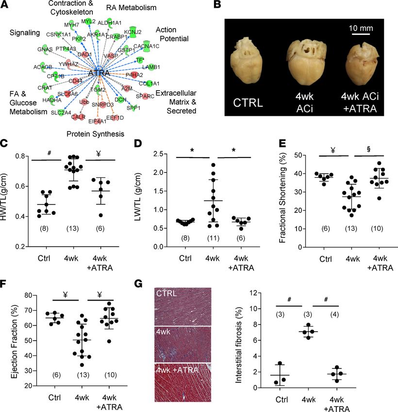
RESEARCH ARTICLE
without affecting left ventricular ejection fraction (LVEF) (19). At the cellular level, RAR-α deletion exacer-
bates myocardial oxidative stress and blunts SERCA-mediated sarcoplasmic reticulum Ca2+-reuptake. Giv-
en that curbing ATRA signaling causes cardiac dysfunction in the adult mouse heart, here we have sought
to understand whether an ATRA signaling defect exists in human and animal models of HF.
ATRA signaling activity in failing human myocardium was assessed by direct quantification of retinol,
ATRA, and other endogenous retinoid metabolites. We show that myocardial ATRA declined in both
human and experimental HF, despite ample local retinol reserves. The decline arose early in HF progres-
sion in guinea pigs, and forestalling it prevented HF. Interestingly, despite sharing low ATRA levels, the
expression of ATRA-metabolizing enzymes differ between human and guinea pig HF, which could have
implications for therapeutic strategy.
Results
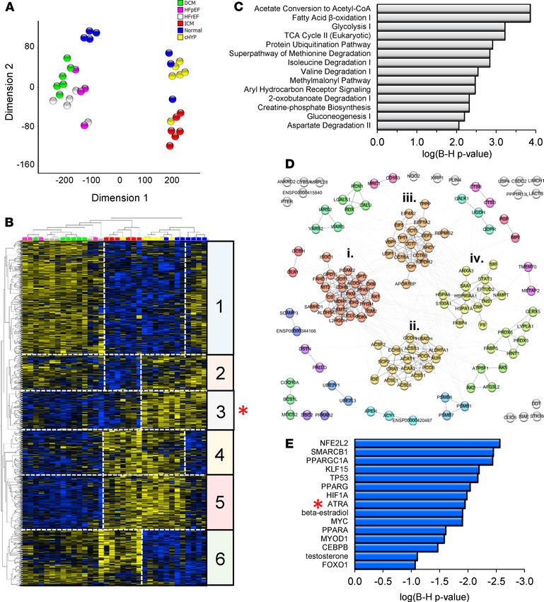
Coordinate protein downregulation in the human HF proteome is consistent with decreased cardiac ATRA. To gauge
the prospects for a retinoid signaling defect, we performed a de novo analysis of a recently published
high-quality proteomic data set (20). The study encompassed patients with HF arising from several patho-
logically or etiologically distinct forms of HF, including HF with reduced ejection fraction (HFrEF, n = 5),
HF with preserved ejection fraction (HFpEF, n = 4), ischemic cardiomyopathy (ICM, n = 6), and idiopathic
dilated cardiomyopathy (IDCM, n = 6). Nonfailing hearts (LVEF > 50%) fell into 2 groups, those with
hypertrophy (cHYP, n = 6) and those without (Normal, n = 7).
Empirical-Bayesian statistical analysis (LIMMA) was performed to identify differentially regulated proteins
in the data set. Trends and similarities among significantly regulated proteins (1144; P < 0.05) are summarized
by dimension reduction (t-distributed stochastic neighbor embedding, t-SNE; Figure 1A) and hierarchical clus-
tering (Figure 1B). HFrEF, HFpEF, and IDCM proteomes shared substantially similar protein profiles at this
depth of coverage (Figure 1B, clusters 1, 4, 5, and 6). HF secondary to ischemia exhibited a distinct proteome
(Figure 1B, cluster 2), differing from other forms of HF, as well as the proteomes of patients with nonfailing
hypertrophic hearts (cHYP) and nonfailing nonhypertrophic hearts (Normal). Nevertheless, hierarchical clus-
tering showed a subset of differentially downregulated proteins common to both ischemic and nonischemic
HF (Figure 1B, cluster 3, 135 proteins, red asterisk). These proteins are associated with processes now broadly
understood to be impaired in human HF, including fatty acid β-oxidation, branched-chain amino acid (BCAA)
metabolism, the Krebs cycle, and creatine phosphate metabolism (Figure 1C). These processes are interconnect-
ed in a bona fide functional network (Figure 1D) consisting of 136 nodes connected by 455 edges (connections),
which is substantially greater than would be expected by chance (139 edges, P < 1 × 10–16). Markov clustering
revealed 12 functional modules (protein nodes with high connectivity) containing at least 3 proteins. The 4 larg-
est modules within cluster 3 are highlighted in Figure 1D. They encompass (i) glycolysis/TCA cycle/creatine
phosphate biosynthesis, (ii) fatty acid oxidation/BCAA metabolism, (iii) protein synthesis/TCP1 ring complex,
and (iv) other chaperones and Ca2+-binding proteins. To gain insight into potential transcriptional programs
that might be consistent with coordinate protein downregulation within this cluster, we performed upstream
regulator analysis across HF types (Ingenuity Pathway Analysis, QIAGEN; Figure 1E). Shown are statistically
significant transcriptional regulators (P ≤ 0.05, Fisher’s exact test, both proteins and metabolites) ranked by
their z scores or level of inferred inhibition. Among transcriptionally active metabolites, the action of ATRA
was inferred to be the most inhibited (z score= –1.95, P = 0.05, red asterisk). Indeed, an ATRA deficit would be
directly consistent with coordinate downregulation of at least 10 other proteins in cluster 3. Moreover, ATRA
has demonstrated relationships with 9 other transcription factors implicated in Figure 1E (see Supplemental Fig-
ure 1; supplemental material available online with this article; https://doi.org/10.1172/jci.insight.137593DS1).
Therefore, coordinate downregulation of cardiac proteins in human HF is consistent with low levels of ATRA.
Mass spectrometry shows ATRA decline in the failing human heart. Levels of ATRA and other endog-
enous retinoids were assessed directly in human HF myocardium using HPLC-UV and LC-multi-
stage tandem mass spectrometry (LC-MRM3) in a blinded manner. The patient characteristics of the
20-patient cohort used in the study are summarized in Table 1. The cohort consisted of men (n = 9) and
women (n = 11), including 13 White patients, 4 African American patients, and 2 patients of unknown
race. Neither age nor BMI differed significantly between nonfailing and failing (IDCM) groups. Non-
failing hearts (n = 10) weighed 368 ± 74 g, whereas failing hearts (IDCM; n = 10) that had undergone
chamber dilation and enlargement were 62% heavier (596 ± 88 g; P < 0.00001). Nonfailing hearts had
an average LVEF of 53% ± 3% compared with 15% ± 4% (P < 0.0001) for failing hearts.
JCI Insight 2021;6(8):e137593 https://doi.org/10.1172/jci.insight.137593 2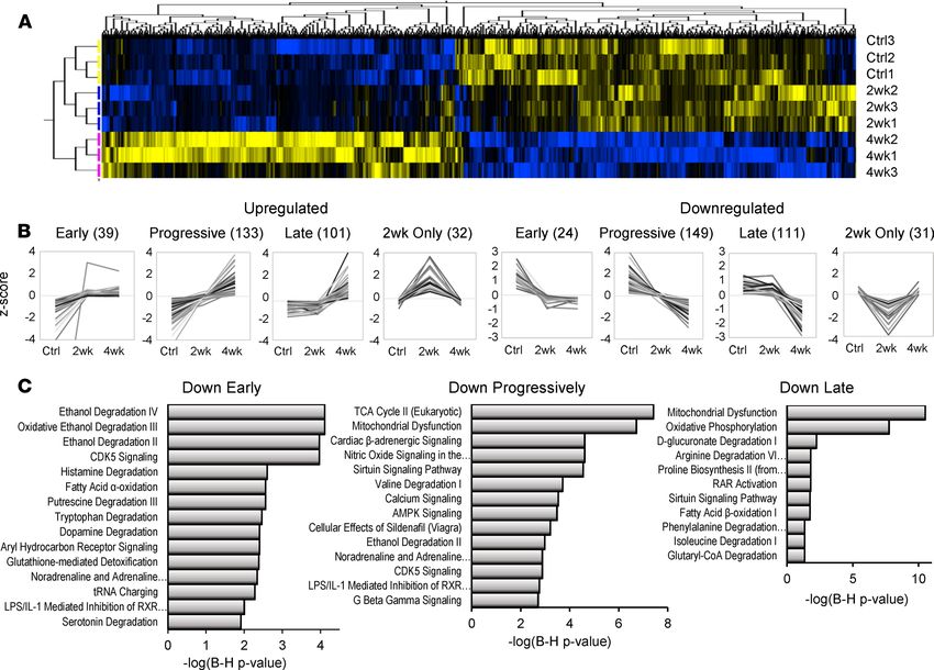
RESEARCH ARTICLE

Figure 1. Human cardiac proteomes are consistent with a decline of the vitamin A metabolite and transcriptionally active hormone, ATRA, across
human heart failure etiologies. (A) Dimension reduction (t-SNE) of significantly regulated proteins across 4 HF etiologies and compensated hypertro-
phy (LIMMA, P < 0.05). Normal, myocardium from healthy donors (blue; n = 7); cHYP, compensated hypertrophy (yellow; n = 6); HFrEF, HF with reduced
ejection fraction (white; n = 5); HFpEF, HF with preserved ejection fraction (pink; n = 4); IDCM, idiopathic dilated cardiomyopathy (green; n = 6); ICM,
ischemic cardiomyopathy (red; n = 6). HFrEF, HFpEF, and IDCM proteomes share substantial similarity, whereas ICM has a distinctive biosignature. (B)
Hierarchical clustering of significantly regulated proteins (blue, downregulated; yellow, upregulated). HF samples follow the color scheme from A. Pro-
tein levels largely correlate across HFpEF, HFrEF, and IDCM (e.g., clusters 1, 4, and 5). Cluster 2 depicts proteins uniquely downregulated in ICM. Clusters
3 and 6 represent proteins similarly regulated across HF etiologies. Specifically, cluster 3 (red asterisk) represents 132 proteins that are downregulated
in most HF patients. (C) Pathway analysis showed that these proteins fall into pathways widely viewed as metabolic hallmarks of HF. (D) Coordinately
downregulated proteins constitute a bona fide multimodular protein association network. (E) Upstream regulator analysis to identify transcriptional
programs that might explain coordinate downregulation and activity of the network. ATRA (red asterisk) activity is inferred to decrease.

                                        In Figure 2A, we show the chemical structures of the retinoid species quantified from human cardiac
                                   tissue, which include retinyl esters (the cellular storage form retinol), retinol, ATRA, and other endogenous
                                   isomers 13-cis-RA and 9,13-cis-RA, whose cellular roles are still not clearly defined. The potent retinoid X
                                   receptor (RXR) agonist, 9-cis RA, was not detectable in human hearts despite a detection limit for the mass
                                   spectrometry assay at the femtomole level (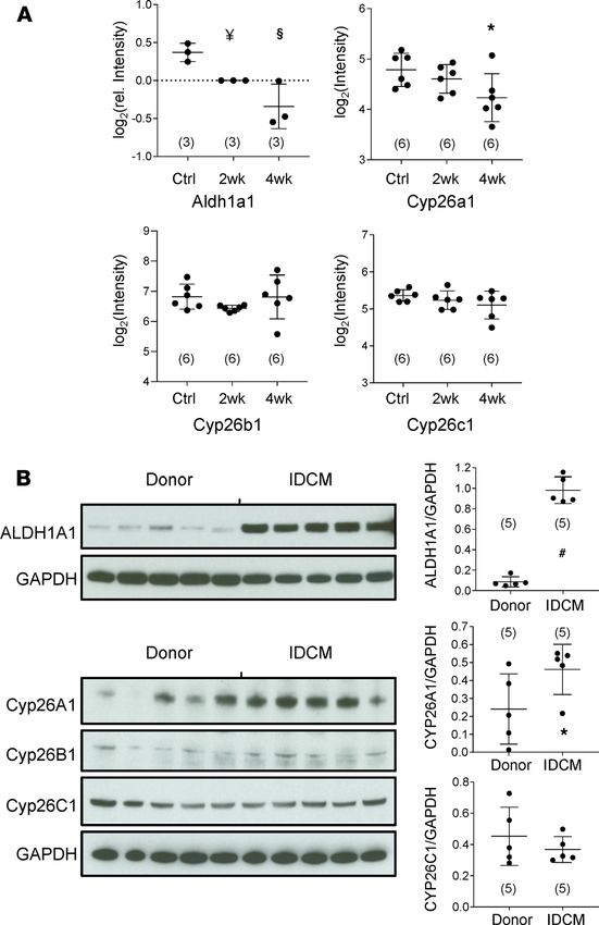
RESEARCH ARTICLE

                                 significantly higher in the IDCM patient group (1.03 ± 0.47 [n = 10] vs. 0.54 ± 0.14 nmol/g tissue [n = 10], P
                                 < 0.05). Levels of retinyl esters perhaps trended higher, though failed to reach significance (1.84 ± 1.18 [n =
                                 10] vs. 2.57 ± 1.08 nmol/g [n = 10], P = 0.16). Notably, however, ATRA was 39% lower in the myocardium
                                 of IDCM patients relative to donor controls (0.79 ± 0.28 [n = 10] vs. 1.30 ± 0.58 pmol/g [n = 9], P < 0.05).
                                 The decline in ATRA coupled with increased retinol is consistent with an impairment in ATRA biosynthesis.
                                 The ATRA decline was mirrored in levels of the 13-cis-RA isomer, which also fell 39% in IDCM (4.26 ± 2.11
                                 [n = 10] vs. 2.58 ± 1.30 pmol/g [n = 10], P < 0.05). Levels of 9,13-cis-RA were unchanged between groups.
                                 Multifactor ANOVA was used to verify that significant differences in ATRA levels were not due to the poten-
                                 tially confounding effect of sex or race (Supplemental Figure 2). HF diagnosis was the only significant factor
                                 contributing to variance in ATRA levels (P = 0.019).
                                      Coordinate protein downregulation across the stages of guinea pig HF implicates retinoid pathways. We sought
                                 to determine the extent to which the human proteomic biosignatures and retinoid profile of HF were reca-
                                 pitulated in an experimental model and over what timescale of HF progression. Here, we used the guinea
                                 pig ACi model (aortic constriction and daily low-dose isoproterenol) that demonstrated a phase of com-
                                 pensated hypertrophy (2 weeks) followed by rapid loss of pump function (decompensation) by 4 weeks. We
                                 previously published an integrated transcriptomic, proteomic, and metabolomic analysis of the guinea pig
                                 ACi model (21), though failed to identify endogenous retinoids in our initial metabolomic screen, which
                                 is not surprising given their relatively low abundance, the limited ability of global metabolomic screens
                                 to detect low abundance analytes, and the inefficiency of typical global metabolomic sample preparation
                                 methodology for the extraction of retinoids. In Figure 3, using empirical Bayesian statistics, hierarchical
                                 clustering, and correlation profiling, we were able to classify the trajectory of protein changes as a function
                                 of HF progression. Briefly, of the 4150 proteins identified in 3 independent iTRAQ proteomics exper-
                                 iments (n = 3 per group), 994 proteins were differentially regulated, clustered in Figure 3A (P < 0.05).
                                 Inspection of Figure 3A, as well as initial k means clustering, revealed that differentially regulated proteins
                                 fell into 8 principal groups, 4 upregulated and 4 downregulated. For both up- and downregulated groups,
                                 some protein levels changed within 2 weeks and remained largely unchanged thereafter (early). There were
                                 also proteins whose changes progressed linearly as a function of HF (progressive) and those whose relative
                                 levels only changed substantially between 2 and 4 weeks (late). Finally, there were a few proteins whose
                                 levels differed from controls substantially at 2 weeks only. Proteins were ranked by correlation to models of
                                 these 8 trajectories to identify proteins that best exemplified each class. Protein levels that correlated highly
                                 (Pearson’s r > 0.975) are depicted in Figure 3B. Briefly, the 8 classes encompassed 620 of 994 significantly
                                 regulated proteins. Progressive changes were the most common (282 proteins), followed by late protein
                                 changes (212 proteins). Comparatively few classified as early movers (63 proteins).
                                      To understand the impact of this regulation, particularly with respect to processes shown to be down-
                                 regulated in Figure 1, pathway analysis was performed on the Down Early, Down Progressively, and Down
                                 Late classes. The Down Early pathways are dominated by pathways that feature aldehyde dehydrogenase
                                 2 (Aldh2) as a common element, including “ethanol degradation” and a retinoid signaling pathway “LPS/
                                 IL1-mediated inhibition of RXRα.” Pathways implicated in the Down Progressively class include the TCA
                                 cycle (specifically succinate dehydrogenase [SDH] subunits), mitochondrial dysfunction (SDH and ATP
                                 synthase subunits), cardiac β-adrenergic signaling (including the major channels of excitation-contraction
                                 coupling), and valine degradation. “Ethanol degradation” is again significant, owing in part to downreg-
                                 ulation of the retinaldehyde-metabolizing enzyme, retinal dehydrogenase 1 (Aaldh1a1), which IPA attri-
                                 butes to this pathway. The aforementioned “LPS/IL1-mediated inhibition of RXRα” pathway is also
                                 implicated. Finally, Down Late pathways include oxidative phosphorylation and fatty acid β-oxidation and
                                 metabolism of amino acids isoleucine, proline, and arginine. Here, however, RAR activation is specifically
                                 implicated. Taken together, pathway analysis of downregulated proteins implicates retinoid-metabolizing
                                 enzymes and retinoid signaling pathways across all phases of HF progression in the guinea pig.
                                      Cardiac ATRA declines early in guinea pig HF. Consistent with implicated pathways, upstream regulator
                                 analysis of downregulated proteins (irrespective of class) indicated that among transcriptionally active metab-
                                 olites, ATRA action was inferred to be inhibited (Figure 4A, z score = –2.18, P = 0.0003). Another implicated
                                 metabolite was L-triiodothyronine (z score = –2.67, P = 0.001), the bioactive form of thyroid hormone, whose
                                 levels were previously known to be deficient in advanced human HF (22, 23). Resident cardiac retinoids in
                                 guinea pig myocardium were quantified in a blinded manner as described in Figure 2. Specifically, guinea
                                 pig control (Ctrl), 2-week, and 4-week HF hearts were analyzed by quantitative mass spectrometry. Levels of

JCI Insight 2021;6(8):e137593 https://doi.org/10.1172/jci.insight.137593                                                                        4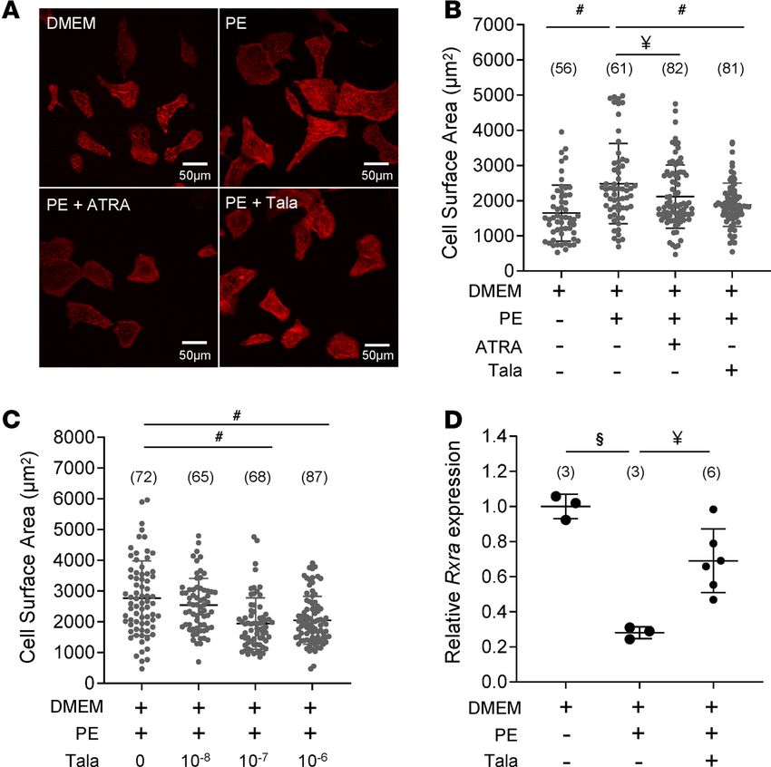
RESEARCH ARTICLE

Table 1. Patient profile

                              Sex                   Race                 Age              Heart weight(g)         BMI               LVEF (%)
                                                                   Nonfailing hearts
        1070                 Male                    W                     71                   404              25.38                 ND
        1106                Female                   AA                    65                   484              42.60                 ND
        1155                Female                   AA                    20                   314              23.73                 53
        1248                 Male                    W                     56                   437              26.20                 53
        1347                Female                   W                     57                   308              23.53                 ND
        1387                Female                   W                     53                   305              24.56                 58
        1413                Female                   W                     49                   436              37.89                 53
        1471                Female                   W                     55                   296              31.99                 53
        1516                Female                   W                     66                   281              20.91                 58
                                                                     Failing hearts
        1045                 Male                   AA                     44                   491              31.25                18
        1169                 Male                    W                     63                   668              28.09                18
        1239                Female                   W                     52                   565              31.22                10
        1264                 Male                    W                     51                   510              27.36                 17
        1275                 Male                    W                     62                   662              24.78                10
        1289                Female                  AA                     67                   516              32.81                20
        1304                Female                   W                     63                   526              23.14                10
        1430                 Male                   UNK                    62                   730              22.60                 15
        1459                 Male                   UNK                    62                   702              27.65
RESEARCH ARTICLE
Figure 2. ATRA but not vitamin A declines in idiopathic dilated cardiomyopathy. (A) Chemical structures of the retinoid
species quantified from myocardial biopsies by reversed-phase chromatography and mass spectrometry. Retinyl esters
represent the in situ retinoid reserve, or storage form, of retinol. Retinol, or vitamin A, is the metabolic precursor of reti-
naldehyde and all-trans retinoic acid (ATRA). Other geometric isomers of retinoic acid include 13-cis-RA and 9,13-cis-RA.
The potent RXR ligand, 9-cis-RA, was not detected in the heart. (B–F) Quantitation of endogenous cardiac retinoids. Dif-
ferences between group means were assessed by a 2-tailed t test (* denotes P < 0.05). N values are given in parentheses.
(B) Retinol levels were significantly elevated in patients with idiopathic dilated cardiomyopathy (IDCM) relative to donor
heart tissue. (C) A similar trend was noted for retinyl esters, though the difference was not significant. (D) ATRA was
significantly lower in IDCM than among donors, as were levels of 13-cis-RA (E). (F) 9,13-cis-RA did not differ significantly.
group to 50.4% ± 10.5% (n = 13) in ACi (Figure 5F). This, too, was prevented by concomitant treatment with
ATRA (64.7% ± 7.0% [n = 10]; P < 0.01). Echocardiographic measurements are summarized in Supplemental
Table 1. ATRA treatment also prevented HF-associated increases in interstitial fibrosis (Figure 5G). Specifically,
the extent of fibrosis increased nearly 4.5-fold at 4 weeks of HF (7.1% ± 0.68% vs. 1.6% ± 1.32% in controls, P
= 0.0001), whereas fibrosis in the ATRA-treated group was limited to 1.7% ± 0.70% (P < 0.0001 with respect to
4-week group). Finally, we confirmed that the ATRA treatment resulted in increased intracardiac ATRA (87%,
P < 0.05) relative to the 4-week HF group (Supplemental Figure 4).
Enzymes responsible for ATRA metabolism are regulated differently between human and guinea pig HF. Given
that resident cardiac ATRA levels are an indicator of heart health, we investigated the origins of low
ATRA in HF. Steady-state ATRA is determined by a bevy of enzymes that influence its rate of biosyn-
thesis from retinol, including the retinol dehydrogenases (RDHs), retinal reductases (e.g., DHRS3), and
retinaldehyde dehydrogenases (RALDHs, e.g., ALDH1A1, ALDH1A2) (25, 26). Its rate of catabolism
is largely determined by the CYP26 family of p450 hydroxylases (CYP26A1, CYP26B1, and CYP26C1)
(26, 27). In the guinea pig proteome, Aldh1a1 (Figure 6A) declined 23% by 2 weeks (n = 3, P < 0.01,
JCI Insight 2021;6(8):e137593 https://doi.org/10.1172/jci.insight.137593 6RESEARCH ARTICLE
Figure 3. Trajectories of proteomic changes in guinea pig heart failure progression implicate retinoid pathways from early to late stages. (A) Hierar-
chically clustered heatmap of differentially regulated proteins in guinea pig HF progression (LIMMA, P < 0.05). Groups include the control (Ctrl; n = 3), 2
weeks of ACi protocol (2wk; n = 3), and 4 weeks of the ACi protocol (4wk; n = 3); 924 of 4150 were deemed significantly regulated. Careful inspection of
the heatmap revealed 8 observable clusters corresponding to trajectories of protein expression. (B) Identification of proteins whose group means (z score
normalized) most closely correlated (Pearson’s r > 0.975) with the 8 observed trajectories of up or downregulation. Numbers in parentheses indicate the
number of proteins classified in each group; 620 of 924 proteins met the strict correlation criterion. (C) Pathway analysis was performed on proteins whose
trajectories were classified as early, progressive, or late movers (Fisher’s exact test). After Benjamini-Hochberg correction, retinoid pathways were identi-
fied (P < 0.05). An RXR pathway was implicated in early and progressive trajectories. Several proteins downregulated at 4 weeks only contributed to the
identification of the RAR activation pathway.
LIMMA-moderated t test) and 39% in HF (n = 3, P < 001). A second RALDH1-like protein and Aldh1a2
trended toward modest declines (12% and 15%) but were not significant (see Supplemental Figure 5).
Finally, Aldh2, the broad specificity aldehyde dehydrogenase, though down early in guinea pig HF (see
Supplemental Table 2), exhibited modest downregulation (16%, n = 3, P < 0.05). Based on enzyme levels
as a first approximation, a decline in the rate of ATRA biosynthesis in guinea pig HF is initially consistent
with reduced Aaldh1a1. CYP26s are poorly represented in trypsin-accessible proteomes owing to their
low abundance, high isoelectric point, and membrane association. Transcript data (Figure 6A) indicated
that neither Cyp26b1 nor Cyp26c1 mRNA levels changed in guinea pig HF. Cyp26a1 transcript levels
were initially unchanged at 2 weeks, but declined significantly by 4 weeks (32%, P < 0.05), perhaps as a
compensatory response to limit cellular ATRA decline.
Although IDCM shared low ATRA with guinea pig HF, levels of ATRA-metabolizing enzymes dif-
fered dramatically. Immunoblot analysis indicated that levels of ALDH1A1 (normalized to GAPDH) were
several-fold higher in IDCM (Figure 6B). Among the CYP26s, CYP26B1 was only marginally detectable.
CYP26C1 levels did not differ between the donor and patient groups. CYP26A1 levels were intriguing
because they were highly variable, particularly in donor hearts. Levels were considerably higher in most
patient samples. The near tripling of CYP26A1 levels was significant (P < 0.05).
JCI Insight 2021;6(8):e137593 https://doi.org/10.1172/jci.insight.137593 7RESEARCH ARTICLE
Figure 4. Low cardiac ATRA is recapitulated early in guinea pig heart failure progression. (A) URA analysis identified
endogenous chemical compounds inferred to be altered, given the differential regulation of proteins in Figure 3A. Select
compounds (Fisher’s exact test, P < 0.05, z score > 1.0) are shown. Low ATRA was specifically inferred from URA of guinea
pig HF. B–F show the results of the analysis of cardiac retinoids. Experimental groups are as defined in Figure 3. Group
means were analyzed by 1-way ANOVA with a post hoc Tukey’s HSD test and P < 0.05 was considered significant. N values
for each group are given in parentheses. (B) Retinol levels were unchanged from controls to HYP and HF. (C) The storage
form of retinol, retinyl esters, declined significantly in HF but was already trending lower in HYP. (D) ATRA was significant-
ly decreased in both HYP and HF, whereas other geometric isomers of retinoic acid did not differ significantly (E and F). As
in human myocardium, 9-cis-RA was not detected. *P < 0.05.
Targeted inhibition of ATRA catabolism limits cardiomyocyte hypertrophy in vitro. High cardiac CYP26A1
expression in human HF could, conceivably, compromise prospects for therapy predicated on ATRA supple-
mentation, as it would likely limit the half-life of administered ATRA in situ. However, inhibition of CYP26
enzymes could limit the rate of ATRA clearance and potentially boost endogenous ATRA. To test this con-
cept, we examined the impact of the pan-CYP26 inhibitor, talarozole, on α-adrenergic hypertrophy in neo-
natal rat ventricular myocytes (NRVMs; Figure 7). In replicated experiments, phenylephrine (PE) treatment
of serum-starved NRVMs increased cross-sectional area on the order of 45%–80% (Figure 7A). As shown in
Figure 7B, PE induced an increase in the cross-sectional area of 48% with respect to cells incubated in DMEM
alone. Cotreatment with 1 μM ATRA blunted the hypertrophy (23% increase relative to DMEM). Talarozole,
likewise at 1 μM, also suppressed PE-induced hypertrophy relative to PE (12% increase relative to DMEM).
Figure 7C illustrates the concentration dependence of talarozole. Cell treatment with 100 nM talarozole
was sufficient to elicit maximum suppression of PE-induced hypertrophy. Finally, we sought to determine
the impact of both the hypertrophic stimulus and concomitant CYP26 inhibition on levels of intracellular
ATRA. Yields of ATRA from NRVMs proved sufficiently low as to preclude direct quantitation by mass
spectrometry. Therefore, we used qPCR to quantify the expression of the ATRA-responsive gene Rxra as a
proxy for ATRA. After 24 hours in culture, PE reduced Rxra mRNA levels by 72% (n = 3, P < 0.001 relative
to minimal media alone). Coadministration of talarozole with PE significantly limited the decline to 33% (n
= 6, P < 0.01 relative to PE alone). Together, the results showed that boosting endogenous ATRA in NRVMs
by inhibiting its catabolism was as effective as treatment with ATRA at limiting cardiomyocyte hypertrophy.
JCI Insight 2021;6(8):e137593 https://doi.org/10.1172/jci.insight.137593 8RESEARCH ARTICLE
Figure 5. ATRA prevents experimental guinea pig heart failure. (A) Proteins differentially regulated in guinea pig HF
(LIMMA P < 0.05), whose modulation by ATRA has been documented (IPA knowledge base), span hallmark processes of
HF, suggesting the therapeutic potential of ATRA supplementation. (B) Representative hearts from sham-operated (Ctrl),
4 weeks after aortic constriction (4wk), and 4 weeks of treatment with 2 mg/kg/d of ATRA (4wk+ATRA). C–G depict struc-
tural and functional analyses of guinea pig hearts and lungs, analyzed by 1-way ANOVA with a Tukey’s post hoc HSD test.
(#: P ≤ 0.0001, §: P < 0.001, ¥: P < 0.01, *: P < 0.05). N values are given in parentheses. (C) ATRA prevented cardiac hypertro-
phy (HW: heart weight; TL: tibia length) and (D) pulmonary congestion (LW: lung weight). (E) ATRA treatment prevented
declines in fractional shortening and (F) ejection fraction. (G) The impact of ATRA on accruing interstitial fibrosis was
assessed. Representative images are shown on the left (10× magnification) and ensemble data on the right.
Discussion
Implication of ATRA decline in HF. The potentially novel primary findings of this study are the following:
(a) there was a resident cardiac ATRA decline in HF patients and a guinea pig model of HF, despite
adequate tissue reserves of retinol; (b) remodeling of the retinoid metabolic program differed between
human and guinea pig HF, with implications for a therapeutic strategy based on normalizing cardiac
ATRA levels; and (c) inhibition of the CYP26 family of the cytochrome p450 hydroxylases with talaro-
zole inhibited myocyte hypertrophy in vitro.
Rigorous isomer-specific quantification of retinoids is challenging, which may explain why the role
of cardiac retinoid metabolism in the adult mammalian heart is understudied. Motivated by studies
showing that low circulating retinoic acid is associated with poor cardiovascular outcomes and mortal-
ity (28), we sought to quantify the state of retinoids in failing human hearts. We showed that nonisch-
emic, dilated, failing hearts exhibited a nearly 40% ATRA decline, even as retinol increased, consistent
with an impairment in ATRA biosynthesis. Interestingly, ATRA has previously been shown to be ele-
vated in a study of patients with severe HF (LVEF < 35%) and advanced coronary artery disease (29).
JCI Insight 2021;6(8):e137593 https://doi.org/10.1172/jci.insight.137593 9RESEARCH ARTICLE
Figure 6. Remodeling of the enzymes of ATRA metabolism differs between guinea pig and human heart failure. (A)
Guinea pig HF: Groups are as defined in Figure 3. N values are given in parentheses. ALDH1A1 protein levels followed a
trend of progressive downregulation in HF that differed significantly from Ctrl at 2 weeks and 4 weeks. LIMMA-moderat-
ed t test as in ref. 21; §: P < 0.001, ¥: P < 0.01. Transcript levels of CYP26A1 fell significantly by 4-week HF, whereas levels
of CYP26B1 and CYP26C1 were unchanged (1-way ANOVA, post hoc Tukey’s HSD; *: P < 0.05). (B) Human IDCM: immu-
noblot signal intensities were analyzed as detailed in Methods. Group mean differences were assessed by a 2-tailed t
test (#: P < 0.0001). Immunoblots indicate that retinaldehyde dehydrogenase 1 (ALDH1A1) was expressed at low levels
in the myocardium of healthy donors but was increased approximately 10-fold in patients with IDCM. CYP26A1 levels
were highly variable and substantially upregulated in IDCM. CYP26B1 was only detected marginally and not quantified.
CYP26C1 was detected in human hearts, but levels did not differ significantly between donors and patients.
The same study reported no difference in nonischemic failing hearts. Discrepancies may be attributable
to differences in retinoid quantitation methodology, disease etiology, and comorbidities, among other
factors. Nevertheless, what is clear is that aberrant intracardiac ATRA metabolism has been implicated
in 2 etiologies of human HF. Notwithstanding that the HFrEF, HFpEF, IDCM, and ICM proteomes are
at least consistent with low cardiac ATRA, systematic characterization of the retinoids across HF types
and severities is warranted.
In this study, we also explicitly demonstrated an ATRA decline in a mammalian model of HF. The result
is consistent with pharmacological studies showing that ATRA ameliorates maladaptive remodeling in a rat
transverse aortic constriction model (30) and in the context of doxorubicin-induced cardiomyopathy (31, 32).
JCI Insight 2021;6(8):e137593 https://doi.org/10.1172/jci.insight.137593 10RESEARCH ARTICLE
Figure 7. Inhibiting ATRA catabolism limits cardiomyocyte hypertrophy. (A) Images: NRVMs incubated in minimal
medium (top left) in the presence of phenylephrine (PE; top right), PE plus ATRA (bottom left), or talarozole (Tala;
bottom right). The scale is provided in the bottom right of the images. Data in B–D were analyzed by 1-way ANOVA with
a post hoc Tukey’s HSD test. N values are given in parentheses. (#: P < 0.0001, §: P < 0.001, ¥: P < 0.01). (B) Ensem-
ble NRVM cross-sectional areas across the experimental groups are shown. ATRA (1 μM) prevented PE-induced cell
enlargement. The CYP26 inhibitor Tala (1 μM) recapitulated mitigation of hypertrophy. (C) Concentration-dependent
suppression of NRVM hypertrophy by Tala; 100 nM was sufficient to achieve maximum suppression. (D) Transcript lev-
els of Rxra (an ATRA-responsive gene) in NRVMs were measured (by qPCR) as a proxy for levels of intracellular ATRA.
PE decreased Rxra expression relative to control levels, but the decline was mitigated by coadministration of 1 μM Tala.
Here, we showed that ATRA prevented HF and pulmonary congestion in a guinea pig model of HF and
sudden cardiac death. How impaired ATRA homeostasis contributes to HF progression is poorly understood.
Some clues may be garnered from in vitro retinoid signaling studies and mouse KO studies of one of ATRA’s
cognate receptors, RAR-α. We showed, as others have (33, 34), that ATRA blunted α-adrenergic hypertro-
phic signaling in NRVMs (Figure 7). ATRA has been shown to upregulate MAPK phosphatases, Mkp1 and
Mkp2, which inactivate Mek upstream of Erk1 and Erk2 (34). In mice, attenuating ATRA signaling by car-
diomyocyte-specific KO of RAR-α leads to increased oxidative stress, altered calcium transients, and diastolic
dysfunction (19). The oxidative stress was notable, as the ratio of reduced to oxidized cellular glutathione
(GSH/GSSG) declined substantially (approximately 40%–50%), owing to the upregulation of ROS-generating
enzymes like NADPH oxidase 2 and 4 (Nox2 and Nox4), and downregulation of ROS scavenging enzymes,
SOD1 and SOD2. Altered Ca2+ transients in RAR-α conditional KO (cKO) hearts were characterized by
slowed Ca2+ reuptake to the SR, which was associated with decreased expression of SERCA2a and decreased
phosphorylation of phospholamban.
Our findings showed that interstitial fibrosis increased in the guinea pig HF model and that the extent
was limited by ATRA treatment (Figure 5G). Levels of fibrosis were not altered in the cardiac RAR-α cKO
model (19), in which RAR-α was specifically depleted in cardiomyocytes. This would suggest that attenua-
tion of cardiomyocyte RAR signaling is insufficient to drastically alter local profibrotic myocyte signaling
to neighboring fibroblasts. Reduction of fibrosis by ATRA in the guinea pig model is then likely due to
direct RAR signaling in cardiac fibroblasts. This would be consistent with evidence that ATRA attenuates
the growth of neonatal cardiac fibroblasts in vitro, the secretion of type I and III collagens, and TGF-β
JCI Insight 2021;6(8):e137593 https://doi.org/10.1172/jci.insight.137593 11RESEARCH ARTICLE
expression in response to angiotensin II (35). Taken together, the benefits of ATRA treatment in the guinea
pig HF model likely stem from activating, or at least offsetting the decline of, protective signaling pathways
across cell types of the heart.
ATRA signaling can be modulated either through changes in the effective cellular concentrations of
ATRA or through altered levels and/or the intrinsic activation status of the RARs. Zhu et al. (19) showed
that models of metabolic stress, such as streptozotocin-induced type I diabetes or a high-fat diet, alter the
expression of RAR-α. In contrast, another study found no significant changes in the mRNA of RARs
in guinea pig HF (21). We suggest that altered cardiac RAR signaling in HF stems, at least in part, from
changes to the local availability of its ligand, ATRA.
Changes in retinoid metabolism in guinea pig HF. From Figures 4 and 6, we conclude that low cardiac
ATRA levels in guinea pig HF did not stem from a deficit of the parent substrate, retinol. Rather, impaired
retinol metabolism, either at the level of retinol conversion to retinaldehyde (RAL) by the RDHs, or RAL
to ATRA by the RALDHs, could lead to low ATRA. RDHs (Rdh5, Rdh13, Rdh14) were detected in the
guinea pig HF proteome but were not significantly regulated. One of the best-characterized RDHs, Rdh10,
was not detected. However, since our iTRAQ proteomics workflow employed data-dependent peptide acqui-
sition, the absence of evidence for Rdh10 should not necessarily be interpreted as evidence of its absence.
At the level of RALDHs, Raldh1 (Aldh1a1) but not Raldh2 (Aldh1a2) was progressively, substantially, and
significantly downregulated in guinea pig HF (39% by 4 weeks). The data presented are initially consistent
with the downregulation of Raldh1 contributing to a bottleneck in ATRA biosynthesis from RAL in the
guinea pig. The role of Raldh1 in the heart has received little scrutiny, perhaps since its expression is weak
and temporally restricted in mouse cardiac development relative to Raldh2 (36), and Raldh2 polymorphisms
have been associated with both congenital heart disease (37) and uncontrolled blood pressure (38). Never-
theless, our study showed that Raldh1 was dynamically regulated, not only in guinea pig but also in human
HF. It bears noting, however, that further factors may contribute to the altered rates of ATRA biosynthesis
in HF, including the levels and intracellular location of cellular retinol-binding proteins and cellular retinoic
acid–binding proteins (CRABPs) (25), as well as the activities of the retinaldehyde reductases that limit the
retinaldehyde pool. Targeted mass spectrometry assays, currently in development, will afford comprehensive
identification and absolute quantification of the core retinoid-metabolizing enzymes of the heart.
Divergent regulation of retinoid-metabolizing enzymes between human and guinea pig HF. It is clear that
despite a common metabolic endpoint (i.e., low ATRA), control of the retinoid metabolic network differs
substantially between humans and guinea pigs. The data in Figure 6 offer a striking contrast in the levels of
ATRA-synthesizing RALDHs and ATRA-catabolizing CYP26s between human HF patients and guinea
pigs. As with the guinea pig HF, RALDH1 levels were dynamically regulated in human IDCM. Counterin-
tuitively though, levels increased in human HF, as did levels of CYP26A1. Notwithstanding that the results
shown in Figure 2 (i.e., decreased ATRA, increased retinol) are consistent with impaired ATRA biosynthe-
sis, high CYP26A1 could also contribute to decreased cardiac ATRA levels in patients by accelerating its
catabolism. The roles of CYP26s in adult physiology, let alone cardiac physiology, are poorly understood,
but recent work with conditional global CYP26A1-KO mice indicates that this high-Vmax form of CYP26
plays a key role in hepatic retinol homeostasis and clearance of exogenous ATRA (39). CYP26B1 is
thought to have a greater role in extrahepatic tissues. Indeed, CYP26B1 has been shown to regulate ATRA
metabolism in human aortic smooth muscle cells (40) and macrophages within atherosclerotic lesions (41).
Moreover, a hyperactive CYP26B1 polymorphism (rs2241057, Leu264Ser) may aggravate atherosclerosis
(41). However, Figure 6 clearly shows that cardiac CYP26A1 was dynamically regulated in both human
and experimental HF, albeit fluctuating in opposite directions. It is unclear whether the divergent regulation
of both RALDH1 and CYP26A1 reflects a genuine species difference in the transcriptional regulation of
these enzymes or whether the etiology of HF may be a factor, since the guinea pig model of HF was elicited
by pressure overload, whereas IDCM individuals had no overt hypertension. Moreover, the impact of the
profound changes to neurohumoral signaling in end-stage human HF on ATRA metabolism is unknown,
as is the impact of comorbidities. Again, deep profiling of the core ATRA metabolic program by mass
spectrometry across classes and stages of HF should prove informative.
Broader changes to retinoid signaling in HF. Proteomic analysis of both human and guinea pig hearts (Figures 1
and 3) implicated RXR-dependent pathways in addition to ATRA/RAR-dependent pathways. RXRs are tran-
scriptional regulatory proteins that interact with several binding partners including RARs, vitamin D receptors,
glucocorticoid receptors, thyroid hormone receptors, and peroxisome proliferator–activated receptors, among
JCI Insight 2021;6(8):e137593 https://doi.org/10.1172/jci.insight.137593 12RESEARCH ARTICLE
others (42). RXRs, particularly RXR-α, are known to regulate the levels of ATRA-metabolizing enzymes. Spe-
cifically, CRABP2, RALDH1, RALDH2, and CYP26A1 have all been shown to be dependent on RXR tran-
scriptional activity (43–47). However, given the binding promiscuity of RXRs, impaired RXR signaling likely
has roles in HF progression that extend beyond a role in cardiac ATRA homeostasis. Prior work has shown that
RXR-α dysregulation has been observed later in the pathogenesis of HF. Specifically, low transcript and protein
levels have been shown to correlate with impaired fatty acid metabolism in tachy-paced dog hearts (48). This
would be consistent with RXR-α’s role, complexed to PPARα, in transcriptional control of enzymes that cata-
lyze fatty acid β-oxidation (e.g., medium-chain acyl-CoA dehydrogenase) (49–52).
In vitro, 9-cis-RA but not ATRA serves as a high-affinity ligand for RXRs that dramatically increases
transcriptional activation of RXR target genes. However, it bears noting that we did not identify 9-cis-RA
in either human or guinea pig hearts. This is consistent with prior retinoid MS quantification in rats, mice,
and nonhuman primates, which showed that the only organ currently known to synthesize detectable levels
of 9-cis-RA is the pancreas (53–55). This suggests that the involvement of the cardiac RXRs, in any stage
of HF progression, may be subordinate to control by RXR-binding partners and their ligands including
RARs/ATRA, the PPARs/essential fatty acids, and others.
Challenges facing the development of retinoid therapy for HF. As a potent hormone, ATRA levels are fine-
ly tuned physiologically, and establishing a therapeutic window could pose a challenge. ATRA has been
used successfully to treat acute promyelocytic leukemia (APL) (56) and is generally well tolerated. About
10%–15% of patients experience a condition formerly known as retinoic acid syndrome (57), characterized
by cardiorespiratory distress similar to acute respiratory distress syndrome, displaying fever, dyspnea, pleu-
ral and pericardial effusion, hypotension, and occasionally renal failure. But the syndrome has since been
renamed APL differentiation syndrome, as it can also be triggered by treatment with arsenic trioxide, and
thus is now believed to reflect the consequence of APL treatment rather than ATRA-specific toxicity at the
commonly used dose (45 mg/m2).
Nevertheless, in studies focused on the hearts of healthy rats, 10 mg/kg/d, although ATRA did not
adversely affect cardiac morphology or function at 2 months, it did elicit significant changes in cardiomyo-
cyte cross-sectional area, as well as changes in indices of oxidative stress (58). Clearly, further animal model
studies are required to determine the minimum effective ATRA dose to ameliorate HF.
Although retinoid quantitation results (Figures 2 and 4) are consistent with an impairment in ATRA
biosynthesis in both human and guinea pig HF, in Figure 6, we also show high levels of CYP26A1 in
IDCM patients. High levels of CYP26A1 enzyme would constitute a reserve of ATRA-clearing activity
that could limit the impact of ATRA supplementation therapy. This scenario, of muted ATRA efficacy
due to high CYP26 activity, has also been documented in APL (59) and has prompted the development
of a class of CYP26 inhibitors, also known as retinoic acid metabolism blocking agents (RAMBAs), that
have the potential to prolong the half-life of pharmacologically administered ATRA when used as adjuvant
therapy in refractory patients (60). Talarozole (R115866 or Rambazole) is a third-generation RAMBA that
displays more than 300-fold greater selectivity for CYP26 over other related cytochrome p450 hydroxylas-
es, such as CYP17 and CYP19 (61, 62).
Intriguingly, CYP26 inhibition is sufficient to boost endogenous ATRA levels without the need for
pharmacologically administered ATRA. Specifically, in rats, talarozole was sufficient to boost endog-
enous ATRA in the serum and liver and to induce expression of ATRA-responsive genes (63). In this
study, we showed that treatment of NRVMs with as little as 100 nM of talarozole recapitulated suppres-
sion of α-adrenergic hypertrophy seen in response to exogenous ATRA (Figure 7). We also confirmed
1 μM talarozole was sufficient to offset the PE-induced decline in Rxra mRNA, which was chosen as
a proxy for NRVM ATRA levels. The precise CYP26 gene targeted by talarozole in NRVMs is unclear
because it is a pan-CYP26 inhibitor (63, 64). However, given that the IC50 for inhibition of hypertro-
phy is less than 100 nM and the IC50 for CYP26C1 inhibition is about 4 μM (64), talarozole likely acts
through either CYP26A1 or CYP26B1.
Conclusion. ATRA is the latest example of a nuclear receptor–binding hormone whose levels are per-
turbed in human HF. Advanced HF in patients is associated with low serum levels of thyroid hormone (22,
65) and local cardiac hypothyroidism (23, 66, 67). The importance of intracardiac thyroid hormone levels
was shown by cardiac-specific overexpression of type 2 triiodothyronine deiodinase, the primary enzyme
responsible for the conversion of thyroid hormone to its active form, T3 (68). Although triiodothyronine
deiodinase expression did not mitigate transverse aortic constriction–induced hypertrophy, it did prevent
JCI Insight 2021;6(8):e137593 https://doi.org/10.1172/jci.insight.137593 13RESEARCH ARTICLE
declines in contractility. Similar approaches, such as cardiac conditional overexpression of the RALDHs or
KO of the CYP26s, will be key to defining the role of perturbed ATRA metabolism in HF.
Methods
Human myocardial tissue
Nonfailing hearts were obtained at the time of organ donation and failing human hearts were procured at
the time of heart transplantation. Nonfailing donor hearts had an LVEF of more than 50%. IDCM hearts
had an LVEF of 20% or less and dilated left ventricles. Hearts were arrested in situ using ice-cold cardio-
plegia solution, transported to the lab on wet ice, and flash-frozen within 4 hours of collection. Transmural
myocardial samples were dissected from the mid-left ventricular free wall.
Guinea pig HF/SCD model and ATRA treatment
Male Hartley guinea pigs (300 g) were obtained from Hilltop Lab Animals Inc. The guinea pig model of
HF and sudden cardiac death (SCD) has been described previously (24). Briefly, the HF and SCD guin-
ea pig model was produced by combining ascending aortic constriction and daily (nonhypertrophic) iso-
proterenol challenge (ACi model). As characterized previously (24), cardiac function of ACi animals is
well compensated in the first 2 weeks (HYP) but declines rapidly thereafter (HF). Hypertrophic hearts were
collected between 1 and 2 weeks after surgery (designated “2wk” in the figures), whereas failing hearts were
collected at 4 weeks (“4wk”). After retrograde perfusion with 20 mL Tyrode’s solution, excised hearts were
snap-frozen in liquid N2 and stored at –80°C. The treatment group consisted of guinea pigs subjected to the
ACi protocols with the surgical implantation of an osmotic pump, which administered ATRA solubilized
in palm oil and DMSO at a dose of 2 mg/kg/d.
Echocardiography
Transthoracic echocardiography was performed on conscious guinea pigs at 4 weeks after surgery using a Vevo
2100 high-resolution in vivo imaging system with a 24 MHz transducer (VisualSonics). Two-dimensional-
ly directed M-mode images were obtained from the short-axis views. Echocardiographic measurements were
made on 3 consecutive cardiac cycles by the leading edge-to-leading edge method. Left ventricular end-diastolic
and end-systolic dimensions and left ventricular end-diastolic posterior wall thickness were measured from the
M-mode images, and left ventricular fractional shortening was calculated with the software VisualSonics v1.3.8.
Histology
Levels of cardiac interstitial fibrosis were assessed by Masson’s trichrome staining. Briefly, hearts were
excised and rapidly immersed in ice-cold saline solution, then fixed with 4% paraformaldehyde overnight.
Tissues were then embedded in paraffin and sectioned into 5 μm slices along the short axis of the heart.
Five slices were collected from the midventricular region of each heart and stained with Masson’s tri-
chrome. Four regions from each slide were used to analyze interstitial collagen by computer-assisted image
analysis (ImageJ, NIH). The data from the 4 regions were averaged to obtain the mean fibrosis level for
each heart. Data were calculated from at least 3 biological replicates (n = 3 for Ctrl, n = 4 for 4-week HF
and 4-week + ATRA).
Quantification of retinoids
Vitamin A metabolites were quantified in a blinded manner. All samples were frozen at collection and
stored at –80°C until extraction. Heart tissues were homogenized in saline and subjected to a 2-step liq-
uid-liquid extraction under yellow lights as described previously (55, 69, 70). Internal standards were
4,4-dimethyl-RA and retinyl acetate. Levels of retinoic acid were determined by LC-MRM3 on a Shimad-
zu Prominence UFLC XR liquid chromatography system coupled to an AB Sciex 5500 QTRAP hybrid
triple-quadrupole mass spectrometer using atmospheric pressure chemical ionization operated in positive
ion mode as previously described (55). Retinol and retinyl esters were quantified via HPLC-UV as before
(69). Retinoic acid, retinol, and total retinyl ester were normalized per gram of tissue. Statistical analysis of
multigroup studies was performed by ANOVA with a post hoc Tukey’s honest significant difference (HSD)
test. Two-group comparisons were performed using a Student’s 2-tailed t test.
JCI Insight 2021;6(8):e137593 https://doi.org/10.1172/jci.insight.137593 14RESEARCH ARTICLE
Analysis of proteomic and microarray data sets
Data summarization and statistics. A proteomic study of cardiac tissue from 34 human subjects was published
and is publicly available from the proteomeXchange database (PXD008934) (20). Proteins were originally
normalized and quantified based on label-free quantitation (LFQ) ion intensities using MaxQuant (71).
The data set was filtered to analyze proteins identified and quantified in at least 50% of patients. Missing
data were interpolated using the k nearest-neighbors algorithm, and the data were transformed (log2) prior
to performing empirical-Bayesian statistical analysis using a LIMMA multigroup comparison (72, 73), as
implemented in Qlucore Omics Explorer 3.5 (Qlucore). Proteins were deemed differentially regulated if P
was less than 0.05 (FDR = 12%).
The guinea pig cardiac proteome data set, available from proteomeXchange (PXD003980), was
acquired using a peptide labeling MS2-based workflow (iTRAQ) and protein levels were determined by
the “median sweep” algorithm for data normalization and protein summarization (21, 73). Missing data
interpolation and statistical analysis were conducted as described for the human data set. At P less than
0.05, the FDR was 22%.
Select guinea pig cardiac mRNA levels were extracted from our guinea pig microarray data set (21)
(e.g., Figure 6A) deposited in NCBI’s Gene Expression Omnibus (GEO), accessible through GEO series
accession number GSE78077. Data were originally normalized and summarized using robust multiarray
averaging and analyzed by 1-way ANOVA with a post hoc Tukey’s test for pairwise comparisons.
Dimension reduction, clustering, and expression profile ranking. Features of the human HF proteomes were
summarized in 2 dimensions using t-SNE (74) and hierarchical clustering, as implemented in Qlucore
Omics Explorer 3.5 (Qlucore), filtered on differentially regulated proteins (P < 0.05 by LIMMA). Specifi-
cally, individual protein abundances were log-transformed and normalized across samples (mean = 0, var =
1) before agglomerative hierarchical clustering using Euclidean distance and complete linkage.
Differentially regulated proteins from the guinea pig HF data set were subjected to profile correlation
ranking in Spotfire Decision Site with Functional Genomics 9.1.2 (TIBCO Spotfire) to determine which
proteins most closely matched models of early, progressive, and late regulation. Specifically, group averages
of each protein’s relative abundance were normalized by z scoring and compared with the models. Mem-
bership in each expression trajectory class was confined to proteins whose correlation with the model was
greater than 0.975 (Pearson’s r).
Pathway and upstream regulator analysis. Select protein clusters (human proteome) and kinetic profiles
(guinea pig HF) were subjected to Ingenuity Pathway Analysis (QIAGEN). The Ingenuity knowledge base
was used as the reference data set for pathway overrepresentation, which was assessed by Fisher’s exact
test. Multihypothesis testing was addressed by reporting Benjamini-Hochberg corrected P values. Upstream
Regulator Analysis (URA) was used to infer potential transcriptional regulators that would be consistent
with coordinate protein regulation (Fisher’s exact test).
Construction of the functional protein association network. Functional protein association/interaction net-
works were constructed by loading the UniProtKB identifiers of proteins downregulated in cluster 3 into
StringApp 1.4.2 (75), embedded in Cytoscape 3.7.1 (76), and then searching the STRING v11 database (77).
The default association/interaction threshold (STRING score > 0.4) was used to map relationships between
proteins. Network modularity was assessed with the Markov clustering function in the ClusterMaker2 1.3.1
app (78) using the STRING score (>0.6) for edge weighting. The granularity parameter (inflation value) was
set empirically. The final network is presented in an edge-weighted, spring-embedded layout using the Mar-
kov cluster (module) number for edge weighting, with modules arranged for maximum clarity.
Culture of NRVMs
NRVMs were enzymatically dissociated from the ventricles of 2-day-old rats with trypsin. Freshly isolated
NRVMs were resuspended in DMEM culture medium supplemented with 10% FBS, glucose, and vitamin
B12. Two preplating steps were performed to enrich cardiac myocyte content in the culture. The final cell
suspension was collected and plated at the desired density for the downstream experiment. For RT-PCR
application, 1 × 10⁶ cells/well were plated in 6-well plates coated with 0.5% gelatin. For microscopy applica-
tions, 1.5 × 10⁵ cells/plate were seeded in 35 mm glass-bottom culture dishes coated with 0.5% gelatin. After
24-hour initial attachment in 10% FBS-containing media, cells were kept in serum-free media containing
DMEM with 1% penicillin/streptomycin and 0.1% insulin-transferrin-selenium-X. After 12 hours of serum
starvation, drug treatments were initiated. Experiments were terminated after 48 hours of drug treatment.
JCI Insight 2021;6(8):e137593 https://doi.org/10.1172/jci.insight.137593 15RESEARCH ARTICLE
Measurement of NRVM hypertrophy
Cells plated on 35 mm glass-bottom microscopy dishes were washed 3 times with prewarmed PBS. The
cells were fixed in 4.7% paraformaldehyde in PBS for 10 minutes, after which cells were washed an addi-
tional 3 times in warmed PBS. The fixed cells were permeabilized in a solution containing 0.1% Triton
X-100 in PBS for 4 minutes and washed with PBS. Cells were blocked in solution containing 1% BSA in
PBS for 25 minutes, then stained with Alexa Fluor 594 phalloidin (Thermo Fisher Scientific) for 25 min-
utes and washed 3 times in PBS. Cells were imaged with an Andor Revolution X1 spinning disk confocal
inverted microscope at 40× magnification. Cell surface area was measured by ImageJ (NIH). Specifically,
images were converted to 8-bit and contrast-enhanced by 1%. Grayscale images were made binary using the
mean auto local threshold. Cell area was determined with the FIJI analyze particles function to detect cells
larger than 100 μm2, excluding cells on the edge of the image. The areas of individual cells were recorded
from ROI Manager. Group area means were analyzed by 1-way ANOVA with a post hoc Tukey’s HSD test.
Quantitation of RXR-α mRNA
Total RNA was extracted from NRVM cells with QIAGEN RNeasy spin columns. Transcripts were quan-
tified using reverse-transcriptase quantitative PCR (RT-qPCR) on a Bio-Rad CFX384 system using 1-Step
RNA to Ct TaqMan Master Mix (Thermo Fisher Scientific). The 10 μL RT-qPCR reactions were multi-
plexed to contain TaqMan assay Rn00441185 for rat RXR-α and Rn99999916 for Gapdh. The levels of
RXR-α transcript were expressed relative to Gapdh using the 2–ΔCt formula. Mean transcript levels were
analyzed by 1-way ANOVA with post hoc Tukey’s HSD test.
Immunoblot analysis
Protein concentrations of human cardiac tissue were determined using a Bio-Rad DC assay. From each
heart, 20 μg protein was resolved by 4%–12% Bis-Tris gel electrophoresis (NuPAGE, Invitrogen). Proteins
were blotted using the Bio-Rad TransBlot Turbo apparatus (7 minutes at 2.5 A). Membranes were blocked
for 1 hour (5% [w/v] nonfat milk in Tris-buffered saline with 1% [w/v] Tween 20) and incubated overnight
with primary antibodies in blocking solution at 4°C. The following antibodies were used: anti-CYP26A1
(Abcam, ab172474, 1/1000), anti-CYP26B1 (Thermo Fisher Scientific, PA5-15214, 1/1000), anti-CYP26C1
(Abcam, ab80226, 1/100) anti-ALDH1A1 (Abcam, ab52492, 1/1000), and anti-GAPDH (Cell Signaling
Technology, 14C10, 1/2000). Membranes were washed and incubated with anti-rabbit antibody conjugated
to horseradish peroxidase (MilliporeSigma, A0545, 1/80,000) antibodies for 1.5 hours at room temperature
and probed for peroxidase activity using SuperSignal West Pico or West Femto Maximum Sensitivity Che-
miluminescent Substrate. Films were digitized (.TIF), converted to 8-bit grayscale in ImageJ (v1.52a, NIH)
and background was subtracted (rolling ball radius: 50). Immunoblot signal intensities of the CYP26 and
ALDH1A1 blots were quantified and normalized to GAPDH intensities. Mean intensities for donors and
IDCM patient groups were compared by 2-tailed Student’s t test.
Statistics
Statistical analysis was conducted as described in each method above and in the figure legends. Error bars
in dot plot graphs represent the standard deviation about the mean. Results were considered statistically
significant if P was less than 0.05, irrespective of the test used.
Study approval
The procurement of deidentified human myocardial tissue was performed under IRB protocols approved
at the University of Pennsylvania (Philadelphia, Pennsylvania, USA) and Johns Hopkins University (Balti-
more, Maryland, USA) as previously described (20, 79). Guinea pigs were housed in an animal care facility
at the Johns Hopkins University School of Medicine, in conformance with the Guide for the Care and Use of
Laboratory Animals published by the NIH (publication no. 85-23, revised 1996, National Academies Press),
with the approval of the Johns Hopkins University Animal Care and Use Committee.
Author contributions
NY, LEP, BOR, MAK, and DBF designed the experiments. KBM provided human heart tissues and data.
NY, LP, JY, JWJ, and TL performed the experiments. NY, LEP, BOR, MAK, and DBF analyzed the data.
DBF wrote the manuscript. NY, LEP, TL, KNP, CCT, KBM, BOR, and MAK edited the manuscript.
JCI Insight 2021;6(8):e137593 https://doi.org/10.1172/jci.insight.137593 16You can also read