Calcium Signaling in Pancreatic Immune Cells In situ
←
→
Page content transcription
If your browser does not render page correctly, please read the page content below
FUNCTION, 2021, 2(1): zqaa026

                                                                               doi: 10.1093/function/zqaa026
                                                                               Advance Access Publication Date: 13 October 2020
                                                                               Original Research

RESEARCH ARTICLE

Calcium Signaling in Pancreatic Immune Cells In situ

                                                                                                                                                        Downloaded from https://academic.oup.com/function/article/2/1/zqaa026/5922605 by guest on 23 November 2020
                                               1,2                                                  1                                  1
Oleksiy Gryshchenko                                 , Julia V. Gerasimenko                              , Ole H. Petersen                  ,
                                                   ,1
Oleg V. Gerasimenko                           ,*
1
 Cardiff School of Biosciences, Cardiff University, Cardiff CF10 3AX, UK, 2Bogomoletz Institute of Physiology,
Kyiv 01024, Ukraine
*Corresponding author. E-mail: GerasimenkoOV@cardiff.ac.uk

Abstract
Immune cells were identified in intact live mouse pancreatic lobules and their Ca2þ signals, evoked by various agents, character-
ized and compared with the simultaneously recorded Ca2þ signals in neighboring acinar and stellate cells. Immunochemistry in
the live lobules indicated that the pancreatic immune cells most likely are macrophages. In the normal pancreas the density of
these cells is very low, but induction of acute pancreatitis (AP), by a combination of ethanol and fatty acids, markedly increased
the number of the immune cells. The principal agent eliciting Ca2þ signals in the pancreatic immune cells was ATP, but these
cells also frequently produced Ca2þ signals in response to acetylcholine and to high concentrations of bradykinin.
Pharmacological studies, using specific purinergic agonists and antagonists, indicated that the ATP-elicited Ca2þ signals were
mediated by both P2Y1 and P2Y13 receptors. The pancreatic immune cells were not electrically excitable and the Ca2þ signals
generated by ATP were primarily due to release of Ca2þ from internal stores followed by store-operated Ca2þ entry through Ca2þ
release-activated Ca2þ channels. The ATP-induced intracellular Ca2þ liberation was dependent on both IP3 generation and IP3
receptors. We propose that the ATP-elicited Ca2þ signal generation in the pancreatic immune cells is likely to play an important
role in the severe inflammatory response to the primary injury of the acinar cells that occurs in AP.

Graphical Abstract

Submitted: 28 August 2020; Revised: 5 October 2020; Accepted: 6 October 2020
C The Author(s) 2021. Published by Oxford University Press on behalf of American Physiological Society.
V
This is an Open Access article distributed under the terms of the Creative Commons Attribution License (http://creativecommons.org/licenses/by/4.0/),
which permits unrestricted reuse, distribution, and reproduction in any medium, provided the original work is properly cited.

                                                                                                                                                   1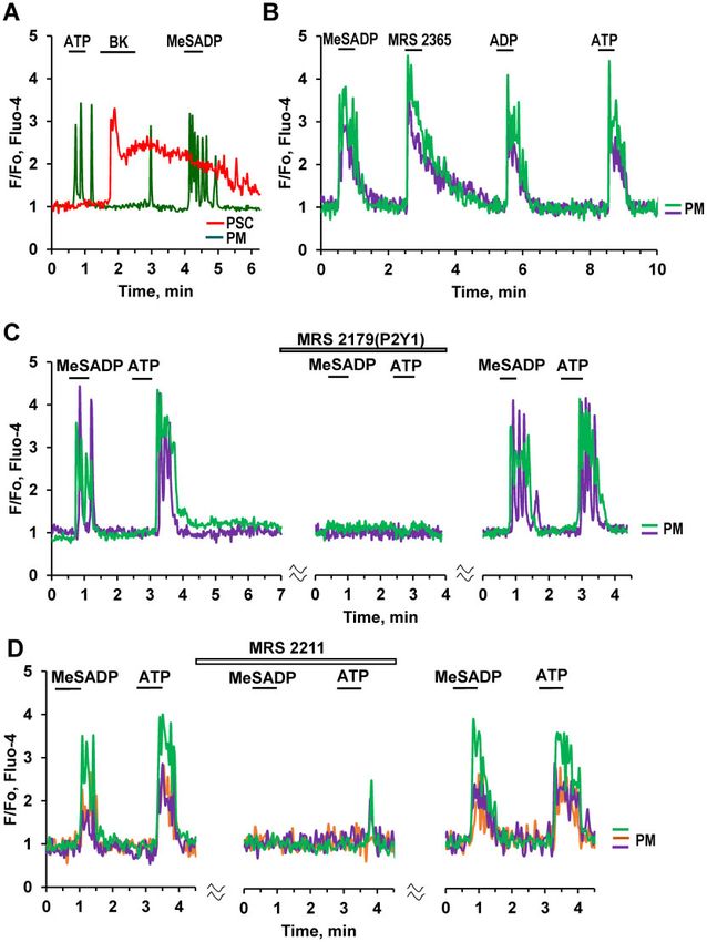
2   | FUNCTION, 2021, Vol. 2, No. 1

Key words: calcium signaling; exocrine pancreas; pancreatic lobules; acute pancreatitis; pancreatic immune cells; pancreatic
macrophages; pancreatic stellate cells; pancreatic acinar cells; P2Y receptors; ATP

Introduction                                                             inflammation, tissue destruction, and high rates of morbidity
                                                                         and mortality.14
Cytosolic Ca2þ signals in the acinar cells of the exocrine                   There have been limited studies in normal pancreatic tissue
pancreas, evoked by acetylcholine (ACh) or cholecystokinin               that possesses different types of tissue-resident myeloid cells.
(CCK), control the physiologically important secretion of diges-         They could be identified by immunohistochemistry using spe-
tive enzymes and fluid.1 The mechanisms responsible for the              cific surface proteins including CD11b and F4/80 that are highly
primary intracellular Ca2þ release and the subsequent Ca2þ               expressed specifically in macrophages.15–17 It was, however, dif-

                                                                                                                                              Downloaded from https://academic.oup.com/function/article/2/1/zqaa026/5922605 by guest on 23 November 2020
release-activated Ca2þ (CRAC) entry of Ca2þ are well estab-              ficult to identify more precisely myeloid cell subpopulations in
lished.1–4 Although the vast majority of Ca2þ signaling studies          the exocrine tissue due to cross-reactivity of conventional den-
have been conducted on acutely isolated mouse acinar cells or            dritic cells and macrophages to the same tissue antigens.17
small acinar cell clusters, the general validity of the results              Here we now characterize the Ca2þ signaling properties of
obtained has been confirmed by studies in more intact prepara-           macrophages in situ, using freshly isolated lobules of exocrine
tions as well as in experiments on human acinar cells.1,5                pancreas. We demonstrate vigorous Ca2þ signal generation in-
    Whereas local repetitive Ca2þ rises regulate physiological           duced by ATP as well as some other agents. As ATP will be re-
secretion, global and sustained elevations of the cytosolic Ca2þ         leased from damaged acinar cells in the very early stage of AP,
concentration ([Ca2þ]i) initiate the disease acute pancreatitis          such Ca2þ signals could play an important role in the generation
(AP).6 Such excessive Ca2þ signals can be elicited by a combina-         of the inflammatory response, which is the major cause of the
tion of ethanol and long-chain fatty acids, bile acids, or be            destruction of the pancreas and surrounding tissues in AP.18
drug-induced, for example by Asparaginase.7 The sustained
global elevation of [Ca2þ]i is generally maintained by open CRAC         Methods
channels,2,6 but can also occur via pressure-induced Piezo1
activation of TRPV4 channels.8                                           Ethical Approval
    Given that the acinar cells constitute the bulk of the exocrine      All animal studies were ethically reviewed and conducted
pancreatic tissue and that these cells synthesize and secrete the        according to the UK Animals (Scientific Procedures) Act, 1986.
digestive enzymes in response to food intake and are responsi-           All animal procedures and experimental protocols were per-
ble for the initiation of AP, it is not surprising that Ca2þ signaling   formed under a Project Licence granted by the UK Home Office
studies in the exocrine pancreas have largely been confined to           and approved by the Animal Care and Ethics Committees at
these cells. However, it has recently become clear that other cell       Cardiff School of Biosciences, Cardiff University. Animals were
types in the exocrine pancreas also play a role, particularly in         maintained in plastic cages supplied with fresh corn cob bed-
the pathophysiology of AP.4,9–12                                         ding, tap water, and commercial pelleted diet.
    Employing isolated lobules of the exocrine pancreas, in which
the normal microscopic structure of the acinar environment is
                                                                         Induction and Evaluation of Experimental AP
preserved, it has been possible to record simultaneously cyto-
solic Ca2þ signals in several different cell types, including stellate   The alcohol-induced experimental model of AP was induced in
cells and intrinsic nerves.10,11 Whereas the role of the Ca2þ            C57BL6/J mice that received two intraperitoneal (IP) injections of
signals evoked by ACh and CCK in the acinar cells is well under-         PBS followed by IP injections of a mixture of ethanol (1.35 g/kg)
stood, the physiological importance of the Ca2þ signals evoked           and palmitoleic acid (150 mg/kg) at 1 h intervals as previously
by bradykinin (BK) in the stellate cells10 is unclear. There is,         described.11 We refer to this AP model as FAEE-AP, since fatty
however, evidence indicating that BK-elicited Ca2þ signals in the        acids and ethanol can react together inside cells to produce
                                                                         fatty acid ethyl esters (FAEEs). Control mice received IP injec-
stellate cells can magnify the damage to the acinar cells caused
                                                                         tions of the PBS solution alone. Mice were humanely killed by
by various agents inducing AP, including the combination of
                                                                         cervical dislocation (Schedule 1) 48 or 72 h after the last injec-
alcohol and fatty acids as well as bile acids.9–11 At least part of
                                                                         tion. For histological assessment pancreatic tissue was fixed in
the damaging effect of the BK-elicited Ca2þ signals in the stellate
                                                                         4% formaldehyde and embedded in paraffin. Fixed slices (4 lm
cells would appear to be mediated by NO generation, due to
                                                                         thickness) stained with hematoxylin and eosin, 10 random
Ca2þ-activation of NO synthase in these cells.12
                                                                         fields of view (magnification: 200), were evaluated by two
    In our recent work on Ca2þ signaling in the peri-acinar envi-
                                                                         blinded independent investigators grading (scale, 0–3; means 6
ronment,11 we identified, in addition to stellate cells and nerve
                                                                         SEM; n ¼ 3 mice per group) edema, inflammatory cell
cells, an unknown cell type, which we called X-cells. These cells
                                                                         infiltration, and necrosis as previously described.19
displayed prominent Ca2þ signals in response to stimulation
with ATP and have been the focus of the present study. We now
show evidence indicating that these X-cells are pancreatic im-           Lobule Preparation
mune cells, most likely macrophages.                                     Pancreatic lobules were freshly isolated from the pancreas of 5-
    Previously, macrophages of the exocrine pancreas have been           to 7-week-old male C57BL6/J mice10 or from mice in which
studied in culture or fixed tissue, primarily under pathological         FAEE-AP had been induced as described above. The pancreas
conditions such as ductal ligation, carbon tetrachloride-induced         was rapidly removed, injected with standard Naþ-Hepes-based
pancreatitis, and experimental pancreatic cancer.13 During pan-          solution containing collagenase and incubated for 5–6 min at
creatic injury, macrophages infiltrate the tissue leading to             37 C. The standard solution was composed of (in mM): NaCl,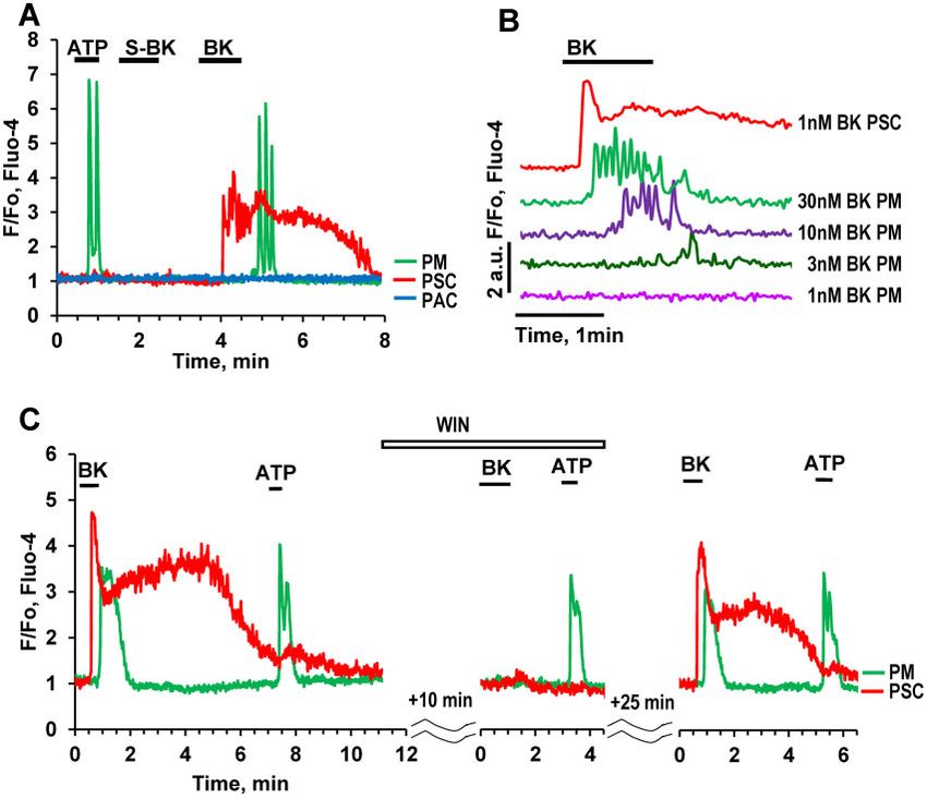
O. Gryshchenko et al.    |   3

140; KCl, 4.8; Hepes, 10; MgCl2, 1; glucose, 10; CaCl2, 1 (unless    cells, as such cells have occasionally been found in
stated otherwise), pH 7.3 (NaOH). The standard solution in           pancreatic tissue.13,17
experiments for investigation of the effects of membrane depo-           To test the hypothesis that the X-cells previously described11
larisation was modified to contain 100 mM KCl and 44.8 mM            are immune cells, we employed immunostaining with different
NaCl. All experiments were carried out with pancreatic lobules       antibodies against surface proteins of immune cells labeled
attached to the coverslip of a perfusion chamber at room tem-        with the fluorescent indicator Alexa Fluor 647 (Figures 1A–C and
perature (23 C).                                                   2D) to allow post staining at the end of functional experiments
                                                                     (Figure1A(iii) and Figure1C(iii)). The fluorescently labeled anti-
Ca21 Measurements                                                    bodies F4/80 as well as CD11b have been commonly used to la-
                                                                     bel macrophages.13,20 Staining confirmed the identity of X-cells
Pancreatic lobules were loaded with 5 lM Fluo-4 acetoxymethyl        as pancreatic macrophages (PMs) in the pancreas (Figures 1A–C
(AM) ester for 20 min at room temperature. The lobules were          and 2E). In addition, the ear-like shape of nuclei, typical for
transferred into a flow chamber and perfused with the standard       macrophages, monocytes, eosinophils, and dendritic cells21–24

                                                                                                                                           Downloaded from https://academic.oup.com/function/article/2/1/zqaa026/5922605 by guest on 23 November 2020
solution alone or containing different chemicals as described in     was very different from the classical round shape of nuclei ob-
the experimental protocols of the result section. Cells were visu-   served in other cells of the pancreas when stained with Hoechst
alized using a Leica SP5 MPII two-photon confocal microscope,        33342 (Figure 1A, B).
with an x63 1.2NA objective lens. The Fluo-4 excitation wave-            The control antibodies IgG are known to induce Ca2þ spikes
length was 488 nm and emission was collected at 500–560 nm           in activated immune cells.25 We applied IgG to control pancre-
with resolution of 256x256 pixels and speed of 0.7 frames/s.         atic lobules, but did not detect any oscillations. Instead we
Images were analyzed using Leica Confocal Software (Leica,           observed occasionally single Ca2þ spikes (Figure 2A), but their
Mannheim, Germany). Fluorescence signals were plotted as             appearance was independent of the presence of IgG. We then
normalized F/F0. Control Immunoglobulin G (IgG) was used in          tested the effect of IgG on PMs in our AP model (FAEE-AP).
concentrations of 0.1–0.25 mg/mL. ANOVA or Student’s t-test          In lobules isolated from FAEE-AP pancreas (48 h), we detected
was performed for statistical analysis.                              IgG-induced Ca2þ oscillations, after a substantial delay, in
                                                                     about 30% of PMs (Figure 2B). Without IgG stimulation, no
Immunostaining in Ex vivo Lobules                                    oscillations were observed in PMs from FAEE-AP pancreas.
Immunostaining of live pancreatic lobules was performed as           These data indicate that PMs are largely quiescent in the pan-
                                                                     creatic tissue of control mice, only displaying the occasional
previously described.10 Mouse F4/80 and mouse CD11b/Integrin
                    R                                                spontaneous Ca2þ spike. However, after AP induction, PMs be-
alpha M Alexa FluorV 647-conjugated monoclonal rat antibodies
                                                                     come activated20 and now close to one-third of the cells re-
were used to label specific surface proteins of immune cells,
                                                                     spond to IgG stimulation with repetitive Ca2þ spiking
usually at the end of Ca2þ measurement experiments, unless
                                                                     (oscillations) (Figure 2B, C). The duration of the spikes in the
otherwise stated. After blocking with 1% BSA and 10% goat
                                                                     FAAE-AP pancreas appeared to be slightly longer than in the
serum containing PBS, the isolated pancreatic lobules were in-
                                                                     control situation, but the difference was not significant
cubated for 1 h at room temperature with the selected antibody.
                                                            R        (Figure 2D). The amplitudes of the Ca2þ spikes in the FAEE-AP
Antibody staining was visualized by exciting Alexa FluorV 647
                                                                     pancreas were not significantly different from those observed
with 633 nm laser at 10% power and emitted light was collected
                                                                     in the control pancreas (P > 0.06).
at 640–700 nm. Hoechst 33342 was used to determine the posi-
                                                                         Immunostaining of PMs with the fluorescently labeled anti-
tion of nuclei using excitation 405 nm and collecting light at
                                                                     bodies F4/80 against surface IC proteins (Figure 1A–C) was used
420–480 nm. Conjugated antibody fluorescence was also over-
                                                                     to calculate the relative density of PMs (Figure 2E, F). Whereas
laid with Fluo-4 staining as described in the Ca2þ measurements
                                                                     the density of pancreatic stellate cells (PSCs) was not different
section. Lobules were attached to the glass coverslips covered
                                                                     in tissues isolated from control and FAEE-AP (72 h) mice (n ¼ 14
with poly-L-lysin.
                                                                     and n ¼ 31, P > 0.54), the density of PMs increased significantly
                                                                     in FAAE-AP mice after 48 h (P < 0.033) and was highly significant
Reagents                                                             after 72 h (P < 0.0001; Figure 2E, F).
BK, S-BK, WIN64338, MeSADP, MRS 2365, MRS 2179, MRS 2211,
ADP, SQ 22536 were purchased from Tocris Biosciences (Bristol,       ATP-elicited Ca21 Signals in PMs
UK). GSK7975A was a gift from GlaxoSmithKline (Stevenage,
                                                                     We have previously reported that X-cells, now identified
UK). Fluo-4 AM and Hoechst 33342 were purchased from
                                                                     as PMs, are highly responsive to stimulation with micromolar
Invitrogen (Life Technologies, Carlsbad, CA, USA). Mouse F4/80
                                                R                    concentrations of ATP.11Figure 3A–D demonstrate that PMs
monoclonal rat Antibody (CI-A3-1) [Alexa FluorV 647] and mouse
                                    VR                               (green traces) are capable of generating substantial Ca2þ signals
CD11b/Integrin alpha M Alexa Fluor 647-conjugated monoclo-
                                                                     in response to ATP (10 lM) (Figure 3A–D).
nal rat antibodies were obtained from Novus Biologicals Europe
                                                                         We tested the possibility that the PMs might be electrically
and R&D Systems Bio-techne, respectively. Other chemicals
                                                                     excitable. In these experiments, the cells in the lobule were
were purchased from Sigma or Calbiochem (Merck, UK).
                                                                     depolarized by exposure to a solution with a high (100 mM) Kþ
                                                                     concentration (HKþ) as previously described.11 This evoked
Results                                                              short-lasting Ca2þ signals in about 30% of the PMs (Figure 3A),
                                                                     but this could be an indirect effect due to release of ACh from
X-cells Identified as Pancreatic Immune Cells
                                                                     depolarized intrinsic nerves (dark blue trace in Figure 3A). In the
In our previous study,11 the unidentified X-cells displayed Ca2þ     presence of atropine (1mM) HKþ failed to evoke any elevation of
signals that were distinct from those observed in the well-          [Ca2þ]i in the PMs, whereas the Ca2þ signals evoked by ATP and
known cells of the pancreatic lobules. One possibility, which we     BK were preserved (Figure 3C). ACh induced Ca2þ signals in a
have now investigated, is that the X-cells might be immune           majority of PMs (70%) (Figure 3B), whereas CCK (Figure 3A) had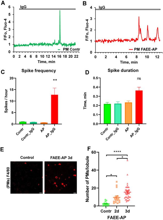
4   | FUNCTION, 2021, Vol. 2, No. 1

                                                                                                                                                                                Downloaded from https://academic.oup.com/function/article/2/1/zqaa026/5922605 by guest on 23 November 2020
Figure 1. Immunostaining after Recording ATP-elicited Ca2þ Signals in pancreatic macrophages (PMs). (A). Representative images of pancreatic lobule loaded with
Fluo-4AM before (Ai) and after ATP (10 mM) application (Aii), the arrow indicates the position of the (n ¼ 8). A corresponding Fluo 4 trace from a PM is shown in Aiii.
Corresponding immunostaining of this lobule with antibodies F4/80 Alexa Fluo 647 is shown below (Aiv). Hoechst 33342 staining of the same area is shown in Av.
Arrow points to ear-like shape of PM nucleus. Overlay of antibody and Hoechst 33342 staining is shown in Avi. Scale bar is 10mm. (B). Immunostaining of another area
in a pancreatic lobule with monoclonal F4/80 antibodies labeled with Alexa Fluor 647 (Bi). Staining of nuclei in the same lobule with Hoechst 33342 (Bii). Overlay of Bi
with Bii is shown in Biii. Scale bar is 10mm. (C). Representative images of a pancreatic lobule loaded with Fluo-4AM before (Ci) and after ATP (10 mM) application (Cii), the
arrow indicates the position of the PM. Corresponding Fluo 4 trace is shown in Ciii. Immunostaining of the same area with monoclonal CD11b antibody conjugated
with Alexa Fluor 647 (n ¼ 8) is shown in Civ. Overlay of Cii and Civ is shown in Cv. Scale bar is 10mm.

no effect. Bradykinin elicited Ca2þ signals in PMs (in 40% of the                       reintroduction of 1 mM Ca2þ to the bath solution and removal of
cases), which were delayed compared to the responses in the                              EGTA (Figure 3D).
neighboring PSCs (Figure 3C).                                                               To investigate the potential involvement of inositol trisphos-
    Depletion of intracellular stores by prolonged stimulation                           phate (IP3) receptors in ATP-elicited Ca2þ signaling in PMs, we
with 10 lM ATP, in the absence of external Ca2þ (and in the                              treated pancreatic lobules with either 100 lM 2-APB (Figure 4A)
presence of ethylene glycol-bis(b-aminoethyl ether)-N,N,N0 ,N0 -                         or the phospholipase C inhibitor U 73122 (10 lM) (Figure 4B).
tetraacetic acid (EGTA) (0.5 mM)), lead to the cessation of Ca2þ                         In both cases this resulted in almost complete inhibition of
signaling (Figure 3D). Ca2þ oscillations resumed after                                   ATP-elicited Ca2þ signal generation (Figure 4). In contrast,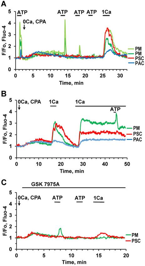
O. Gryshchenko et al.          |   5

                                                                                                                                                                             Downloaded from https://academic.oup.com/function/article/2/1/zqaa026/5922605 by guest on 23 November 2020
Figure 2. IgG-elicited Ca2þ Spikes in PMs. (A). Single short Ca2þ spike occurring after application of IgG (0.1–0.25 mg/mL) in a PM from a control pancreatic lobule. This
was an infrequent observation (5 out of 29 cells tested) and is most likely not an IgG-elicited Ca2þ signal as such single spikes have been also observed in 3 out of 15
cells in the absence of IgG stimulation. (B). Representative trace of IgG (0.1–0.25 mg/mL)-induced Ca2þ signals in PMs in pancreatic lobules isolated from mice with AP
(FAEE-AP model—48 h). Such oscillations were observed in 9 out of 31 cells. Single short spikes have been observed in 4 out of 31 cells. No oscillations were observed in
the absence of stimulation with IgG (n ¼ 14), while single short spikes have been observed in 2 out of 14 cells. (C). Average Ca2þ spike frequencies in PMs displaying
Ca2þ signals under the conditions indicated. The frequencies in control PMs, both stimulated with IgG (blue bar) and unstimulated (green), as well as in unstimulated
PMs from the FAEE-AP model (48 h, orange bar) were much lower than in PMs from the FAEE-AP model stimulated with IgG (red bar, P < 0.007). (D). Average Ca2þ spike
duration in PMs displaying Ca2þ signals under the conditions indicated. Although the average spike duration was longer in the PMs from the FAEE-AP mice stimulated
with IgG than under the other conditions, the difference was not statistically different (P > 0.2). (E). Representative images of immunostaining of PMs in lobules using
antibodies F4/80 conjugated with Alexa Fluor 647. Lobules were isolated from control and FAEE-AP 3-day mice (72 h in vivo FAEE-AP model). Scale bar is 20mm.
(F). Comparison of the average density of PMs in lobules from control and FAEE-AP 2-day and 3-day mice (48 h and 72 h in vivo FAEE-AP model, respectively). Control,
2.36 6 0.6 SEM, n ¼ 14; FAEE-AP 2 day, 9.56 6 1.86 SEM, *P < 0.033, n ¼ 16; FAEE-AP 3 days, 15.37 6 1.51 SEM, *P < 0.038 as compared to FAEE-AP 2-day, n ¼ 35. The differ-
ence between control and FAEE-AP 3-day was very highly significant (****P < 0.0001).

application of an inhibitor of adenylyl cyclase, SQ 22536 (200 lM),                         As mentioned above (Figure 3D), the ATP-elicited Ca2þ signals
only had a partial but nevertheless significant inhibitory effect on                    in PMs depend predominantly on Ca2þ release from internal
Ca2þ signals evoked by 1 lM ATP, but had no effect on the                               stores that can be depleted by prolonged stimulation with ATP
responses to higher doses of ATP, i.e. 3 and 10 mM (Figure 4C).                         in the absence of external Ca2þ. To study the replenishment of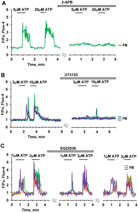
6 | FUNCTION, 2021, Vol. 2, No. 1
Downloaded from https://academic.oup.com/function/article/2/1/zqaa026/5922605 by guest on 23 November 2020
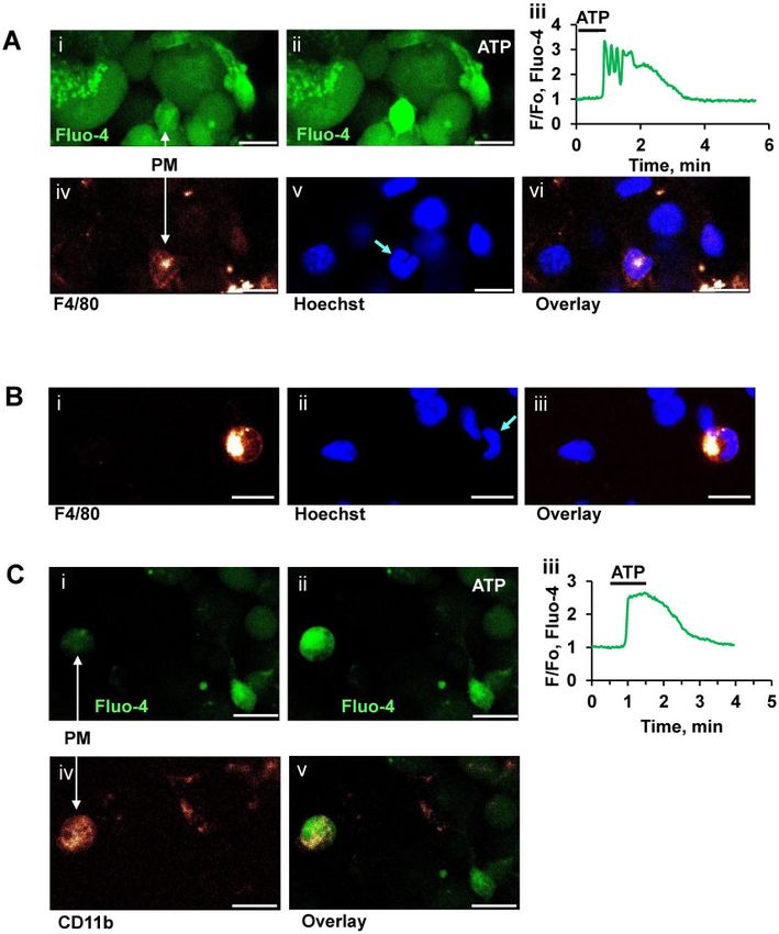
Figure 3. Effects of Stimulation with ATP, Ach, and High Kþ Concentration.
(A). Representative traces (normalized [Ca2þ]i traces, F/F0) of simultaneous
recordings of [Ca2þ]i changes in PM (green trace), PSC (red), pancreatic neuron
(PN, black), and PAC (blue) in the same lobule. ATP (10 mM) only evoked a Ca2þ
signal in the PM, whereas a low concentration of BK (20 nM) only elicited a Ca2þ
signal in the PSC. CCK (at a high concentration, 100 nM) evoked a response in
the PAC, but not in the other cells. Exposure to a high Kþ concentration (100 Figure 4. ATP-induced Ca2þ Signaling in PMs Depends on IP3. (A). Ca2þ signals
mM, HKþ) resulted in an increase in [Ca2þ]i in all the cells except the PSC. HKþ induced by ATP at different concentrations (5 mM, 20 mM) are abolished by
increased [Ca2þ]i in 31 out of 98 PMs. In the PSC, the HKþ-induced depolarisation 100 mM 2-APB (n ¼ 5). (B). ATP-elicited Ca2þ signals are markedly reduced or abol-
markedly reduced the BK-induced elevated [Ca2þ]i plateau. This is a conse- ished by the phospholipase C inhibitor U 73122 (10 lM) (abolished in 36 out of
quence of the severely reduced driving force for store-operated Ca2þ entry, due 45 cells). (C). Application of SQ 22536 (200 lM), an inhibitor of adenylyl cyclase,
to the diminished membrane potential. (B). ACh (100 nM) evokes Ca2þ signals in resulted in partial inhibition of Ca2þ signals elicited by a low (1 mM) ATP concen-
two PMs (n ¼ 23 out of 33 tested cells) following ATP-elicited Ca2þ signals, tration (area under the curve 30–120 s, P < 0.02, n ¼ 18). Complete inhibition of
whereas trypsin had no effect (although trypsin did occasionally induce the response was observed in three cells (brown trace). There was no significant
responses in PMs (3 out of 25 cells). (C). In the presence of atropine (1 mM) HKþ reduction of Ca2þ signals evoked by higher concentrations of ATP (3 mM (n ¼ 18)
(n ¼ 20) failed to increase [Ca2þ]i in two PMs, whereas the effects of bradykinin or 10 mM ATP (n ¼ 12)).
on both PMs (n ¼ 5) and a PSC were retained. (D). Addition of 0.5 mM EGTA, in
the absence of extracellular Ca2þ, first reduced and then abolished the response
to ATP (10 mM) in a PM (n ¼ 5). The ATP response was recovered after removal of
and continuous external readmissions of 1 mM CaCl2. After
EGTA and restoration of the external [Ca2þ] to 1 mM. readmission of external Ca2þ there was also recovery of the
ATP-induced response in the PM (Figure 5B). The most likely
explanation for the observed rise in [Ca2þ]i immediately upon
external Ca2þ readmission is that CRAC channels are open as a
internal stores we have used a standard cyclopiazonic acid
result of the intracellular Ca2þ store depletion following block-
(CPA) protocol for Ca2þ store depletion, while observing simul-
ade of the SERCA pumps by CPA.2 Antigen stimulation of im-
taneously four different cells (two PMs, pancreatic stellate cells
mune cells is known to trigger Ca2þ entry through CRAC
(PSCs), and Pancreatic acinar cells (PACs); Figure 5A).
channels, promoting the immune response to pathogens.26
Application of ATP at the beginning of the experiment, before
GSK 7975A (10 lM), a well-known CRAC channel blocker,2 abol-
CPA addition, induced typical Ca2þ signals in both PMs, a
ished Ca2þ readmission-induced Ca2þ entry in the PM
smaller response in the PACs and no response in the PSCs
(Figure 5C, green trace), whereas there was an incomplete, but
(Figure 5A). After addition of CPA and removal of external Ca2þ,
substantial, inhibition of Ca2þ entry in the PSC (Figure 5C, red
there was no response to ATP in the PAC and the ATP-elicited
trace), in agreement with our previous findings.11
Ca2þ signals in the PMs were gradually reduced and finally al-
most disappeared (Figure 5A). Readmission of external Ca2þ (1
mM) induced rapid Ca2þ elevations in all cells (Figure 5A) most
Pharmacology of ATP-elicited Ca21 Signaling in PMs
likely due to store-operated Ca2þ entry. In a similar type of ex- The purinergic agonist MeSADP, which mainly acts on P2Y1,
periment (Figure 5B), elevations of [Ca2þ]i in three different P2Y12, and P2Y13 receptors,27 elicited Ca2þ signals in PMs
cells (PMs, PACs, and PSCs) were observed after both short (Figure 6A–D). MRS 2179, a selective antagonist of P2Y1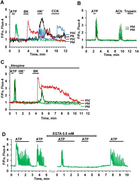
O. Gryshchenko et al.      |   7

                                                                                        BK-elicited Ca21 Signals in PMs
                                                                                        We have previously reported that a low concentration of BK
                                                                                        (1 nM), which typically elicits clear Ca2þ signals in PSCs did not
                                                                                        induce any changes in the cytosolic Ca2þ concentration ([Ca2þ]i)
                                                                                        in X-cells (PMs).11 However, in the present study, we found that
                                                                                        in about 40% of PMs tested, there was a Ca2þ signal in response
                                                                                        to higher concentrations of BK (Figure 7A, B). Figure 7B shows
                                                                                        typical Ca2þ signals in PMs elicited by BK at concentrations from
                                                                                        3 nM to 30 nM, but not at 1 nM BK. The BK-induced Ca2þ signals
                                                                                        in PMs were delayed as compared to those in PSCs (Figure 7A,
                                                                                        C). The delay depended on the BK concentration and became
                                                                                        progressively shorter at higher concentrations (Figure 7B). The

                                                                                                                                                                    Downloaded from https://academic.oup.com/function/article/2/1/zqaa026/5922605 by guest on 23 November 2020
                                                                                        B2 receptor antagonist WIN 64338 reversibly inhibited the
                                                                                        responses to BK (Figure 7C) in both PMs and PSCs, suggesting
                                                                                        that, similar to our previous finding for PCSs,11 PMs possess the
                                                                                        type 2 BK (B2) receptor. Atropine (Figure 3C) had no effect on the
                                                                                        BK-induced responses in PMs.

                                                                                        Discussion
                                                                                        In the quest to understand the mechanisms governing the func-
                                                                                        tion of an organ, it is natural initially to focus on the dominant cell
                                                                                        type, particularly if it is clear that it is the executor of the organ’s
                                                                                        primary physiological task. In the exocrine pancreas, it has been
                                                                                        clear for a long time that the quantitatively dominating acinar cells
                                                                                        synthesize the whole range of enzymes required for the digestion
                                                                                        of food entering the gut and that these cells secrete these enzymes
                                                                                        in a manner precisely controlled by local repetitive cytosolic Ca2þ
                                                                                        spikes.1 It is also clear that AP is initiated in the acinar cells by ex-
                                                                                        cessive global and sustained elevations of [Ca2þ]i.6 However, in re-
                                                                                        cent years it has become clear that there are other cell types in the
                                                                                        peri-acinar environment that can be regarded as “background
                                                                                        actors” and there is increasing interest in understanding their
                                                                                        function and how they may interact with the principal cells.28
                                                                                        Given that cytosolic Ca2þ signal generation is one of the most fun-
                                                                                        damental mechanisms regulating physiological processes29–31 and
                                                                                        that such signals can now be readily recorded in the quasi-intact
                                                                                        pancreas,10,11 Ca2þ measurements in the peri-acinar cells could be
                                                                                        an important tool for obtaining information about the signaling
                                                                                        functions of these accessory and less common cell types.
Figure 5. Ca2þ Re-entry in PMs after Store Depletion. (A). ATP (10 mM)-elicited Ca2þ        Having recently characterized the Ca2þ signaling properties
signals in PMs (light green and dark green traces) are gradually lost following expo-
                                                                                        of the pancreatic stellate cells (PSCs) in pancreatic lobules and
sure to the SERCA inhibitor CPA (20 mM) in the absence of external Ca2þ. [Ca2þ]i
traces from a PAC (blue) as well as from an ATP-insensitive PCS (red) from the
                                                                                        assessed their potential influence on the acinar cells,10,11 we
same pancreatic lobule are included for comparison. A short period of external          have now turned our attention to the immune cells in the exo-
Ca2þ (1 mM) readmission resulted in a transient [Ca2þ]i increase in all four cells (n   crine pancreatic tissue. We show that these cells exist in low
¼ 6). (B). Transient or sustained [Ca2þ]i rises following short-lasting or permanent    density in the normal pancreas and confirm that their number
readmission of external Ca2þ (1 mM) after intracellular Ca2þ store depletion by CPA
                                                                                        increases markedly in the first few days following induction of
in PM (green trace), PSC (red trace) and PAC (blue trace). ATP (1 lM) elicited a Ca2þ
                                                                                        AP by a combination of ethanol and fatty acids (Figure 2).
signal in the PM on the top of the elevated [Ca2þ]i plateau following the maintained
Ca2þ readmission (n ¼ 7). (C). The CRAC channel blocker GSK7975A (20 lM) abol-              Our data for the first time demonstrate functional responses
ished store-operated Ca2þ re-entry in a PM following Ca2þ readmission (n ¼ 7).          of immune cells in their natural environment, namely in live
                                                                                        pancreatic tissue. Ca2þ measurements in ex vivo lobules have
                                                                                        been complemented by immunocytochemistry to test the iden-
receptors, completely blocked Ca2þ transients in PMs induced                            tity of the monitored cells.10,11 This technique has allowed us to
by either ATP or MeSADP (Figure 6C). However, MRS 2211, a                               study both physiological and pathological responses related to
competitive antagonist of P2Y13 receptors also blocked both                             AP. Live immunocytochemistry is ideal for primary antibodies la-
ATP and MeSADP responses in PMs (Figure 6D). A selective                                beled with a fluorescent marker, in our case Alexa 647, but could
P2Y1 agonist, MRS 2365, as well as ADP, induced Ca2þ signals                            also be carried out with the use of secondary antibodies as previ-
in PMs (Figure 6B), whereas suramin, a potent blocker of P2Y                            ously reported.10,11 The positive staining with antibodies against
purinergic receptors, blocked Ca2þ signals in response to appli-                        F4/80 and CD11b (Figures 1 and 2) indicates that these immune
cation of 10 lM ATP in PMs (n ¼ 3). We suggest that this may be                         cells are most likely PMs,32 which have previously been reported
due to co-operation between P2Y1 and P2Y13 in PMs, as previ-                            to increase in numbers in chronic pancreatitis in mice.20
ously reported for mesenchymal stromal cells in adipose                                     Macrophages are known to express a variety of purinergic
tissue.27                                                                               P2Y receptors33 and here we show that both ADP and ATP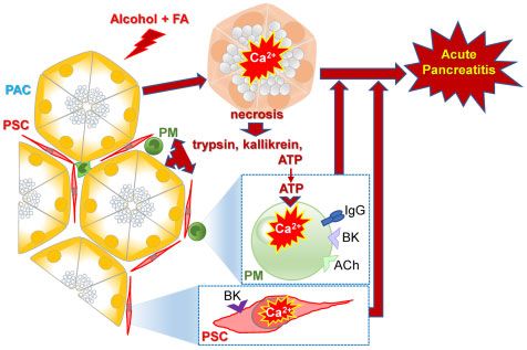
8   | FUNCTION, 2021, Vol. 2, No. 1

                                                                                                                                                                            Downloaded from https://academic.oup.com/function/article/2/1/zqaa026/5922605 by guest on 23 November 2020

Figure 6. Pharmacology of ATP-elicited Ca2þ Signals in PMs. (A). Both ATP (10 lM) and MeSADP (0.1 lM) evoke Ca2þ signals in a PM (n ¼ 64), but not in a BK-sensitive
PSC. MeSADP is a potent purinergic agonist displaying selectivity for P2Y1, P2Y12, and P2Y13 (pEC50 ¼ 8.29 and 9.05 for P2Y1 and P2Y12, EC50 ¼ 19 nM for P2Y13). (B).
Ca2þ signals in two PMs induced by subsequent addition of purinergic agonists: MeSADP (n ¼ 64), the selective P2Y1 agonist MRS 2365 (0.2 lM, 10 out of 15 cells), ADP
(20 lM, n ¼ 16), and ATP (10 lM). (C). MRS 2179, a selective antagonist of P2Y1 receptors, reversibly blocks Ca2þ transients evoked by activation of purinergic receptors
by 0.3 lM MeSADP (8 out of 10 cells) and 10 lM ATP. (D). MRS 2211(10 lM), a competitive antagonist of P2Y13 receptors reversibly blocks Ca2þ signals evoked by both
ATP (10 lM, 13 out of 14 cells) and MeSADP (0.3 lM, 11 out of 12 cells) in PMr.

activate P2Y receptors blocked by suramin and specific inhibi-                         described for ADP in mesenchymal stromal cells,27 which re-
tors of either P2Y1 or P2Y13 receptors (Figure 6). The effects of                      quired both P2Y1 and P2Y13 receptors, while specific inhibition
ATP and MeSADP (agonist of P2Y1, P2Y12, and P2Y13 receptors)                           of either of them abolished the responses to ADP.
were abolished by either MRS2179, a selective antagonist of                                It has been known for a long time that macrophages can
P2Y1 receptors, or MRS 2211, a competitive antagonist of                               generate cytosolic Ca2þ signals via activation of IP3 receptors34
P2Y13.27 Similar synergistic responses have previously been                            and here we show that the ATP-induced Ca2þ signals in PMs are
O. Gryshchenko et al. | 9
Downloaded from https://academic.oup.com/function/article/2/1/zqaa026/5922605 by guest on 23 November 2020
Figure 7. BK Concentration–Response Relationship in PMs. (A). Representative [Ca2þ]i trace shows that 20 nM BK can evoke a Ca2þ signal in a PM (but delayed compared
to the Ca2þ signal simultaneously recorded in a neighboring PSC) (81 out of 207 tested cells) while the B1 type receptor agonist S-BK has no effect. (B). Representative
traces from the same cell shows concentration-dependence of BK-induced Ca2þ signals in PM. For comparison, the upper trace (red) shows a BK (1 nM)-induced
Ca2þ signal in a PSC. (C). The bradykinin B2 type receptor antagonist WIN 64338 reversibly blocks Ca2þ signals evoked by BK (20 nM) in both a PM (n ¼ 8) and a PSC, with
no effect on the ATP responses in the PM.
primarily due to intracellular Ca2þ release (Figure 3) and can be NO production.36 It is therefore possible that ATP-elicited Ca2þ
very markedly reduced by the phospholipase C inhibitor U73122 signals in the PMs, via NO formation, could participate in the
or the inhibitor of store-operated Ca2þ entry 2-APB (Figure 4), vicious circle previously described in relation to the interaction
which can also inhibit IP3-elicited Ca2þ release.35 between acinar and stellate cells.11 The cytokine storm is likely
It is well established that macrophages produce and secrete to be amplified by the BK storm, since we have shown in
a wide range of inflammatory cytokines and Ca2þ signals in this study that the PMs are not only activated by ATP, but also
these cells have been linked to both production and secretion of by BK.
these inflammatory mediators.33,36–38 Although we currently Initial damage of the acinar cells, elicited by combinations
have no data about the consequence(s) of Ca2þ signal genera- of fatty acids and ethanol, by bile acids or by asparagi-
tion specifically in the macrophages in the intact pancreas, it is nase6,7,11 would release ATP into the acinar environment and
likely—based on what is generally known about macrophage elicit Ca2þ signals in the pancreatic macrophages. Via NO for-
function,33,36–38 that the Ca2þ signals described in our study mation (and possibly also other mechanisms), further dam-
could play an import role in the generation of the cytokine age of the pancreatic acinar cells would occur with additional
storm that is such an important feature of severe AP.39 release of ATP establishing a vicious circle. An initial element
It has very recently been noted that there are interesting of inflammation would induce entry of more PMs into the
similarities between the multi-organ failures and patterns of pancreatic tissue, amplifying the overall impact of Ca2þ signal
elevated cytokines observed in severe cases of COVID-19 and generation in these cells and further worsening the severity
AP.39 Furthermore, it has been suggested that in addition to of AP.
the cytokine storm, there is also a BK storm involved in severe Clearly, we are still at an early stage of understanding the
cases of COVID-19.40 This may also be the case in severe cases initiation of the inflammatory response that in severe cases of
of AP. Plasma levels of BK are elevated in AP,41 and this causes AP results in destruction of the pancreas and other organs, but
Ca2þ signal-mediated NO formation in the PSCs.10–12 There is we can now begin to appreciate the importance of the role that
evidence indicating that the NO formation amplifies acinar ne- cells in the peri-acinar environment, such as stellate cells and
crosis.11,12 In macrophages, intracellular Ca2þ can also activate PMs, could play in this process.10 | FUNCTION, 2021, Vol. 2, No. 1
Acknowledgments 14. Russo MW, Wei JT, Thiny MT, et al. Digestive and liver dis-
eases statistics, 2004. Gastroenterology 2004;126:1448–1453.
O.G., J.V.G., O.H.P., and O.V.G. designed the study; O.G., J.V.G., 15. Gundra UM, Girgis NM, Ruckerl D, et al. Alternatively
and O.V.G. conducted and analyzed the experiments; O.G., activated macrophages derived from monocytes and tissue
J.V.G., O.H.P., and O.V.G. wrote the manuscript. macrophages are phenotypically and functionally distinct.
Blood 2014;123 (20):e110–e122.
Funding 16. Schulz C, Gomez Perdiguero E, Chorro L, et al. A lineage of
myeloid cells independent of Myb and hematopoietic stem
The work was supported by grants from the Medical cells. Science 2012;336(6077):86–90.
Research Council (UK) (MR/J002771/1 and G19/22/2 to O.H.P.) 17. Weisberg SP, Carpenter DJ, Chait M, et al. Tissue-resident
and Children with Cancer UK grants (2014/167 and 2019/288 memory T cells mediate immune homeostasis in the human
to O.V.G. and J.V.G.). pancreas through the PD-1/PD-L1 pathway. Cell Rep 2019;29,
3916–3932.
Downloaded from https://academic.oup.com/function/article/2/1/zqaa026/5922605 by guest on 23 November 2020
18. Rakonczay Z, Hegyi P, Takacs T, et al. The role of NF-jB acti-
Conflict of interest statement
vation in the pathogenesis of acute pancreatitis. Gut 2008;
None declared. 57(2):259–267.
19. Wen L, Voronina S, Javed MA al. Inhibitors of ORAI1 prevent
cytosolic calcium-associated injury of human pancreatic aci-
References
nar cells and acute pancreatitis in 3 mouse models.
1. Petersen OH, Tepikin A. Polarized calcium signaling Gastroenterology 2015;149(2):481–492.
in exocrine gland cells. Annu Rev Physiol 2008;70:273–299. 20. Xue J, Sharma V, Hsieh MH. Alternatively activated macro-
2. Gerasimenko JV, Gryshchenko O, Ferdek PE, et al. Ca2þ phages promote pancreatic fibrosis in chronic pancreatitis.
release-activated Ca2þ channel blockade as a potential tool in Nat Commun 2015;6:71582014.
anti-pancreatitis therapy. Proc Natl Acad Sci U S A 2013;110: 21. Nikolic T, Bunk M, Drexhage HA, et al. Bone marrow precur-
13186–13191. sors of nonobese diabetic mice develop into defective
3. Gerasimenko JV, Charlesworth RM, Sherwood MW, et al. Both macrophage-like dendritic cells in vitro. J Immunol 2004;
RyRs and TPCs are required for NAADP-induced intracellular 173(7):4342–4351.
Ca2þ release. Cell Calcium 2015;58(3):237–245. 22. Rostam HM, Reynolds PM, Alexander MR, et al. Image based
4. Pallagi P, Madacsi T, Varga A, et al. Intracellular Ca2þ Machine Learning for identification of macrophage subsets.
signalling in the pathogenesis of acute pancreatitis: recent Sci Rep 2017,7:3521.
advances and translational perspectives. Int J Mol Sci 2020; 23. Skinner BM, Johnson EEP. Nuclear morphologies: their
21(11):4005. diversity and functional relevance. Chromosoma 2017;126:
5. Murphy JA, Criddle DN, Sherwood M, et al. Direct activation of 195–212.
cytosolic Ca2þ signaling and enzyme secretion by cholecysto- 24. Trescos Y, Tessier E, Rougeaux C, et al. Micropatterned
kinin in human pancreatic acinar cells Gastroenterology 2008; macrophage analysis reveals global cytoskeleton constraints
135(2):632–641. induced by bacillus anthracis edema toxin. Infect Immun 2015,
6. Gerasimenko JV, Gerasimenko OV, Petersen OH. The role of 83:3114–3125.
Ca2þ in the pathophysiology of pancreatitis. J Physiol 2014; 25. Myers JT, Swanson JA. Calcium spikes in activated
592(2):269–280. macrophages during Fcc receptor-mediated phagocytosis.
7. Peng S, Gerasimenko JV, Tsugorka TM, et al. Galactose J Leukocyte Biol 2002;72:677–684.
protects against cell damage in mouse models of acute 26. Feske S, Gwacket Y, Prakriya M, et al. A mutation in Orai1
pancreatitis. J Clin Invest 2018;128(9):3769–3778. causes immune deficiency by abrogating CRAC channel func-
8. Swain SM, Romac JM-J, Shahid R, et al. TRPV4 channel open- tion. Nature 2006;441:179–185.
ing mediates pressure-induced pancreatitis initiated by 27. Kotova PD, Bystrova MF, Rogachevskaja OA, et al. Coupling
Piezo1 activation. J Clin Invest 2020;130(5): 2527–2541. of P2Y receptors to Ca2þ mobilization in mesenchymal
9. Ferdek PE, Jakubowska MA, Gerasimenko JV, et al. Bile acids stromal cells from the human adipose tissue. Cell Calcium
induce necrosis in pancreatic stellate cells dependent on cal- 2018;7:1–14.
cium entry and sodium-driven bile uptake. J Physiol 2016; 28. Kusiak AA, Szopa MD, Jakubowska MA, et al. Signaling in the
594(21):6147–6164. physiology and pathophysiology of pancreatic stellate cells –
10. Gryshchenko O, Gerasimenko JV, Gerasimenko OV, et al. Ca2þ a brief review of recent advances. Front Physiol 2020;11:78.
signals mediated by bradykinin type 2 receptors in normal 29. Berridge MJ. The inositol trisphosphate/calcium signaling
pancreatic stellate cells can be inhibited by specific Ca2þ pathway in health and disease. Physiol Rev 2016;96:1261–1296.
channel blockade. J Physiol 2016;594(2):281–293. 30. Petersen OH, Verkhratsky A. Calcium and ATP control multi-
11. Gryshchenko O, Gerasimenko JV, Peng S, et al. Calcium sig- ple vital functions. Philos Trans R Soc Lond B Biol Sci 2016;
nalling in the acinar environment of the exocrine pancreas: 371(1700):20150418.
physiology and pathophysiology. J Physiol 2018;596(14): 31. Rizzuto R, Pozzan T. Microdomains of intracellular Ca2þ: mo-
2663–2678. lecular determinants and functional consequences. Physiol
12. Jakubowska MA, Ferdek PE, Gerasimenko OV, et al. Nitric ox- Rev 2006;86(1):369–408.
ide signals are interlinked with calcium signals in normal 32. Galli SJ, Borregaard N, Wynnet TA. Phenotypic and functional
pancreatic stellate cells upon oxidative stress and inflamma- plasticity of cells of innate immunity: macrophages, mast
tion Open Biol 2016;6(8):160149. cells and neutrophils. Nat Immunol 2011;12(11):1035–1044.
13. Calderon B, Carrero JA, Ferris ST, et al. The pancreas anatomy 33. Desai BN, Leitinger N. Purinergic and calcium signaling in
conditions the origin and properties of resident macro- macrophage function and plasticity. Front Immunol 2014;5:
phages. J Exp Med 2015;212:1497–1512. 580.O. Gryshchenko et al. | 11
34. Randriamampita C, Bismuth G, Trautmann A. Ca2þ-induced IL-10 production in human macrophages. J Immunol 2010;184:
Ca2þ release amplifies the Ca2þ response elicited by inositol 5545–5552.
trisphosphate in macrophages. Cell Regul 1991;2:513–522, 39. Hegyi P, Szakacs Z, Sahin-Toth M. Lipotoxicity and cytokine
35. Bootman MD, Collins TJ, Mackenzie L, et al. 2- storm in severe acute pancreatitis and COVID-19.
Aminoethoxydiphenyl borate (2-APB) is a reliable blocker of Gastroenterology 2020;824–827.
store-operated Ca2þ entry but an inconsistent inhibitor of 40. Garvin MR, Alvarez C, Miller JI, et al. A mechanistic
InsP3-induced Ca2þ release. FASEB J 2002;16(10):1145–1150. model and therapeutic interventions for COVID-19
36. Feske S, Wulff H, Skolnik EY, et al. Ion channels in innate and involving a RAS-mediated bradykinin storm. eLife 2020;9:
adaptive immunity. Annu Rev Immunol 2015;33:291–353 e59177.
37. Murray RZ, Stow JL. Cytokine secretion in macrophages: SNAREs, 41. Hirata M, Hayashi I, Yoshimura K, et al. Blockade of bradyki-
Rabs, and membrane trafficking. Front Immunol 2014;5:538. nin B2 receptor suppresses acute pancreatitis induced by ob-
38. Kelly EK, Wang L, Ivashkiv LB. Calcium-activated pathways struction of the pancreaticobiliary duct in rats. Br J Pharmacol
and oxidative burst mediate zymosan-induced signalling and 2002;135:29–36.
Downloaded from https://academic.oup.com/function/article/2/1/zqaa026/5922605 by guest on 23 November 2020You can also read