Basal cell carcinoma not always the 'good guy': Case report of a life threatening basosquamous carcinoma and review of the literature - Spandidos ...
←
→
Page content transcription
If your browser does not render page correctly, please read the page content below
EXPERIMENTAL AND THERAPEUTIC MEDICINE 22: 1158, 2021

                   Basal cell carcinoma‑not always the ‘good guy’:
                   Case report of a life‑threatening basosquamous
                       carcinoma and review of the literature
                          CORNEL ADRIAN PETREANU1,2, ELENA‑DANIELA ȘERBAN3,
               MARIA‑MAGDALENA CONSTANTIN3,4, CORNEL SAVU1,2, ALEXANDRU VICTOR ZARIOSU2,
                   OANA CLAUDIA DELEANU5,6, STEFANA BUCUR3,4 and TRAIAN CONSTANTIN7,8

           1
            Department of Thoracic Surgery, ‘Carol Davila’ University of Medicine and Pharmacy, 050474 Bucharest;
       2
         1st Department of Thoracic Surgery, ‘Prof. Dr. Marius Nasta’ Institute of Pneumophtisiology, 050159 Bucharest;
       3
         2nd Department of Dermatology, ‘Colentina’ Clinical Hospital, 020125 Bucharest; Departments of 4Dermatology
               and 5Pneumophtisiology, ‘Carol Davila’ University of Medicine and Pharmacy, 050474 Bucharest;
     6
      2nd Department of Pneumophtisiology, ‘Prof. Dr. Marius Nasta’ Institute of Pneumophtisiology, 050159 Bucharest;
               7
                 Department of Urology, ‘Carol Davila’ University of Medicine and Pharmacy, 050474 Bucharest;
                    8
                      Department of Urology, ‘Prof. Dr. Th. Burghele’ Hospital, 050659 Bucharest, Romania

                                           Received June 25, 2021; Accepted July 28, 2021

                                                     DOI: 10.3892/etm.2021.10592

Abstract. Cutaneous basosquamous carcinoma is a variant                Introduction
of basal cell carcinoma that is characterized by histopatho‑
logical features of both basal and squamous cell carcinoma.            Basosquamous carcinoma (BSC), also called metatypical
Due to its local invasiveness, high frequency of recurrence,           basal cell carcinoma, is a subtype of basal cell carcinoma
and its metastatic potential, it is considered to be one of the        (BCC), displaying histological features of both BCC and
most aggressive subtypes of basal cell carcinoma. We present           squamous cell carcinoma (SCC) (1). This tumor may
the case of an 81‑year‑old male who was admitted to the                have a more aggressive behavior that resembles more
hospital with incessant hemorrhage arising from a cutaneous            an SCC than a BCC in terms of invasiveness and poor
tumor that later proved to be a basosquamous carcinoma. Due            prognosis, with a greater likelihood of recurrence and
to the COVID‑19 pandemic at the time, the patient did not              metastasis (2).
seek medical attention as soon as the bleeding was observed,               As with any other BCC, it presents as a plaque, papule,
although he did present when the symptom increased in                  or nodule with an ulcerating potential. Elderly, fair‑skinned
intensity and became incessant. To our knowledge, this is              white men are the most affected, especially on the head and
the first case report of a cutaneous basosquamous carcinoma            neck area (3). Dermoscopy shows polymorphous vascular
that presents with a massive life‑threatening hemorrhage               structures typical of BCC combined with scaling, ulcer‑
tumor, thus endangering the patient's life. The clinical and           ation, white circles and white structureless areas (4,5).
histopathological features, the behavior and the treatment of          Histopathologically, it is characterized by islands of basa‑
cutaneous basosquamous carcinoma are further reviewed in               loid cells mixed with atypical squamous cells (1). According
this article.                                                          to the National Comprehensive Cancer Network (NCCN)
                                                                       Clinical Practice Guidelines in Oncology, BSC is included
                                                                       in the high‑risk BCC category (2). This is due to the higher
                                                                       metastatic rate when compared with conventional BCC
                                                                       (5‑8.4 vs.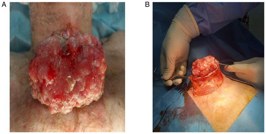
2 PETREANU et al: CASE REPORT OF A LIFE-THREATENING BASOSQUAMOUS CARCINOMA AND REVIEW
Case presentation be explained in regards to the inverse association of BCC
with the country's geographic latitude and the Fitzpatrick skin
An 81‑year‑old male presented with a hemorrhagic tumor phototype of its inhabitants (16).
located in the suprasternal notch. He had chronic atrial fibril‑ Literature indicates that BCC is a heterogeneous entity and
lation and was under chronic anticoagulation treatment with that specific histological variants can arise in particular areas,
apixaban. There was no personal clinical history and family exhibiting different clinical behavior, have a disparate etiology,
history for other tumors, skin or otherwise The tumor had and a distinct response to treatment (18). The latest edition of
appeared approximately 12 years ago and had a rapid growth the World Health Organization (WHO) Classification of Skin
during the previous year, but the patient did not seek medical Tumors recognizes the following 10 histological subtypes of
care due to the COVID‑19 pandemic crisis at the moment. BCC: nodular, superficial, micronodular, infiltrating, scle‑
Physical examination revealed a bleeding vegetating tumor rosing/morphoeic, basosquamous, pigmented, fibroepithelial,
(Fig. 1A), measuring 14x12 cm. The perilesional skin appeared BCC with sarcomatoid differentiation, and BCC with adnexal
erythematous and edematous. Clinically, there was no differentiation (1). Although the pathogenesis of basosquamous
evidence of lymphadenopathy. The clinical differential diag‑ carcinoma (BSC) has historically been a subject of debate, as
nosis included SCC, BCC, Merkel cell carcinoma, amelanotic some authors considered it to be a subtype of BCC and others
melanoma, or cutaneous metastasis of unknown origin. The an independent tumor with a different evolution, WHO finally
patient was controlled using mechanical hemostatic methods identifies it as a BCC variant (1). In any case, it is rather rare,
such as direct pressure and gauze pack and local hemostatic and data concerning the precise percentage are also missing.
agents, while also stopping the anticoagulant. To further plan It is estimated that BSC represents approximately 1.2 to 2.7%
the surgical excision, especially in consideration of the tumor's of all malignancies in the NMSC group (7,19‑22).
great size, a whole‑body computed tomography (CT) scan was Ultraviolet radiation (UVR) is known to be the prevalent
performed which did not reveal any metastasis or neoplastic risk factor for BCC, SCC, and therefore BSC (23) with a pref‑
infiltration of the muscle fascia. Therefore, standard surgical erential localization on the sun‑exposed areas, especially the
excision with 2‑cm margins was performed with the imme‑ head and neck region (3). Moreover, it appears that BSC occurs
diate repair of the surgical defect using adjacent tissue flaps more frequently in men (3,24) in the six to eight decades of
(Fig. 1B). The histopathology examination revealed a large life (24) with a mean age at diagnosis of 72±11.5 years (3).
ulcerated eso‑endophytic lesion invading the dermis and the It is difficult to make a diagnosis of BSC using only naked
subcutis composed of atypical basaloid strands and islands eye examination as it resembles other subtypes of BCC. It
commingled with islands of polygonal atypical squamous presents as a pink or flesh‑colored plaque, papule, or nodule.
cells with abundant eosinophilic cytoplasm, embedded in a Ulceration is usually present. The lesion can have a pearly
cellular fibrotic stroma and chronic inflammatory infiltrate. or translucent feature with telangiectatic vessels being seen
Lymphovascular invasion was present. No perineural inva‑ within it (3).
sion was observed. Circumferential, peripheral and deep While clinical differentiation from other BCC variants
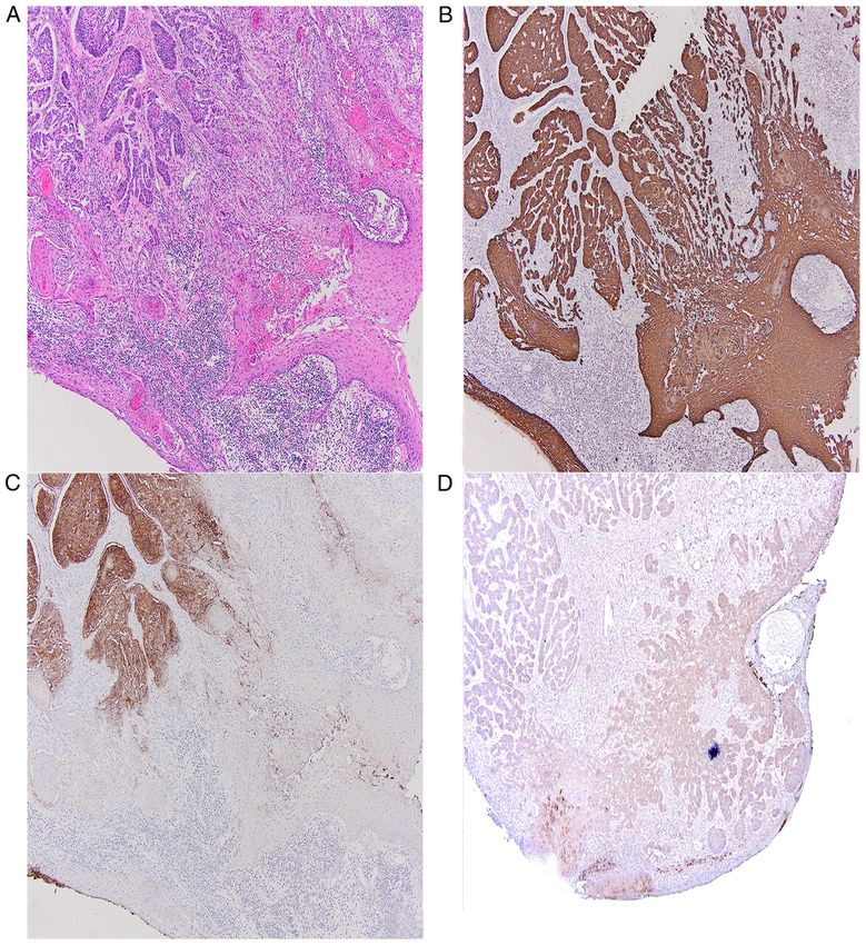
margin assessment was negative. At immunohistochemistry, is demanding, dermoscopy can provide some important
there was diffuse positivity for cytokeratin 34betaE12. The hints allowing a more appropriate treatment planning. The
basaloid component stained positive for Ber‑EP4 with loss dermoscopic evaluation of BSC detects features of both BCC
of immunoreactivity of Ber‑EP4 in areas showing squamous and SCC such as unfocused arborizing vessels, blue‑gray
differentiation, whereas epithelial membrane antigen (EMA) blotches, ulceration or blood crusts, white structures and white
and p53 were partially immunoreactive only in the squamous structureless areas. Identification of only one overlapping
differentiated areas (Fig. 2). The Ki‑67 immunoreactivity was dermoscopic criterion both of BCC and SCC is an alarming
about 60‑70%. Based on the data gathered, a diagnosis of clue for BSC (4,5).
basal cell carcinoma, basosquamous subtype, was made. The The definitive diagnosis of BSC is made only by histo‑
TNM according to the Eighth Edition of the American Joint pathological examination. The latest edition of the WHO
Committee on Cancer Classification (AJCC) was pT3Nx (14). Classification of Skin Tumors describes BSC as having
Since there was no sign of residual disease, no further treatment features of both BCC and SCC such as islands of basaloid
was indicated and follow‑up was recommended. cells mixed with focal or scattered atypical squamous cells
At six‑month follow‑up, there was no clinical sign of local and transition zones containing cells with features interme‑
recurrence or metastasis. diate between the two. The malignant cells are embedded in
a cellular fibrotic stroma. The basaloid component expresses
Discussion BerEP4 with a gradual loss of reactivity towards the squamous
cell component. The squamous cells stain positively for EMA.
Non‑melanoma skin cancer (NMSC) is the most common There is also a high cyclin D1 expression as well as a low
group of cutaneous neoplasms in the Caucasian population BCL2 expression (1).
with basal cell carcinoma (BCC) being the prevalent entity, Before treatment, assessment of recurrence risk is manda‑
although precise epidemiologic data are limited, as in most tory. According to the NCCN Clinical Practice Guidelines
countries there are no cancer registries that collect data in Oncology concerning the risk of recurrence, there are two
regarding it (15‑17). main categories of BCC: the low‑risk group and the high‑risk
However, the NMSC incidence rate is on a steady increase group (2). The following features are associated with lesions
in the USA, most European countries, and Australia, with the with a low risk of recurrence potential: immunocompetent
latter registering the highest growth (15). These trends could patient, no history of radiation therapy at the site, not aEXPERIMENTAL AND THERAPEUTIC MEDICINE 22: 1158, 2021 3
Figure 1. (A) Clinical presentation. (B) Intra‑operatory findings.
recurrent tumor (a primary tumor); well‑defined clinical This category includes topical therapies such as imiquimod
borders;4 PETREANU et al: CASE REPORT OF A LIFE-THREATENING BASOSQUAMOUS CARCINOMA AND REVIEW
Figure 2. (A) Histopathological examination using hematoxylin and eosin staining at a magnification of x4. (B) 34 betaE2 immunostaining at a magnification
of x4. (C) BerEP4 immunostaining at a magnification of x4. (D) EMA immunostaining at a magnification of x4.
considered one of the most aggressive subtypes because of the Funding
high‑risk of local invasiveness, high frequency of recurrences,
and metastases. The diagnosis is made almost exclusively No funding was received.
by histopathological examination and the treatment is based
mainly on surgery. Availability of data and materials
In this article, we presented the case of an 81‑year‑old‑male
with an uncontrolled hemorrhage deriving from a basosqua‑ Any further information concerning the case report is avail‑
mous carcinoma. As mentioned before, this entity has an able upon request of the corresponding author.
aggressive behavior, presenting with ulceration and infiltra‑
tion, but this is the first case report, at least to the best of Authors' contributions
our knowledge, that presented as a massive hemorrhage
endangering the patient's life. All authors had equally contributed to writing and editing the
manuscript. TC contributed in all the stages of the article; he
Acknowledgements designed the article and revised the manuscript for impor‑
tant intellectual content. MMC, EDS and CAP contributed
Not applicable. to the conception of the work, revision of the language andEXPERIMENTAL AND THERAPEUTIC MEDICINE 22: 1158, 2021 5
contributed to drafting the final paper. CS, AVZ, SB and OCD 12. Wetzig T, Woitek M, Eichhorn K, Simon JC and Paasch U:
Surgical excision of basal cell carcinoma with complete margin
revised the work critically in light of the literature data. The control: Outcome at 5‑year follow‑up. Dermatology 220: 363‑369,
final manuscript for publication was approved by all authors. 2010.
13. Likhacheva A, Awan M, Barker CA, Bhatnagar A, Bradfield L,
Brady MS, Buzurovic I, Geiger JL, Parvathaneni U, Zaky S and
Ethics approval and consent to participate Devlin PM: Definitive and postoperative radiation therapy for
basal and squamous cell cancers of the skin: Executive summary
The patient provided informed written consent prior to inclu‑ of an American society for radiation oncology clinical practice
guideline. Pract Radiat Oncol 10: 8‑20, 2020.
sion in the present article. No ethics committee approval was 14. Keohane SG, Proby CM, Newlands C, Motley RJ, Nasr I, Mohd
necessary. Mustapa MF; British Association of Dermatologists (Squamous
and Basal Cell Carcinoma Guideline Development Groups),
Slater DN; Royal College of Pathologists (Skin Cancer Lead):
Patient consent for publication The new 8th edition of TNM staging and its implications for skin
cancer: a review by the British Association of Dermatologists
The patient provided written informed consent for the and the Royal College of Pathologists, U.K. Br J Dermatol 179:
824-828, 2018.
publication of any associated data and accompanying images. 15. Lomas A, Leonardi‑Bee J and Bath‑Hextall F: A systematic
review of worldwide incidence of nonmelanoma skin cancer. Br
Competing interests J Dermatol 166: 1069‑1080, 2012.
16. Verkouteren JAC, Ramdas KHR, Wakkee M and Nijsten T:
Epidemiology of basal cell carcinoma: Scholarly review. Br
The authors declare that they have no competing interests and J Dermatol 177: 359‑372, 2017.
they have no financial relationships to disclose. 17. Solovastru LG, Vata D, Statescu L, Constantin MM and
Andrese E: Skin cancer between myth and reality, yet ethically
constrained. Rev Rom Bioet 12: 47‑52, 2014.
References 18. Scrivener Y, Grosshans E and Cribier B: Variations of basal cell
carcinomas according to gender, age, location and histopatho‑
1. Elder DE, Massi D, Scolyer RA and Willemze R: Basal cell logical subtype. Br J Dermatol 147: 41‑47, 2002.
carcinoma. In: WHO Classification of Skin Tumors. 4th edition. 19. Martin RC II, Edwards MJ, Cawte TG, Sewell CL and
International Agency for Research on Cancer, Lyon, France, McMasters KM: Basosquamous carcinoma: Analysis of prog‑
pp26‑34, 2018. nostic factors influencing recurrence. Cancer 88: 1365‑1369,
2. Bichakjian CK, Olencki T, Aasi SZ, Alam M, Andersen JS, 2000.
Berg D, Bowen GM, Cheney RT, Daniels GA, Glass LF, et al: 20. Garcia C, Poletti E and Crowson AN: Basosquamous carcinoma.
Basal cell skin cancer, version 1.2016, NCCN clinical practice J Am Acad Dermatol 60: 137‑143, 2009.
guidelines in oncology. J Natl Compr Canc Netw 14: 574‑597, 21. Bowman PH, Ratz JL, Knoepp TG, Barnes CJ and Finley EM:
2016. Basosquamous carcinoma. Dermatol Surg 29: 830‑832, 2003.
3. Ciążyńska M, Sławińska M, Kamińska‑Winciorek G, Lange D, 22. Leibovitch I, Huilgol SC, Selva D, Richards S and Paver R:
Lewandowski B, Reich A, Pabianek M, Szczepaniak K, Basosquamous carcinoma: Treatment with mohs micrographic
Hankiewicz A, Ułańska M, et al: Clinical and epidemiological surgery. Cancer 104: 170‑175, 2005.
analysis of basosquamous carcinoma: Results of the multicenter 23. Gallagher RP, Hill GB, Bajdik CD, Fincham S, Coldman AJ,
study. Sci Rep 10: 18475, 2020. McLean DI and Threlfall WJ: Sunlight exposure, pigmentary
4. Akay BN, Saral S, Heper AO, Erdem C and Rosendahl C: factors, and risk of nonmelanocytic skin cancer. I. Basal cell
Basosquamous carcinoma: Dermoscopic clues to diagnosis. carcinoma. Arch Dermatol 131: 157‑163, 1995.
J Dermatol 44: 127‑134, 2017. 24. Oldbury JW, Wain RAJ, Abas S, Dobson CM and Iyer SS:
5. Giacomel J, Lallas A, Argenziano G, Reggiani C, Piana S, Basosquamous carcinoma: A single centre clinicopathological
Apalla Z, Ferrara G, Moscarella E, Longo C and Zalaudek I: evaluation and proposal of an evidence‑based protocol. J Skin
Dermoscopy of basosquamous carcinoma. Br J Dermatol 169: Cancer 3: 6061395, 2018.
358‑364, 2013. 25. Gulleth Y, Goldberg N, Silverman RP and Gastman BR: What
6. Alam M, Desai S, Nodzenski M, Dubina M, Kim N, Martini M, is the best surgical margin for a basal cell carcinoma: A meta‑
Fife D, Reid D, Pirigyi M, Poon E, et al: Active ascertainment of analysis of the literature. Plast Reconstr Surg 126: 1222‑1231,
2010.
recurrence rate after treatment of primary basal cell carcinoma 26. Smeets NW, Krekels GA, Ostertag JU, Essers BA, Dirksen CD,
(BCC). J Am Acad Dermatol 73: 323‑325, 2015. Nieman FH and Neumann HA: Surgical excision vs Mohs'
7. Tan CZ, Rieger KE and Sarin KY: Basosquamous carcinoma: micrographic surgery for basal‑cell carcinoma of the face:
Controversy, advances, and future directions. Dermatol Surg 43: Randomised controlled trial. Lancet 364: 1766‑1772, 2004.
23‑31, 2017. 27. Bath‑Hextall F, Ozolins M, Armstrong SJ, Colver GB, Perkins W,
8. Wermker K, Roknic N, Goessling K, Klein M, Schulze HJ Miller PS and Williams HC; Surgery versus Imiquimod for
and Hallermann C: Basosquamous carcinoma of the head and Nodular Superficial basal cell carcinoma (SINS) study group:
neck: Clinical and histologic characteristics and their impact on Surgical excision versus imiquimod 5% cream for nodular
disease progression. Neoplasia 17: 301‑305, 2015. and superficial basal‑cell carcinoma (SINS): A multicentre,
9. Zhu GA, Danial C, Liu A, Li S and Chang AL: Overall and non‑inferiority, randomised controlled trial. Lancet Oncol 15:
progression‑free survival in metastatic basosquamous cancer: A 96‑105, 2014.
case series. J Am Acad Dermatol 70: 1145‑1146, 2014. 28. Love WE, Bernhard JD and Bordeaux JS: Topical imiquimod or
10. Peris K, Fargnoli MC, Garbe C, Kaufmann R, Bastholt L, fluorouracil therapy for basal and squamous cell carcinoma: A
Seguin NB, Bataille V, Marmol VD, Dummer R, Harwood CA, systematic review. Arch Dermatol 145: 1431‑1438, 2009.
et al: Diagnosis and treatment of basal cell carcinoma: European 29. Voiculescu VM, Lisievici CV, Lupu M, Vajaitu C, Draghici CC,
consensus‑based interdisciplinary guidelines. Eur J Cancer 118: Popa AV, Solomon I, Sebe TI, Constantin MM and Caruntu C:
10‑34, 2019. Mediators of inflammation in topical therapy of skin cancers.
11. Ad Hoc Task Force; Connolly SM, Baker DR, Coldiron BM, Mediators Inflamm 10: 8369690, 2019.
Fazio MJ, Storrs PA, Vidimos AT, Zalla MJ, Brewer JD, 30. R o o z e b o o m M H , A r i t s A H , N e l e m a n s PJ a n d
Begolka WS, et al: AAD/ACMS/ASDSA/ASMS 2012 appro‑ Kelleners‑Smeets NW: Overall treatment success after treatment
priate use criteria for mohs micrographic surgery: A report of of primary superficial basal cell carcinoma: A systematic review
the American academy of dermatology, American college of and meta‑analysis of randomized and nonrandomized trials. Br
mohs surgery, American society for dermatologic surgery asso‑ J Dermatol 167: 733‑756, 2012.
ciation, and the American society for mohs surgery. J Am Acad
Dermatol 67: 531‑550, 2012.You can also read