Bacteriological Analytical Manual (BAM) Chapter 10: Detection of Listeria monocytogenes in Foods and Environmental Samples, and Enumeration of ...
←
→
Page content transcription
If your browser does not render page correctly, please read the page content below
Bacteriological Analytical Manual (BAM)
Chapter 10: Detection of Listeria
monocytogenes in Foods and
Environmental Samples, and Enumeration
of Listeria monocytogenes in Foods

Authors: Anthony D. Hitchins (ret.) and Karen Jinneman and Yi Chen

April 2022

https://www.fda.gov/food/laboratory-methods-food/bacteriological-analytical-manual-bam
Table of Contents

Revision History ............................................................................................................................. 3
Introduction ..................................................................................................................................... 4
A. Equipment and Materials ........................................................................................................... 4
B. Media and Reagents ................................................................................................................... 5
C. Control Cultures ......................................................................................................................... 6
D. Sample Preparation .................................................................................................................... 7
E. Enrichment Procedure ................................................................................................................ 8
F. Alternate Screening Methodologies ........................................................................................... 8
G. Isolation Procedure .................................................................................................................. 11
H. Identification Procedure ........................................................................................................... 12
I. Subtyping of L. monocytogenes Isolates (required) .................................................................. 17
J. Enumeration (required) ............................................................................................................. 18
References ..................................................................................................................................... 20

                                                                                                                                                  2
Revision History
• April 2022: Section E.2 correction to additives amounts, Section J. clarification on
enumeration homogenate preparations
• February 2022: Section E.2, correction to additives amounts; Section J, clarification on
enumeration homogenate preparations; editing of other sections.
• March 2017: Addition of the sample matrix for environmental samples.
• January 2016: More specific sample preparation and analytical set-up instructions for
qualitative detection or quantitative enumeration.
• January 2016: Reorganization and editing of all sections.
• February 2013: update to Table 1; update for BAM Media M52:
buffered Listeria enrichment broth (BLEB) in the Media section.
• November 2011: Addition of PCR confirmation for Listeria
monocytogenes and Listeria spp. isolates other than L. grayi.
• April 2011: Section E. Diagram describing the Henry Optical System for examination of
colonies added; references for Listeria monocytogenes Risk Assessment and Guidance
updated.
• August 2002: Section J. Enumeration: Added instructions for positive result on all MPN
tubes.
• April 2001: Section H. CAMP test: Updated address for ATCC and added a link to its
web page.
3Introduction
The genus Listeria contains 6 species: L. monocytogenes, L. innocua, L. seeligeri, L.
welshimeri, L. ivanovii, and L. grayi (Table 1). L. grayi (28, 40) and L. ivanovii (13, 27) each
contain two subspecies, which do not need to be specified in this analysis. A taxonomic review
of the genus by Rocourt (41) in 1999 updated the previous reviews (11, 43). In recent years,
many new species were proposed. However, these new species are not widely adopted and the
number of type strains for the newly proposed species are very limited. L. ivanovii and L.
monocytogenes are pathogenic for mice and other animals. However, only L. monocytogenes is
commonly associated with human listeriosis. Listeriosis associated infection by L. ivanovii, and
even by L. seeligeri, is extremely rare in humans. The universal occurrence of L.
monocytogenes in food (42) and the risk of contracting foodborne listeriosis (47,48) have been
thoroughly reviewed recently. This chapter describes the detection and enumeration of L.
monocytogenes in foods and detection from food processing environment.
This standard methodology and alternative rapid methodologies are intended to be used for
detection and isolation of L. monocytogenes from foods and environmental samples. Analytical
sample size for foods is generally 25 g, and this can be from individual units or as part of a
sample composite.
Alternatively, rapid test kits with their respective enrichment media approved as AOAC Official
Methods of Analysis (OMA) may be conditionally used to screen for the presence
of Listeria contaminants. Putative Listeria isolates on selective agars from standard or screen
positive enrichments are purified on non-selective agars and confirmed by conventional
identification tests or by a battery of such tests in kit form. Isolates may be rapidly confirmed
as L. monocytogenes (or not) by using specific test kits or PCR procedures. Subtyping of L.
monocytogenes isolates is generally expected, which includes serological typing and whole
genome sequencing. Optional pathogenicity testing of L. monocytogenes isolates is described in
Section H.
Enumeration of L. monocytogenes in positive food samples is performed on reserve sample by
colony count on L. monocytogenes differential selective agars in conjunction with MPN
enumeration using selective enrichment in buffered Listeria enrichment broth with subsequent
plating on L. monocytogenes differential selective agars as described below.
A. Equipment and Materials
FDA does not specifically endorse any of commercial products listed. Equivalent products may
be available.
1. Sterile swab: The following or equivalent:
a. 3M™ Swab-sampler in 10 ml D/E neutralizing broth (catalog#
RS96010DE, www.mmm.com) (Polyester).
4b. Puritan® dry cotton swab (catalog# 25-806 1PC, 25-806
2PC, www.puritanmedproducts.com) (Cotton).
c. World Bioproducts PUR-Blue™ swab sampler with (catalog# BLU-10DE) or
without (catalog# BLU-DRY, www.worldbioproducts.com) 10 ml D/E neutralizing
broth (Polyurethane).
d. Healthlink® dry swab transporter (catalog# 4159BX, www.hardydiagnostics.com)
(Polyester).
2. Sterile sponge: The following or equivalent:
a. World Bioproducts EZ Reach™ sponge sampler with (catalog# EZ-10DE-PUR) or
without (catalog# EZ-DRY-PUR, www.worldbioproducts.com) 10 ml D/E neutralizing
broth (Polyurethane).
b. Nasco Whirl-Pak® dry sponge probe (catalog# B01475WA, www.enasco.com)
(Cellulose).
c. 3M™ Sponge-sticks with (catalog# SSL10DE, www.mmm.com) or without (catalog#
SSL100) 10 ml D/E neutralizing broth (Cellulose).
If available, dry samplers can be obtained and D/E neutralizing broth added later, or pre-
moistened samplers can be obtained. We do not recommend less than 10 ml D/E neutralizing
broth due to the possible presence of sanitizer residues on environmental surfaces.
3. Balance for weighing sample to 0.1 g
4. Incubators, 30, 35, and 37°C
5. Water bath, 80 ± 2°C
6. Phase-contrast microscope with oil immersion phase objective (100×)
7. Blender motor and jars or stomacher and bags
8. Vortex mixer
B. Media and Reagents
FDA does not specifically endorse any of commercial products listed. Equivalent products may
be available.
1. Buffered Listeria enrichment broth (BLEB) (M52)
2. Acriflavine monohydrochloride
3. Nalidixic acid (sodium salt)
4. Cycloheximide
5. Natamycin (Pimaricin)
6. Dey/Engley (D/E) neutralizing broth (M193)
7. Oxford medium (OXA) (M118)
8. Polymyxin acriflavine lithium chloride ceftazidime aesculin mannitol (PALCAM)
(M118a)
59. Modified Oxford MOX agar (M103a)
10. Lithium chloride-phenylethanol-moxalactam (LPM) agar (M81) with added esculin and
iron (M82)
11. R&F® Listeria monocytogenes Chromogenic Plating Medium (R&F Laboratories,
Downers Grove, IL) (M17a)
12. Agar Listeria according to Ottaviani and Agosti (M10a)
13. Oxoid™ chromogenic Listeria agar (Oxoid Ltd., Basingstoke, England) (M40b)
14. Rapid’L.mono™ (Bio-Rad Laboratories Inc.) (M131a)
15. CHROMagar™ Listeria (CHROMagar, Paris, France) (M40a)
16. Trypticase soy agar with 0.6% yeast extract (TSAye) (M153)
17. Sheep blood agar (M135)
18. Hydrogen peroxide solution, 3% for catalase test (R12)
19. Gram stain kit
20. Motility test medium (MTM, Difco™) (M103)
21. Trypticase soy broth with 0.6% yeast extract (TSBye) (M157)
22. Purple carbohydrate fermentation broth base (M130), containing 0.5% solutions of
dextrose, esculin, maltose, rhamnose, mannitol, and xylose
23. Physiological saline solution, 0.85% (R63)
24. Fluorescent antibody (FA) buffer (Difco™)
25. Listeria-typing sera Type 1 (Difco™ catalog# 223031) and Type 4 (Difco™ catalog#
223041)
26. Listeria Antisera Set (Denka Seiken™ catalog# 294616)
27. Optional: Nitrate reduction medium (M108) and nitrate detection reagents (R48)
Note: Alternative companies may be used when the products are equivalent.
C. Control Cultures
1. Listeria monocytogenes ATCC 19115
2. Listeria innocua ATCC 33090
3. Listeria seeligeri ATCC 35967
4. Listeria ivanovii ATCC 19119
5. Rhodococcus equi ATCC 6939
6. Staphylococcus aureus ATCC 25923 or ATCC 49444
Green fluorescent protein (GFP) control strains which fluoresce under UVA light, at
wavelengths between 360 to 400 nm, have been developed by FDA and have been licensed by
FDA to Microbiologics for distribution (https://www.microbiologics.com; 200 Cooper Avenue
North St. Cloud, MN 56303, 1-800-599-2847). The following cultures may be purchased from
Microbiologics:
Listeria monocytogenes (1/2a) / FDALS808, catalog# 01249UV
6D. Sample Preparation Sample transport and storage practices should maintain the recommended storage conditions for the food commodity. Sample analysis should be initiated as soon as possible upon sample receipt. If sample analysis must be delayed, store frozen samples frozen (-20 ± 5°C); store nonperishable, canned or low-moisture foods at room temperature, and store refrigerated, unfrozen perishable foods at 4 ± 2°C until sample analysis is initiated. Basic analytical options include: 1. Qualitative detection (limit of detection
that are not suitable to be collected by regular sized swabs/sponges mentioned in
           section A, refer to applicable sampling compliance guidance documents for additional
           instructions. After sampling, swab/sponge can be maintained in D/E neutralizing
           broth at 4°C for up to 48 h before analysis. Swab/sponge submerged in D/E
           neutralizing broth is then added to 90 ml (or more to fully submerge the swab or
           sponge) of basal BLEB (M52). Sponge in D/E neutralizing broth can also be added to
           225 ml of basal BLEB. Thoroughly massage or stomach to expel the collection broth
           into enrichment broth. Continue enrichment as described in Section E or F.

       d. Interpret result: Confirmation of one or more L. monocytogenes isolates from an
          enrichment indicates that L. monocytogenes is present at ≥ 1 CFU per sample size
          analyzed or present on environmental swab or sponge sample.
2. Quantitative determination from foods:
Enumeration is performed using a combination of MPN and direct plating. Refer to Section J for
details. Surveillance samples are generally first analyzed by qualitative detection and reserve
portions of the positive samples are then enumerated. For outbreak response situations, all
samples may be directly enumerated. Refer to Section J and applicable sampling compliance
guidance documents.

E. Enrichment Procedure
1. Incubate food samples or environmental samples homogenized in basal BLEB (M52, 43) at
30°C, for 4 h.
2. Aseptically add the three filter sterilized selective agents (M52) to achieve final concentrations
of 10 mg/L acriflavin, 50 mg/L cycloheximide and 40 mg/L sodium nalidixic acid in the BLEB
pre-enrichments.
3. Mix the enrichment with additives and continue incubation at 30°C for the remainder of the 24
to 48 h enrichment period.

F. Alternate Screening Methodologies
The following alternative screening methodologies may be used to screen samples for the
presence of Listeria. Follow the manufacturers' package insert making certain they have not
deviated from the approved versions of the AOAC International Official Methods Manual
protocols (Section F.1). The kits are only approved for the specified food and environmental
matrices, claimed in the OMA method, which vary from kit to kit. For other matrices that are not
validated a matrix extension validation is necessary. Negative results obtained with the products
are considered definitive and no further testing is required. Presumptive positive results with
these rapid screening methods must be confirmed by streaking to the selective agars and
confirming isolates to the species level by the procedures described in Sections G-I.

                                                                                                   8
1. Rapid screening methods for Listeria spp.:
a. AOAC Official Method 993.09. Colorimetric deoxyribonucleic acid hybridization
method (GENE-TRAK Listeria Assay) (3, 16).
(Applicable to dairy products, meats, and seafoods)
Collaborative study: milk (2%), brie cheese, cooked crab meat, frankfurters, roast beef,
raw ground pork
Pre-collaborative: crab meat, raw shrimp, cheddar cheese, cottage cheese, ice cream,
chocolate milk, nonfat dried milk, fish fillet, ground raw pork, fermented sausage, raw
ground turkey
b. AOAC Official Method 994.03. Colorimetric Monoclonal Enzyme-Linked
Immunosorbent Assay Method (Listeria-Tek) (4, 17, 31).
(Applicable to dairy products, seafoods, meats)
Collaborative study: frankfurters, roast beef vacuum packed, brie cheese, 2% milk, raw
frozen shelled shrimp, cooked frozen crab
Pre-collaborative: crab meat, ice cream, milk, chocolate milk, non-fat dried milk, raw
fish, cooked beef, roast beef, cured ham, raw sausage, raw oyster, raw chicken, raw
turkey
c. AOAC Official Method 995.22. 2000. Colorimetric polyclonal enzyme immunoassay
screening method (TECRA™ Listeria Visual Immunoassay) (5, 29).
(Applicable to dairy foods, seafoods, poultry, meats (not raw ground chuck), leafy
vegetables)
Collaborative study: fish fillets, ice cream, lettuce, chicken, ground turkey
Pre-collaborative: crabmeat, shrimp, soft cheese, chocolate milk, non-fat dried milk, raw
beef, roast beef, frankfurters, bologna, oysters, chicken
d. AOAC Official Method 2002.09. TECRA™ Listeria Visual Immunoassay Using
TECRA Listeria Enrichment Broth (32, 5, 29).
(Applicable to raw and processed meats, cultured and non-cultured dairy products)
Note: Method is based upon 995.22 but with altered enrichment, omission of
cycloheximide and additional foods.
Collaborative study: fish fillets, turkey, raw ground beef, ice cream, lettuce
e. AOAC Official Method 996.14. Assurance® Polyclonal Enzyme Immunoassay Method
(EIA) (6, 19).
(Applicable to dairy foods, red meats, pork, poultry products, fruits, nutmeats, seafood,
pasta, vegetables, cheese, animal meal, chocolate, and eggs, bone meal and from
environmental surfaces)
Collaborative study: nonfat dried milk, ice cream, raw poultry, raw shrimp, cooked roast
beef, green beans
Pre-collaborative: crabmeat, soft cheese, dry egg, egg liquid frozen, milk, chocolate milk,
raw fish, bone meal, raw beef, raw pork, scallops, chocolate, nuts, pasta, raw chicken,
coleslaw
f. AOAC Official Method 997.03. Visible Immunoprecipitate Assay (VIP) (7, 20).
(Applicable to dairy foods, red meats, pork, poultry and poultry products, seafood, fruits,
vegetables, nutmeats, pasta, chocolate, eggs, and bone meal, and environmental surfaces)
9Collaborative study: nonfat dried milk, ice cream, raw poultry, raw shrimp, cooked roast
beef, green beans, environmental surfaces
g. AOAC Official Method 999.06. Enzyme Linked Immunofluorescent Assay (ELFA)
VIDAS® LIS Assay Screening Method (8, 21).
(Applicable to dairy products, vegetables, seafoods, raw meats and poultry, and
processed meats and poultry)
Collaborative study: ice cream, green beans, fish, turkey, cheese, roast beef
h. AOAC Official Method 2004.06. Modified VIDAS® LIS Assay Screening Method (36).
(Applicable to dairy products, vegetables, seafood, raw meats and poultry, and processed
meats and poultry)
Collaborative study: brie cheese, ice cream, fish, green beans, roast beef
i. AOAC Official Method 2010.02. VIDAS® LSX Assay Screening Method (35).
(Applicable to dairy products, vegetables, seafood, raw meats and poultry, and processed
meats and poultry)
Collaborative study: vanilla ice cream, cheddar cheese, raw ground beef, frozen green
beans, deli turkey, cooked shrimp
j. AOAC Official Method 2013.10. VIDAS® UP Listeria (LPT) Assay Kit (36).
(Applicable to deli ham (25 and 125 g), pepperoni (25 g), beef hot dogs (25 g), chicken
nuggets (25 g), chicken liver pâté (25 g), ground beef (125 g), deli turkey (125 g), cooked
shrimp (25 g), smoked salmon (25 g), whole cantaloupe melon, bagged mixed salad (25
g), peanut butter (25 g), black pepper (25 g), vanilla ice cream (25 g), queso fresco (25
and 125 g), stainless steel, plastic, ceramic and concrete environmental surfaces)
Collaborative study: queso fresco
See supplemental data, Tables 2A–D, for detailed results of the collaborative study on J.
AOAC Int. website, http://aoac.publisher.ingentaconnect.com/content/aoac/jaoac.
2. Rapid screening methods for Listeria monocytogenes: Please note that these methods do not
screen for Listeria spp. and therefore may not be suitable for situations in which the
identification of Listeria spp. is desired.
a. AOAC Official Method 2003.12. BAX® Automated System (9).
(Applicable to dairy products, fruits and vegetables [except radishes], seafoods, raw and
processed meats, and poultry)
Collaborative study: frankfurters, soft cheese, smoked salmon, ground beef, radishes,
peas
b. AOAC Official Method 2004.02. Enzyme Linked Immunofluorescent Assay (ELFA)
VIDAS® LMO2 Assay Screening Method (33).
(Applicable to L. monocytogenes in dairy products, vegetables, seafood, raw meats and
poultry, and processed meats and poultry)
Collaborative study: vanilla ice cream, brie cheese, coked roast beef, frozen green beans,
frozen tilapia fish
c. AOAC Official Method 2013.11. VIDAS® Listeria monocytogenes (LMX) Assay (37).
(Applicable to deli ham (25 and 125 g), fermented sausage (25 g), liver pâté (25 g),
10processed cheese (25 g), vanilla ice cream (25 g), cooked shrimp (25 g), smoked white
fish (25 g), frozen spinach (25 g), peanut butter (25 g), deli turkey (25 and 125 g), queso
fresco (125 g), and ground beef (125 g))
Collaborative study: queso fresco
G. Isolation Procedure
1. At 24 h and 48 h, streak BLEB enrichments onto one esculin-based and one chromogenic
selective agar from each of the categories listed in Sections G.1.A and G.1.B. Incubate plates for
up to 48 h. Check plates at both 24 h and 48 h.
A. Esculin based Listeria selective agars:
a. Oxford agar (OXA) (18) (M118): After 24 h incubation at 35°C
typical Listeria species colonies are approximately 1 mm diameter, gray to black
colonies surrounded by a black halo. Following 48 h incubation
typical Listeria species colonies are approximately 2-3 mm diameter, black with a
black halo and sunken center.
b. PALCAM (50) (M138a): Incubation conditions and appearance
of Listeria species colonies are the same as for Oxford agar except that the
background plate color is red.
c. Modified Oxford Agar (MOX) (46) (M103a): Incubation conditions and
appearance of Listeria species colonies are the same as for Oxford agar.
d. LPM (30) (M81) fortified with esculin and Fe3+: Incubate at 30°C.
Typical Listeria species colony appearance is similar to Oxford agar.
B. Chromogenic L. monocytogenes-L. ivanovii agars:
a. R&F® Listeria monocytogenes Chromogenic Plating Medium (R&F® LMCPM)
(39, 25) (M17a): Incubate plates at 35°C, L. monocytogenes and L.
ivanovii produce a 1-3 mm diameter, smooth, convex, blue/green colony and
small blue/green halo. All other Listeria species produce a 1-2 mm, smooth,
convex white colony with no halo. Typical L. monocytogenes and L.
ivanovii colonies can be distinguished using a commercial Confirmatory Medium
(R&F laboratories) or by the identification methods described in Section H.
b. RAPID’L.mono™ (M131a): Incubate plates at 37°C, L. monocytogenes and L.
ivanovii produce a 1-3 mm diameter, smooth, convex, blue/green colony. Typical
colonies appear dark blue/green in the red background of RAPID’L.mono™ agar
and appear blue/green when the background flora change the background color of
the agar to yellow. Additionally, a yellow halo will surround L. ivanovii colonies.
However, yellow halo may be obvious in areas of at least moderate growth.
Heavy growth of L. ivanovii could turn the entire plate to yellow. In addition,
caution has to be taken for L. monocytogenes-L. ivanovii differentiation because
background flora in some food commodities can change the color of certain areas
of this agar to yellow, and this could make L. monocytogenes appear as L.
11ivanovii. All other Listeria species produce a 1-2 mm, smooth, convex white
colony with or without a yellow halo.
c. Agar Listeria according to Ottaviani and Agosti (44, 49) (M10a), or Oxoid™
chromogenic Listeria agar (OCLA) (M40b): Incubate plates at 37°C,
all Listeria species appear as 1-3 mm diameter blue/green colonies.
Additionally, L. monocytogenes and L. ivanovii have an opaque white halo
surrounding the colony.
d. CHROMagar™ Listeria (M40a): Incubation conditions and appearance
of Listeria colonies are the same as for Agar Listeria according to Ottaviani and
Agosti except that the background plate color is light blue.
Note: The chromogen used in both the R&F® LMCPM and RAPID’L.mono™
agars is indicative of phosphatidylinositol-specific phospholipase C (PI-PLC)
activity. On these agars Listeria species with PI-PLC activity, L.
monocytogenes and L. ivanovii, will appear blue-green and all other Listeria
species will not develop the blue-green color and remain white in appearance. In
the case of Agar Listeria according to Ottaviani and Agosti and CHROMagar™
the presence of a Listeria species is based on a specific β-glucosidase enzyme
activity detected by the chromogen, therefore, all Listeria species will appear
blue-green on these agars. The phospholipase activity specific for L.
monocytogenes and L. ivanovii is determined by the additional opaque white halo
surrounding the colony.
For the approved rapid methods, use the selective isolation agar recommended by
the manufacturer but auxiliary use of chromogenic L. monocytogenes-L.
ivanovii differential agars is also recommended.
2. Select up to 5 typical colonies from each esculin based agar and streak for purity to TSAye
(M153) and incubate plates at 30°C for 24 to 48 h. Select up to 2 typical colonies for streaking if
using L. monocytogenes-L. ivanovii differential chromogenic agars. The plates may be incubated
at 35°C if colonies will not be used for wet-mount motility observations.
3. If isolated colonies are available use remaining colony growth to stab a 5% sheep blood agar
(M135) plate. Incubate at 35°C for 24 to 48 h.
H. Identification Procedure
Identify purified isolates from growth on the TSAye plate by the following classical tests
(Section H.1.a-e). Alternatively, rapid biochemical test kits or PCR analyses may be used to
confirm isolates to the species level (Section H.2.a-c).
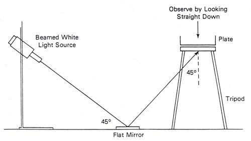
12Examine TSAye plates for typical 1-3 mm diameter smooth convex white colonies. Observation
with Henry oblique transmitted illumination (Figure 1) (23) can be helpful at this stage but is not
mandatory.
Figure 1. Henry Optical System for examination of colonies
1. Standard classification:
a. Hemolysis:
Inoculate heavily (from TSAye colony) 5% sheep blood agar by stabbing plates that have
been poured thick and dried well (check for moisture before using). Draw grid of 20-25
spaces on plate bottom. Stab one culture per grid space. Always stab positive controls (L.
ivanovii and L. monocytogenes) and negative control (L. innocua). Incubate for 24 to 48 h
at 35°C. Attempt to stab as near to bottom of agar layer as possible, without actually
touching bottom of agar layer and possibly fracturing the agar.
Examine blood agar plates containing culture stabs brightly lit from behind the plate. L.
monocytogenes and L. seeligeri produce a slightly cleared zone around the stab. L.
innocua shows no zone of hemolysis, whereas L. ivanovii produces a well-defined clear
zone around the stab. If mixed culture was observed on the TSAye plate repeat the
hemolysis test with an isolated colony.
CAMP test: Resolve questionable reactions by the Christie-Atkins-Munch-Peterson
(CAMP) (15) test. CAMP test strains are available from culture collections, including the
American Type Culture Collection (ATCC), Manassas, VA, http://www.atcc.org
i. Streak weakly β-hemolytic S. aureus (FDA strain ATCC 49444 (CIP 5710;
NCTC 7428) or ATCC 25923) and R. equi (ATCC 6939; NCTC 1621)
vertically on sheep blood agar.
13ii. Separately streak test strains horizontally between the S. aureus and R.
equi streaks without quite touching them. Incubate plate 24 to 48 h at 35°C.
Figure 2 shows the arrangement of the culture streaks on a CAMP plate.
iii. Examine plates for hemolysis in the zone of influence of the vertical streaks.
Hemolysis of L. monocytogenes and L. seeligeri is enhanced near the S.
aureus streak; L. ivanovii hemolysis is enhanced near the R. equi streak. Other
species are non-hemolytic and do not react in this test (Table 1).
iv. Alternatively, a factor easily prepared from S. aureus cultures can be used to
enhance hemolysis by L. monocytogenes and L. seeligeri in sheep blood agar
plates. Disks impregnated with the β-lysin of S. aureus (Remel, Lenexa, KS)
can be used.
Table 1. Differentiation of Listeria species
Species Mannitol Rhamnose Xylose Virulencea β- Hemolysis Hemolysis
Hemolysisb enhancement with enhancement
Staphylococcus with
aureus (S) Rhodococcus
equi (R)
L. - +c - + + + -d
monocytogenes
L. ivanoviie - - + + + - +
L. innocua - Vf - - - - -
L. welshimeri - V + - - - -
L. seeligeri - - + - +g + -
L. grayih + V - - -
a
Mouse test
b
Sheep blood agar stab
c
Some lineage III strains of L. monocytogenes, which are primarily associated with animal
listeriosis, are rhamnose negative.
d
Rare strains are S+ and R+. The R+ reaction is less pronounced than that of L. ivanovii.
e
Ribose fermenting strains are classified as L. ivanovii subsp. ivanovii and ribose non-fermenters
as L. ivanovii subsp. londiniensis.
f
V, variable biotypes, greater than 10% of strains for this trait.
g
Weakly hemolytic L. seeligeri strains may appear non hemolytic.
h
Includes two subspecies - L. grayi subsp. murrayi reduces nitrate. L. grayi subsp. grayi does
not reduce nitrate.
14Figure 2. CAMP test for Listeria monocytogenes: Inoculation pattern of the sheep blood
agar plate. Horizontal lines represent streak inoculations of 5 test strains. Vertical lines
represent streak inoculations of Staphylococcus aureus (S) and Rhodococcus equi (R).
Hatched lines indicate (diagrammatically only) the locations of hemolysis enhancement
regions.
b. Motility: Pick typical colony from TSAye and examine by wet mount, using 0.85% saline
for suspending medium and oil immersion objective of phase-contrast microscope.
Choose a colony with enough growth to make a fairly heavy suspension; emulsify
thoroughly. Listeria spp. are slim, short rods with slight rotating or tumbling motility.
Always compare with known culture. Cocci, large rods, or rods with rapid, swimming
motility are not Listeria spp. Alternatively, stab tube of MTM (M103) from TSAye.
Incubate for up to 7 days at room temperature (20-25°C). Observe daily until the isolate
growth pattern is obvious. Listeria is motile, giving a typical umbrella-like growth
pattern.
c. Catalase: Test typical colonies for catalase by placing some colony growth in a drop of
3% hydrogen peroxide. Listeria species are catalase-positive.
d. Gram stain: Use 16 to 24 h growth from TSAye plates. All Listeria spp. are short, Gram-
positive rods; however, with older cultures the Gram stain reaction can be variable and
also cells may appear spheroidal. The cells have a tendency to palisade in thick-stained
smears. This can lead to false rejection as a diphtheroid.
e. Carbohydrate fermentation series: Pick typical colony to a tube of TSB for inoculating
carbohydrate fermentation and other test media. Incubate at 35°C for 24 h. This culture
may be kept at 4°C several days and used repeatedly as inoculum.
15i. From TSBye culture, inoculate the following carbohydrates in 0.5% solutions
in purple carbohydrate broth with Durham tubes: dextrose, esculin, maltose,
rhamnose, mannitol, and xylose. Incubate 7 days at 35°C.
ii. Positive reactions will be indicated by the production of acid and the media
turning a yellow color with no gas production. All Listeria spp. should be
positive for dextrose, esculin, and maltose. All Listeria spp. except L.
grayi should be mannitol-negative. If pigmentation of the isolate on OXA,
PALCAM, MOX or LPM plus esculin/Fe3+ is unequivocal, the esculin test
may be omitted. Consult Table 1 for interpretations of the results.
f. Optional: Nitrate reduction test: Only L. grayi subsp. murrayi reduces nitrates. The test
distinguishes L. grayi subsp. murrayi from L. grayi subsp. grayi.
i. Use a TSAye culture to inoculate nitrate broth (M108). Incubate at 35°C for 5
days.
ii. Add 0.2 ml reagent A, followed by 0.2 ml reagent B (R48). A red-violet color
indicates presence of nitrite, i.e., nitrate has been reduced. If no color
develops, add powdered zinc and hold for 1 h. A developing red-violet color
indicates that nitrate is still present and has not been reduced.
iii. As an alternative procedure, add 0.2 ml reagent A followed by 0.2 ml reagent
C. An orange color indicates reduction of nitrate. If no color develops, add
powdered zinc as above. Development of an orange color indicates unreduced
nitrate.
g. Optional: Since all Listeria species test negative for indole, oxidase, urease, and H2S
production from organic sulfur compounds (H2S is produced from thiosulfate in the
MICRO-ID™ test kit) and test positive for methyl red and Voges-Proskauer, these tests
are discretionary. Brochothrix, which is phylogenetically closely related to Listeria, is
distinguishable from Listeria by its inability to grow at 35°C and by its lack of motility.
Distinguishing features of the Gram-positive non-sporeforming
rods, Erysipelothrix and Kurthia, which occur rarely in Listeria analysis, can be found
elsewhere (11, 43).
h. Optional: Immunocompromised mouse pathogenicity test: The classical tests
for Listeria pathogenicity are the Anton conjunctivitis test (rabbits), inoculation of mice,
and inoculation of embryonated eggs. The immunocompromised mouse test, using intra-
peritoneal injection, is recommended because of its greatly improved sensitivity (38).
Confirmation of L. monocytogenes animal pathogenicity is not needed for clinical isolates
and is optional for food isolates. An isolate should be identified as L. monocytogenes if it
meets all the other criteria outlined in this chapter. See link for detailed protocol.
Biochemical and pathogenicity data are summarized in Table 1. All data collection must be
completed before species identities are determined and subsequent subtyping performed.
Atypical Listeria strains exist that could confuse the identification. For example, there are
undocumented references (46) to hemolytic L. innocua isolates. Some L. monocytogenes and L.
16welshimeri isolates are rhamnose-negative. Some L. seeligeri isolates have very weak hemolytic
reaction and can be confused with non-hemolytic species. Sometimes aberrant Listeria strains
are isolated which are extremely difficult to speciate (26) (See Guideline for BAM Users
on Identification of Atypical Hemolytic Listeria Isolates). If such an aberrant Listeria isolate is
obtained, contact Karen Jinneman.
2. Alternate rapid identification:
Purified isolates may be rapidly identified by using commercial kits or real-time PCR. Follow
manufacturer instructions for inoculation and interpretation.
a. API® Listeria (bioMerieux, Durham, NC) which requires an additional β-hemolysis
test for final isolate identification (12). CAMP test is optional.
b. MICRO-ID™ Listeria Identification System (Remel, Lenexa, KS) which requires an
additional CAMP test and β-hemolysis test. (2, 22).
c. VITEK® 2 Automatic Gram Positive card (bioMerieux, Hazelwood, MO), which
requires an additional CAMP test and β-hemolysis test (38).
d. Real-time PCR, which requires an additional β-hemolysis test. CAMP test is optional.
• Protocol: Simultaneous Confirmation of Listeria species and L.
monocytogenes isolates by real-time PCR.
• Attachment 1: Single Lab Validation (SLV) - Individual Ct values for each isolate
by each enzyme mix. (pdf, 58Kb)
• Attachment 2: Multi Lab validation (MLV) - Individual Ct values for each isolate
by laboratory. (pdf, 60Kb)
• Listeria species identification includes L. monocytogenes, L. innocua, L.
ivanovii, L. seeligeri and L. welshimeri. Identification of L. grayi has not been
verified with this assay. Isolates that are identified as Listeria species but not L.
monocytogenes can be fully speciated as described in Section H.1.a-e or H.2.a-c.
Alternative AOAC OMA rapid methods for the detection of L. monocytogenes listed in section
F.2 can be used to confirm pure culture. Depending on the kit, isolates may be identified in pure
culture or from OXA or the other selective isolation agars. Purified isolates identified as Listeria
monocytogenes by these tests should be retained for regulatory reference.
For environmental samples, refer to applicable sampling compliance guidance documents to
determine if identification to the genus level is sufficient and if further differentiation between L.
monocytogenes and other Listeria species is necessary.
I. Subtyping of L. monocytogenes Isolates (required)
Confirmed L. monocytogenes isolates should be typed serologically and genetically.
1. Serological typing: Serology is useful when addressing epidemiological considerations.
Most L. monocytogenes isolates obtained from patients, foods, and the environment are type 1 or
4. More than 90% of L. monocytogenes isolates can be serotyped with commercially available
sera. However, all nonpathogenic Listeria species, except L. welshimeri, share one or more
17somatic antigens with L. monocytogenes (43). Therefore, serotyping alone without thorough
isolate characterization is not adequate for identification of L. monocytogenes.
Use commercial sera (Difco™ Type 1 catalog# 223031 and Type 4 catalog# 223041) to
characterize isolates as type 1, type 4 or not type 1 or 4 (types 3, 5, 6, etc.) at a minimum. Use a
TSBye culture to inoculate Tryptose broth. Incubate for 24 h at 35°C, at which temperature
flagella (H) antigen expression is reduced. Transfer to Tryptose agar slants and incubate for 24 h
at 35°C. Wash both slants in a total of 3 ml Difco™ fluorescent antibody (FA) buffer and
transfer to a sterile 16 × 125-mm screw-cap tube. Heat in a water bath at 80°C for 1 h. Sediment
cells by centrifugation at 1600 g for 30 min. Remove 2.2-2.3 ml of supernatant fluid and
resuspend the pellet in the remainder of buffer. Follow manufacturer's recommendations for sera
dilution and agglutination.
Complete serological characterization can also be done (Denka Seiken™ catalog# 294616). Pure
cultures of L. monocytogenes isolates should be cultured for 24 h at 35°C on non-selective agar
such as BHI agar. Colony growth is then resuspended, heat inactivated and tested for
agglutination as recommended by the antisera manufacturer.
2. Genetic subtyping: Whole genome sequencing of food and environmental isolates should be
submitted to GenomeTrakr. Retain all isolates as additional subtyping techniques may also be
requested.
J. Enumeration (required)
If a food sample tests positive for L. monocytogenes, use a reserve portion of sample for
enumeration. Enumeration is performed using a combination of MPN and direct plating.
1. MPN procedure:
a. Prepare a homogenate of a 50-g amount of reserve food sample in 450 ml pre-
warmed basal BLEB with selective agents.
b. Prepare a 4-dilution, 3-tube MPN using dilutions that will deliver equivalent to 10, 1,
0.1, and 0.01 g sample per aliquot at each respective dilution.
c. Incubate all twelve aliquots as described in Section E.
d. At 48 h streak each aliquot as described in Section G followed by isolation and
confirmation according to sections G-H. Alternatively each of the enrichments can be
rapidly screened by one of the approved methods described in Section F with
confirmation of all presumptive positives by the isolation and confirmation steps as
described in Sections G-H.
e. Interpret results based on the number of tubes with confirmed positive L.
monocytogenes using the tables in BAM Appendix 2 (14).
f. If all the MPN tubes are Listeria positive, refer to direct plating for enumeration
results.
In situations that would require narrower confidence limits, the number of replicate
tubes for certain dilutions could be increased. One ml of homogenate of complete
18BLEB and sample can be added and diluted by multi-channel pipette or robotically, in
96-well plates.
2. Direct plating procedure:
a. For solid food, prepare a homogenate of a 25-g amount of reserve food sample in 225
ml basal BLEB without selective agents. Perform another 10-fold dilution if
necessary. Certain foods may require different sample set-up and dilution procedures,
refer to applicable compliance assignment documents.
b. Direct plate 1 ml of liquid food or 1 ml homogenate of solid food prepared in step a
onto 3 to 5 plates of one of the L. monocytogenes differential chromogenic agars as
described in section G.
c. If the colonies on the plates are too numerous to count, use reserve sample, perform
additional 10-fold serial dilutions and proceed with direct plating.
d. Confirm 5 representative colonies per plate.
e. Alternatively, spiral plating method described in Chapter 3 can be used for direct
plating.
Notes: Guideline for BAM Users on Identification of Atypical Hemolytic Listeria Isolates
19References
1. Anonymous. 2001. Validation certificate for alternative method according to the standard
EN ISO
16140:2003. http://www.chromagar.com/fichiers/1450365640CHR_21_01_12_01_en_V
2013.pdf
2. AOAC Official Method 992.18. 2000. MICRO-ID Listeria. Chapter 17.10.02, pp. 141-
144 In: Official Methods of Analysis of AOAC INTERNATIONAL. 17th Edition. W.
Horwitz (ed.). Volume 1. Agricultural Chemicals, Contaminants and Drugs. AOAC
INTERNATIONAL, Gaithersburg, MD.
3. AOAC Official Method 993.09. 2000. Listeria in dairy products, seafoods, and meats.
Colorimetric deoxyribonucleic acid hybridization method (GENE-TRAK Listeria Assay).
Chapter 17.10.04, pp. 147-150 In: Official Methods of Analysis of AOAC
INTERNATIONAL. 17th Edition. W. Horwitz (ed.). Volume 1. Agricultural Chemicals,
Contaminants and Drugs. AOAC INTERNATIONAL, Gaithersburg, MD.
4. AOAC Official Method 994.03. 2000. Listeria monocytogenes in dairy products,
seafoods, and meats. Colorimetric monoclonal enzyme-linked immunosorbent assay
method (Listeria-Tek). Chapter 17.10.05, pp. 150-152 In: Official Methods of Analysis
of AOAC INTERNATIONAL. 17th Edition. W. Horwitz (ed.). Volume 1. Agricultural
Chemicals, Contaminants and Drugs. AOAC INTERNATIONAL, Gaithersburg, MD.
5. AOAC Official Method 995.22. 2000. Listeria in foods. Colorimetric polyclonal enzyme
immunoassay screening method (TECRA Listeria Visual Immunoassay [TLVIA]).
Chapter 17.10.06, pp. 152-155 In: Official Methods of Analysis of AOAC
INTERNATIONAL. 17th Edition. W. Horwitz (ed.). Volume 1. Agricultural Chemicals,
Contaminants and Drugs. AOAC INTERNATIONAL, Gaithersburg, MD.
6. AOAC Official Method 996.14. 2000. Assurance Polyclonal Enzyme Immunoassay
Method. Chapter 17.10.07, pp. 155-158 In: Official Methods of Analysis of AOAC
INTERNATIONAL. 17th Edition. W. Horwitz (ed.). Volume 1. Agricultural Chemicals,
Contaminants and Drugs. AOAC INTERNATIONAL, Gaithersburg, MD.
7. AOAC Official Method 997.03. 2000. Visual Immunoprecipitate Assay (VIP). Chapter
17.10.08, pp. 158-160 In: Official Methods of Analysis of AOAC INTERNATIONAL.
17th Edition. W. Horwitz (ed.). Volume 1. Agricultural Chemicals, Contaminants and
Drugs. AOAC INTERNATIONAL, Gaithersburg, MD.
8. AOAC Official Method 999.06. 2000. Enzyme Linked Immunofluorescent Assay
(ELFA) VIDAS LIS Assay Screening Method. Chapter 17.10.09, pp. 160-163. In:
Official Methods of Analysis of AOAC INTERNATIONAL. 17th Edition. W. Horwitz
(ed.). Volume 1. Agricultural Chemicals, Contaminants and Drugs. AOAC
INTERNATIONAL, Gaithersburg, MD.
9. AOAC Official Method 2003.12. 2005. Evaluation of BAX® Automated System for the
Detection of Listeria monocytogenes in Foods. Chapter 17.10.10, pp. 222-225. In:
Official Methods of Analysis of AOAC INTERNATIONAL. 18th Edition. W. Horwitz
(ed.). AOAC INTERNATIONAL, Gaithersburg, MD.
2010. Asperger, H., H. Heistinger, M. Wagner, A. Lehner and E. Brandl. 1999. A contribution
of Listeria enrichment methodology - growth of Listeria monocytogenes under varying
conditions concerning enrichment broth composition, cheese matrices and competing
microflora. Microbiology 16:419-431.
11. Bille, J., J. Rocourt, and B. Swaminathan. 1999. Listeriae, Erysipelothrix, and Kurthia,
pp. 295-314. In: Manual of Clinical Microbiology. 7th Edition. P. R. Murray (ed.).
American Society for Microbiology, Washington, DC.
12. 2008 draft. Bille, J. B. Catimel, E. Bannerman, C. Jacquet, M.N. Yersin, I. Camiaux, D.
Monget and J. Rocourt. 1992. API Listeria, a new and promising one-day sysem to
identify Listeria isolates. Appl. Environ. Microbiol. 58(6):1857-1860.
13. Boerlin et al. 1992. L. ivanovii subsp. londoniensis subsp. novi. Int. J. Syst.
Bacteriol. 42:69-73.
14. Blodgett, R. 2006. Appendix 2. Most Probable Number from Serial Dilutions. In U.S.
Food and Drug Administration Bacteriological Analytical Manual Online.
15. Christie, R., N. E. Atkins, and E. Munch-Petersen. 1944. A note on the lytic phenomenon
shown by group B streptococci. Aust. J. Exp. Biol. Med. Sci. 22: 197-200.
16. Curiale, M. S., T. Sons, L. Fanning, W. Lepper & D. McIver. 1994. Deoxyribonucleic
acid hybridization method for the detection of Listeria in dairy products, seafoods, and
meats: collaborative study. J. AOAC INTERNATIONAL 77:602-617.
17. Curiale, M. S., W. Lepper & B. Robison. 1994. Enzyme-linked immunoassay for
detection of Listeria monocytogenes in dairy products, seafoods, and meats: collaborative
study. J. AOAC INTERNATIONAL 77:1472-1489.
18. Curtis, G. D. W., R. G. Mitchell, A. F. King, and J. Emma. 1989. A selective differential
medium for the isolation of Listeria monocytogenes. Lett. Appl. Microbiol. 8:95-98.
19. Feldsine, P. T., A. H. Lienau, R. L. Forgey, and R. D. Calhoon. 1997. Assurance
polyclonal enzyme immunoassay (EIA) for detection of Listeria monocytogenes and
related Listeria species in selected foods: collaborative study. J. AOAC
INTERNATIONAL 80:775-790.
20. Feldsine, P. T., A. H. Lienau, R. L. Forgey & R. G. Calhoon. 1997. Visual
immunoprecipitate assay (VIP) for Listeria monocytogenes and related Listeria species
detection in selected foods: collaborative study. J. AOAC INTERNATIONAL 80:791-805.
21. Gangar, V., M. S. Curiale, A. D'Onorio, A. Schultz, R. L. Johnson, and V. Atrache. 2000.
VIDAS® Enzyme-linked immunofluorescent assay for detection of Listeria in foods:
collaborative study. J. AOAC INTERNATIONAL 83:903-918.
22. Higgins, D. L., and B. J. Robison. 1993. Comparison of MICRO-ID Listeria method with
conventional biochemical methods for identification of Listeria isolated from food and
environmental samples: collaborative study. J. AOAC INTERNATIONAL 76:831-838.
23. Hitchins, A. D. 1998. Listeria monocytogenes. Chapter 10. In: G. J. Jackson
(Coordinator) Bacteriological Analytical Manual. 8th Edition. Revision A. AOAC
INTERNATIONAL, Gaithersburg, MD.
24. Hitchins, A. D., and R. E. Duvall. 2000. Feasibility of a defined microflora challenge
method for evaluating the efficacy of foodborne Listeria monocytogenes selective
enrichments. J. Food Protect. 63:1064-1070.
2125. Jinneman, K., J. M. Hunt, C. A. Eklund, J. S. Wernberg, P. N. Sado, J. M. Johnson, R. S.
Richter, S. T. Torres, E. Ayotte, S. J. Eliasberg, P. Istafanos, D. Bass, N. Kexel-
Calabresa, W. Lin,, and C. N. Barton. 2003. Evaluation and Interlaboratory Validation of
a Selective Agar for Phosphatidylinositol-Specific Phospholipase C Activity Using
Chromogenic Substrate to Detect Listeria monocytogenes from Foods. J. Food
Protect. 66:441-445.
26. Johnson, J.M., K. Jinneman, G. Stelma, B. G. Smith, D. Lye, J. Messer, J. Ulaszek, L.
Evsen, S. Gendel, R. W. Bennett, B. Swaminathan, J. Pruckler, A. Steigerwalt, S.
Kathariou, S. Yildirim, D. Volokhov, A. Rasooly, V. Chizhikov, M. Wiedmann, E.
Fortes, R. E. Duvall, and A. D. Hitchins. 2004. Natural Atypical Listeria innocua Strains
with Listeria monocytogenes Pathogenicity Island 1 Genes. Appl. Environ.
Microbiol. 70:4256-4266.
27. Jones, D., and H.P.R. Seeliger. 1986. International committee on systematic bacteriology.
Subcommittee the taxonomy of Listeria. Int. J. Syst. Bacteriol. 36:117-118.
28. Jones, D. 1992. Current classification of the genus Listeria. In: Listeria 1992. Abstracts
of ISOPOL XI, Copenhagen, Denmark). p. 7-8.
29. Knight, M. T., M. C. Newman, M. Joseph-Benziger Jr., J. R. Agin, M. Ash, P. Sims, and
D. Hughes. 1996. TECRA Listeria Visual Immunoassay [TLVIA] for detection
of Listeria in foods: collaborative study. J. AOAC INTERNATIONAL 79:1083-1094.
30. Lee, W. H., and D. McClain. 1986. Improved L. monocytogenes selective agar. Appl.
Environ. Microbiol. 52:1215-1217.
31. Mattingly, J. A., B. T. Butman, M. C. Plank, and R. J. Durham. 1988. A rapid
monoclonal antibody-based ELISA for the detection of Listeria in food products. J.
AOAC INTERNATIONAL 71:669-673.
32. Official Methods of Analysis of AOAC INTERNATIONAL AOAC INTERNATIONAL,
Gaithersburg, MD, USA Official Method 2002.09
33. Official Methods of Analysis of AOAC INTERNATIONAL AOAC INTERNATIONAL,
Gaithersburg, MD, USA Official Method 2004.02
34. Official Methods of Analysis of AOAC INTERNATIONAL AOAC INTERNATIONAL,
Gaithersburg, MD, USA Official Method 2004.06
35. Official Methods of Analysis of AOAC INTERNATIONAL AOAC INTERNATIONAL,
Gaithersburg, MD, USA Official Method 2010.02
36. Official Methods of Analysis of AOAC INTERNATIONAL AOAC INTERNATIONAL,
Gaithersburg, MD, USA Official Method 2012.02
37. Official Methods of Analysis of AOAC INTERNATIONAL AOAC INTERNATIONAL,
Gaithersburg, MD, USA Official Method 2013.10
38. Official Methods of Analysis of AOAC INTERNATIONAL AOAC INTERNATIONAL,
Gaithersburg, MD, USA Official Method 2013.11
39. Restaino, L., E. W. Frampton, R. M. Irbe, G. Schabert, and H. Spitz. 1999. Isolation and
detection of Listeria monocytogenes using fluorogenic and chromogenic substrates for
phosphatidylinositol-specific phospholipase C. J. Food Protect. 62:244-251.
2240. Rocourt, J., P. Boerlin, F.Grimont, C. Jacquet, and J-C. Piffaretti. 1992. Assignment
of Listeria grayi and Listeria murrayi to a single species, Listeria grayi, with a revised
description of Listeria grayi. Int. J. Syst. Bacteriol. 42:171-174.
41. Rocourt, J. 1999. The genus Listeria and Listeria monocytogenes: phylogenetic position,
taxonomy, and identification. In: Listeria, Listeriosis and Food Safety. E. T. Ryser and E.
H. Marth (Eds). 2nd edition, pp. 1-20. Marcel Dekker, Inc., New York, NY.
42. Ryser, E. T., and E. H. Marth. 1999. Listeria, Listeriosis and Food Safety. 2nd edition.
Marcel Dekker, Inc., New York, NY.
43. Seeliger, H.P.R., and D. Jones. 1986. Listeria. pp. 1235-1245. In: Bergey's Manual of
Systematic Bacteriology, Vol. 2, 9th ed. P.H.A. Sneath, N.S. Mair, M.E. Sharpe, and J.G.
Holt (Eds). Williams & Wilkins Co., Baltimore, MD.
44. Shaw S., Nundy D. and Blais B.: Performance of the ALOA medium in the detection of
hemolytic Listeria species in food and environmental samples. Laboratory Services
Division, Canadian Food Inspection Agency, Ottawa, Ontario, Canada K1A 0C6.
45. Stelma, G.N., Jr., A. L. Reyes, J. T. Peeler, D.W. Francis, J.M. Hunt, P L. Spaulding,
C.H. Johnson, and J. Lovett. 1987. Pathogenicity testing for L. monocytogenes using
immunocompromised mice. J. Clin. Microbiol. 25:2085-2089.
46. USDA/FSIS. 1999. Isolation and identification of Listeria monocytogenes from red meat,
poultry, egg and environmental samples. Ch. 8. Microbiology Laboratory Guidebook. 3rd
Edition, Revision 2.
47. US FDA/CFSAN. 2008. Guidance for Industry: Control of Listeria monocytogenes in
Refrigerated or Frozen Ready-To-Eat Foods (Draft Guidance). (accessed 04/14/2011).
48. US DHHS/FDA/CFSAN and USDA/FSIS. 2003. Listeria monocytogenes Risk
Assessment: Quantitative Assessment of Relative Risk to Public Health from
Foodborne Listeria monocytogenes among Selected Categories of Ready-to-Eat Foods.
(accessed 04/14/2011).
49. Vlaemynck G., Lafarge V., Scotter S. (2000): Improvement of the detection of Listeria
monocytogenes by the application of ALOA, a diagnostic, chromogenic isolation
medium. Journal of Applied Microbiology, 88 : 430-441.
50. Van Netten et al. 1989. Liquid and solid selective differential media for the detection and
enumeration of Listeria monocytogenes. Int. J. Food Microbiol. 8:299-316.
51. Wang, S-Y. and A. D. Hitchins. 1994. Differential enrichment kinetics of severely and
moderately injured Listeria monocytogenes cell fractions of heat injured populations. J.
Food Safety 14:259-27.
23You can also read