Atypical Peripheral Ossifying Fibroma of the Mandible - MDPI
←
→
Page content transcription
If your browser does not render page correctly, please read the page content below
dentistry journal
Case Report
Atypical Peripheral Ossifying Fibroma of the Mandible
Tomislav Katanec *, Lea Budak, Davor Brajdić and Dragana Gabrić
Department of Oral Surgery, School of Dental Medicine, University of Zagreb, 10000 Zagreb, Croatia;
lbudak@sfzg.hr (L.B.); dbrajdic@kbd.hr (D.B.); dgabric@sfzg.hr (D.G.)
* Correspondence: tkatanec@sfzg.hr
Abstract: Peripheral ossifying fibroma (POF) is a benign localized lesion originating from gingival
and alveolar oral mucosa. Its origin can be cells of periodontal ligament. The lesions usually develop
in women in their twenties. POF is a complex clinical and histological diagnosis due to its shared
characteristics with many other conditions. In this paper, we presented a case of an atypical peripheral
ossifying fibroma (POF) in the left lateral part of the mandible in a 70-year-old male patient who had
two semicircular bridges supported on four implants in the upper and lower jaws. A review of CBCT
and orthopedic imaging showed no visible intraosseous changes. Histological analysis revealed the
diagnosis of POF. The case in question is interesting, as elaborated on in the discussion section of this
paper because POF is usually found in female patients aged between 20 and 30 years.
Keywords: peripheral ossifying fibroma; irritation fibromatosis; gigantocellular lesions
1. Introduction
Peripheral ossifying fibroma (POF) is a non-malignant localized lesion originating
from gingival and alveolar oral mucosa, clinically manifesting as a painless, slow-growing,
hard nodule, usually smaller than 2 cm. It is histologically characterized by a fibrous tissue
affected by a variable number of fibroblastic cells. The presence of well-defined islands
Citation: Katanec, T.; Budak, L.; of metaplastic bone can be extremely helpful when setting a differential diagnosis [1,2].
Brajdić, D.; Gabrić, D. Atypical
Various sources name this pathology differently, e.g., peripheral cementifying fibroma,
Peripheral Ossifying Fibroma of the
peripheral fibroma with cementogenesis, peripheral fibroma with osteogenesis, periph-
Mandible. Dent. J. 2022, 10, 9.
eral fibroma with calcification, calcified or ossified fibrous epulis and calcified fibroblastic
https://doi.org/10.3390/dj10010009
granuloma [1]. The lesions usually develop in women in their twenties and can appear
Academic Editor: Jiiang-Huei Jeng anywhere in the mouth, including the tongue, lips, mouth floor, palate or maxillary and
mandibular alveolar crest [3]. A mutation of the SATB2 gene inactivates the gene using
Received: 2 November 2021
different molecular mechanisms, which can lead to the development of this mass. Proliferat-
Accepted: 4 January 2022
Published: 6 January 2022
ing cell nuclear antigen-positive cells in POF, indicating the high proliferative activity of the
lesion, may influence the treatment modalities. The result is a so-called syndrome related
Publisher’s Note: MDPI stays neutral to SATB2 [4], a condition characterized by neurodevelopmental and behavioral disabilities,
with regard to jurisdictional claims in palatal clefts, teeth and bone anomalies that rarely cause defects in other organ systems [5].
published maps and institutional affil-
POF is a complex clinical and histological diagnosis because it shares characteristics with
iations.
many other conditions, such as pyogenic granuloma, peripheral gigantocellular granuloma,
irritation fibroma or the non-bone metastasis of some tumors [6]. However, POF is a focal
reactive, non-neoplastic tumorous mass of the soft tissue, primarily originating from the
Copyright: © 2022 by the authors.
interdental papilla. It can be soft and has a smooth surface. The color can vary from a light
Licensee MDPI, Basel, Switzerland.
rosy color to a dark cherry red [7].
This article is an open access article
2. Case Study
distributed under the terms and
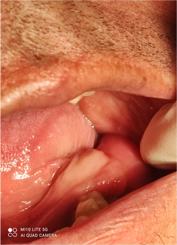
conditions of the Creative Commons A male 70-year-old patient came to the Department of Oral Surgery, University Hospi-
Attribution (CC BY) license (https:// tal Centre Zagreb with a voluminous fibrous mass in the distal region of the left mandible
creativecommons.org/licenses/by/ (Figure 1). The patient has two acrylic bridges on four implants. The implants were placed
4.0/). six months prior to admission to the clinic, before the patient noticed the appearance of
Dent. J. 2022, 10, 9. https://doi.org/10.3390/dj10010009 https://www.mdpi.com/journal/dentistry
Dent. J. 2022, 10, 9                                                                                                     2 of 9

                                   the mass. The acrylic bridges are 3 months old. The patient states feeling “swelling in the
                                   back part of the left mandible three months before coming in for a checkup”. Panoramic
                                   radiograph and CBCT did not show any radiolucency, radiopaque areas or signs of periim-
                                   plantitis around the implants in the bone (Figure 2). The lesion has a smooth surface, with
                                   no ulcerations. It was on a broad base connected, with the sublingual anatomical region.
                                   The patient has poor oral hygiene, smokes and consumes alcohol: about two to three glasses
                                   of wine or beer a day. The palpation of the mass indicated that it was fixed to the alveolar
                                   crest of the left mandible on a wide base and was spreading to the left sublingual area.
                                   The measured size of the mass was 3.5 × 2 cm. After conducting a clinical examination,
                                   the differential diagnosis was possible irritation fibromatosis, peripheral gigantocellular
                                   fibroma or peripheral ossifying or non-ossifying fibroma, as well as a malignant mass, and
                                   the final diagnosis will be reached after the final PHD analysis. The final decision was an
                                   excision in toto (Figure 3). The mass was approached and the layers of submucosa were
                                   divided with a scalpel and an electro knife. During operation, it was noticed that the mass
                                   has a belonging artery connected to the left sublingual area. The artery was ligated with a
                                   resorbing thread 4/0 and the mass underwent complete excision (Figure 4). Parts of the
                                   flap were left to heal per secundam, but most of the incision towards the sublingual region
                                   was stitched with a non-resorbing silk thread 4/0 (Figures 5 and 6). A full hemostasis was
                                   achieved by electrocauterization of the bleeding areas. The excison was performed under
                                   local anesthesia. Clinical, medical examination and removal of sutures were performed
 Dent. J. 2021, 9, x FOR PEER REVIEW                                                                                    3 of 10
                                   seven days after surgery (Figure 7).

                               Figure 1.
                               Figure 1. Clinical
                                         Clinical appearance
                                                  appearance of
                                                             of the
                                                                the mass
                                                                    mass during
                                                                         during first
                                                                                first visit.
                                                                                      visit.

                                    The mass was then sent for pathohistological (PHD) analysis, which showed that
                               the mass has a reactively changed multilayered epithelium with abundant mononuclear
                               linear inflammatory infiltrate underneath. There were no elements of Lichen. Fragments
                               resembling cemento-osseous lacunae were found inside the thick fiber stroma. A restricted
                               area with clusters of hemosiderin and gigantocellular cells was found in the middle of the
                               sample. Interestingly, a small salivary gland with multiplied intra- and interlobular tissue
                               with widened performing ducts was found in the sample. The outer edges of the sample
                               also contained part of the mucocele wall (Figure 8).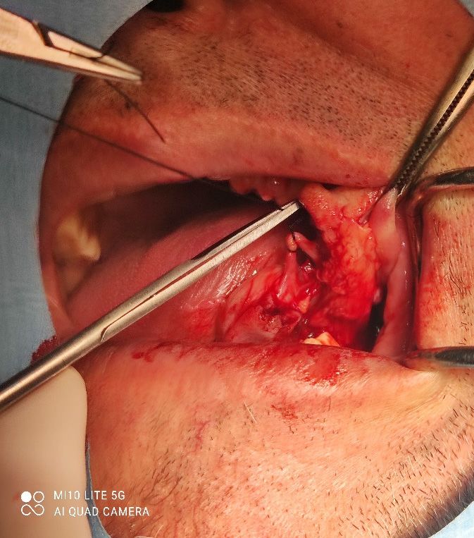
Dent. J. 2022, 10, 9                                                                                                 3 of 9

                                       Figure 1. Clinical appearance of the mass during first visit.

     Dent. J. 2021, 9, x FOR PEER REVIEW                                                                   4 of 10
                                       Figure
     Dent. J. 2021, 9, x FOR PEER REVIEWFigure 2. Orthopantomogram
                                               2. Orthopantomogram of of
                                                                      thethe lower
                                                                          lower jaw jaw   ofpatient.
                                                                                     of the the patient.   4 of 10

                                       Figure 3. Removal of the mass.
                                      Figure 3. Removal of the mass.
                                       Figure 3. Removal of the mass.

                                       Figure 4. Ligation of the artery.
                                       Figure 4. Ligation of the artery.
                                      Figure 4. Ligation of the artery.
Dent.
 Dent.J.J.2022,
           2021,10,
                 9, x9 FOR PEER REVIEW                                  5 4of
                                                                            of 10
                                                                               9
 Dent. J. 2021, 9, x FOR PEER REVIEW                                    5 of 10

                                     Figure5.5.Size
                                    Figure      Sizeof
                                                     ofthe
                                                        themass.
                                                           mass.
                                     Figure 5. Size of the mass.

                                     Figure 6. Right after suturing.
                                     Figure6.6.Right
                                    Figure      Rightafter
                                                      aftersuturing.
                                                            suturing.
Dent.
Dent.J.J.2022,
2021,10,
9, x9 FOR PEER REVIEW 6 5ofof10
9
Dent. J. 2021, 9, x FOR PEER REVIEW 6 of 10
Figure7.
Figure 7. Seven
Seven days
days after
after operation,
operation,medical
medicalexamination
examinationand
andremoval
removalof
ofsutures.
sutures.
Figure 7. Seven days after operation, medical examination and removal of sutures.
Figure 8. Histological finding of the preparation at magnification 4×.
Figure 8.
Figure Histological finding
8. Histological finding of
of the
the preparation
preparation at
at magnification ×.
magnification 44×.
3. Discussion
Poorly coordinated intermaxillary relations between the upper and lower acrylic
3. Discussion
The appearance of POF is of unknown etiology and can be linked to different suscep-
bridge, as well as parafunctional movements, can be considered distinct predisposing
tibility
The factors such as
appearance ofbruxism,
POF of is of dental
unknown plaque and inadequately
etiology and can be linked set orto adjusted
different prosthetic
suscep-
factors for the development this formation.
teeth. Different
tibility factors such names for POF dental
as bruxism, are used in theand
plaque literature, such asset
inadequately fibrous epulisprosthetic
or adjusted or calcify-
ing
teeth.
3. fibroblastic
Different names
Discussion granuloma for POF [8]. are
Peripheral
used in fibroma and fibromatosis
the literature, such as fibrous are epulis
found or in calcify-
relation
to CD
ing 34,
fibroblastic α-smooth
granuloma muscle [8]. actine
Peripheral(α-SMA),fibroma vimentin,
and Ki-67
fibromatosis
The appearance of POF is of unknown etiology and can be linked to different suscepti- (Mib1) are and
found transforming
in relation
growth
to CDfactors
bility factor-α
34, such(TGF-α).
α-smooth muscle
as bruxism, TGF-α actine is presumably
dental (α-SMA),
plaque and connected
vimentin,
inadequately to fibroblast
Ki-67 (Mib1) and
set or adjustedproliferation
transforming
prostheticand
fibroblastic
growth activity
factor-α [9].
(TGF-α). POF TGF-α forms is 3.1% of
presumably all oral tumors
connected and
to
teeth. Different names for POF are used in the literature, such as fibrous epulis or calcifying 9.6%
fibroblast of all gingival
proliferation lesions
and
[10]. As a reactive
fibroblastic
fibroblastic activity
granuloma benign [8].lesion
[9]. POF forms of3.1%
Peripheral the fibroma
fibrous
of all oral tissue,
andtumorsPOFand is not
fibromatosis 9.6% aare
counterpart
offound
all gingival of lesions
the soft
in relation to
tissue
[10].
CD Asto
34, athe
α-smooth central
reactive ossifying
benign
muscle lesion
actine fibroma,
(α-SMA), which
of the fibrous represents
vimentin,tissue,
Ki-67 anisosteogenic
POF(Mib1) notand neoplasm.
a counterpart
transforming Central
of the soft
growth
ossifying
tissue
factor-α fibroma
to (TGF-α).
the central originates
ossifying
TGF-α from
fibroma,
is presumably endosteum or to
which represents
connected periodontal ligamentneoplasm.
an osteogenic
fibroblast proliferation near and the root apex
Central
fibroblastic
activity [9]. POF forms 3.1% of all oral tumors and 9.6% of all gingival lesions [10]. As of
and then
ossifying spreads
fibroma in the
originates medullar
from bone
endosteumcavity. or On the other
periodontal hand,
ligament the peripheral
near the roottype
apexa
the ossifying
and thenbenign
reactive fibroma,
spreads in theof
lesion asmedullar
well
the as peripheral
fibrous bone cavity.
tissue, gigantocellular
POF Onisthe
notother granuloma
hand,
a counterpart (PGCG),
theofperipheral
the collides
type
soft tissue of
to
with
the
the the periodontal
ossifying
central fibroma,
ossifying ligament
as wellwhich
fibroma, from
as which
peripheral
representsit gigantocellular
develops. It can granuloma
an osteogenic be found solely
neoplasm. (PGCG),
Centralin soft tissues
collides
ossifying
above
with
fibromathethe alveolar from
periodontal
originates crest.
ligamentClinically,
endosteum from whichPOF resembles
it develops.
or periodontal a solitary,
It can benear
ligament slow-growing
found thesolely inand
root apex soft well-re-
andtissues
then
stricted
above
spreads thenodular
in alveolar mass
crest.
the medullar with
bone a smooth
Clinically,
cavity.POF surface,
On the usually
resembles
other a accompanied
hand,solitary, by normal
slow-growing
the peripheral type of colored
and mu-
thewell-re-
ossify-
cosa.
stricted
ing It nodular
fibroma,has aas wide
mass
well base
aswithand is usually
a smooth
peripheral of a solid
surface,
gigantocellular consistency
usually accompanied
granuloma [11]. by normal
(PGCG), collidescoloredwithmu-the
cosa. Intraoral
It has a ossifying
wide base fibroma
and is has
usually been
of a described
solid in
consistency the
periodontal ligament from which it develops. It can be found solely in soft tissues above literature
[11]. since the late 1940s.
Various
the alveolar names
Intraoral were
ossifying
crest. given
fibroma
Clinically, toPOFcomparable
has lesions,
been described
resembles such
a solitary, as epulis,
in slow-growing
the literature peripheral
since thefibroma with
late 1940s.
and well-restricted
calcification,
Various namesperipheral
were givenfibroma to comparable with osteogenesis,
lesions, such as calcified fibroblasticfibroma
epulis, peripheral granuloma,with
calcification, peripheral fibroma with osteogenesis, calcified fibroblastic granuloma,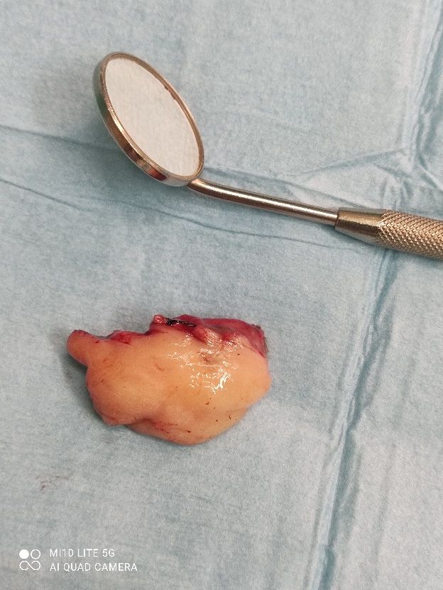
Dent. J. 2022, 10, 9                                                                                             6 of 9

                       nodular mass with a smooth surface, usually accompanied by normal colored mucosa. It
                       has a wide base and is usually of a solid consistency [11].
                             Intraoral ossifying fibroma has been described in the literature since the late 1940s.
                       Various names were given to comparable lesions, such as epulis, peripheral fibroma with
                       calcification, peripheral fibroma with osteogenesis, calcified fibroblastic granuloma, pe-
                       ripheral cementifying fibroma, peripheral fibroma with cementogenesis and peripheral
                       cemento-ossifying fibroma [8,12]. Around 60% of similar lesions are found in the upper jaw
                       and more than half of all cases impact the region of incisors and canines, to be exact, the
                       interdental papilla. Usually, children and adolescents aged from 10 to 30 are affected [13,14],
                       but mostly 20-year-olds, with a decreasing incidence after the age of 30 [15]. Only 0.5% of
                       cases occur in older age groups [16]. Due to hormonal influences, women are more likely
                       to be affected by the growth of this lesion [17]. Kfir et al. concluded that the size of POF
                       is usually smaller than 1.5 cm in diameter. A case of a gigantic POF was recorded in the
                       literature, which measured 9 cm in diameter [18]. Therefore, this case is quite interesting
                       because POF was found in a male patient aged 70 who had elevated levels of parathyroid
                       hormone (PTH).
                             Ogbureke et al. presented a case of a 44-year-old male who came into the emergency
                       room and complained about swelling in the back segment of the right mandible that had
                       been going on for three months. His family history included diabetes mellitus type 2,
                       cardiovascular diseases and hypertension. He had two implants in the right distal quadrant
                       near the lesion three months before the appearance of the mass. It was concluded that it
                       was quite hard to clinically differentiate peripheral gigantocellular lesion and peripheral
                       ossifying fibroma. The difference can be established histologically. Gigantocellular cells are
                       present in both POF and peripheral gigantocellular granuloma, whereas cemento-ossifying
                       lacunae is only present in POF [19].
                             Gulati et al. reported a case of a female, 56-year-old patient with a mass of hard
                       consistency upon palpation. The dimensions were 3.5 cm × 4 cm × 3 cm. The mass
                       was located near teeth 13–23 and the teeth were completely periodontally compromised
                       (stage III). A mass had a wide base and was erythematous and painless. The panoramic
                       radiograph showed a clean radiographic status. 940-nm diode laser (Biolase® , Foothill
                       Ranch, CA, USA) was used to remove the mass. Pathohistological analysis showed fibro-
                       cellular stroma with scattered islands of osteoid tissue varying in size (both mature lamellar
                       bone and immature bone). Calcification of the bone tissue showed peripheral osteoblastic
                       margins in some places. Some proliferations of the endothelial and inflammatory cells were
                       present in the tissue. Overall, the pathohistological characteristics indicated POF [20].
                             Prasad et al. believe that POF can develop as a pyogenic granuloma in the beginning,
                       which later goes through maturation of the fibers and calcification [21].
                             Satish et al. claim that POF is a consequence of inflammatory hyperplasia of the
                       periodontal cells or periost. Metaplasia of the fibrous tissue leads to dystrophic calcification
                       and the formation of the bone [22].
                             Rallan et al. show the case of a twelve-year-old boy who had taken notice of the
                       swelling that started a month before his visit to the department of pediatric dentistry
                       and observed that it increased in size. The patient’s medical history did not point to any
                       significant health indications. An oval-shaped gingival mass was discovered in the palatal
                       region of maxillary incisors upon completion of the intraoral examination. The mass was
                       impeding his bite and distressed the patient. The swelling was well-circumscribed, sessile,
                       erythematous, firm on palpation and measured approximately 2 × 2 cm in dimensions.
                       The lesion was asymptomatic with no clinically visible ulcerations.
                             They concluded that the mass is a reactive lesion of the connective tissue. Predisposi-
                       tion to such lesions is most common in the anterior maxilla of young women. The standard
                       treatment protocol involves excisional biopsy, followed by histopathological evaluation.
                       As the lesion has a tendency for recurrence, follow-up is of the utmost importance in most
                       cases [23].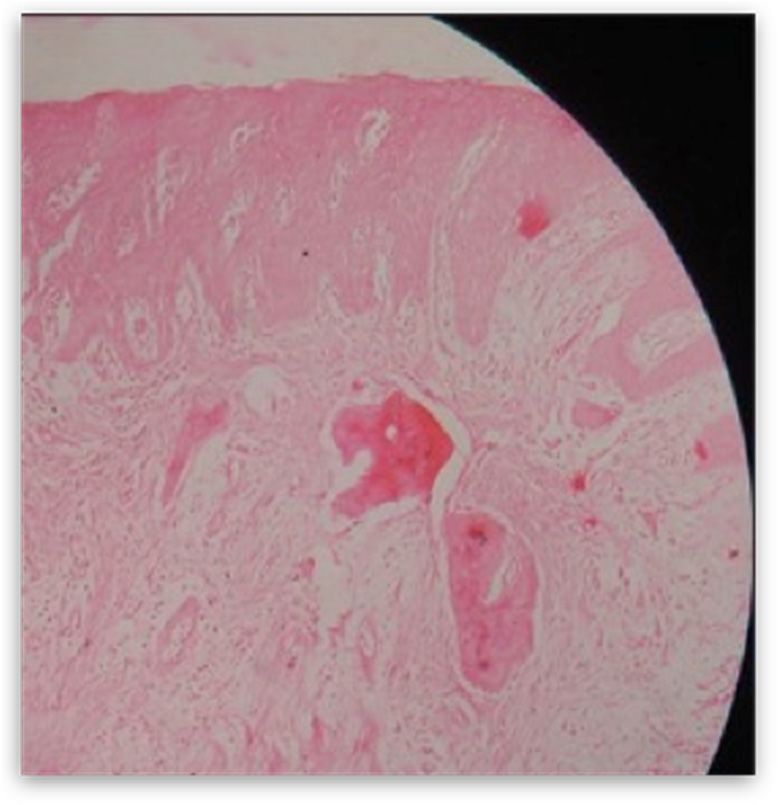
Dent. J. 2022, 10, 9                                                                                               7 of 9

                             Nadimpalli and Kadakampally describe the case of a 23-year-old female patient with
                       recurrent peripheral ossifying fibroma located in the right lower premolar region. Clinical,
                       radiographic and histologic features of POF, including differential diagnosis, treatment
                       and follow-up, are explained in the report. Due to the possible recurrence of POF, early
                       diagnosis, followed by surgical excision and the curettage of surrounding tissue, are
                       essential measures of recurrence prevention. They noted the importance of early diagnosis
                       and conservative management of such lesions, as they can become more destructive over
                       time if left untreated. Regular follow-up is essential after excision due to the high growth
                       potential of the lesion (8–20% recurrence rate) [24].
                             Interestingly, Sudhakar et al. show the case of a 55-year-old woman with a mass
                       in the upper jaw in the second left incisor. An intra-oral periapical radiograph (IOPAR)
                       and orthopantomography did not reveal any pathological changes except for generalized
                       horizontal bone loss. Similar to the pathohistological finding in the patient shown in
                       our case, the underlying connective tissue was highly cellular, with plump fibroblasts
                       intermingled in a delicate fibrillar stroma associated with areas of woven tra-becullar bone
                       and osteoids.
                             Furthermore, due to the gender of the patient and the second decade predilection, the
                       role of hormones has also been questioned as the predisposing factor of POF [25].
                             It is important to point out that, in our case the patient, is a man in his seventies, with
                       the appearance of POF in the area of the left lateral part of the mandible. In the literature that
                       we have already mentioned, the occurrence of POF as well as other ossifying or cemento-
                       ossifying fibromas is more common in females aged from 20 to 50 years. Predilection sites
                       of neoplasms are most common in the front of the maxilla or in the area of the hard palate
                       in the distal parts of the maxilla.
                             Agarwal et al. Present a 68-year-old female patient troubled by a soft tissue growth on
                       her left palate. The patient noticed a small nodule that grew to the present size in 4 months.
                       The patient had no significant medical and personal history. Clinical intraoral examination
                       discovered a shiny, rounded and elongated pink enlargement on the left side posterior
                       of the palate in projection of the cementoenamel junction of 26. It extended from 23 to
                       27 anteroposteriorly and was 1 cm lateral to palatal midline to the occlusal surface of the
                       left maxillary molars, buccolingually. Histological examination revealed connective tissue
                       stroma with a rich fibroblastic nature and overlying epithelium comprising bony trabeculae
                       with osteoblastic rimming of mature bone with a mostly lamellar structure, confirming the
                       lesion as POF. On regular follow-up, the lesion healed without any complications [26].
                             Katanec et al. published a similar case of symmetrical fibrous hyperplasia of the palate.
                             In that case, a 47-year-old patient developed a bilateral mass in the hard palate,
                       spreading to the junction of the hard and soft palate. A fibromatous mass appeared 3 years
                       before the visit to the clinic. One year before the clinical examination, the nodule grew to
                       form a voluminous fibromatous mass larger than 5 cm in diameter on both sides. The mass
                       affected the area of the upper canines on both sides to the border with the soft palate. The
                       formation was hard to palpation and connected at a wide base to the palatal artery. No
                       signs of acute inflammation were present. Excision of the formation without interference
                       with healthy tissue, followed by the removal of the affected periosteum and periodontal
                       ligament, was the most suitable treatment. It is of the utmost importance to limit the
                       irritating factors and possible trauma to the tissue [27].
                             Dutra et al. concluded that the incidence of hyperplastic lesions in all oral pathogenesis
                       is high. It is more common in the female population on the gingiva in the anterior part
                       of alveolar ridge. Inflammatory fibrous hyperplasia is the most common lesion (72.09%),
                       followed by oral pyogenic granuloma (11.79%), giant cell fibroma (7.30%) and peripheral
                       ossifying fibroma (5.24%) [28].
                             Borghesi et al. present the appearance of four benign formations in the same hemi-
                       mandible, diagnosed by CBCT in a 50-year-old female patient. That was the first case in
                       the literature showing peripheral osteoma (PO), compound odontoma (CO), focal cemento-
                       osseous dysplasia (FocCOD), and cemento-ossifying fibroma (COF) together in the same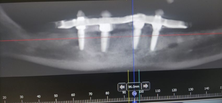
Dent. J. 2022, 10, 9                                                                                                                8 of 9

                                  patient mandible. This research, as well as our case, is of great importance for the case of
                                  differential diagnosis of neoplasms in the oral cavity and bones [29].
                                       In a retrospective survey study and literature review, Sangle et al. suggest that
                                  traumatic fibromas are the clinically most common lesions in oral cavity. As we have
                                  already stated, Sangle et al. concluded that irritating factors are important predisposing
                                  elements for the occurrence of fibromatous formations [30].

                                  4. Conclusions
                                        We presented a case report of a 70-year-old male patient who had histologically
                                  confirmed POF in left mandible lingually. As elaborated in the discussion of this paper,
                                  this case is interesting because POF is more often found in female patients aged from 20 to
                                  30 years. This presented case report is interesting as the patient is an adipose man in his
                                  seventies, who has two Toronto bridges placed on four implants in his upper and lower
                                  jaw. The occlusion of the bite surfaces of the bridges was not adequately aligned, which
                                  led to irritation and was a possible predisposing factor for the development of irritative
                                  fibromatosis, i.e., POF in this case.
                                        Overall blood count showed elevated parathyroid hormone levels. This fact may also
                                  be related to the formation of POF. After surgical excision of the formation, the operated
                                  area of the patient healed properly. There are no signs of recurrence at present, and the
                                  occlusion of the existing bridges is properly coordinated.

                                  Author Contributions: T.K. has determined the clinical diagnosis of the presented case, and he has
                                  developed the main idea of the paper. He performed the surgery. He actively participated in the
                                  process of writing and processing of the work, as well as in the collection of references. L.B. has
                                  participated in establishing a clinical diagnosis, and assisted in surgical removal of hyperplasia. She
                                  actively wrote the paper, collected references, and participated in text writing and photo processing.
                                  D.B. as a full university professor, she mentored the preparation of the paper, participated in the
                                  clinical diagnosis, and the preparation of the paper. D.G. as a university professor, he coordinated the
                                  preparation of the paper, participated in the clinical diagnosis, and in the writing and editing of the
                                  paper. All authors have read and agreed to the published version of the manuscript.
                                  Funding: This research received no external founding.
                                  Institutional Review Board Statement: Not applicable.
                                  Informed Consent Statement: The patient assigned informed consent statement.
                                  Data Availability Statement: For data information contact corresponding author.
                                  Conflicts of Interest: The authors declare no conflict of interest.

References
1.    Buchner, A.; Hansen, L.S. The histomorphologic spectrum of peripheral ossifying fibroma. Oral Surg. Oral Med. Oral Pathol. 1987,
      63, 452–461. [CrossRef]
2.    Bhaskar, S.N.; Jacoway, J.R. Peripheral fibroma and peripheral fibroma with calcification: Report of 376 cases. J. Am. Dent. Assoc.
      1966, 73, 1312–1320. [CrossRef]
3.    Maturana-Ramírez, A.; Adorno-Farías, D. A retrospective analysis of reactive hyperplastic lesions of the oral cavity: Study of
      1149 cases diagnosed between 2000 and 2011, Chile. Acta Odontol. Latinoam. 2015, 28, 103–107.
4.    Zarate, Y.A.; Fish, J.L. SATB2-associated syndrome: Mechanisms, phenotype, and practical recommendations. Am. J. Med.
      Genet. A 2017, 173, 327–337. [CrossRef] [PubMed]
5.    Zarate, Y.A.; Kalsner, L. Genotype and phenotype in 12 additional individuals with SATB2-associated syndrome. Clin. Genet.
      2017, 92, 423–429. [CrossRef]
6.    Pal, S.; Hegde, S.; Ajila, V. The varying clinical presentations of peripheral ossifying fibroma: A report of three cases.
      Rev. Odonto Ciênc. 2012, 27, 251–255. [CrossRef]
7.    Neville, B.W.; Damm, D.D.; Allen, C.M.; Bouquot, J.E. Oral and Maxillofacial Pathology; WB Saunders, Co.: Philadelphia, PA, USA,
      1995; pp. 374–376.
8.    Lee, K.W. The fibrous epulis and related lesions. Periodontics 1968, 6, 277–292.
9.    Rotaru, H.; Choi, J.Y.; Hong, S.P.; Lee, Y.C.; Yun, K.L.; Kim, S.G. Transforming growth factor-a and oral fibroma: Immunohisto-
      chemical and in situ hybridization study. J. Oral Maxillofac. Surg. 2003, 61, 1449–1454. [CrossRef] [PubMed]
Dent. J. 2022, 10, 9 9 of 9
10. Walters, J.D.; Will, J.K.; Hatfield, R.D.; Cacchillo, D.A.; Raabe, D.A. Excision and repair of the peripheral ossifying fibroma: A
report of 3 cases. J. Periodontol. 2001, 72, 939–944. [CrossRef] [PubMed]
11. Baumgartner, J.C.; Stanley, H.R.; Salomone, J.L. Peripheral ossifying fibroma. J. Endod. 1991, 17, 182–185. [CrossRef]
12. Gardner, D.G. The peripheral odontogenic fibroma: An attempt at clarification. Oral Surg. Oral Med. Oral Pathol. 1982, 54, 40–48.
[CrossRef]
13. Kfir, Y.; Buchner, A.; Hansen, L.S. Reactive lesions of the gingival: A clinicopathologic study of 741 cases. J. Periodontol. 1980, 51,
655–661. [CrossRef] [PubMed]
14. Kendrick, F.; Waggoner, W.F. Managing a peripheral ossifying fibroma. ASDC J. Dent. Child. 1996, 63, 135–138.
15. Jain, A.; Deepa, D. Recurrence of peripheral ossifying fibroma: A case report. People’s J. Sci. Res. 2010, 3, 23–25.
16. Effiom, O.A.; Adeyemo, W.L.; Soyele, O.O. Focal reactive lesions of the Gingiva: An analysis of 314 cases at a tertiary Health
Institution in Nigeria. Niger. Med. J. 2011, 52, 35–40. [PubMed]
17. Shetty, D.C.; Urs, A.B.; Ahuja, P.; Sahu, A.; Manchanda, A.; Sirohi, Y. Mineralized components and their interpretation in the
histogenesis of peripheral ossifying fibroma. Indian J. Dent. Res. 2011, 22, 56–61. [CrossRef] [PubMed]
18. Poon, C.K.; Kwan, P.C.; Chao, S.Y. Giant peripheral ossifying fibroma of the maxilla: Report of a case. J. Oral Maxillofac. Surg.
1995, 53, 695–698. [CrossRef]
19. Ogbureke, E.I.; Vigneswaran, N.; Seals, M.; Frey, G.; Johnson, C.D.; Ogbureke, K.U. A peripheral giant cell granuloma with
extensive osseous metaplasia or a hybrid peripheral giant cell granuloma-peripheral ossifying fibroma: A case report. J. Med.
Case Rep. 2015, 9, 14. [CrossRef]
20. Gulati, R.; Khetarpal, S.; Ratre, M.S.; Solanki, M. Management of massive peripheral ossifying fibroma using diode laser. J. Indian
Soc. Periodontol. 2019, 23, 177–180. [CrossRef]
21. Prasad, S.; Reddy, S.B.; Patil, S.R.; Kalburgi, N.B.; Puranik, R.S. Peripheral ossifying fibroma and pyogenic granuloma. Are they
interrelated? N. Y. State Dent. J. 2008, 74, 50–52.
22. Satish, B.N.; Kumar, P. Peripheral ossifying fibroma of hard palate: A case report. Int. J. Dent. Clin. 2010, 2, 30–34.
23. Rallan, M.; Pathivada, L.; Rallan, N.S.; Grover, N. Peripheral ossifying fibroma. BMJ Case Rep. 2013, 2013, bcr2013009010.
[CrossRef]
24. Nadimpalli, H.; Kadakampally, D. Recurrent peripheral ossifying fibroma: Case report. Dent. Med. Probl. 2018, 55, 83–86.
25. Das, U.M.; Azher, U. Peripheral ossifying fibroma. J. Indian Soc. Pedod. Prev. Dent. 2009, 27, 49–51. [CrossRef]
26. Agarwal, P.; Chug, A.; Kumar, S.; Jain, K. Palatal peripheral ossifying fibroma: A rare occurrence. Int. J. Health Sci. 2019, 13, 63–66.
27. Katanec, T.; Bakula, A.; Filipović-Zore, I.; Kuna, T. Symmetrical Fibrous Hyperplasia of the Palate. Acta Stomatol. Croat. 2021, 55,
207–211. [CrossRef] [PubMed]
28. Dutra, K.L.; Longo, L.; Grando, L.J.; Rivero, E.R.C. Incidence of reactive hyperplastic lesions in the oral cavity: A 10 year
retrospective study in Santa Catarina, Brazil. Braz. J. Otorhinolaryngol. 2019, 85, 399–407. [CrossRef] [PubMed]
29. Borghesi, A.; Tonni, I.; Pezzotti, S.; Maroldi, R. Peripheral osteoma, compound odontoma, focal cemento-osseous dysplasia, and
cemento-ossifying fibroma in the same hemimandible: CBCT findings of an unusual case. Radiol. Case Rep. 2017, 12, 756–759.
[CrossRef] [PubMed]
30. Sangle, V.A.; Pooja, V.K.; Holani, A.; Shah, N.; Chaudhary, M.; Khanapure, S. Reactive hyperplastic lesions of the oral cavity: A
retrospective survey study and literature review. Indian J. Dent. Res. 2018, 29, 61–66. [CrossRef]You can also read