ANATOMY AND DEVELOPMENTAL BIOLOGY - 2022 HONOURS PROGRAM - Monash ...
←
→
Page content transcription
If your browser does not render page correctly, please read the page content below

MONASH
          MEDICINE,
          NURSING AND
          HEALTH SCIENCES

ANATOMY AND
DEVELOPMENTAL
BIOLOGY
2022
HONOURS
PROGRAM
2022 Honours Program in Anatomy & Developmental
                       Biology
The Honours programs in Anatomy and Developmental Biology is an excellent opportunity for students
to undertake research in one of the Department’s key research areas. By enrolling in Honours students
will increase employment opportunities, develop research skills and critical thinking, learn to work
collaboratively in a team and will develop new discoveries in biomedical science.

STRUCTURE OF HONOURS COURSE
The course is divided into two units
   1. BMH4100 = 75% of overall course mark
   2. BMH4200 = 25% of overall course mark

BMH4100 – Biomedicine research project
Synopsis
Students will undertake a supervised research project of a publishable standard. Students will research
literature relevant to their topic, carry out a research project and present the results of their study in both
written and oral form.

Outcomes
On completion of this unit, students will be able to:
   1. Critically review the scientific literature that underpins the area of the research project;
   2. Undertake a supervised research project and contribute to project design and management;
   3. Apply appropriate laboratory techniques, research methodologies and data analysis methods to
      collect, interpret and report research findings;
   4. Effectively present research and findings orally showing a firm grasp of the area;
   5. Analyse research undertaken in the context of the discipline area and report findings in an
      extended written report.

BMH4200 – Advanced studies in biomedicine
Synopsis
Students will develop analytic abilities and critical thinking skills in specific areas of Biomedical
Science. Each module within the unit BMH4200 will include common coursework activities and a
common assessment regime.

Outcomes
On completion of this unit, students will be able to:
   1. Critically review scientific literature in the discipline area of research;
   2. Apply knowledge of current methodologies and concepts to appraise scientific literature in the
      discipline area;
   3. Apply analytical and data analysis techniques relevant to the discipline area of research;
   4. Effectively communicate concepts in the discipline area of research both in writing and orally.
                                                                                                      1
Course Components-BMS Students
 BMH4100 (75%) - Biomedicine research project
Components                     Assessment (%)
Literature Review & Project    10
Outline
Seminar 1                      Pass/Fail
Thesis                         80
Seminar 2                      10
Thesis Review                  *Contributes to thesis mark
Total                          100

BMH4200 (25%) - Biomedicine research project
Components                     Assessment (%)
Written Critical Review Exam   30
Journal Club presentation      20
Biostatistics Module           30
2000 Word Essay                20
Total                          100

Course Components-Science Students

BMH4100 (75%) - Biomedicine research project
Components                     Assessment (%)
Literature Review & Project    10
Outline
Seminar 1                      5
Thesis                         80
Seminar 2                      5
Thesis Review                  *Contributes to thesis mark
Total                          100

BMH4200 (25%) - Biomedicine research project
Components                     Assessment (%)
Written Critical Review Exam   30
Journal Club presentation      30
Biostatistics Module           40
Total                          100

                                                             2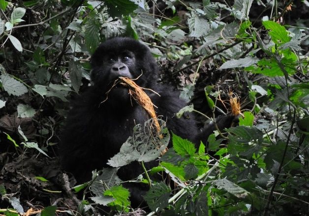
How do I apply?
There is a 3-Step application process for Anatomy and Developmental Biology entry:
   1. Discuss the projects of interest with the potential supervisors by appointment.
   2. Submission of a project application signed by the supervisors to Dr Olga Panagiotopoulou
       (olga.panagiotopoulou@monash.edu)
   3. Formal application to the faculty. The application form can be downloaded from the Monash
       Biomedicine Discovery Institute website:
       https://www.monash.edu/discovery-institute/honours/so-how-do-i-apply

If you have a query regarding eligibility, please submit an enquiry online via ask.monash or call 1800
MONASH (1800 666 274).

Entry Criteria
Bachelor of Science (Honours)
Bachelor of Science students wishing to undertake an Honours degree in the School of Biomedical
Science (SOBS) have increased flexibility to complete an Honours degree in the Department of Anatomy
and Developmental Biology. Any major in the School of Biomedical Science will allow students to
undertake an Honours degree within the Department of Anatomy and Developmental Biology.

A distinction grade average (70%) in 24 points of relevant 3rd year units, of which normally 18 points
are developmental biology or biochemistry, human pathology, immunology, microbiology,
pharmacology and physiology units. In addition to the requirements listed above, students must meet the
entry requirements for the Science honours program relevant to their course of enrolment. Enrolment in
an honours project is subject to approval of the supervisor and the Honours Convenor.

Bachelor of Biomedical Science (Honours)
An average of 70% or higher in at least 24 points at 3rd year (including 12 points in Biomedical Science
core units).

The closing date for Bachelor of Biomedical Science and the Faculty of Science applications is usually
mid-November.

                                                                                                         3
Key research areas
The Department of Anatomy & Developmental Biology at Monash University is very active in a
variety of research areas. It boasts several of the world’s leading research scientists in the field of
Developmental biology and Anatomy.

Major areas of research include:

◼ Blood cell homeostasis: cell death and disease- Prof Benjamin Kile lab (Dr Dominic De Nardo)
◼ Bone biomechanics, implant design, oral and maxillofacial trauma (Dr Olga Panagiotopoulou)
◼ Brain development, neuroplasticity and stem cells (Prof Roger Pocock)
◼ Cellular physiology (Dr Senthil Arumugam)
◼ Comparative development and evo-devo (A/Prof Craig Smith)
◼ Developmental neuroscience and multiple sclerosis (Dr David Gonsalvez)
◼ Development and disease (Dr Alex Combes)
◼ Education Research (A/Prof Michelle Lazarus)
◼ Epithelial regeneration (Prof Helen Abud)
◼ Epigenetics and Reprogramming (Prof José Polo)
◼ Gene Regulation (Dr Partha Das)
◼ Integrated morphology and palaeontology (A/Prof Justin Adams)
◼ Kidney development and disease (Prof Ian Smyth)
◼ Kidney development, programming and disease (Prof John Bertram)
◼ Morphology, Ontogeny and Evolution (Dr Jason Massey)
◼ Nervous system development (Dr Brent Neumann)
◼ Neurogenesis and neuroregeneration (Prof Zhi-Cheng Xiao)
◼ Oocyte and embryo development (Prof John Carroll)
◼ Ovarian biology (A/Prof Karla Hutt)
◼ Organogenesis and cancer (Prof Kieran Harvey)
◼ Palaeodiet research (A/Prof Luca Fiorenza)
◼ Prostate Cancer Research (Prof Gail Risbridger)
◼ Prostate Cancer Research (Dr Luc Furic)
◼ Sensory perception and ageing (Dr Jie Liu)
◼ Stem Cells and Translational Immunology (A/Prof Tracy Heng)
◼ 3D Cancer Model Lab (A/Prof Daniela Loessner)

                                                                                                          4
CONTACTS IN THE DEPARTMENT

Dr Olga Panagiotopoulou
Principal Honours Convenor
Department of Anatomy and Developmental Biology
10 Chancellors Walk (13C Building), Level 1, Room C149
Tel: 9905 0262
Email: olga.panagiotopoulou@monash.edu

Associate Professor Craig Smith
Honours Co-Convenor
Department of Anatomy and Developmental Biology
Building 76, Level 3, Room 355
Tel: 9905 0203
Email: craig.smith@monash.edu

Associate Professor Tracy Heng
Honours Co-Convenor
Department of Anatomy and Developmental Biology
Building 75, Level 3, Room 314
Tel: 9905 0629
Email: tracy.heng@monash.edu

                                                         5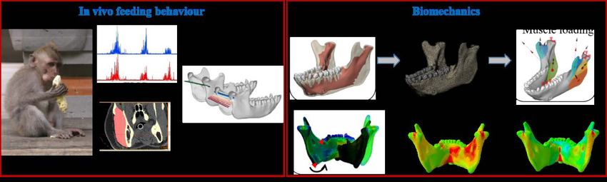
MONASH BIOMEDICINE DISCOVERY INSTITUTE
          School of Biomedical Sciences

ABOUT THE MONASH BIOMEDICINE DISCOVERY INSTITUTE
(encompassing the School of Biomedical Sciences)

WHO WE ARE
  • An institute with the scale and scope to tackle major
    research questions
  • 120+ internationally-renowned research teams committed
    to addressing global health priorities

WHAT WE DO
  • Discovery research to accelerate our ability to prevent,
     diagnose and treat disease
  • Innovate through national and international collaborations
     and partnerships with researchers, health precincts and
     industry

                                                                 6
MONASH BIOMEDICINE DISCOVERY INSTITUTE
             School of Biomedical Sciences

With more than 120 internationally-renowned research teams, the Monash Biomedicine
Discovery Institute (BDI) is one of the largest and highest-quality biomedical research institutes
in Australia. Monash BDI works with national and international collaborators on global health
priority areas, including cancer, cardiovascular disease, development and stem cells, infection
and immunity, metabolism, diabetes and obesity, and neuroscience.

Our discoveries accelerate the ability to prevent, diagnose and treat disease by leveraging our
strong partnerships with researchers, health precincts and industry, together with our access
to unparalleled, world-leading research infrastructure.

The Monash BDI encompasses the School of Biomedical Sciences, and is part of Monash’s
Faculty of Medicine, Nursing and Health Sciences. The School of Biomedical Sciences delivers
biomedical sciences education to more than 2,000 undergraduate students and 300
postgraduate students.

Based at Monash’s Clayton campus, the Monash BDI is structured to include six health-
focused discovery programs and five discipline-specific departments. This allows for the cross-
pollination of ideas needed to tackle the big questions in biomedical research − it is at the
intersection of these global health issues that truly innovative discoveries will be made.

DISCOVERY PROGRAMS                                  RESEARCH | EDUCATION | ENGAGEMENT
   • Cancer
   • Cardiovascular Disease
   • Development & Stem Cells
   • Infection & Immunity
   • Metabolism, Diabetes & Obesity
   • Neuroscience
DEPARTMENTS
   • Anatomy & Developmental Biology
   • Biochemistry & Molecular Biology
   • Microbiology
   • Pharmacology
   • Physiology
CENTRES
   • Centre for Human Anatomy Education

                                                                                                7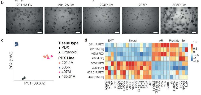
BDI PROGRAM: CANCER

Prostate Cancer Research Group

             https://www.monash.edu/discovery-institute/prostate-cancer-research-group

Project Title                      Identifying new treatments for drug-resistant cancer
Main Supervisor                    Prof Gail Risbridger gail.risbridger@monash.edu 9902 9558

Other Supervisors                  Dr Mitchell Lawrence, A/Prof Renea Taylor
Location                           Clayton Campus, 19 Innovation Walk, Level 3
Outline of project

Background:
Over the last decade, new treatments have extended the survival of
men with advanced prostate cancer. Unfortunately, patients
eventually develop resistance to all current treatments. Therefore,
effective new therapies are needed for drug-resistant cancers.

Our group is tackling this challenge in a new way - by growing
tumours from different patients in the lab. Using this novel technique,
we can compare how tumours respond to novel treatments. Our results
are already showing that some tumours are more sensitive than others
to specific treatments. These exciting results are helping us prioritise
drugs for further clinical validation.

Project aim:
In this project, we will test the response of different patients’ tumours   Patient prostate cancer cells
to novel drugs targeting the epigenome and DNA damage repair.               stained in pink before drug
Subsequently, we will identify molecular features that distinguish           treatment (top) and after
between sensitive and resistant tumours. These important results will        drug treatment (bottom).
provide new insight into better treatments for tumours that have failed       Below, prostate cancer
current therapies.                                                          organoids before treatment

Techniques to be used:
This project will use techniques a range of techniques including
organoid     cell      culture,    patient-derived   xenografts,
immunohistochemistry and pathology.

                                                                                                            8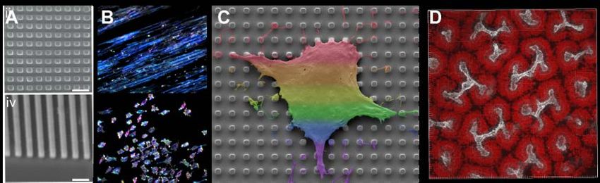
Prostate Cancer Research Group

            https://www.monash.edu/discovery-institute/prostate-cancer-research-group
Project Title                   Investigating the progression of neuroendocrine prostate cancer
Main Supervisors                Prof Gail Risbridger    gail.risbridger@monash.edu       9902 9558

Other Supervisors               Dr Roxanne Toivanen

Location                        Clayton Campus, 19 Innovation Walk, Level 3
Outline of Project

Background: Prostate cancer is the most commonly diagnosed cancer
in Victoria. As most prostate tumours rely on androgen hormones for
growth, the most common therapies for metastatic prostate cancer are
drugs which inhibit androgen signalling. However, some patients
develop neuroendocrine prostate tumours, which are highly aggressive
and resistant to androgen inhibitors. Currently, there is limited
understanding of how neuroendocrine tumours emerge in patients, and      Immunohistochemistry of a
therapeutic options are limited.                                         xenograft of neuroendocrine
                                                                              prostate cancer.
Our laboratory has successfully developed a model where patient
samples of neuroendocrine prostate cancer can be grown and studied
in the laboratory. These samples represent an invaluable resource for
studying the biology of these tumours and testing novel therapeutics.

Project Aims:
The goal of this project is to use patient-derived models, including
xenografts and organoids, of neuroendocrine prostate cancer to study
disease progression and test effective therapeutic strategies.

Techniques:
The project will involve a variety of techniques including working
with human tumours, tissue culture of organoids and                       Prostate cancer organoids
immunohistochemistry.                                                   before (top) and after (bottom)
                                                                                drug treatment

                                                                                                          9
Prostate Cancer Research Group
https://www.monash.edu/discovery-institute/prostate-cancer-research-group
Project Title Treating prostate cancer with high dose testosterone
Main Supervisor Dr Mitchell Mitchell.lawrence@monash.edu 9902
Lawrence 9558
Other Supervisors Prof Gail Gail.risbridger@monash.edu 9902
Risbridger 9558
Other Supervisors
Location 19 Innovation Walk, Monash Clayton Campus
Outline of project
Background:
Patients with aggressive prostate cancer usually receive drugs that block hormones. The first clinical trial
of this treatment was in 1941. Despite 80 years of progress, and potent new drugs, these treatments are only
temporarily effective. Patients inevitably develop drug-resistant tumours that are even more aggressive.
The current drugs can also be expensive and produce side-effects that reduce quality of life.
Project aim:
To address these challenges, our goal is to develop a safe, effective and inexpensive new treatment for
prostate cancer known as “Bipolar Androgen Therapy” (BAT). BAT differs from current treatments
because it overstimulates hormone activity rather than blocking it. Clinical trials of BAT in Australia and
the United States are producing promising results.
Techniques to be used:
Our plan is to use BAT as the backbone of new combination treatments – combining it with other drugs to
control prostate cancer cells for longer.
This raises the question: what is the best drug to combine with BAT? To resolve this question, this project
will use organoids to screen different combination treatments. The techniques will involve 3D cell culture,
microscopy, histology, and quantitative PCR.
(Phase microscope images of different organoid models of prostate cancer)
10BDI PROGRAM: NEUROSCIENCE
Sensory Perception and Ageing
Lab
https://www.monash.edu/discovery-institute/liu-lab
Project Title Mitochondrial calcium homeostasis
Main Supervisor Jie Liu Jie.liu1@monash.edu 99050622
Other Supervisors
Other Supervisors
Location 3rd floor, Building 75, 15 Innovation Walk
Outline of project
Background:
Calcium ions (Ca2+) are one of the most critical signalling molecules which play an important role in
many physiological processes, prominently including muscle contraction, neuronal excitation and
cellular metabolism, therefore, Intracellular Ca2+ dysregulation is a major cause of many diseases.
Mitochondria, an intracellular Ca2+ store, directly regulates cellular Ca2+ dynamics. Balancing the
mitochondrial Ca2+ depends on the specific ion channel on mitochondrial membrane. In past decade,
most of the molecular machineries modulated mitochondrial Ca2+ homeostasis have been identified. Ca2+
influx into mitochondria is mediated by mitochondrial calcium uniporter (MCU) complex, which resides
on inner membrane to guarantee Ca2+ uptake upon cytoplasmic Ca2+ increase. So far, it is widely believed
that MCU complex is the main route for Ca2+ flux into mitochondria. However, in our preliminary study
we found that an undefined MCU-independent transporter might also contribute to the mitochondrial
Ca2+ uptake in C. elegans.
Project aim/s:
In this project, a typical ethyl methanesulfonate
(EMS) screen will be conducted to identify the
potential mitochondrial calcium transporter. B KCl (70 mM)
This project aims to understand the mechanisms
of mitochondrial Ca2+ regulation, which
ΔF/Fo
provides the potential pharmacological targets
of mitochondrial Ca2+ machineries for the
treatment of related diseases in humans
Techniques to be utilised: KCl depolarization produced an increase in
We will investigate the regulatory mechanism mitochondrial calcium response in C. elegans
through a multidisciplinary approach which body wall muscle cells.
combines calcium imaging, patch-clamp,
genetic manipulation and behavioural analysis
with biochemical methods.
11BDI PROGRAM: DEVELOPMENT & STEM CELLS
Epithelial Regeneration
Laboratory
https://www.monash.edu/discovery-institute/abud-lab
Project Title Expression profile and impact of EGF ligands on intestinal stem
cells
Main Supervisor Thierry Jarde Thierry.jarde@monash.edu 99029208
Other Supervisors Helen Abud Helen.abud@monash.edu 99029113
Other Supervisors
Location Clayton Campus, 19 Innovation Walk, Level 3
Outline of project
Background:
Intestinal stem cells are regulated by essential cellular pathways,
including EGF signalling, that drive stem cell maintenance and
proliferation. The EGF signalling pathway is activated locally by
one of the eleven EGF family of ligands produced by supporting
niche cells. Interestingly, a complete understanding of cellular
sources and impact of individual EGF ligands on gastrointestinal
stem cells is currently missing.
Project Aim:
This project aims to understand the function of the EGF family of
ligands in the human and murine gastrointestinal tract.
Techniques:
This project will involve characterising the expression profile of EGF family of ligands in the human and
murine gastrointestinal tract using single cell RNA sequencing datasets and immunofluorescence. The
function of identified ligands will be investigated using human and mouse organoids and in vivo models.
12Epithelial Regeneration
Laboratory
https://www.monash.edu/discovery-institute/abud-lab
Project Title Expression profile and impact of EGF ligands on intestinal stem
cells
Main Supervisor Dr Rebekah rebekah.engel@monash.edu 99029196
Engel
Other Supervisors Dr Christine ckoulis@cabrini.com.au 95083547
Koulis
Other Supervisors Prof Helen Abud helen.abud@monash.edu
Location Clayton Campus, 19 Innovation Walk, Level 3
Outline of project
Background:
Colorectal cancer is one of the leading causes of cancer-
related deaths worldwide. Patients diagnosed with colorectal
cancer often experience different clinical outcomes and drug
responses, even when controlled for similar pre-operative
features, tumour stage and pathological characteristics.
Project Aim:
This project aims to determine predictive and prognostic
biomarkers in colorectal cancer through the use of tissue
microarrays and organoid technology. Specifically, we will
identify biomarkers including molecular subtypes that
distinguish between sensitive and resistant tumours. These important results will provide new insights into
personalised cancer treatments.
Techniques:
This project will involve a range of techniques including working with human colorectal tumours, tissue
microarray construction, patient-derived organoid cell culture, drug sensitivity assays,
immunohistochemistry and pathology.
13Egg and Embryo Development
Group
https://www.monash.edu/discovery-institute/carroll-lab
Project Title Investigation of mRNA levels in aging oocytes
Main Supervisor John Carroll j.carroll@monash.edu 99024381
Other Supervisors Wai Shan Yuen wai.yuen@monash.edu 99020390
Other Supervisors
Location Clayton Campus, 19 Innovation Walk, Level 3
Outline of project
Background:
Reproduction success is highly dependent upon the age at which
women attempt to conceive, which is progressively increasing
worldwide. This age-dependent decrease in fertility is
associated with an increase in the incidence of miscarriage and
the prevalence of chromosomal abnormalities. In IVF, maternal
age is among the strongest predictors of success. Together with
social trends that see the age of first pregnancy between 1991
and 2011 increase by 35% in the 35-39 year-old age bracket and
by over 70% in the 40-45 year old age group, point to a major
medical problem that affects a significant proportion of the
population. We previously performed single oocyte mRNA
sequencing in mice eggs and found that the biggest difference is
the diminished presence of mRNA levels in old oocytes (Top
Figure). In addition we found that the mRNA profile of oocytes
from young and old mice presented either into an “Aged” or a
“Young” state (Bottom figure). CL0
“Young”
‘
Project Aim:
The goal of this project is to validate our single oocyte mRNA
sequencing by determining the protein levels of key age-related
genes, and to assess their contribution to the ageing process in
oocytes. “Aged”
Techniques:
The project will involve a variety of techniques including mouse
egg collection and culture, in vitro fertilisation, embryo
development, live-cell imaging and immunofluorescence.
14Egg and Embryo Development
Group
https://www.monash.edu/discovery-institute/carroll-lab
Project Title Molecular mechanisms behind cortical polarity in eggs
Main Supervisor John Carroll j.carroll@monash.edu 99024381
Other Supervisors Wai Shan Yuen wai.yuen@monash.edu 99020390
Other Supervisors
Location Clayton Campus, 19 Innovation Walk, Level 3
Outline of project
Background:
Asymmetric cell division underlies essential developmental processes in all
eukaryotes. An extreme form of this occurs during the meiotic divisions of
the oocyte. The mammalian oocyte undergoes two consecutive cell divisions
to form a haploid oocyte and two small polar bodies. These extremely
asymmetric divisions allow the oocyte to retain cytoplasmic components
essential for fertilisation and early embryo development. In mammalian
oocytes, asymmetry is coupled to the migration of the centrally placed
meiosis I spindle along its long axis towards the nearest cortex. A critical
event required for asymmetric division in the oocyte is the establishment of
cortical polarity. Cortical polarisation results in formation of an F-actin
domain near the spindle during late metaphase I and in metaphase II arrested
oocytes. The differentiation of the polar cortex in metaphase of both meiotic
divisions contrasts with mitotic cell divisions where the recruitment and
activation of Rho and associated actin polymerisation only occurs after
anaphase when the central spindle forms. In our previous study, we
determined that polo- kinase 1 (Plk1) is essential for establishing cortical
polarity in meiosis I but not in maintaining it during meiosis II.
Project Aim: Fig 1 – Plk1’s unique
This goal of this project is to determine the molecular mechanism behind localisation during
the Plk1-mediated polarity pathway. Candidate genes which are involved in meiosis I
this are hypothesised to be Ran-GTP, RanBP1.
Techniques:
The project will use a variety of techniques such
as mouse egg collection and culture, live-cell
imaging, microinjection of fluorescently-labelled
probes, and immunofluorescence.
Fig 2 – Plk1 inhibition abolishes cortical polarity
through actin pathway
15Egg and Embryo Development
Group
https://www.monash.edu/discovery-institute/carroll-lab
Project Title Molecular mechanisms behind cortical polarity in eggs
Main Supervisor John Carroll j.carroll@monash.edu 99024381
Other Supervisors Deepak Adhikari deepak.adhikari@monash.edu 99020120
Other Supervisors
Location Clayton Campus, 19 Innovation Walk, Level 3
Outline of project
Background:
Our research is focused on understanding the mechanisms of oocyte development, maturation and
fertilization in mammals. Early embryo development in mammals is driven and underpinned by a healthy
fertile oocyte. How the oocyte acquires this developmental potential is not understood and problems in
oocyte quality are largely responsible for the high rates of infertility and miscarriage in the population.
Project Aim:
In this project, we will examine how mitochondria are
formed during oocyte development and how mitochondrial
quantity and quality impact the quality of oocyte. We will
eventually identify the pathways that might be impacted by
mitochondrial dysfunctions. These important findings will
provide insights into potential ways of improving egg
quality and fertility outcome.
Techniques:
This project will use a range of techniques including in vitro
oocyte and embryo culture, live cell imaging,
immunofluorescence and molecular biology techniques.
16Stem Cell Models
Progenitor regulation
Combes Lab
https://www.monash.edu/discovery-institute/combes-lab
Project Title Stem Cell Models of Kidney Disease
Main Supervisor Alex Combes alex.combes@monash.edu 99056219
Other Supervisors
Other Supervisors Ana Núñez Nescolarde, Rachel Lam
Location 19 Innovation Walk, Clayton Campus
Outline of project
Background:
Kidney organoids are miniature human tissues produced from
stem cells in culture. Kidney organoids are amenable to high
throughput screening and have shown promise as a model of
genetic kidney disease. However, it is unclear whether
organoids can be used to model more complex and common
aspects of chronic kidney disease, which affects 10% of the
global population.
Project Aim:
This project aims to broaden the impact of organoid technology
on kidney disease by testing how human kidney organoids
respond to factors known to initiate kidney injury and chronic
kidney disease in humans and animal models.
Techniques:
Developing stem cell models of chronic kidney disease will involve the culture and differentiation of
human pluripotent stem cells to generate kidney organoids. Organoids will be challenged with
environmental, metabolic, and signalling factors in vitro and assessed by gene expression profiling,
histology and confocal microscopy. An optional bioinformatics component is available for this project.
17Stem Cell Models
Progenitor regulation
Combes Lab
https://www.monash.edu/discovery-institute/combes-lab
Project Title Nephron Progenitor regulation in Kidney Development and
Disease
Main Supervisor Alex Combes alex.combes@monash.edu 99056219
Other Supervisors
Other Supervisors
Location 19 Innovation Walk, Clayton Campus
Outline of project
Background:
Kidney development is controlled to a large extent by
the balance between self-renewal and differentiation of
a multipotent nephron progenitor (NP) population.
Self-renewing NP cells produce factors that drive
kidney growth, while differentiating NP cells build the
filtration capacity of the organ by forming nephrons.
As such, knowledge of NP regulation has a major
bearing on both chronic kidney disease and the
production of nephrons in culture.
Project Aims:
This project aims to identify novel mechanisms of nephron progenitor regulation and dynamics in knockout
mouse models. These efforts may lead to fundamental advances in progenitor cell biology and practical
outcomes including improved production of nephrons for disease modelling and drug screening
applications.
Techniques:
This project has scope to cater for students wanting to develop laboratory and/or bioinformatics skills.
Laboratory techniques will include dissection and culture of mouse embryonic kidneys, histology,
immunofluorescence, confocal microscopy, gene expression profiling. Bioinformatic techniques will
include analysis of single cell gene expression profiling data (no prior coding experience necessary).
18POCOCK GROUP
https://www.monash.edu/discovery-institute/pocock-lab
Project Title Neuronal Regulation of Mitochondrial Health.
Main Supervisor Prof. Roger roger.pocock@monash.edu 99050658
Pocock
Other Supervisors Rebecca Cornell
Other Supervisors
Location 15 Innovation Walk, Clayton Campus
Outline of project
Background: Mitochondrial damage is a hallmark of obesity, diabetes and cardiovascular disease. Cells
combat mitochondrial damage by triggering protective stress responses to prevent a breakdown in
metabolic homeostasis. Recent evidence demonstrate that the nervous system can regulate stress
responses in distal tissues.
In unpublished work, we discovered that neuropeptide signalling from two sensory neurons regulate
the mitochondrial stress response in distal fat storage cells. This project will use state-of-the-art
genetic and imaging tools dissect how the brain controls mitochondrial stress.
Project Aims: In this project, we will identify molecular mechanisms through which the brain controls
mitochondrial stress responses in distal tissues. Based on our preliminary data we believe that we have
discovered a new means of controlling systemic mitochondrial stress, which may be useful in therapies
for multiple diseases.
Techniques to be utilised: This project will utilise techniques in genetics, in vivo neurodevelopmental
dissection, molecular biology (CRISPR), biochemistry, microscopy.
Animal transgenically expressing green
fluorescent protein to enable
visualisation of mitochondrial stress.
19Brain Development,
Neuroplasticity and Stem Cells
Laboratory
https://www.monash.edu/discovery-institute/pocock-lab
Project Title Transcriptional control of stem cell development
Main Supervisor Prof. Roger roger.pocock@monash.edu
Pocock
Other Supervisors Dr Wei Cao
Other Supervisors
Location 15 Innovation Walk, Clayton Campus
Outline of project
Background: Stem cells have great potential for regenerative studies and disease therapeutics. However,
we do not fully understand how stem cells develop in vivo, limiting their therapeutic potential. To study
stem cell development in vivo, the well-defined Caenorhabditis elegans stem cell niche has proven an
excellent model. Using RNA sequencing and gene knockout studies we have identified multiple
transcription factors that are important for stem cell development. This project will use state-of-the-art
techniques to dissect the mechanistic functions of these transcription factors in stem cells.
Project Aims: In this project, we will decipher the function of conserved transcription factors in C.
elegans stem cell development. We believe that this work will identify novel mechanisms through which
stem cells may be manipulated.
Techniques to be utilised: This project will utilise techniques in genetics, in vivo stem cell analysis,
confocal microscopy, biochemistry and CRISPR-Cas9.
Image of the Caenorhabditis elegans
germline highlighting germ cell nuclei
(red) and cell membranes (green).
20Brain Development,
Neuroplasticity and Stem Cells
Laboratory
https://www.monash.edu/discovery-institute/pocock-lab
Project Title How do you make a brain?
Main Supervisor Prof. Roger roger.pocock@monash.edu
Pocock
Other Supervisors Dr Pedro Moreira
Other Supervisors
Location 15 Innovation Walk, Clayton Campus
Outline of project
Background: Neurons in the human brain communicate with each other through ~850,000 kilometres of
cables. These cables are called axons, and they are guided to their correct positions during development
by an array of cell-surface and secreted molecules. How the expression of these axon guidance
molecules is regulated during brain development is a fundamental knowledge gap. We have
identified a transcription factor controls that controls axon guidance in Caenorhabditis elegans.
Project Aims: In this project, we will identify how the transcription factor controls gene expression to
regulate axon guidance of specific neurons. Based on our preliminary data we believe that we have
discovered a new means of controlling brain development, which may be useful in therapies for brain
disorders.
Techniques to be utilised: This project will utilise techniques in genetics, in vivo neurodevelopmental
dissection, molecular biology (CRISPR), biochemistry, microscopy.
Animal transgenically expressing
fluorescent proteins to enable
visualisation of the nervous system.
21Stem Cells and Translational
Immunology (Heng Lab)
https://www.monash.edu/discovery-institute/heng-lab/home
Project Title Mechanisms of mesenchymal stem cell therapy
Main Supervisor A/Prof Tracy Tracy.Heng@monash.edu 99050629
Heng
Other Supervisors
Location Level 3, 15 Innovation Walk
Outline of project
Background:
Multipotent mesenchymal stromal cells (MSCs) are fibroblastic precursor cells that have the stem cell-
like ability to differentiate into a variety of cell types. MSCs are endowed with potent anti-inflammatory
and immunosuppressive properties, and are being used in over 500 clinical trials to treat various
inflammatory conditions. However, MSCs undergo programmed cell death (apoptosis) shortly after
infusion and it remains unclear how these short-lived cells mediate their therapeutic effects.
Live MSCs secrete immunosuppressive molecules
while infused MSCs undergo apoptosis in the lung,
triggering an anti-inflammatory immune response.
Project aim/s:
This project aims to elucidate how the innate and adaptive immune responses to apoptotic MSCs impact
therapeutic efficacy. The findings will have broad implications for the future development of MSC-based
therapies.
Techniques to be utilised: This project will utilise techniques applicable to both immunology and stem
cell research, including stem and stromal cell isolation, tissue culture, flow cytometry, qPCR,
bioluminescence imaging and mouse disease models.
22Kidney Development and Disease
(Smyth Lab)
https://www.monash.edu/discovery-institute/smyth-lab
Project Title Characterising novel genes which cause congenital kidney disease
Main Supervisor Ian SMYTH ian.smyth@monash.edu 99029119
Location 19 Innovation Walk
Background:
Our group is involved in an Australia-wide program
(www.kidgen.org.au) which aims to identify novel
causative genes in patients with kidney disease. Individuals
with inherited kidney disease who do not have mutations in
known genes will have their genomes sequenced to identify
novel genetic causes for their conditions.
Project Aim/s:
We use CRISPR/Cas9 genome engineering approaches to
model disease causing mutations in mice. Using these
models, honours students will have a unique opportunity to
establish how novel disease genes function in the kidney,
how their protein products regulate cell biology and how
their mutation leads to congenital renal malformations.
Techniques:
This project will utilise CRISPR/Cas9 approaches to introduce changes in mice which we will then
study using histology, OPT imaging, immunostaining and RNA sequencing. Causative genes will be
studied in vitro using tissue culture and various biochemical approaches will be employed to best
understand gene and protein function.
23Kidney Development and Disease
(Smyth Lab)
https://www.monash.edu/discovery-institute/smyth-lab
Project Title Understanding the pathogenesis of polycystic kidney disease
Main Supervisor Ian SMYTH ian.smyth@monash.edu 99029119
Location 19 Innovation Walk
Background:
Polycystic kidney disease (PKD) is the most common
potentially lethal Mendelian disease, affecting around
1/1000 people. It arises when cells of the kidney tubules
over-proliferate, forming cysts which gradually expand
and ablate normal kidney tissue. We have shown that the
Aurora A Kinase is a central regulator of the
development of cystic disease in a number of in vivo
models of PKD.
Project Aim/s:
In this project we will examine a number of different
mechanisms by which Aurka might be regulating the
growth of renal cysts to better understand the how the
protein functions, and to investigate it as a possible
therapeutic target for treating the disease.
Techniques:
This project will examine mouse and cellular models of PKD and the biochemical functions of AURKA.
It will utilise a broad range of techniques to do so including histology, immunohistochemistry and
transcriptional profiling. Specific cell signalling pathways will be studied in vitro using tissue culture
and various biochemical approaches to best understand gene and protein function.
24Harvey Laboratory
https://www.monash.edu/discovery-institute/harvey-lab/home
Project Title Watching the Hippo pathway in real time in growing organs
Main Supervisor Kieran Harvey kieran.harvey@monash.edu
Other Supervisors Samuel Manning sam.manning@monash.edu
Other Supervisors Benjamin Kroeger benjamin.kroeger@monash.edu
Location Anatomy and Developmental Biology Department
Outline of project
Background
A new frontier in biomedical research will involve watching individual
proteins work in real time, in living organs. Traditionally, researchers have
drawn conclusions about gene function using indirect techniques that only
allow us to infer what a gene normally does, without actually watching it
work. For example, we create organisms that lack a particular gene and
determine whether something goes wrong. If the loss of gene X causes organs
to overgrow then we assume that gene X normally limits organ size. This has
been an extraordinarily powerful approach for interrogating gene function but
it cannot substitute the ability to watch gene products executing their function
in real time, which allows determination of exactly when, where and how they
work.
Project aim/s
We will investigate the role of the Hippo tumour suppressor pathway in organ growth by watching, for
the first time, its activity, in growing organs, in real time. This will provide novel insights into normal
organ growth and pathogenic organ growth in diseases such as cancer.
We aim to observe Hippo pathway activity in real time in the following situations:
a) When organs are actively growing
b) When organs stop growing
c) In regions of organs that are subject to mechanical compression
d) Throughout the cell cycle
Techniques
You will be taught an array of techniques including ex vivo organ culture, live multi-photon microscopy,
image analysis and Drosophila genetics.
25Comparative Development and
Evo-Devo Laboratory
https://www.monash.edu/discovery-institute/smith-lab
Project Title Role of Pax2 in chicken embryonic gonadal development
Main Supervisor A/Prof Craig craig.smith@monash.edu 9905 0203
Smith
Other Supervisors Dr. Andrew andrew.major@monash.edu
Major
Location Clayton Campus, 19 Innovation Walk, Level 3
Outline of project
Background
Embryonic gonads are unique because they have a developmental choice: testis or ovary formation. Our
lab studies how this choice is executed at the molecular genetic level. We recently identified novel
expression of the Pax2 transcription factor gene in the embryonic chicken gonad (see image below; pink
immunofluorescence).
Project aim/s
This project will explore the role of Pax2 in the gonad, using gene over-expression and knockdown
approaches.
Techniques
Methods used will centre around experimental developmental biology; PCR, RT-PCR, gene cloning, in
situ hybridisation, immunofluorescence and organ culture.
26Kidney Development,
Programming and Disease
Research Laboratory
https://www.monash.edu/discovery-institute/bertram-lab
Project Title Maternal nutrition and offspring kidney development
Main Supervisor Dr Luise Cullen- luise.cullen- 99029106
McEwen mcewen@monash.edu
Other Supervisors Prof John Bertram john.bertram@monash.edu 99029101
Location Clayton Campus, 19 Innovation Walk, Level 3
Outline of project
Background:
The intrauterine environment is critical
for fetal growth and organ
development. Kidney development is
especially vulnerable to a suboptimal
maternal environment, with long-term
adverse consequences for kidney and
cardiovascular health. We have
recently shown that a simple
modification of the maternal diet can
boost, and even rescue, nephron
endowment in offspring destined to
develop kidneys with a low nephron
endowment. Just how this diet achieves these clinically-relevant outcomes is unclear.
Project aim:
In this project, you will investigate how the maternal diet can boost and rescue nephron endowment.
Techniques: Mouse embryonic and neonatal dissection, immunohistochemistry/immunofluorescent
labelling techniques, confocal microscopy, stereology, 3D imaging.
27Gene Regulation Laboratory (PPD
LAB)
https://www.monash.edu/discovery-institute/partha-das-lab
Roles of epigenetic and epi-transcriptomic modifications in embryonic
Project Title
stem cells, neurodevelopment, and neurodevelopmental disorders (NDDs)
Main Supervisor Dr Partha Das partha.das@monash.edu 99024009
Other Supervisors Dr Pratibha Pratibha.tripathi@monash.edu 9902 9111
Tripathi
Location Level 3, 15 Innovation Walk,
Outline of project
Background:
Our laboratory research interests focus on how epigenetic (DNA and histone modifications) and epi-
transcriptomic (RNA modifications) changes control gene expression either at transcriptional or post-
transcriptional levels. We use various
experimental approaches and cutting-edge
technologies including- Cell and
Molecular Biology, Biochemistry,
CRISPRs, CRISPR screens, high-
throughput sequencing technologies
(ChIP-seq, RNA-seq, WGS, ATAC-seq,
RRBS, Hi-C), proteomics, bioinformatics
and computational biology to understand
the gene regulation that control embryonic
stem cells (ESCs) state, differentiation,
Fig. Distribution of predominant RNA modifications development and diseases.
Project aim/s:
1. Investigating the roles of m6A RNA modification in ESCs
2. Examining the roles of m6A RNA modification in developing brain using human brain organoids
3. Dissecting the roles of m5C RNA modification in Williams Beuren Syndrome (WBS) and Autism
spectrum disorders (ASD) using human brain organoids
4. Roles of epigenetic factors in neurodevelopment and ASD using brain organoids
5. Establishing epigenetic and epi-transcriptomic connections for gene regulation in ESCs
Techniques to be utilised:
CRISPRs, RNA-seq, m5C and m6A-RNAseq, RIP-seq, RIBO-seq, ChIP-seq, scRNAseq,
immunoprecipitation, western blots, mass spectrometry, immunostaining, human and mouse ESCs/iPSCs
culture, brain organoids.
28Organoid engineering and stem cell
differentiation
https://research.monash.edu/en/persons/jess-frith https://frithlab.com/
https://research.monash.edu/en/persons/alex-combes https://www.monash.edu/discovery-
https://research.monash.edu/en/persons/victor-cadarso-busto https://www.appliedmicronanolab.com/
Project Title Effects of the microenvironment on kidney development
Main Supervisor Jess Frith Jessica.frith@monash.edu 9905 1967
Other Supervisors Alex Combes Alex.combes@monash.edu
Victor Cadarso Victor.Cadarso@monash.edu
Location New Horizons, 20 Research Way/19 Innovation Walk
Outline of project
Background:
Human kidney organoids, miniature organs grown in vitro, have great potential to accelerate disease
modelling and drug screening for the 750 million individuals affected by chronic kidney disease world-
wide. However, the utility of these stem cell-derived tissues is limited by variability between organoid
batches and a lack of control over tissue patterning.
During differentiation, the fate of cells fate is determined by their response to a complex milieu of signals
from their surrounding microenvironment, including mechanical and biochemical cues as well as soluble
growth factors. Current protocols to produce human kidney tissue from stem cells use soluble factors
alone to direct differentiation. However, previous work by our team has shown how changing physical
cues, for example by culturing cells on surfaces with microscale topographies, or patterned ligands to
which the cells adhere can have important effects on cell differentiation and function. The influence of
these signals on kidney cell specification and organisation is not yet well understood, but offers new
avenues to improve control over differentiation and tissue structure, both of which are required to improve
kidney organoids for disease modelling and drug screening applications.
A) Examples of engineered surfaces with specific micro topographies. B) Cell morphology changes with surface
patterning C) A cell on micropillars D) Nephron patterning in vitro. E) Patterning in organoids (left) vs a developing
kidney (right).
Project aim/s:
The aim of this project is to determine the effects of surface patterning, through changes in surface
topography and/or ligand presentation on kidney cell specification and development.
Techniques: This project will involve making engineered surfaces, with variations in topography and/or
surface chemistry. Kidney explants, dissociated kidneys, and/or induced pluripotent cells will be cultured
on these surfaces and their response to these microenvironmental changes monitored by immunostaining,
high-content imaging and image-based analyses of cell fate and mechanosignalling.
29Moving Morphology & Functional
Mechanics
Panagiotopoulou Lab
https://www.monash.edu/discovery-institute/panagiotopoulou-lab/home
Project Title Jaw Fracture Repair and Biomechanics of Chewing
Main Supervisor Dr Olga olga.panagiotopoulou@monash.edu 9905
Panagiotopoulou 0262
Other Supervisors
Location 10 Chancellors Walk, Level 1, Room C142
Outline of project
Background:
Mandible (lower-jaw) fractures account for ~30-40% of all instances of maxillofacial trauma and
can occur due to a variety of incidents (e.g. assault, oropharyngeal/congenital disorders, cancer,
vehicular accidents, sporting accidents or falls). Current treatment aimed at restoring mandibular
function and aesthetics by immobilizing the fracture with fixation mini-plates and screws is not
free of morbidity. Approximately 10–30% of mandibular fracture patients experience
postoperative complications, such as mal-union (bone segments not fusing properly),
malocclusion (tooth misalignment), infection, and joint dysfunction. Moreover, there is no
consensus on the optimal surgical interventions for certain types of mandible fracture. We
propose that a significant cause of post-surgical complication is the appearance of strain
environments in the fracture zone and around the implants, which are not conductive to healing.
Project Aims:
To study the biomechanical function of the jaw during a complete chewing cycle in healthy
controls and post fracture fixation.
Techniques:
3D virtual reconstruction of CT scans, 3D implant design, Dynamic Finite Element Modelling,
Theoretical mathematical models.
30Moving Morphology & Functional
Mechanics
Panagiotopoulou Lab
https://www.monash.edu/discovery-institute/panagiotopoulou-lab/home
Feeding mechanics and bite force performance in white sharks during
Project Title
development
Main Supervisor Dr Olga olga.panagiotopoulou@monash.edu 9905 0262
Panagiotopoulou
Other Supervisors Dr David Reser david.reser@monash.edu 9902 7393
A/Prof Charlie Huveneers charlie.huveneers@flinders.edu.au
Location 10 Chancellors Walk, Level 1, Room C142
Outline of project
Background:
Sharks are essential top predators in marine ecosystems that are ecologically, economically, and culturally
important. Their predatory success relies upon the unique anatomy of their jaws, which produce extremely
high bite forces while withstanding
damaging tissue stresses.
Large sharks, such as the white shark, alter
their diets during development, with
young individuals predominantly pursuing
fish, while adults preferentially target
large, more energy-dense prey. This
preference requires extreme bite force
capacity, which develops during ontogeny
from juveniles to full-size adults (4–6
metre length).
It is currently unknown whether the
increase in bite force is due to increases in
head size and muscles, or whether
additional maturational factors are
involved. To better understand bite force scaling within and between species during ontogeny and its
implications for the ecological role of large sharks, in-depth studies on jaw feeding mechanics are
required.
Project Aims
To study the biomechanical function of white shark jaws during growth.
Techniques
Cadaveric dissections, muscle physiological cross-sectional area analyses, optical microscopy, cryo-
electron microscopy, 3D virtual reconstruction of CT scans and synchrotron data, finite element
modelling, and theoretical mathematical models.
31Moving Morphology & Functional
Mechanics
Panagiotopoulou Lab
https://www.monash.edu/discovery-institute/panagiotopoulou-lab/home
Feeding mechanics and mandible shape in non-human primates during
Project Title
ontogeny
Main Supervisor Dr Olga olga.panagiotopoulou@monash.edu 9905
Panagiotopoulou 0262
Other Supervisors A/ Prof Luca Luca.fiorenza@monash.edu 990
Fiorenza 59809
Location 10 Chancellors Walk, Level 1, Room C142
Outline of project
Background:
Lower jaw (mandible) shape has yet to yield definitive information on hominid diet, due to our
poor understanding on primate jaw biomechanics during development. While current thinking
attributes mandible shape to functional adaptation to food consistency (what we eat) our lab’s
work suggests a different hypothesis: that, rather than food consistency per se, primate jaws are
shaped by the interaction between the developing teeth and feeding behaviour (how they eat).
Testing this hypothesis is hampered by almost complete lack of knowledge about how individual
feeding behaviour changes jaw structure during development.
We have recently published a novel multi-pronged approach integrating in vivo experiments and
bioengineering, which proposes to provide the missing piece in understanding the function of the
jaw during growth.
Project Aims
To study the biomechanical function of the macaque jaw during growth.
Techniques
Cadaveric dissections, muscle physiological cross-sectional area analyses, optical microscopy,
cryo-electron microscopy, 3D virtual reconstruction of CT scans, finite element modelling, and
theoretical mathematical models.
32Moving Morphology & Functional
Mechanics
Panagiotopoulou Lab
https://www.monash.edu/discovery-institute/panagiotopoulou-lab/home
Project Title Feeding mechanics of fossil lungfish from the Gogo Formation
Main Supervisor Dr Olga olga.panagiotopoulou@monash.edu 9905 0262
Panagiotopoulou
Other Supervisors Dr Alice alice.clement@flinders.edu.au
Clement
Prof John Long john.long@flinders.edu.au
Location 10 Chancellors Walk, Level 1, Room C142
Outline of project
Background:
Lungfish are a group of lobe-finned fish (Sarcopterygians) that first appeared in the Devonian Period over
400 million years and still survive to the present day. They are the closest living group of fish to tetrapods
(terrestrial vertebrates) and as such, provide great insight into the changes that occurred in the lead up to
the fish-tetrapod transition. Fossil material from the world-famous Gogo Formation, (Australia’s ancient
“first great barrier reef”) in northern WA, is exceptional for its preservation (lagerstätten deposit) and
diversity of vertebrates, with over 50 species of fish already described. Whilst contemporaneous deposits
around the world typically feature one or two species of lungfish, the lungfish at Gogo are particularly
diverse with >10 species. These species display highly variable jaw morphologies which we hypothesise
reflect diverse dietary niches and is a likely driver of their highly successful adaptive radiation.
Project Aims
To study the links between feeding behaviour and jaw shape in Gogo lungfishes (6 species). We
hypothesise that the extreme morphological diversity of the Gogo lungfish jaws was a major driver in
their highly successful radiation.
Techniques
3D virtual reconstruction of CT scans, Finite Element Modelling, Theoretical mathematical models.
33Palaeodiet Research Lab
https://www.monash.edu/discovery-institute/fiorenza-lab
Ecological diversity in Sumatran and Bornean orangutans from dental
Project Title
wear analysis
Main Supervisor A/Prof Luca luca.fiorenza@monash.edu 990 59809
Fiorenza
Other Supervisors Dr Jason S. Jason.massey@monash.edu 9902 9111
Massey
Location Building 13C, Clayton campus
Outline of project
Background:
Orangutans are extraordinary primates. They use less energy than almost
any other mammal to cope with the very unpredictable environment that
they inhabit. They also breastfeed their young for up to eight years. No
other mammals, including humans, lactate their offspring for a such long
period of time. However, their extremely low reproductive rate makes
their population highly vulnerable, meaning that orangutans take a long
time to recover from population decline. Both Bornean and Sumatran
orangutans are listed as critically endangered species by the IUCN Red
List of Threatened Species lists.
Project aim/s:
Bornean and Sumatran orangutans are closely-related species with some
dietary differences. While Sumatran orangutans spend more time feeding on ripe fruit pulp, Bornean
orangutans rely more on low-quality foods including barks, leaves and other vegetation. In this project
we will investigate how cranio-dental anatomy of these two orangutan species correlate with their diet
and ecology.
Specifically, the Honours project has two key aims:
1. Examine if tooth morphology of Bornean and Sumatran orangutans is functionally linked to the
exploitation of hard and tough foods.
2. Reconstruct the diets of these two species from molar macrowear analysis
Techniques to be utilised:
The Honours project will be based on a multidisciplinary approach that include advanced 3D computer
methods, dental anthropology, biomechanics, functional morphology, palaeontology and statistics.
Ultimately, this method can contribute to unravelling how early humans morphologically evolved and
adapted to fast changing environments.
34Integrated Morphology and
Palaeontology
https://www.monash.edu/discovery-institute/adams-lab
Mapping the Marsupial Mind: Does brain shape and size
Project Title
correlate with behavioural adaptations?
Main Supervisor Dr J W Adams justin.adams@monash.edu 03 9902 4280
Other Supervisors Dr A Evans alistair.evans@monash.edu 03 9905 3110
Other Supervisors
Location C154 (10 Chancellor’s Walk, Clayton Campus)
Outline of project
Background:
There are substantial differences between
marsupial and placental mammal brains, including
a fundamentally different relationship between
brain and body size. Recent research has explored
the question of overall marsupial brain size and the
potential relationship to behavioural adaptations,
the issue of brain size shape within Australian
marsupials remains largely unexplored. Exploring
the proportion and shape of major cerebral cortical
areas (particularly as expressed by impressions
within the skull) may shed light on how the brains
of kangaroos, possums, quolls and other marsupials
reflect their particular adaptive niches; and will
allow for consideration of recently extinct
marsupials like the thylacine (‘Tasmanian tiger’).
Project aim/s:
In this project, we will investigate the shape of the brain within living marsupial species to determine
what associations exist between brain size, discrete cerebral regions, and behavioural categories from
locomotion to diet to sociality. By developing new methods and approaches based on interpreting the
endocranial surfaces of the skull to establish interpretable, 3D data on brain shape, this project will
develop critical data on living marsupials that will be directly applied to interpret brain shape in the
thylacine. This will include the first analysis of the thylacine brain shape through combined computerised
tomography (CT) and magnetic resonance imaging (MRI).
Techniques to be utilised:
This project will utilise techniques applicable to a range of comparative anatomy and modern biological
studies including medical imaging-based reconstruction of organs from CT and MRI data, 3D
morphometrics, 3D data processing and 3D printing, and statistical analysis of shape. Equally this project
will develop new methods and approaches to quantify brain shape that are not reliant on brain tissue
itself, which will expand comparative approaches across larger datasets and diversity of living and extinct
marsupial species.
35You can also read